j turgut ozal med cent - · pdf filestruma ovarii originates from ovarian primitive germ...
TRANSCRIPT

91
Papillary carcinoma of the thyroid in a mature cystic teratoma of the ovary: a case report
Derya Burkankulu Agirbas1, Vehbi Yavuz Tokgoz2, Mehmet Sipahi2, Recep Erin1, Ayten Livaoglu3, Ramazan Kozan4
1Trabzon Training and Research Hospital, Clinic of Obstetrics and Gynecology, Trabzon, Turkey 2Giresun University, Department of Obstetrics and Gynecology, Giresun, Turkey
3Trabzon Training and Research Hospital, Clinic of Pathology Trabzon, Turkey 4Eren Private Hospital, Clinic of General Surgery, İstanbul, Turkey
Abstract Mature cystic teratomas (MCT) are composed of a mixture of tissues derived from all three germ layers and are the most common ovarian tumor in both adolescents and women of reproductive age. Although most of these tumors are benign, malignant transformation of MCT occurs only in about 1-2% of cases. Any of the tissues within the MCT, can change malignant transformation. Struma ovarii is a rare ovarian tumor, reported as %0.5-1 of all ovarian masses and <3% of MCT’s. Less than 5% of struma ovarii present malignant transformation. We reported a 66 year-old postmenopausal woman who was admitted for routine control. We revealed solid-cystic mass in the right adnexa that suggested teratoma. We performed total abdominal hysterectomy and bilateral salpingooophorectomy and thyroid papillary carcinoma was determined in pathological assesment. It is difficult to determine the diagnosis prior to surgery, final pathological diagnosis was confirmed as struma ovarii based on the typical morphology of the thyroid follicles and the results of immunohistochemical staining.
Keywords: Mature Cystic Teratoma; Struma Ovarii; Thyroid Papillary Carcinoma; Malign Transformation.
INTRODUCTION
Struma ovarii originates from ovarian primitive germ cells, and is described as a single layer, highly specific mature ovarian teratoma. This is the most common form of monodermal teratoma. Struma ovarii is diagnosed if the thyroid component of the tumor consists >50% of all tumor tissue, or <50% but with symptoms of hyperthyroidism(1). Teratomas have a wide range of age distribution, with approximately 50% occurring in women between 20 and 40 years of age and at least half of these patients are asymptomatic at the time of diagnosis (2). CA-125, CA19-9, and CEA may be elevated. Most patients with struma ovarii are in their fifth decade and are asymptomatic; however, they may present with a palpable abdominal mass or rarely with ascites, Meigs syndrome, hyperthyroidism. Elevated serum levels of CA-125 may also be detected (2). In this manuscript, we present a case study of a papillary thyroid carcinoma arising within mature cystic teratoma.
CASE REPORT
A 66-year-old woman, post-menopausal for 16 years was admitted to gynecology clinic for routine menopausal controls, without any vaginal discharge or post-menopausal bleeding at April 2014. Gynecological examination revealed atrophic vulva, vagen and cervix, a normal uterine size (diameter, ~50 mm), a cystic and solid mass of ~8 cm in diameter in the right adnexal region, and a normal left adnexa. Mass was mobile. There was no family history similar to this condition. Ultrasound showed a mixed echogenicity, solid-cystic abdominopelvic mass with thin septations at right ovary with 80x70 mm diameter and no blood flow in Doppler examination. Ascites was absent. Biochemical evaluations and tumour markers CEA (Carsinoembrionic Antigen), CA125, hCG (human Chorionic Gonadotrophin) and AFP (Alpha Fetoprotein) was in normal range. Thyroid function tests were not observed preoperatively because there was no complaint or any symptom which may be related with goitre. During surgery the uterus was observed to be of normal size, left ovary was normal and the tumour on the right ovary was approximately 70x80 mm in diameter, unilobuler and had a complete capsule with a smooth surface. There was no ascites in abdomen. A lesion on omentum with 0,5x1 cm was detected. Total abdominal hysterectomy with bilateral salpingo-oopherectomy(TAH+BSO), omental biopsy was performed. Frozen section examination during surgery
Received: 17.10.2016 Accepted: 29.11.2016
Corresponding Author Vehbi Yavuz Tokgöz Giresun University, Department of Obstetrics andGynecology, Giresun, Turkey E-mail: [email protected]
J Turgut Ozal Med Cent 2017;24(1):91-3 CASE REPORT DOI: 10.5455/jtomc.2016.10.110

92
J Turgut Ozal Med Cent 2017;24(1):91-3 Case Report DOI:10.5455/jtomc.2016.10.110
was determined as benign. After operation histological and immunological analysis were performed, which revealed the pathological diagnosis of struma ovarii in the right ovary. On gross examination, the tumour was cystic with a smooth wall. Cut section showed multiple cystic areas with thin septations. The cavity of the cyst contains a pultaceous mixture of shed epithelium, fluid fat, and hair shafts. An area of 5 cm in diameter had gelatinous consistency, with brilliant white color.
Microscopy revealed papillary projections between smooth muscle fibers constituting follicular structures through the cyst lumen (Figure 1).
Figure 1. Thyroid tissue with neoplastic properties and calcification
The nuclei are typically large and round to oval with “ground glass” appearance at epithelium lining thyroid follicles which are characteristics of papillary carcinoma (Figure 2).
Figure 2. Papillary thyroid carcinoma
Respiratory type ciliary epithelium at surface, mucous glands in the wall structure, fatty tissue and smooth muscle tissue areas were observed (Figure 3).
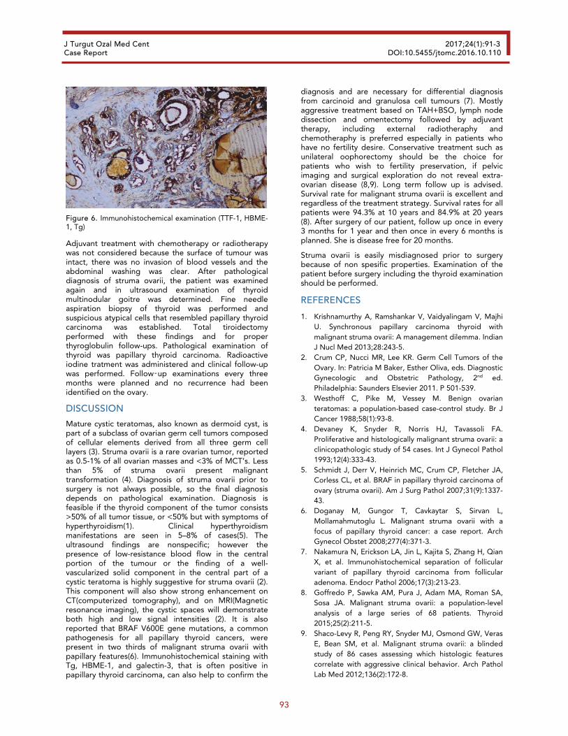
Diagnosis of struma ovarii arising within the teratoma was made and confirmed on immunohistochemistry, which was positive for TTF-1 (thyroid transcription
factor-1), HBME-1 (Hector Battifora Mesothelial-1) and Tg (thyroglobulin) at epithelium of papillas (Figure 4,5,6).
Figure 3. Respiratory silier ephitelium at surface, mucous gland structures at wall, lipid tissue and smooth muscle areas
Figure 4. Immunohistochemical examination (TTF-1, HBME-1, Tg)
Figure 5. Immunohistochemical examination (TTF-1, HBME-1, Tg)
93
J Turgut Ozal Med Cent 2017;24(1):91-3 Case Report DOI:10.5455/jtomc.2016.10.110
Figure 6. Immunohistochemical examination (TTF-1, HBME-1, Tg)
Adjuvant treatment with chemotherapy or radiotherapy was not considered because the surface of tumour was intact, there was no invasion of blood vessels and the abdominal washing was clear. After pathological diagnosis of struma ovarii, the patient was examined again and in ultrasound examination of thyroid multinodular goitre was determined. Fine needle aspiration biopsy of thyroid was performed and suspicious atypical cells that resembled papillary thyroid carcinoma was established. Total tiroidectomy performed with these findings and for proper thyroglobulin follow-ups. Pathological examination of thyroid was papillary thyroid carcinoma. Radioactive iodine tratment was administered and clinical follow-up was performed. Follow‑up examinations every three months were planned and no recurrence had been identified on the ovary.
DISCUSSION
Mature cystic teratomas, also known as dermoid cyst, is part of a subclass of ovarian germ cell tumors composed of cellular elements derived from all three germ cell layers (3). Struma ovarii is a rare ovarian tumor, reported as 0.5-1% of all ovarian masses and <3% of MCT’s. Less than 5% of struma ovarii present malignant transformation (4). Diagnosis of struma ovarii prior to surgery is not always possible, so the final diagnosis depends on pathological examination. Diagnosis is feasible if the thyroid component of the tumor consists >50% of all tumor tissue, or <50% but with symptoms of hyperthyroidism(1). Clinical hyperthyroidism manifestations are seen in 5–8% of cases(5). The ultrasound findings are nonspecific; however the presence of low-resistance blood flow in the central portion of the tumour or the finding of a well-vascularized solid component in the central part of a cystic teratoma is highly suggestive for struma ovarii (2). This component will also show strong enhancement on CT(computerized tomography), and on MRI(Magnetic resonance imaging), the cystic spaces will demonstrate both high and low signal intensities (2). It is also reported that BRAF V600E gene mutations, a common pathogenesis for all papillary thyroid cancers, were present in two thirds of malignant struma ovarii with papillary features(6). Immunohistochemical staining with Tg, HBME-1, and galectin-3, that is often positive in papillary thyroid carcinoma, can also help to confirm the
diagnosis and are necessary for differential diagnosis from carcinoid and granulosa cell tumours (7). Mostly aggressive treatment based on TAH+BSO, lymph node dissection and omentectomy followed by adjuvant therapy, including external radiotheraphy and chemotheraphy is preferred especially in patients who have no fertility desire. Conservative treatment such as unilateral oophorectomy should be the choice for patients who wish to fertility preservation, if pelvic imaging and surgical exploration do not reveal extra-ovarian disease (8,9). Long term follow up is advised. Survival rate for malignant struma ovarii is excellent and regardless of the treatment strategy. Survival rates for all patients were 94.3% at 10 years and 84.9% at 20 years (8). After surgery of our patient, follow up once in every 3 months for 1 year and then once in every 6 months is planned. She is disease free for 20 months.
Struma ovarii is easily misdiagnosed prior to surgery because of non spesific properties. Examination of the patient before surgery including the thyroid examination should be performed.
REFERENCES
1. Krishnamurthy A, Ramshankar V, Vaidyalingam V, Majhi U. Synchronous papillary carcinoma thyroid with malignant struma ovarii: A management dilemma. Indian J Nucl Med 2013;28:243-5.
2. Crum CP, Nucci MR, Lee KR. Germ Cell Tumors of the Ovary. In: Patricia M Baker, Esther Oliva, eds. Diagnostic Gynecologic and Obstetric Pathology, 2nd ed. Philadelphia: Saunders Elsevier 2011. P 501-539.
3. Westhoff C, Pike M, Vessey M. Benign ovarian teratomas: a population-based case-control study. Br J Cancer 1988;58(1):93-8.
4. Devaney K, Snyder R, Norris HJ, Tavassoli FA. Proliferative and histologically malignant struma ovarii: a clinicopathologic study of 54 cases. Int J Gynecol Pathol 1993;12(4):333-43.
5. Schmidt J, Derr V, Heinrich MC, Crum CP, Fletcher JA, Corless CL, et al. BRAF in papillary thyroid carcinoma of ovary (struma ovarii). Am J Surg Pathol 2007;31(9):1337-43.
6. Doganay M, Gungor T, Cavkaytar S, Sirvan L, Mollamahmutoglu L. Malignant struma ovarii with a focus of papillary thyroid cancer: a case report. Arch Gynecol Obstet 2008;277(4):371-3.
7. Nakamura N, Erickson LA, Jin L, Kajita S, Zhang H, Qian X, et al. Immunohistochemical separation of follicular variant of papillary thyroid carcinoma from follicular adenoma. Endocr Pathol 2006;17(3):213-23.
8. Goffredo P, Sawka AM, Pura J, Adam MA, Roman SA, Sosa JA. Malignant struma ovarii: a population-level analysis of a large series of 68 patients. Thyroid 2015;25(2):211-5.
9. Shaco-Levy R, Peng RY, Snyder MJ, Osmond GW, Veras E, Bean SM, et al. Malignant struma ovarii: a blinded study of 86 cases assessing which histologic features correlate with aggressive clinical behavior. Arch Pathol Lab Med 2012;136(2):172-8.