j. zool. lond. 243:417-433. - ohio university
TRANSCRIPT
J. Zool., Lond. (1997) 243, 417—433
Sprawling locomotion in the lizard Sceloporus clarkii: the effects of speed on gait,hindlimb kinematics, and axial bending during walking
S. M. REILLY AND M. J. DELANCEY
Department of Biological Sciences, Ohio University, Athens OH 45701, U.S.A.
(Accepted 15 January 1997)
(With 4 figures in the text)
Although the hindlimb is widely considered to provide the propulsive force in lizard locomotion, no
study to date has analysed kinematic patterns of hindlimb movements for more than one stride for a
single individual and no study has considered limb and axial kinematics together. In this study,
kinematic data from several individuals of the Scetoporus clarkii are used to describe the movement
patterns of the axial skeleton and hindlimb at different speeds, to analyse how kinematics change withspeed, and to compare and contrast these findings with the inferred effects of speed cited in theliterature. Angular limb movements and axial bending patterns (standing wave with nodes on the
girdles) did not change with speed. Only the relative speed of retracting the femur and flexing the kneeduring limb retraction changes with speed. Based on these data and similar results from a recent studyof salamanders, it appears that, over a range of speeds involving a walking trot, sprawling vertebratesincrease speed by simply retracting the femur relatively faster, thus this simple functional adjustment
may be a general mechanism to increase speed in tetrapods. The demonstration that femoral retractionalone is the major speed effector in Sceloporus clarkii lends strong functional support to ecomorphological implications of limb length (and especially femur length and caudifemoralis size) inlocomotory ecology and performance in phrynosomatid lizards. It also lends support to inferencesabout the caudifemoralis muscle as a preadaptation to terresthal locomotion and as a key innovation inthe evolution of bipedalism.
Introduction
A large body of literature on the ecological morphology of lizard locomotion has focused on thephysiological basis and energetics of locomotion and has emphasized the ecological and evolutionarysignificance of locomotor performance (reviewed in: Huey, Pianka & Schoener, 1983; Pianka, 1986;Bennett, 1989; Bennett & Huey, 1990; Miles, 1994; Garland & Losos, 1994). A unifying thread in thisresearch is that locomotory performance depends on the integration of organismal systems thatcombines a certain energetic capacity, that drives a certain morphological system, with a particularmuscle physiology, to move the limbs to propel the lizard at speeds that ensure that it will survive,reproduce and pass on its genes in a particular environment or niche. In lizards, each component(energetics, morphology, muscle physiology, ecology) of this integrated organism has seen considerable study except on how the lizard moves the limbs to propel itself at different speeds. Most studies oflimb movements in lizards have focused on stride and gait characteristics (Snyder, 1952; Urban, 1965;Daan & Belterman, 1968; Sukhanov, 1974; Rewcastle, 1981, 1983; Brinkman, 1981; Peterson, 1984;Avery et at., 1987; White & Anderson, 1994), and on inferences of limb movements from anatomicalstudies (Schaeffer, 1941; Snyder, 1952; Brinkman, 1980; Rewcastle, 1980, 1983), but few studies havepresented information on kinematic movements of axial or limb segments (Snyder, 1952; Urban, 1965;Landsmeer, 1984; Peterson, 1984; Bels et at., 1992; Ritter, 1992). Only two studies have described
417
© 1997 The Zoological Society of London
418 S. M. REILLY AND M. J. DELANCEY
motor and kinematic patterns during lizard locomotion; one on the forelimb during walking in Varanus(Jenkins & Goslow, 1983) and one for the hindlimb during walking in Sceloporus clarkii (Reilly,1995). From these studies, a basic understanding of lizard gaits and a gross description of limbmovements have emerged but they are largely based on anecdotal and qualitative descriptions ofindividual strides of lizards that are usually accelerating or decelerating as they run past a stationarycamera. Although the hindlimb is widely considered to provide the propulsive force in lizardlocomotion (Snyder, 1952; Gray, 1968; Sukhanov, 1974), no study to date has analysed kinematicpatterns of hindlimb movements for more than one stride for a single individual, and no study hasconsidered limb and axial kinematics together. The goals of this paper are to use kinematic data fromseveral individuals of the same species to describe the movement patterns of the axial skeleton andhindlimb at different speeds, to analyse how kinematics change with speed, and to compare andcontrast these findings with the inferred effects of speed cited in the literature. Details of thekinematics of limb movements compared to those of other tetrapods are presented in Reilly &DeLancey (1997).
Materials and methods
Kinematic recordings were obtained from Sceloporus clarkii collected in Molino Basin, Santa CatalinaMountains, 10 miles N. of Tucson, Arizona. Lizards were housed in 10 gallon aquaria in a temperature controlledroom (25 °C ) and fed water and crickets ad libitum for the duration of the experiments. Kinematic data for 3lizards moving at 3 speeds were analysed for speed effects. Sceloporus clarkii was used because it is believed touse generalized sprawling locomotion (Sukhanov, 1974), and because this species is morphologically generalized,possessing the primitive morphology of the family Phrynosomatidae (Miles, 1994). Detailed descriptions of themotor patterns (Reilly, 1995) and kinematics for one speed (Reilly & DeLancey, 1997) are presented elsewhere.This analysis is the first study of lizard locomotion which analyses the kinematics of multiple strides at severalknown speeds.
Kinematic analysis
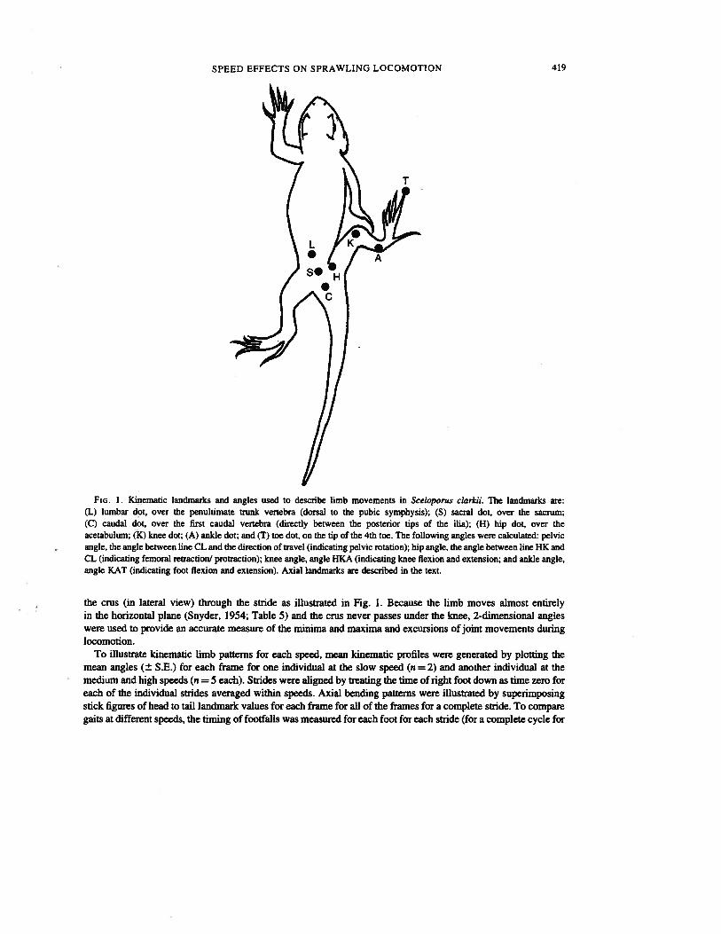
The lizards were filmed under strobe lights at 200 fields per second using a NAC HSV-400 high-speed videosystem. Elapsed time in milliseconds was recorded on each video frame during filming. Lateral and dorsal viewsof the lizards running were filmed (using mirrors) on a 70 cm long canvas treadmill with a background markedwith lines every 10 cm. A series of quadrupedal running bouts was elicited by pinching the tail when the lizardmoved out of the video field. The body temperature of the lizards during the runs was 27—30 C. Reflectivelandmarks were painted on the lizards (Fig. 1) to mark the vertebral column and the sacrum, pelvis, acetabulum,knee, ankle and tip of the longest (fourth) toe. The limb landmarks were visible in both the lateral and dorsalviews. The vertebral column was marked using the following landmark points (based on obvious points, paintdots, or intersections of chevron markings along the back): the tip of the snout, the occiput (midline posterior ofskull), the pectoral girdle (midline between the arms), 4 equally spaced trunk segments, the penultimate trunkvertebra (directly dorsal to the pubic symphysis), the sacral vertebra, the first caudal vertebra (directly between theposterior tips of the ilia), and several caudal segments.
Lizards were run repeatedly at each of 3 preset speeds encompassing a 3-fold increase in speed from about 1 to3km/h (exact speeds below). A total of 22 strides of the right leg during which the lizards exactly matched thetreadmill speed was used in the analysis: 2 strides from one individual (SVL = 90 mm) at 0.270 ms’ (0.972 kmlh); 5 strides each for 2 additional individuals (SVLs = 92, 91 nun) at both 0.476 (1.71 km/h) and 0.833 m(3 km/h). For each video field for each stride, the co-ordinates of each landmark were digitized using MeasurementTV (Updegraff, 1990). The co-ordinate data were then used to calculate 2 dimensional angles for each video fieldindicating movements of the pelvis, hip joint and knee joint (in dorsal view) and movements of the foot relative to
SPEED EFFECTS ON SPRAWLING LOCOMOTION 419
FIG. I. Kinematic landmarks and angles used to describe limb movements in Sceloporus clarkii. The landmarks are:(L) lumbar dot, over the penultimate trunk vertebra (dorsal to the pubic symphysis); (S) sacral dot, over the sacrum;(C) caudal dot, over the first caudal vertebra (directly between the posterior tips of the ilia); (H) hip dot, over theacetabulum; (K) knee dot; (A) anlde dot; and (T) toe dot, on the tip of the 4th toe. The following angles were calculated: pelvicangle, the angle between line CL and the direction of travel (indicating pelvic rotation); hip angle, the angle between line HK andCL (indicating femoral retraction/ protraction); knee angle, angle HKA (indicating knee flexion and extension; and ankle angle,angle KAT (indicating foot flexion and extension). Axial landmarks are described in the text.
the crus (in lateral view) through the stride as illustrated in Fig. 1. Because the limb moves almost entirelyin the horizontal plane (Snyder, 1954; Table 5) and the crus never passes under the knee, 2-dimensional angleswere used to provide an accurate measure of the minima and maxima and excursions of joint movements duringlocomotion.
To illustrate kinematic limb patterns for each speed, mean kinematic profiles were generated by plotting themean angles (± S.E.) for each frame for one individual at the slow speed (n = 2) and another individual at themedium and high speeds (n = 5 each). Strides were aligned by treating the time of right foot down as time zero foreach of the individual strides averaged within speeds. Axial bending patterns were illustrated by superimposingstick figures of head to tall landmark values for each frame for all of the frames for a complete stride. To comparegaits at different speeds, the timing of footfalls was measured for each foot for each stride (for a complete cycle for
T
420 S. M. REILLY AND M. J. DELANCEY
each of the 4 feet) and mean gait diagrams for each speed were plotted using mean footfall values for all 4 feet foreach speed.
Kinematic variables
Limb and pelvic movements
To assess the effects of speed on hindlimb kinematics quantitatively, a series of angular and timing variableswere taken from each stride to describepelvic and limb movements. The variables were chosen to capture theangles and timing of minimum and maximum positions of the pelvis and each of the 3 major joints of the hindlimb,the hip (H), knee (K), and ankle (A), as described in Fig. 1. Angular variables were as follows. The angles of eachjoint (H, K, A) were taken at the time of right foot down (DN) and right foot up (UP). These angles indicate thepositions of the 3 joints at the beginning (DN) and end (UP) of the stance phase (ST: from foot down to foot up),and conversely, the swing phase (SW: from foot up to the next foot down). Movement of the limb as a whole interms of the direction of movement of the foot is termed protraction and retraction when the foot is being movedanteriorly and posteriorly, respectively (which are sometimes termed recovery and transport phases, respectively).Because protraction and retraction of the limb do not often correspond to the stance and swing phases (maximumprotraction occurs Q—20 ms before foot down: see THMAXPROT vs. STRIDEDUR in Table II), we alsomeasured the angle of the hip at maximum limb protraction (HMAXPROT), the angle of the hip at maximum limbretraction (HMAXRET), and the angle of excursion (EXC) of the hip during retraction of the limb (HRETEXC).The knee is flexed to a minimum (MIN) and then extended to a maximum (MAX) during both the stance andswing phases, thus angles were taken to quantify the minimum, maximum and excursion of the knee joint duringboth phases (KMINST, KMAXST, KEXCST, KMINSW, KMAXSW, KEXCSW). The ankle is maximallyextended (EXT) just prior to foot down and this angle was measured (AMAXEXT). The anide is then flexed to aminimum during the stance phase and then extended to a maximum just after foot up and this period delineates thepower stroke (PS) of the retraction phase. Thus, we measured the minimum, maximum and excursion of the ankleduring the power stroke (AMINPS, AMAXPS, AEXCPS). The last angular variable is the maximum of rotation ofthe pelvis relative to the direction of travel (PELVMAX).
Timing variables were taken to describe aspects of the stride and the timing of joint movements. The durations(DUR) of the stance phase, swing phase, and entire stride were measured (STANCEDUR, SWINGDUR,STRIDEDUR, respectively). The rest of the timing variables are either times to (T) various joint angles (fromtime 0 at right foot down), or the durations (DUR) of angular excursions described above. These are: the time tohip maximum retraction (THMAXRET), protraction (at the end of the stride, THMAXPROT), and hip protractionduration (THRETDUR); the time to minimum and maximum knee positions during the stance (TKMINST,TKMAXST) and swing (TKMINSW, TKMAXSW) phases and their durations (KDURST, KDURSW); the timeof maximal angle extension prior to foot down (TAMAXEXT) and the time to the minimum and maximum ankleangle during the power stroke (TAMINPS, TAMAXPS) and this excursion (ADURPS); and the time to themaximum pelvic rotation (TPELVMAX). Detailed descriptions of each variable are listed in Appendix 1.
Limb adduction
The knee and toe displacement loops (from co-ordinate data) were examined and their lateral widths weremeasured to examine the amount of adduction of the limb with speed. Displacement loops circumscribe themovements of the landmark in one plane for a single stride. For example, in dorsal view a narrow displacementloop indicates that the landmark is moving in a more or less longitudinal plane with the stance and swingtrajectories of the landmark staying in the same longitudinal plane. A wider loop indicates that the landmarkmoves more medially during the stance phase than the swing phase, indicating greater adduction of the limb. Achange in the width of the knee or toe displacement loops with speed indicates that the amount of limb adductionchanges with speed and, thus, that the animal is shifting from a more sprawling posture to a more erect one.
SPEED EFFECTS ON SPRAWLING LOCOMOTION 421
Changes in limb adduction with speed were quantified using the lateral width of the knee, and toe displacementloops (from dorsal view), as well as the vertical depth of the knee loop from lateral view.
Axial bending
Axial kinematics were quantified using 8 variables describing the maximum amplitudes and longitudinalpositions of landmarks along the vertebral column. Because roughly consistent standing waves with 2 nodes werefound at all speeds, minima and maxima of wave peaks and nodes were measured from each stride. The maximumamplitude of lateral movements of the snout, the trunk region, and the tail were. measured. The minimumamplitude in the pectoral region (the pectoral node) and the pelvic region (the pelvic node) were taken and thepositions of these nodes along the relative to the vertebral landmarks was taken as well. Axial data were digitizedfor one individual at each speed (individual 3 at 0.270 mst (n = 2) and individual 1 at the other 2 speeds (n = 5each).
Statistical analyses
To identify kinematic variables that varied significantly with speed, values for each variable were averaged foreach individual and the individual means were regressed against speed. Significant regressions of angularvariables will indicate joint movements that vary with speed. Timing variables were regressed against speed inboth real time and with timing variables for each stride scaled to the stride duration for that stride. Timingvariables are expected to change with speed in real time, however, significant regressions of timing variablesscaled to stride duration will indicate how the relative timing ofjoint movements varies with speed. Variables thatdiffer with speed will then be used to describe how lizards modulate limb use to increase their velocity.
Results
Limb movements
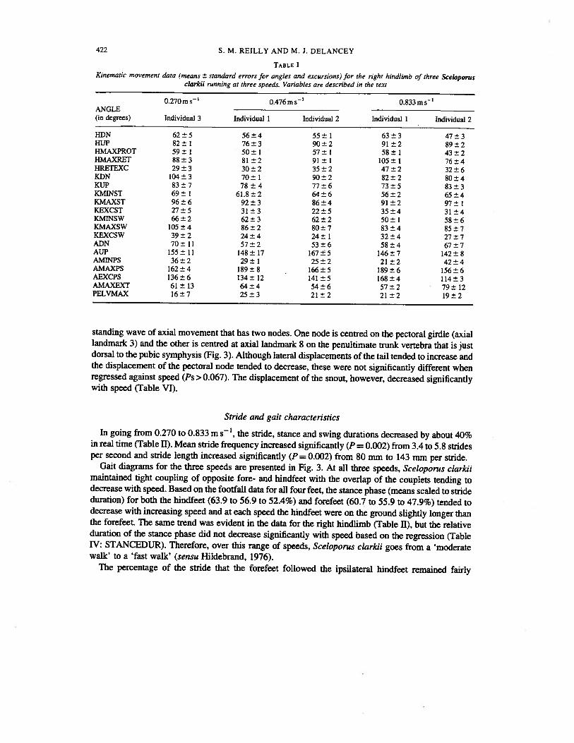
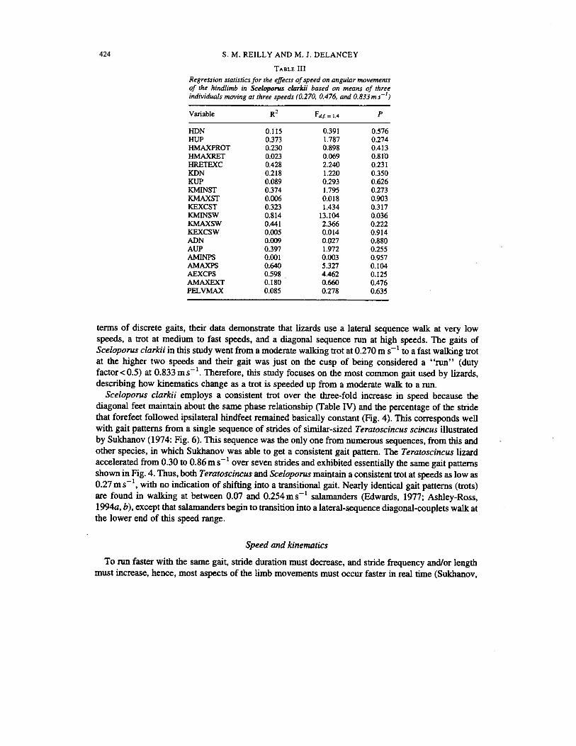
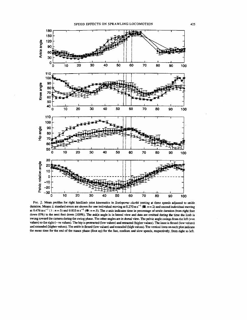
Mean angular data for limb positions and excursions for each of the three individuals are presentedin Table I. Mean timing data are presented in Table II. Representative kinematic profiles for each jointand the rotation of the pelvis are presented scaled to stride duration in Fig. 2. Regression statistics forthe effects of speed on angular data (Table III) revealed that none of the angular measures varied withspeed at the alpha level (P=0.0l).
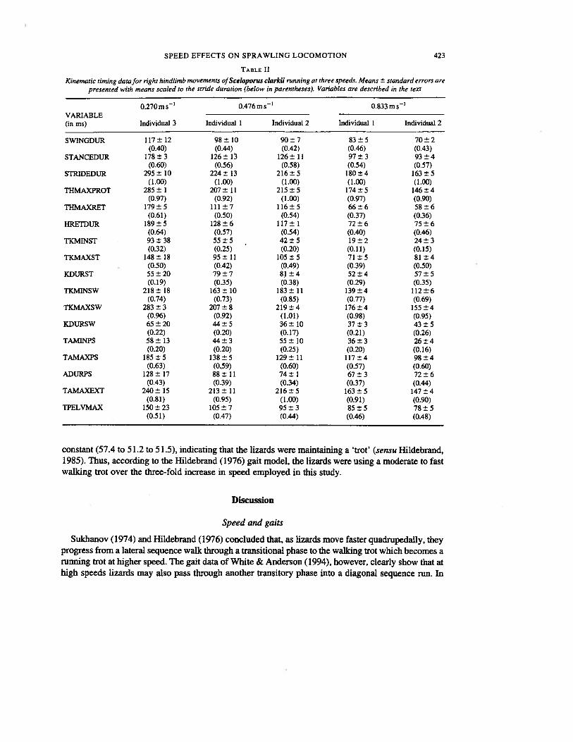
Regression statistics for the timing variables scaled to stride duration are presented in Table IV. Inreal time, 13of the 17 timing variables varied significantly with speed (Ps 0.036) with all involvingfaster movements as speed increases (Table II). When the timing of kinematic movements are scaled tostride length, only three variables vary significantly with speed at the alpha level of 0.02 (Table IV).The hip retraction time (HRETDUR), time from foot down to maximum hip retraction (THMAXRET), and the time to minimum knee angle during the stance phase (TKMINST) decrease as the lizardmoves faster.
Mean widths of displacement loops for the knee and toe are presented in Table V. Comparisons ofthe means and standard deviations shows that the displacement loops for the knee (in both lateral anddorsal view) and the toe in dorsal view are very consistent between speeds, indicating that the degreeof adduction of the femur and the entire limb does not change with speed.
Axial movements
Axial kinematic data are presented in Fig. 3 and Table VI. Sceloporus clarkii walks using a rough
422 S. M. REILLY AND M. J. DELANCEY
TABLE IKinematic movement data (means ± standard errors for angles and excursions) for the right hindlimb of three Sceloporus
clarkii running at three speeds. Variables are described in the text
0.270ms’ 0.476ms 0.833msANGLE(in degrees) Individual 3 Individual 1 Individual 2 Individual 1 Individual 2
HDN 62±5 56±4 55±1 63±3 47±3HUP 82±1 76±3 90±2 91±2 89±2HMAXPROT 59±1 50±1 57±1 58±1 43±2HMAXRET 88±3 81±2 91±1 105±1 76±4HRETEXC 29±3 30±2 35±2 47±2 32±6KDN 104±3 70±1 90±2 82±2 80±4KUP 83±7 78±4 77±6 73±5 83±3KMINST 69±1 61.8±2 64±6 56±2 65±4KMAXST 96±6 92±3 86±4 91±2 97±1KEXCST 27±5 31±3 22±5 35±4 31±4KMINSW 66±2 62±3 62±2 50±1 58±6KMAXSW 105±4 86±2 80±7 83±4 85±7KEXCSW 39±2 24±4 24±1 32±4 27±7ADN 70±11 57±2 53±6 58±4 67±7AUP 155±11 148±17 167±5 146±7 142±8AMINPS 36±2 29±1 25±2 21±2 42±4AMAXPS 162±4 189±8 166±5 189±6 156±6AEXCPS 136±6 134±12 141±5 168±4 114±3AMAXEXT 61±13 64±4 54±6 57±2 79±12PELVMAX 16±7 25±3 21±2 21±2 19±2
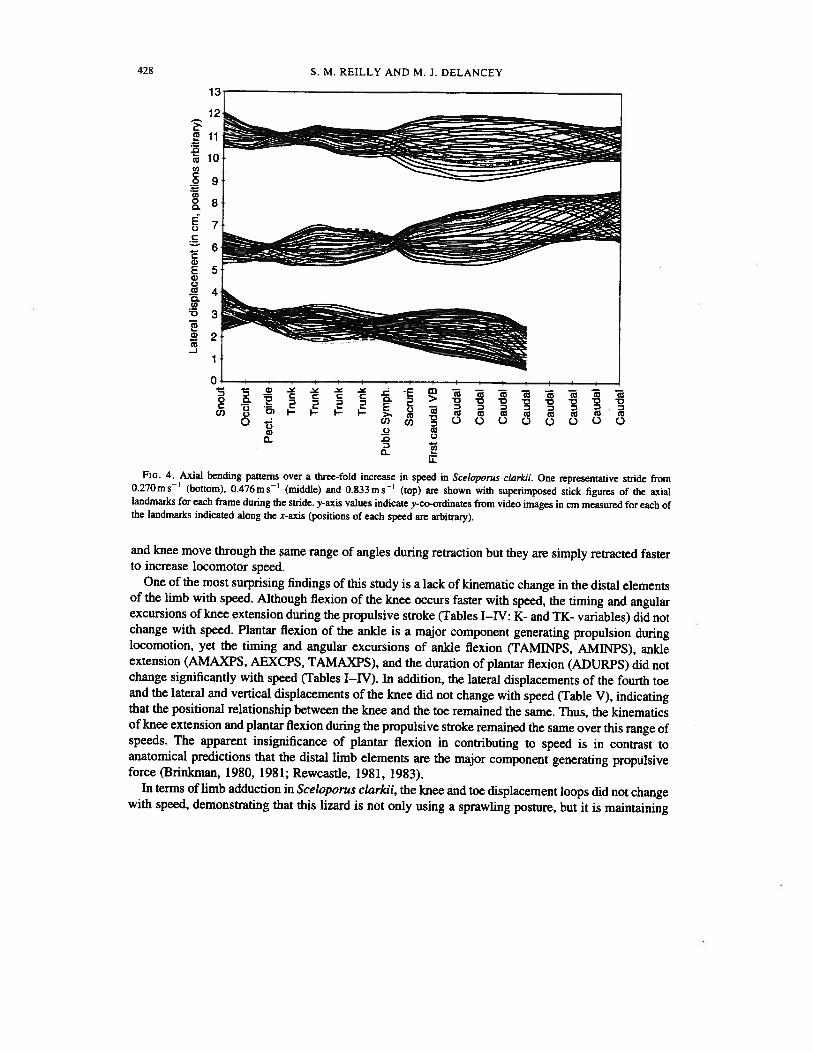
standing wave of axial movement that has two nodes. One node is centred on the pectoral girdle (axiallandmark 3) and the other is centred at axial landmark 8 on the penultimate trunk vertebra that is justdorsal to the pubic symphysis (Fig. 3). Although lateral displacements of the tail tended to increase andthe displacement of the pectoral node tended to decrease, these were not significantly different whenregressed against speed (Ps >0.067). The displacement of the snout, however, decreased significantlywith speed (Table VI).
Stride and gait characteristics
In going from 0.270 to 0.833 ms’, the stride, stance and swing durations decreased by about 40%in real time (Table II). Mean stride frequency increased significantly (P = 0.002) from 3.4 to 5.8 stridesper second and stride length increased significantly (P = 0.002) from 80 mm to 143 mm per stride.
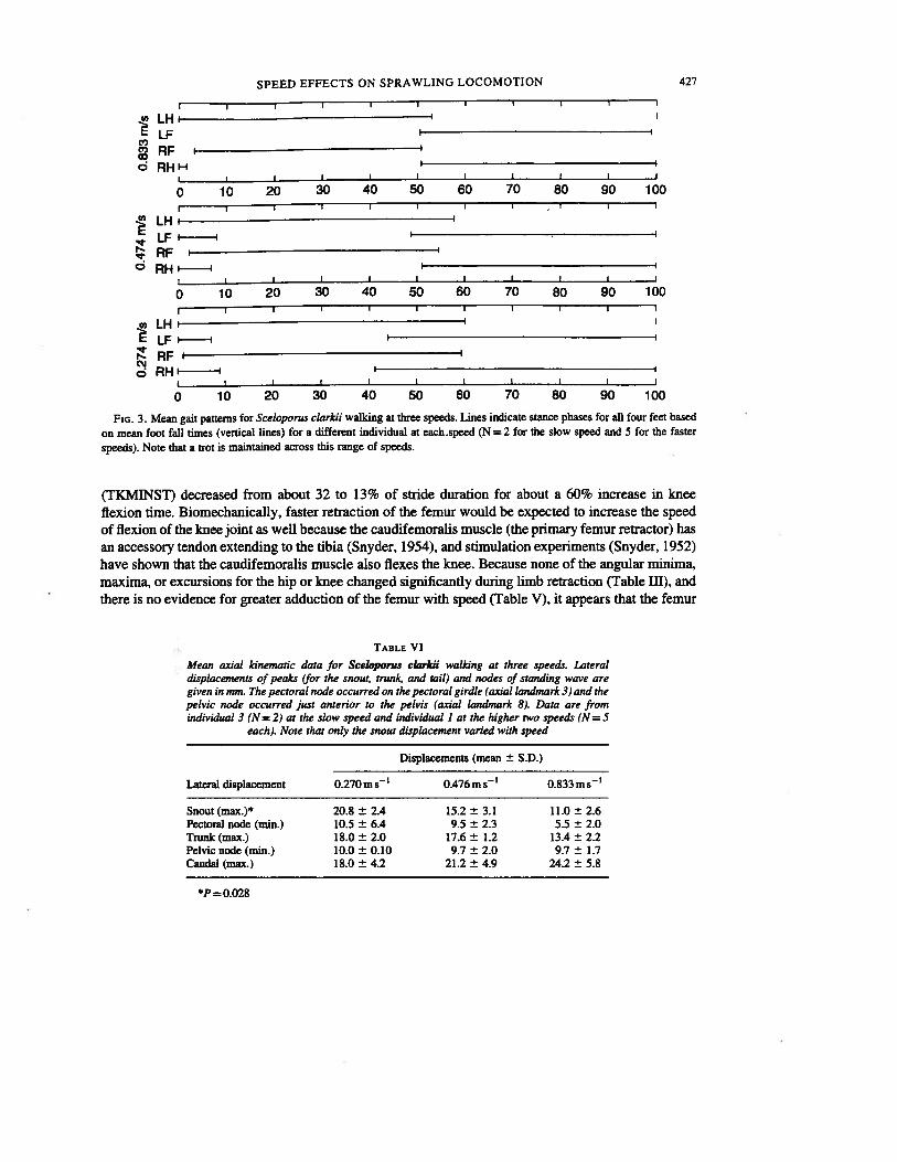
Gait diagrams for the three speeds are presented in Fig. 3. At all three speeds, Sceloporus clarkiimaintained tight coupling of opposite fore- and hindfeet with the overlap of the couplets tending todecrease with speed. Based on the footfall data for all four feet, the stance phase (means scaled to strideduration) for both the bindfeet (63.9 to 56.9 to 52.4%) and forefeet (60.7 to 55.9 to 47.9%) tended todecrease with increasing speed and at each speed the hindfeet were on the ground slightly longer thanthe forefeet. The same trend was evident in the data for the right hindlimb (Table II), but the relativeduration of the stance phase did not decrease significantly with speed based on the regression (TableTV: STANCEDUR). Therefore, over this range of speeds, Sceloporus clarkii goes from a ‘moderatewalk’ to a ‘fast walk’ (sensu Hildebrand, 1976).
The percentage of the stride that the forefeet followed the ipsilateral hindfeet remained fairly
SPEED EFFECTS ON SPRAWLING LOCOMOTION 423
TABLE II
Kinematic timing data for right hindlimb movements ofSceloporus clarkii running at three speeds. Means ± standard errors arepresented with means scaled to the stride duration (below in parentheses). Variables are described in the text
0.270ms 0.476ms’ 0.833ms’VARIABLE(in ms) Individual 3 Individual 1 Individual 2 Individual I Individual 2
SWINGDUR 117±12 98±10 90±7 83±5 70±2(0.40) (0.44) (0.42) (0.46) (0.43)
STANCEDUR 178±3 126±13 126± II 97±3 93±4(0.60) (0.56) (0.58) (0.54) (0.57)
STRIDEDUR 295 ± 10 224 ± 13 216 ± 5 180 ± 4 163 ± 5(1.00) (1.00) (1.00) (1.00) (1.00)
THMAXPROT 285 ± 1 207 ± 11 215 ± 5 174 ± 5 146 ± 4(0.97) (0.92) (1.00) (0.97) (0.90)
THMAXRET 179±5 111±7 116±5 66±6 58±6(0.61) (0.50) (0.54) (0.37) (0.36)
HRETDUR 189±5 128±6 117±1 72±6 75±6(0.64) (0.57) (0.54) (0.40) (0.46)
TKMINST 93±38 55±5 42±5 19±2 24±3(0.32) (0.25) (0.20) (0.11) (0.15)
TKMAXST 148±18 95±11 105±5 71±5 81±4(0.50) (0.42) (0.49) (0.39) (0.50)
KDURST 55±20 79±7 81±4 52±4 57±5(0.19) (0.35) (0.38) (0.29) (0.35)
TKMINSW 218± 18 163± 10 183± 11 139±4 112±6(0.74) (0.73) (0.85) (0.77) (0.69)
TKMAXSW 283±3 207±8 219±4 176±4 155±4(0.96) (0.92) (1.01) (0.98) (0.95)
KDURSW 65±20 44±5 36±10 37±3 43±5(0.22) (0.20) (0.17) (0.21) (0.26)
TAMINPS 58±13 44±3 55±10 36±3 26±4(0.20) (0.20) (0.25) (0.20) (0.16)
TAMAXPS 185±5 138±5 129±11 117±4 98±4(0.63) (0.59) (0.60) (0.57) (0.60)
ADURPS 128±17 88±11 74±1 67±3 72±6(0.43) (0.39) (0.34) (0.37) (0.44)
TAMAXEXT 240± 15 213± 11 216±5 163±5 147±4(0.81) (0.95) (1.00) (0.91) (0.90)
TPELVMAX 150± 23 105±7 95±3 85±5 78±5(0.51) (0.47) (0.44) (0.46) (0.48)
constant (57.4 to 51.2 to 51.5), indicating that the lizards were maintaining a trot’ (sensu Hildebrand,1985). Thus, according to the Hildebrand (1976) gait model, the lizards were using a moderate to fastwalking trot over the three-fold increase in speed employed in this study.
Discussion
Speed and gaits
Sukhanov (1974) and Hildebrand (1976) concluded that, as lizards move faster quadrupedally, theyprogress from a lateral sequence walk through a transitional phase to the walking trot which becomes arunning trot at higher speed. The gait data of White & Anderson (1994), however, clearly show that athigh speeds lizards may also pass through another transitory phase into a diagonal sequence run. In
424 S. M. REILLY AND M. J. DELANCEY
TABLE III
Regression statistics for the effects of speed on angular movementsof the hindlimb in Sceloporus clarkii based on means of threeindividuals moving at three speeds (0.270, 0.476, and 0.833 m s’)
Variable R Fdf = 1,4 P
HDN 0.115 0.391 0.576HUP 0.373 1.787 0.274HMAXPROT 0.230 0.898 0.413HMAXRET 0.023 0.069 0.810HRETEXC 0.428 2.240 0.23 1KDN 0.218 1.220 0.350KUP 0.089 0.293 0.626KMINST 0.374 1.795 0.273KMAXST 0.006 0.018 0.903KEXCST 0.323 1.434 0.3 17KMINSW 0.814 13.104 0.036KMAXSW 0.441 2.366 0.222KEXCSW 0.005 0.014 0.914ADN 0.009 0.027 0.880AUP 0.397 1.972 0.255AMINPS 0.001 0.003 0.957AMAXPS 0.640 5.327 0.104AEXCPS 0.598 4.462 0.125AMAXEXT 0.180 0.660 0.476PELVMAX 0.085 0.278 0.635
terms of discrete gaits, their data demonstrate that lizards use a lateral sequence walk at very lowspeeds, a trot at medium to fast speeds, and a diagonal sequence run at high speeds. The gaits ofSceloporus clarkii in this study went from a moderate walking trot at 0.270 m s to a fast walking trotat the higher two speeds and their gait was just on the cusp of being considered a “run” (dutyfactor<0.5) at 0.833 ms1. Therefore, this study focuses on the most common gait used by lizards,describing how kinematics change as a trot is speeded up from a moderate walk to a run.
Sceloporus clarkii employs a consistent trot over the three-fold increase in speed because thediagonal feet maintain about the same phase relationship (Table IV) and the percentage of the stridethat forefeet followed ipsilateral hindfeet remained basically constant (Fig. 4). This corresponds wellwith gait patterns from a single sequence of strides of similar-sized Teratoscincus scincus illustratedby Sukhanov (1974: Fig. 6). This sequence was the only one from numerous sequences, from this andother species, in which Sukhanov was able to get a consistent gait pattern. The Teratoscincus lizardaccelerated from 0.30 to 0.86 m s’ over seven strides and exhibited essentially the same gait patternsshown in Fig. 4. Thus, both Teratoscincus and Sceloporus maintain a consistent trot at speeds as low as0.27 ms’, with no indication of shifting into a transitional gait. Nearly identical gait patterns (trots)are found in walking at between 0.07 and 0.254 ms salamanders (Edwards, 1977; Ashley-Ross,1 994a, b), except that salamanders begin to transition into a lateral-sequence diagonal-couplets walk atthe lower end of this speed range.
Speed and kinematics
To run faster with the same gait, stride duration must decrease, and stride frequency and/or lengthmust increase, hence, most aspects of the limb movements must occur faster in real time (Sukhanov,
SPEED EFFECTS ON SPRAWLING LOCOMOTION 425
180
150
. 120Ca,a,
C
a,
CCua, 70a,
60
110
100
90
cu 80
70
60
50
- 30C,Ca,c 100
Cu0
C-)>a,°- -30
Fm. 2. Mean profiles for right hindlimb joint kinematics in Sceloporus clarkii trotting at three speeds adjusted to strideduration. Means ± standard errors are shown for one individual moving at 0.270 ms (: n = 2) and second individual movingat 0.476m s (I: n = 5) and 0.833 m s (•: n = 5). The x-axis indicates time in percentage of stride duration from right footdown (0%) to the next foot down (100%). The ankle angle is in lateral view and data are omitted during the time the limb isswung toward the camera during the swing phase. The other angles are in dorsal view. The pelvic angle swings from the left (+vevalues) to the right (—ye values). The hip is protracted (low values) and retracted (higher values). The knee is flexed (low values)and extended (higher values). The ankle is flexed (low values) and extended (high values). The vertical lines on each plot indicatethe mean time for the end of the stance phase (foot up) for the fast, medium and slow speeds, respectively, from right to left.
0 10 20 30 40 50 60 70 80 90 100
0 10 20 30 40 50 60 70 80 90 100
426 S. M. REILLY AND M. J. DELANCEY
TABLE IV
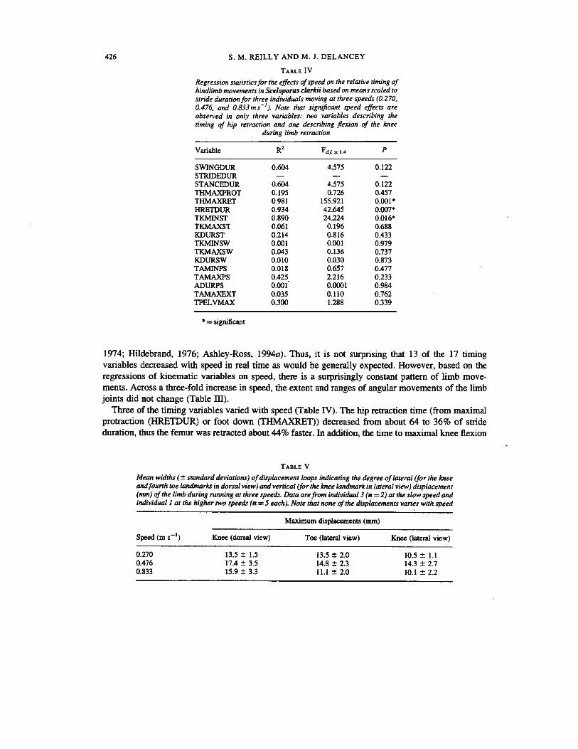
Regression statisticsfor the effects ofspeed on the relative timing ofhindlimb movements in Sceloporus clarkii based on means scaled tostride duration for three individuals moving at three speeds (0.270,0.476, and 0.833ms’). Note that significant speed effects areobserved in only three variables: two variables describing thetiming of hip retraction and one describing flexion of the knee
during limb retraction
Variable R2 Ff—
j4 P
SWINGDUR 0.604 4.575 0.122STRJDEDU1 — — —
STANCEDUR 0.604 4.575 0.122TI-IMAXPROT 0.195 0.726 0.457THMAXRET 0.981 155.921 0.001*HEErDUR 0.934 42.645 0.007*TKMINST 0.890 24.224 0.016*TKMAXST 0.061 0.196 0.688KDURST 0.214 0.816 0.433TKMINSW 0.001 0.001 0.979TKMAXSW 0.043 0.136 0.737KDURSW 0.010 0.030 0.873TAMINPS 0.018 0.657 0.477TAMAXPS 0.425 2.2 16 0.233ADURPS 0.001 0.0001 0.984TAMAXEXT 0.035 0.110 0.762TPELVMAX 0.300 1.288 0.339
* = significant
1974; Hildebrand, 1976; Ashley-Ross, 1 994a). Thus, it is not surprising that 13 of the 17 timingvariables decreased with speed in real time as would be generally expected. However, based on theregressions of kinematic variables on speed, there is a surprisingly constant pattern of limb movements. Across a three-fold increase in speed, the extent and ranges of angular movements of the limbjoints did not change (Table III).
Three of the timing variables varied with speed (Table IV). The hip retraction time (from maximalprotraction (HRETDUR) or foot down (THMAXRET)) decreased from about 64 to 36% of strideduration, thus the femur was retracted about 44% faster. In addition, the time to maximal knee flexion
TABLE V
Mean widths (± standard deviations) of displacement loops indicating the degree of lateral (for the kneeandfourth toe landmarks in dorsal view) and vertical (for the knee landmark in lateral view) displacement(mm) of the limb during running at three speeds. Data are from individual 3 (n = 2) at the slow speed andindividual I at the higher two speeds (n = 5 each). Note that none of the displacements varies with speed
Maximum displacements (mm)
Speed (m s’) Knee (dorsal view) Toe (lateral view) Knee (lateral view)
0.270 13.5 ± 1.5 13.5 ± 2.0 10.5 ± 1.10.476 17.4 ± 3.5 14.8 ± 2.3 14.3 ± 2.70.833 15.9 ± 3.3 11.1 ±. 2.0 10.1 ± 2.2
SPEED EFFECTS ON SPRAWLING LOCOMOTION 427
I I I I I I I I
LHi I
ELFC,,RF I
RHH I II I I I I I
0 10 20 30 40 50 60 70 80 90 100I I I I I
: : : : : :: JFio 3. Mean gait patterns for Sceloporus clarkii walking at three speeds. Lines indicate stance phases for all four feet based
on mean foot fall times (vertical lines) for a different individual at each .speed (N = 2 for the slow speed and 5 for the faster
speeds). Note that a trot is maintained across this range of speeds.
(TKMJNST) decreased from about 32 to 13% of stride duration for about a 60% increase in kneeflexion time. Biomechanically, faster retraction of the femur would be expected to increase the speedof flexion of the knee joint as well because the caudifemoralis muscle (the primary femur retractor) hasan accessory tendon extending to the tibia (Snyder, 1954), and stimulation experiments (Snyder, 1952)have shown that the caudifemoralis muscle also flexes the knee. Because none of the angular minima,maxima, or excursions for the hip or knee changed significantly during limb retraction (Table III), andthere is no evidence for greater adduction of the femur with speed (Table V), it appears that the femur
TABLE VI
Mean axial kinematic data for Sceloporus clarkii walking at three speeds. Lateraldisplacements of peaks (for the snout, trunk, and tail) and nodes of standing wave aregiven in mm. The pectoral node occurred on the pectoral girdle (axial landmark 3) and thepelvic node occurred just anterior to the pelvis (axial landmark 8). Data are fromindividual 3 (N= 2) at the slow speed and individual 1 at the higher two speeds (N = 5
each). Note that only the snout displacement varied with speed
Displacements (mean ± S.D.)
Lateral displacement 0.270 m s_I 0.476 ms1 0.833 m s’
Snout (max.)* 20.8 ± 2.4 15.2 ± 3.1 11.0 ± 2.6Pectoral node (mm.) 10.5 ± 6.4 9.5 ± 2.3 5.5 ± 2.0Trunk (max.) 18.0 ± 2.0 17.6 ± 1.2 13.4 ± 2.2Pelvic node (mjs.) 10.0 ± 0.10 9.7 ± 2.0 9.7 ± 1.7Caudal (max.) 18.0 ± 4.2 21.2 ± 4.9 24.2 ± 5.8
*p0 028
428 S. M. REILLY AND M. J. DELANCEY
Ce
CuC’)
0
U)00.
EC.)C
C
Ea)C.)Ce
0.U)V
cu
Ce-J
FIG. 4. Axial bending patterns over a three-fold increase in speed in Sceloporus clarkii. One representative stride from0.270m s1 (bottom), 0.476m s’ (middle) and 0.833 m s1 (top) are shown with superimposed stick figures of the axiallandmarks for each frame during the stride. y-axis values indicate y-co-ordinates from video images in cm measured for each ofthe landmarks indicated along the x-axis (positions of each speed are arbitrary).
and knee move through the same range of angles during retraction but they are simply retracted fasterto increase locomotor speed.
One of the most surprising findings of this study is a lack of kinematic change in the distal elementsof the limb with speed. Although flexion of the knee occurs faster with speed, the timing and angularexcursions of knee extension during the propulsive stroke (Tables I—TV: K- and TK- variables) did notchange with speed. Plantar flexion of the anide is a major component generating propulsion duringlocomotion, yet the timing and angular excursions of anide flexion (TAMTNPS, AMINPS), ankleextension (AMAXPS, AEXCPS, TAMAXPS), and the duration of plantar flexion (ADURPS) did notchange significantly with speed (Tables 1—IV). In addition, the lateral displacements of the fourth toeand the lateral and vertical displacements of the knee did not change with speed (Table V), indicatingthat the positional relationship between the knee and the toe remained the same. Thus, the kinematicsof knee extension and plantar flexion during the propulsive stroke remained the same over this range ofspeeds. The apparent insignificance of plantar flexion in contributing to speed is in contrast toanatomical predictions that the distal limb elements are the major component generating propulsiveforce (Brinkman, 1980, 1981; Rewcastle, 1981, 1983).
In terms of limb adduction in Sceloporus clarkii, the knee and toe displacement loops did not changewith speed, demonstrating that this lizard is not only using a sprawling posture, but it is maintaining
0) X . . . —-. C C C C -E • • . - . . .
0 0. 2 D 2 . D > V V V V V V V V—•o) l I I— > cu Cu ce cu cu Cu CU CU
0— Cl) Q) 00000000a) C.)
C)0 .3D
U-
SPEED EFFECTS ON SPRAWLING LOCOMOTION 429
this basic posture as speed increases. There is no transition to a more erect posture as had beenobserved with increased speed and gait change in the alligator (Gatesy, 1991), which is commonlyinferred to be a key reason for the evolutionary transition to erect postures and faster locomotion inmammals and archosaurs (Hildebrand, 1976, 1985; Gatesy, 1990).
Pelvic and axial movements remained constant as well. Maximum pelvic rotation from the directionof travel was highly conserved (Table IV, Fig. 2) ranging only from 16 to 25 degrees over allindividuals and speeds (Table I) which matches the ranges of values for the alligator (Gatesy, 1991), asalamander (Ashley-Ross, 1994a, b) and several other lizard species (Ritter, 1992). The time of peakpelvic rotation occurred at from 44 to 51% of stride duration (Table IV), consistently just before footup (54 to 60% of stride duration) and thus, maximum pelvic rotation occurs at from 8 1—85% of thestance phase. A similar pattern for peak pelvic rotation is seen in the trotting salamander, Dicamptodon(Ashley-Ross, 1 994b).
As speed increases, the lizards maintain a loose standing wave of axial bending with nodes on thepectoral girdle and the anterior end of the pelvis (Fig. 3). This verifies other observations of standingwaves in lizards (Ritter, 1992) and Reilly’s (1995) prediction of a standing wave based on the fact thatSceloporus clarkii exhibits simultaneous unilateral onset of axial muscles during locomotion at thehigh speed. The positions of the nodes did not shift with locomotor speed and the node (minimum) andpeak (maximum) amplitudes did not change (except for the snout). This matches Daan & Belterman’ s(1968) report that lizards tended to show little or no change in lateral bending with speed. Lateraldisplacement of the tip of snout decreased with speed (Table VI). This tendency to focus the head moreforward as speed increases has been shown in other lizards (Ritter, 1992) and a marsupial using lateralundulation (Pridmore, 1992). A standing wave pattern with nodes near the girdles is the same as seenin walking salamanders (Ashley-Ross, 1 994b) and axial patterns of several lizards shown by Ritter(1992).
The functional basis of increasing speed
Previous descriptions of the sprawling limb cycle differ on which components of the limb generatethe majority of propulsive force. Some hypothesize that the proximal elements are more important ingenerating propulsive force (Snyder, 1952; Sukhanov, 1974; Hildebrand, 1985; Ashley-Ross, 1994b)than the distal elements (Rewcastle, 1981, 1983). It seems obvious that axial bending, femoralretraction, knee extension, and plantar flexion of the foot have important synergistic contributions togenerating propulsion during the limb cycle, but which aspects change with speed? Although force andkinematic data are needed to pinpoint key components producing propulsive force, our kinematicresults suggest that, for this species, there may be a fairly simple functional explanation of how speedis increased.
Over the range of speeds we studied virtually all of the axial, pelvic, and limb kinematics remainedthe same; with only a few timing variables changing. This finding corresponds with studies of asalamander (Ashley-Ross, 1994b) and mammals (Goslow, et al., 1973, 1981; Grillner, 1975;Halbertsma, 1983) which report little to no change in angular excursions of the limbs with speed,but some timing changes. Speed in Sceloporus clarkii was increased by simply retracting the femurand flexing the knee faster relative to stride length while everything else remains the same. Althoughthe limb is retracted in a fundamentally different way (Reilly & DeLancey, 1997), a similar patternoccurs in the similarly-sized salamander Dicamptodon tenebrosus (Ashley-Ross, 1994a). Quantitativeanalysis of kinematic speed effects in this salamander showed that no angular variables changed andthat only one swing phase limb timing variable was significantly different across speeds (time to
430 S. M. REILLY AND M. J. DELANCEY
minimum pelvic-femur angle during the swing phase). This variable can be subtracted from the dutyfactor (Ashley-Ross, 1994b: Table 1) to show that relative femoral retraction time decreasessignificantly with speed as well. The observation that femoral retraction increases with speed in thefirst two sprawling vertebrates to be studied quantitatively, suggests that simply increasing femoralretraction rate may be a general mechanism by which speed is increased at walking speeds. This idea issupported by functional inferences made by Rewcastle (1983) and Snyder (1952). Rewcastle arguesthat the velocity of limb retraction and hence the velocity of the animal is related to femur lengthbecause femur length is proportionately the greatest in species showing the greatest celerity. Snyder(1952) argued convincingly (based on stimulation experiments) that the caudifemoralis muscle is themajor retractor of the entire limb through its action in retracting the femur and flexing the knee.Furthermore, he showed that, in six species of lizards, the caudifemoralis muscle (which is relativelylarge in Sceloporus clarkii) comprises up to 36% of the hindlimb muscle mass and up to 56% of themass of the muscles spanning the hip joint (Snyder, 1954), pointing to a major influence of this muscleand therefore femoral retraction as the primary component producing propulsive force in lizards.Gatesy (1990) has expanded these arguments to make inferences about the evolution of locomotion intheropods; and a similar basis for increasing speed has been shown in the vervet monkey (Vilensky &Gankiewicz, 1990). In addition, Reilly & DeLancey (1997) relate the novel caudifemoralis morphology of lizards to functional differences in limb retraction compared to the amphibians and mammalsand hypothesize that these correlated traits may be the functional basis for a fundamental dichotomy inthe functional morphology of erect locomotion in mammals vs. saunans.
An additional timing adjustment that appears to be general is that of the onset of limb retractionrelative to the timing of foot down. In Dicamptodon (Ashley-Ross, 1994a, b), Sceloporus(This study;In prep.) and vervet monkeys (Vilensky & Gankiewicz, 1990), the onset of limb retraction beginsearlier relative to foot down as speed increases. Thus, the foot hits the substratum after the limb beginsto retract, reducing braking impulses and loss of momentum that may occur and speeding up limbretraction.
Ecomorphological implications of hindlimb function in lizards
Mean maximum sprint speed in Sceloporus clarkii averages 1.89 m s1 (on racetracks: Miles, 1994)and the average speed over a 2-metre track averages l.24ms (Miles, unpubi. data). The range ofspeeds used in this study therefore extends from 21 to 67% of average track speed and 14 to 44% ofmaximum sprint speed. Thus, this study describes the function of the hindlimb that occurs over aconsiderable portion of the lizard’s locomotory acceleration to average and sprint speeds. That lizardsrun fast is the keystone of a large foundation of research on the ecological, morphological andphysiological bases of locomotor performance (Garland & Losos, 1994). How lizards run faster hasprimarily been the subject of inference. In general, lizards are thought to increase speed by: 1)changing the stride length via changes in the amplitude of standing axial bending waves (Snyder,1952; Sukhanov, 1974); 2) switching from standing to travelling waves of axial bending (Edwards,1977; Ritter, 1992); or 3) increasing the force of limb propulsion (Snyder, 1954; Gatesy, 1990).Obviously, our data for Sceloporus clarkii support the third hypothesis only, and pinpoint femoralretraction as the primary mechanism to increase speed. However, we have looked at increases in speedover only about the first half of maximal speeds used by these lizards. At higher speeds, lizards maybegin to change the amplitude of their standing wave of axial bending or modulate axial bending into atravelling wave starting from the pelvic node. It is also possible that other aspects of the limbpropulsion kinematics will change as well. First, the femur may continue to be retracted relatively
SPEED EFFECTS ON SPRAWLING LOCOMOTION 431
faster. This would involve earlier offset times but larger amplitudes of motor activity in thecaudifemoralis muscle (simultaneous EMG data for strides used in this study are presently understudy to test this hypothesis). Second, lizards could begin to use greater femoral excursions to increasestride length and speed as do birds (Gatesy, 1990). Third, whereas plantar flexion kinematics did notchange in our study, any increase in forces produced by this component could easily increase overallpropulsive forces at higher speeds. Finally, the limb could be adducted with associated changes in kneeand anide kinetics to create a more erect posture. Only quantitative studies over a wider range ofspeeds will identify whether axial bending changes as speed is increased further and what aspects oflimb movement are changed to propel the lizard up to maximal sprint speed.
A key assumption in lizard ecomorphology is that limb length and its relationship to body length arerelated to climbing ability, running speed, substratum use, and the use of bipedal locomotion(Sukhanov, 1974; Rewcastle, 1981; Pounds, 1989; Losos & Sinervo, 1989; Sinervo & Losos, 1991;Miles, 1994). The demonstration that femoral retraction alone is the major speed effector over amoderate range of ecologically relevant speeds, lends strong functional support to the ecomorphological implications of limb length (and especially femur length and caudifemoralis size) inlocomotory ecology and performance studies in lizards. It also lends support to inferences about thecaudifemoralis muscle as a preadaptation to terrestrial locomotion (Peters & Goslow, 1983; Russell &Bauer, 1992) and as a key innovation in the evolution of bipedalism and erect postures (Bakker, 1971;Charig, 1972; Biewener, 1989; Gatesy, 1990). Considerable further study of locomotion in othersprawling vertebrate species is needed to develop a sound functional foundation from whichhypotheses and inferences about the evolution of locomotor performance and the evolution of ereàtand bipedal locomotory postures can be forged and tested. However, based on the few studies to dateon sprawling locomotion, it appears that sprawling and erect locomotion may be functionally moresimilar than has been thought in the past (Gray, 1968; Hildebrand, 1985; Gatesy, 1991).
We thank Don Miles, Willem Roosenburg and Audrone Biknevicius for assistance in many aspects of thisresearch and for comments on the manuscript. Undergraduates Dorothy Glass, Pat McCabe, Mike Lyons, MikeBamett, and Cindy Hamen assisted in data collection, digitizing, and data analysis. This research was supported byOhio University Research Challenge Grant RC 95—025 to S. M. Reilly and NSF BSR 861788 to D. B. Miles.
REFERENCES
Ashley-Ross, M. A. (1994a). Metamorphic and speed effects on hindlimb kinematics during terrestrial locomotion in thesalamander Dicamptodon tenebrosus. J. exp. Biol. 193: 285—305.
Ashley-Ross, M. A. (1994b). Hindlimb kinematics during terrestrial locomotion in a salamander (Dicamptodon tenebrosus).J. exp. Biol. 193: 255—283.
Avery, R. A., Mueller, C. F., Smith, J. A. & Bond, D. J. (1987). The movement patterns of lacertid lizards: speed, gait, and pausesin Lacerta vivipara. J. ZooI. (Lond.) 211: 47—63.
Bakker, R. T. (1971). Dinosaur physiology and the origin of mammals. Evolution 25: 636—658.Bels, V. L., Theys, J. P., Bennett, M. R. & Legrand, L. (1992). Biomechanical analysis of jumping in Anolis carolinensis
(Reptilia: Iguanidae). Copeia 1992: 492—504.Biewener, A. (1989). Mammalian terrestrial locomotion and size. Bioscience 39: 776—783.Bennett, A. F. (1989). Integrated studies of locomotor performance. In Complex organismalfrnctions: integration and evolution
in vertebrates: 191—202. Wake, D. B. & Roth, G. (Eds). London: John Wiley & Sons.Bennett, A. F. & Huey, R. B. (1990). Studying the evolution of physiological performance. Oxford Surv. Evol. Biol. 7: 251—284.Brinkman, D. (1980). Structural correlates of tarsal and metatarsal functioning in Iguana (Lacertilia: Iguanidae) and other
lizards. Can. J. Zool. 58: 277—289.Brinkman, D. (1981). The hind limb step cycle of Iguana and primitive reptiles. J. ZooL (Lond.) 193: 91—103.Charig, A. J. (1972). The evolution of the archosaur pelvis and hindlimb: an explanation in functional terms. In Studies in
vertebrate evolution: 121—155. Joysey, K. A. & Kemp, T. S. (Eds). Edinburgh: Oliver & Boyd.
432 S. M. REILLY AND M. J. DELANCEY
Daan, S. & Belterman, T. (1968). Lateral bending in locomotion of some lower tetrapods, I and II. Proc. Ned. Akad. Wetten. C.71: 245—266.
Edwards. J. L. (1977). The evolution of terrestrial locomotion. In Major patterns in vertebrate evolution: 553—576. Hecht, M. K.,Goody, P. C. & Hecht, B. M. (Eds). New York: Plenum Publishing Corp.
Garland, T. & Losos, J. B., Jr (1994). Ecological morphology of locomotor performance in reptiles. In Ecological morpho1og:integrative organismal biology: 240—302. Wainwright, P. C. & Reilly, S. M. (Eds). Chicago: University of ChicagoPress.
Gatesy, S. M. (1990). Caudifemoralis musculature and the evolution of theropod locomotion. Paleobiology 16: 170—186.Gatesy. S. M. (1991). Hind limb movements of the American alligator (Alligator mississippiensis) and postural grades. J. Zool.
(Lond.) 224: 577—588.Gray, J. (1968). Animal locomotion. New York: W. W. Norton & Co. Inc.Goslow, G. E., Jr, Reinking, R. M. & Stuart, D. G. (1973). The cat step cycle: hindlimb joint angles and muscle lengths during
unrestrained locomotion. J. Morphol. 141: 1—42.Goslow, G. E., Jr, Seeherman, H. J., Taylor, C. R., McCutchin, M. N. & Heglund, N. C. (1981). Electrical activity and relative
length changes of dog limb muscles as a function of speed and gait. J. exp. Biol. 94: 15—42.Grillner, S. (1975). Locomotion in vertebrates: central mechanisms and reflex action. Physiol. Rev. 55: 247—304.Halbertsma, J. M. (1983). The stride cycle of the cat: the modeling of locomotion by computerized analysis of automatic
recordings. Ada physiol. scand. (Suppl.) 521: 1—75.Hildebrand, M. (1976). Analysis of tetrapod gaits: general considerations and symmetrical gaits. In Neural control of
locomotion: 181—201. Herman, R. M., Griliner, S., Stein, P. S. G. & Stuart, D. G. (Eds). New York: Plenum Press.Hildebrand, M. (1985). Walking and running. In Functional vertebrate morphology: 38—57. Hildebrand, M., Bramble, D. M.,
Liem, K. F. & Wake, D. B. (Eds). Cambridge, MA: Harvard University Press.Huey, R. B., Pianka, E. R., Schoener, T. W. (Eds) (1983). Lizard ecology: studies ofa model organism. Cambridge MA: Harvard
University Press.Jenkins, F. A., Jr & Goslow, G. E., Jr (1983). The functional anatomy of the shoulder of the savannah monitor lizard (Varanus
exanthamaticus). J. Morphol. 175: 195—216.Landsmeer, I. M. F. (1984). Morphology of the anterior limb in relation to sprawling gait in Varanus. Symp. zool. Soc. Land. No.
52: 27—45.Losos, 3. B. & Sinervo, B. (1989). The effects of morphology and perch diameter on sprint performance of Anolis lizards. J. exp.
Biol. 145: 23—30.Miles, D. 8. (1994). The covariance between morphology and locomotion in Sceloporine lizards. In Lizard ecology: the third
generation: 135—145. Vitt, L. J. & Pianka, E. R. (Eds). Princeton: Princeton University Press.Peters, S. E. & Goslow, G. E., Jr (1983). From salamanders to mammals: continuity in musculoskeletal function during
locomotion. Brain Behav. Evol. 22: 191—197.Peterson. J. A. (1984). The locomotion of Chamaeleo (Reptilia: Sauna) with particular reference to the forelimb. J. Zool. (Land.)
202: 1—42.Pianka, E. R. (1986). Ecology and natural history of desert lizards. Princeton: Princeton University Press.Pounds, J. A. (1989). Ecomorphology, locomotion, and microhabitat structure: patterns in a tropical mainland Anolis
community. Ecol. Monogr. 58: 299—320.Pndmore, P. A. (1992). Trunk movements during locomotion in the marsupial Monodelphis domestica (Didelphidae). J.
Morphol. 211: 137—146.Reilly, S. M. (1994/95). Quantitative electromyography and muscle function of the hind limb during quadrupedal running in the
lizard Sceloporus clarkii. Zoology 98: 263—277. Jena ZACS: Gustav Fischer Verlag.Reilly, S. M. & DeLancey, M. 3. (1997). Sprawling locomotion in the lizard Sceloporus clarkii: quantitative kinematics of a
walking trot. J. Exp. Biol. 200: 753—765.Rewcastle, S. C. (1980). Form and function in lacertilian knee and mesotarsal joints: a contribution to the analysis of sprawling
locomotion. J. Zool., Land. 191: 147—170.Rewcastle, S. C. (1981). Stance and gait in tetrapods: an evolutionary scenario. Symp. zool. Soc. Land. No. 48: 239—267.Rewcastle, S. C. (1983). Fundamental adaptations in the lacertilian hind limb: A partial analysis of the sprawling limb posture
and gait. Copeia 1983: 476—487.Ritter, V. (1992). Lateral bending during lizard locomotion. J. exp. Biol. 173: 1—10.Russell, A. P. & Bauer, A. M. (1992). The m. caudjfemoralis longus and its relationship to caudal autotomy and locomotion in
lizards (Reptilia: Sauna). J. ZooL, LonL 227: 127—143.Schaeffer, B. (1941). The morphological and functional evolution of the tarsus in amphibians and reptiles. Bull. Am. Mus. Nat.
Hist. 78: 395—472.
SPEED EFFECTS ON SPRAWLING LOCOMOTION 433
Sinervo, B. & Losos, J. B. (1991). Walking the tight rope: A comparison of arboreal sprint performance among populations of
Sceloporus occidentalis. Ecology 72: 1225—1233.Snyder, R. C. (1952). Quadrupedal and bipedal locomotion of lizards. Copeia 1952: 64—70.
Snyder, R. C. (1954). Anatomy and function of the pelvic girdle and hindlimb in lizard locomotion. Am. J. Anat. 95: 1—41.
Sukhanov, V. B. (1974). General system of symmetrical locomotion of terrestrial vertebrates and some features of movement of
lower zetrapods. (Translated by M. lvi. Hague). New Delhi: Amerind Publishing Co. Pvt. Ltd.
Updegraff, G. (1990). Measurement TV: video analysis software. San Clemente, CA: Data Crunch.
Urban, E. K. (1965). Quantitative study of locomotion in Teiid lizards. Anim. Behav. 13: 513—529.
Vilensky, J. A. & Gankiewicz, E. (1990). Effects of growth and speed on hindlimb joint angular displacement patterns in vervet
monkeys (Cercopithecus aethiops). Am. J. Phys. Anthropol. 81: 441—449.
White, T. D. & Anderson, R. A. (1994). Locomotory patterns and costs as related to body size and form in teiid lizards. J. Zool.
(Lond.) 233: 107—128.
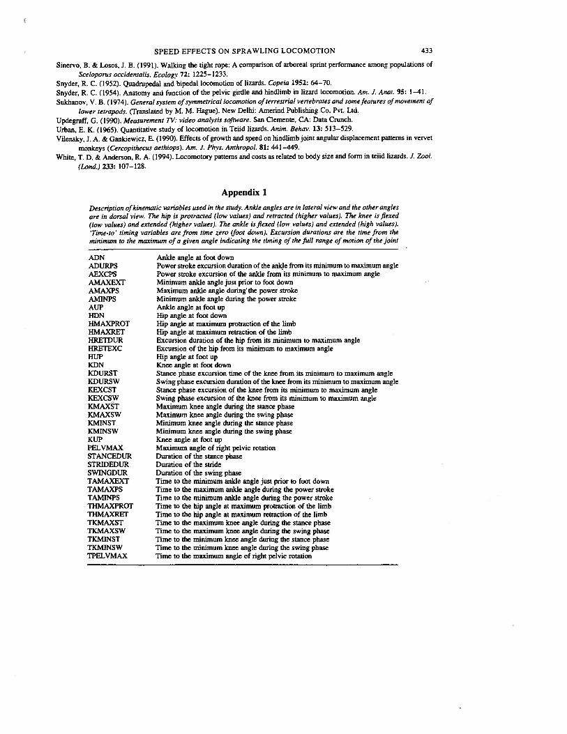
Appendix 1
Description ofkinematic variables used in the study. Ankle angles are in lateral view and the other anglesare in dorsal view. The hip is protracted (low values) and retracted (higher values). The knee is flexed(low values) and extended (higher values). The ankle is flexed (low values) and extended (high values).‘Time-to’ timing variables are from time zero (foot down). Excursion durations are the time from theminimum to the maximum of a given angle indicating the timing of the full range of motion of the joint
ADN Ankle angle at foot downADURPS Power stroke excursion duration of the anide from its minimum to maximum angleAEXCPS Power stroke excursion of the anlde from its minimum to maximum angleAMAXEXT Minimum ankle angle just prior to foot downAMAXPS Maximum anlde angle during the power strokeAMINPS Minimum ankle angle during the power strokeAUP Anlde angle at foot upHDN Hip angle at foot downHMAXPROT Hip angle at maximum protraction of the limbHMAXRET Hip angle at maximum retraction of the limbHRETDUR Excursion duration of the hip from its minimum to maximum angleHRETEXC Excursion of the hip from its minimum to maximum angleHUP Hip angle at foot upKDN Knee angle at foot downKDURST Stance phase excursion time of the knee from its minimum to maximum angleKDURSW Swing phase excursion duration of the knee from its minimum to maximum angleKEXCST Stance phase excursion of the knee from its minimum to maximum angleKEXCSW Swing phase excursion of the knee from its minimum to maximum angleKMAXST Maximum knee angle during the stance phaseKMAXSW Maximum knee angle during the swing phaseKMINST Minimum knee angle during the stance phaseKMINSW Minimum knee angle during the swing phaseKUP Knee angle at foot upPELVMAX Maximum angle of right pelvic rotationSTANCEDUR Duration of the stance phaseSTRIDEDUR Duration of the strideSWINGDUR Duration of the swing phaseTAMAXEXT Time to the minimum ankle angle just prior to foot downTAMAXPS Time to the maximum anide angle during the power strokeTAMINPS Time to the minimum anide angle during the power strokeTHMAXPROT Time to the hip angle at maximum protraction of the limbTHMAXRET Time to the hip angle at maximum retraction of the limbTKMAXST Time to the maximum knee angle during the stance phaseTKMAXSW Time to the maximum knee angle dunng the swing phaseTKMINST Time to the minimum knee angle during the stance phaseTKMINSW Time to the minimum knee angle during the swing phaseTPELVMAX Time to the maximum angle of right pelvic rotation