jane b. reece, lisa a. urry, michael l. cain, steven a ... · chapter 5 (continued) ... source of...
TRANSCRIPT
LECTURE PRESENTATIONSFor CAMPBELL BIOLOGY, NINTH EDITION
Jane B. Reece, Lisa A. Urry, Michael L. Cain, Steven A. Wasserman, Peter V. Minorsky, Robert B. Jackson
© 2011 Pearson Education, Inc.
Lectures byErin Barley
Kathleen Fitzpatrick
The Structure and Function of Proteins
Chapter 5 (continued)
Lectures modified by Garrett Dancik

Concept 5.4: Proteins include a diversity of structures, resulting in a wide range of functions
• Proteins account for more than 50% of the dry mass of most cells
• Protein functions include structural support, storage, transport, cellular communications, movement, and defense against foreign substances
© 2011 Pearson Education, Inc.
2
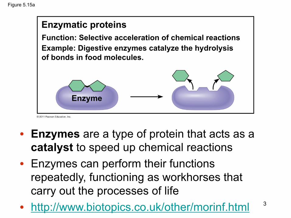
Figure 5.15a
Enzymatic proteins
Enzyme
Example: Digestive enzymes catalyze the hydrolysisof bonds in food molecules.
Function: Selective acceleration of chemical reactions
3
• Enzymes are a type of protein that acts as a catalyst to speed up chemical reactions
• Enzymes can perform their functions repeatedly, functioning as workhorses that carry out the processes of life
• http://www.biotopics.co.uk/other/morinf.html
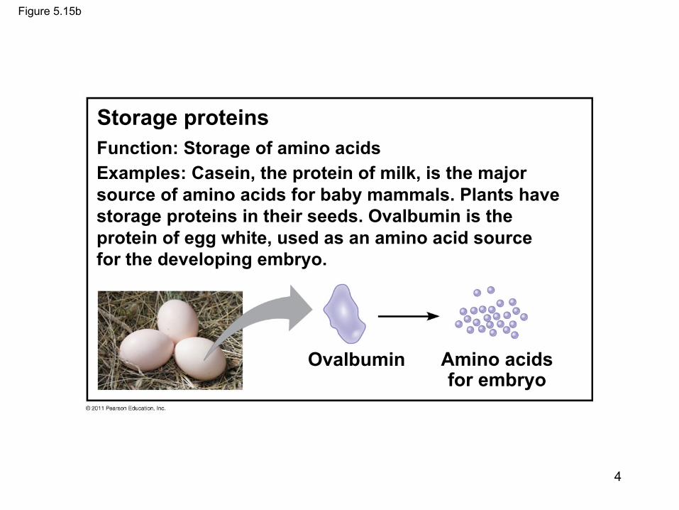
Figure 5.15b
Storage proteins
Ovalbumin Amino acidsfor embryo
Function: Storage of amino acidsExamples: Casein, the protein of milk, is the majorsource of amino acids for baby mammals. Plants havestorage proteins in their seeds. Ovalbumin is theprotein of egg white, used as an amino acid sourcefor the developing embryo.
4

Figure 5.15c
Hormonal proteinsFunction: Coordination of an organism’s activitiesExample: Insulin, a hormone secreted by thepancreas, causes other tissues to take up glucose,thus regulating blood sugar concentration
Highblood sugar
Normalblood sugar
Insulinsecreted
5
Insulin protein entry: https://www.ncbi.nlm.nih.gov/protein/AAA59172.1

6
Insulin and Glucose transport

Figure 5.15d
Muscle tissue
Actin Myosin
100 µm
Contractile and motor proteinsFunction: MovementExamples: Motor proteins are responsible for theundulations of cilia and flagella. Actin and myosinproteins are responsible for the contraction ofmuscles.
7
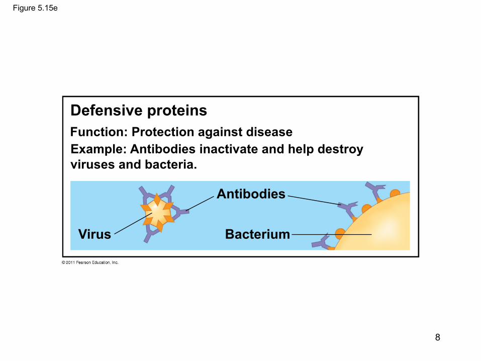
Figure 5.15e
Defensive proteins
Virus
Antibodies
Bacterium
Function: Protection against diseaseExample: Antibodies inactivate and help destroyviruses and bacteria.
8
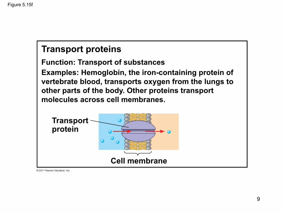
Figure 5.15f
Transport proteins
Transportprotein
Cell membrane
Function: Transport of substancesExamples: Hemoglobin, the iron-containing protein ofvertebrate blood, transports oxygen from the lungs toother parts of the body. Other proteins transportmolecules across cell membranes.
9
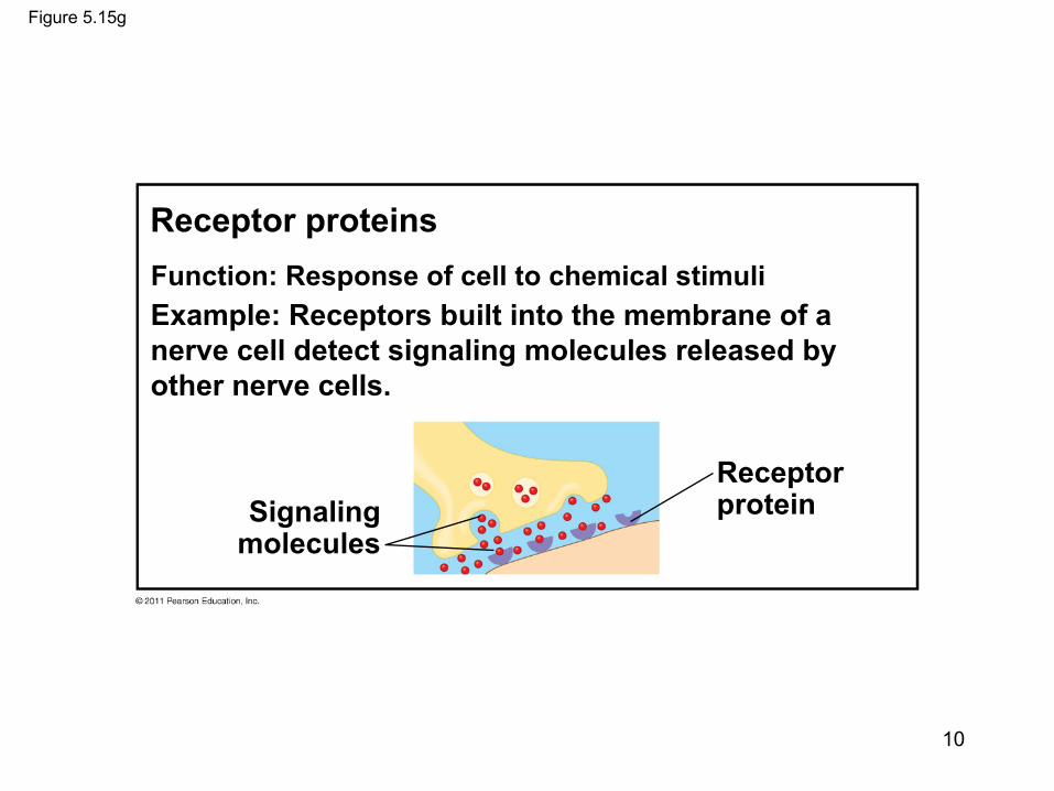
Figure 5.15g
Signalingmolecules
Receptorprotein
Receptor proteinsFunction: Response of cell to chemical stimuliExample: Receptors built into the membrane of anerve cell detect signaling molecules released byother nerve cells.
10
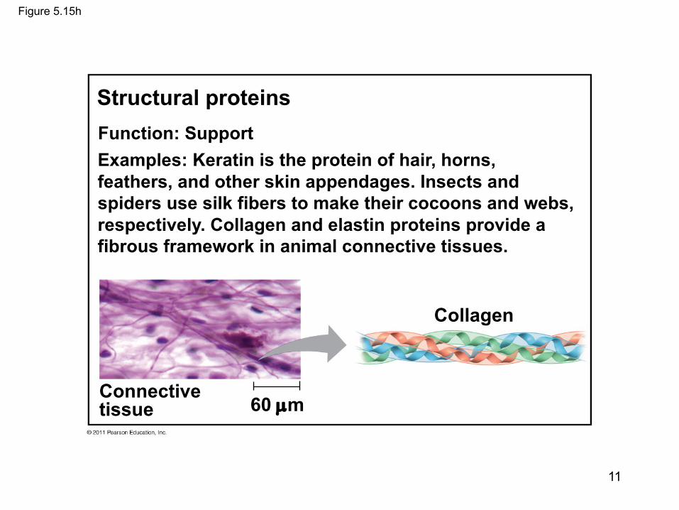
Figure 5.15h
60 µm
Collagen
Connectivetissue
Structural proteinsFunction: SupportExamples: Keratin is the protein of hair, horns,feathers, and other skin appendages. Insects andspiders use silk fibers to make their cocoons and webs,respectively. Collagen and elastin proteins provide afibrous framework in animal connective tissues.
11

12Rajagopalan S et al. PNAS 2010;107:8587-8592
DNA repair proteinsExamples include p53 and BRCA2

• Amino acids are the building blocks (monomers) of proteins– Amino acids are organic molecules with carboxyl and
amino groups– Amino acids differ in their properties due to differing
side chains, called R groups (see next slide)• Polypeptides are unbranched polymers built
from the same set of 20 amino acids• A protein is a biologically functional molecule
that consists of one or more polypeptides
© 2011 Pearson Education, Inc.
13
Proteins

Figure 5.UN01
Side chain (R group)
Aminogroup
Carboxylgroup
a carbon
14
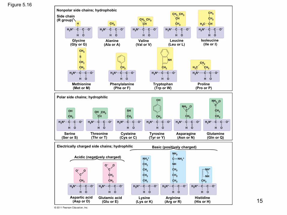
Figure 5.16Nonpolar side chains; hydrophobicSide chain(R group)
Glycine(Gly or G)
Alanine(Ala or A)
Valine(Val or V)
Leucine(Leu or L)
Isoleucine(Ile or I)
Methionine(Met or M)
Phenylalanine(Phe or F)
Tryptophan(Trp or W)
Proline(Pro or P)
Polar side chains; hydrophilic
Serine(Ser or S)
Threonine(Thr or T)
Cysteine(Cys or C)
Tyrosine(Tyr or Y)
Asparagine(Asn or N)
Glutamine(Gln or Q)
Electrically charged side chains; hydrophilic
Acidic (negatively charged)
Basic (positively charged)
Aspartic acid(Asp or D)
Glutamic acid(Glu or E)
Lysine(Lys or K)
Arginine(Arg or R)
Histidine(His or H) 15

Amino Acid Polymers
• Amino acids are linked by peptide bonds• A polypeptide is a polymer of amino acids• Polypeptides range in length from a few to
more than a thousand monomers (amino acids)
• Each polypeptide has a unique linear sequence of amino acids, with a carboxyl end (C-terminus) and an amino end (N-terminus)
© 2011 Pearson Education, Inc.
16
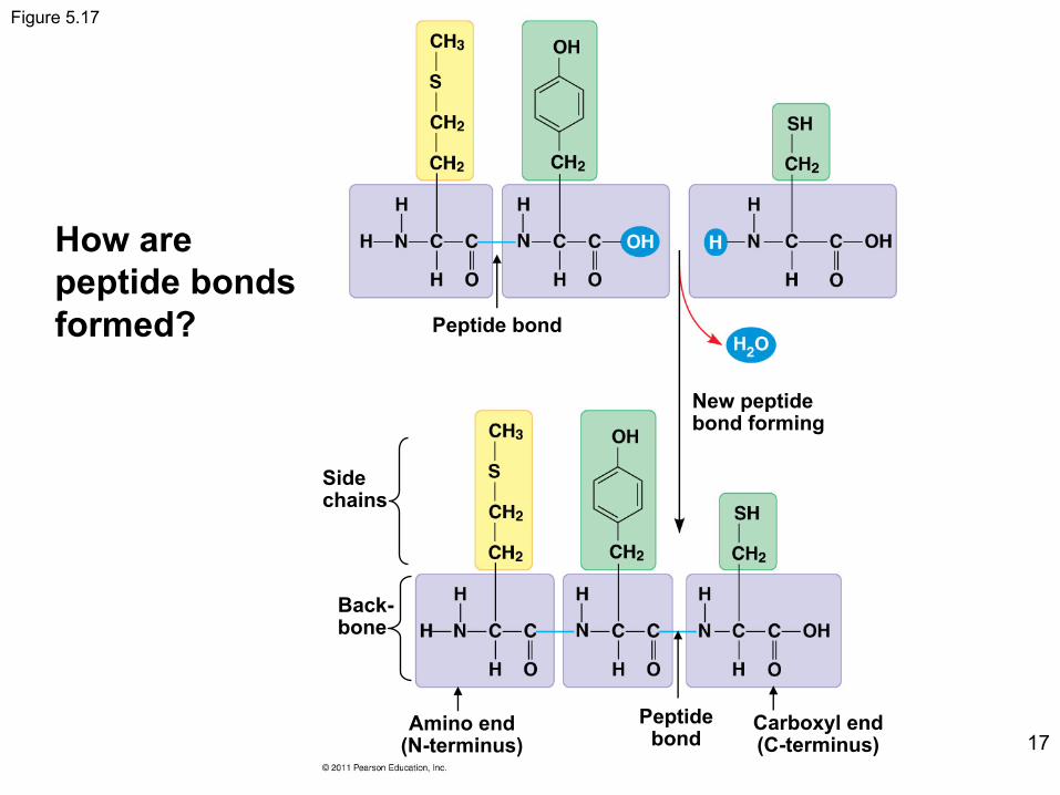
Figure 5.17
Peptide bond
New peptidebond forming
Sidechains
Back-bone
Amino end(N-terminus)
Peptidebond
Carboxyl end(C-terminus) 17
How are peptide bonds formed?
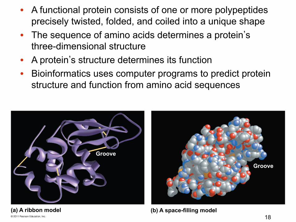
(a) A ribbon model (b) A space-filling model
Groove
Groove
18
• A functional protein consists of one or more polypeptides precisely twisted, folded, and coiled into a unique shape
• The sequence of amino acids determines a protein’s three-dimensional structure
• A protein’s structure determines its function• Bioinformatics uses computer programs to predict protein
structure and function from amino acid sequences

Figure 5.19
Antibody protein Protein from flu virus
19
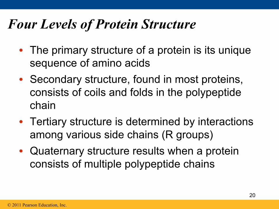
Four Levels of Protein Structure
• The primary structure of a protein is its unique sequence of amino acids
• Secondary structure, found in most proteins, consists of coils and folds in the polypeptide chain
• Tertiary structure is determined by interactions among various side chains (R groups)
• Quaternary structure results when a protein consists of multiple polypeptide chains
© 2011 Pearson Education, Inc.
20
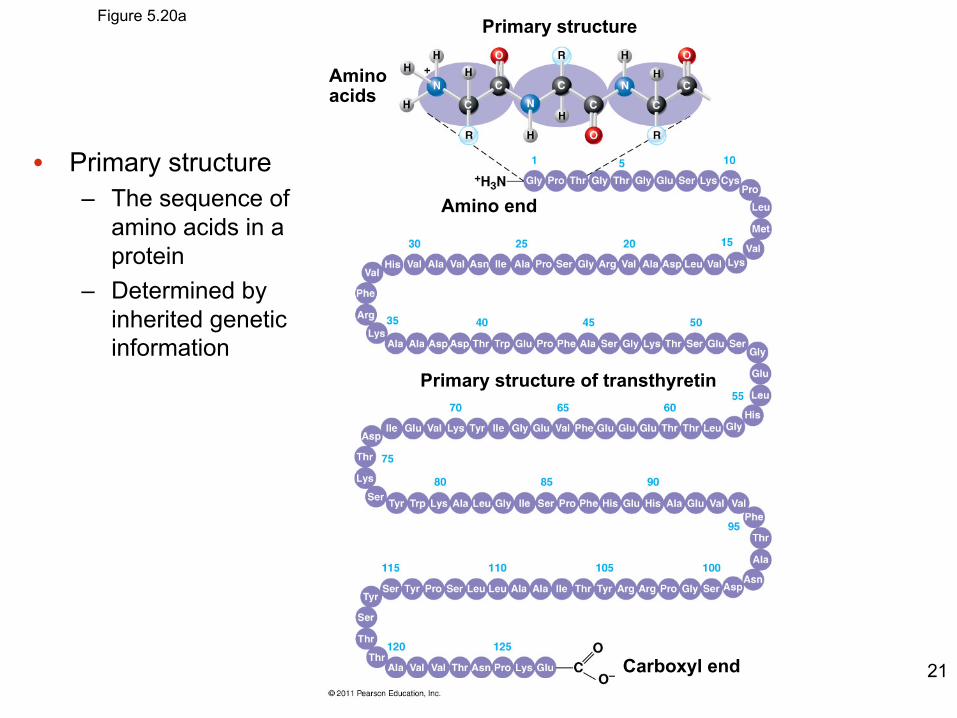
Figure 5.20a Primary structure
Aminoacids
Amino end
Carboxyl end
Primary structure of transthyretin
21
• Primary structure– The sequence of
amino acids in a protein
– Determined by inherited genetic information
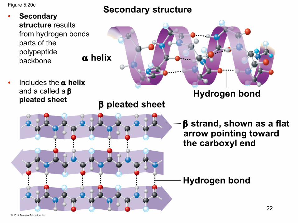
Secondary structure
Hydrogen bond
a helix
b pleated sheet
b strand, shown as a flatarrow pointing towardthe carboxyl end
Hydrogen bond
Figure 5.20c
22
• Secondary structure results from hydrogen bonds parts of the polypeptide backbone
• Includes the a helix and a called a bpleated sheet

Figure 5.20e
Transthyretinpolypeptide
23
• Tertiary structure is the shape of a polypeptide in three dimensions

Hemoglobin
HemeIron
a subunit
a subunit
b subunit
b subunit
Figure 5.20i
24
• Quaternary structure results when two or more polypeptide chains form one macromolecule
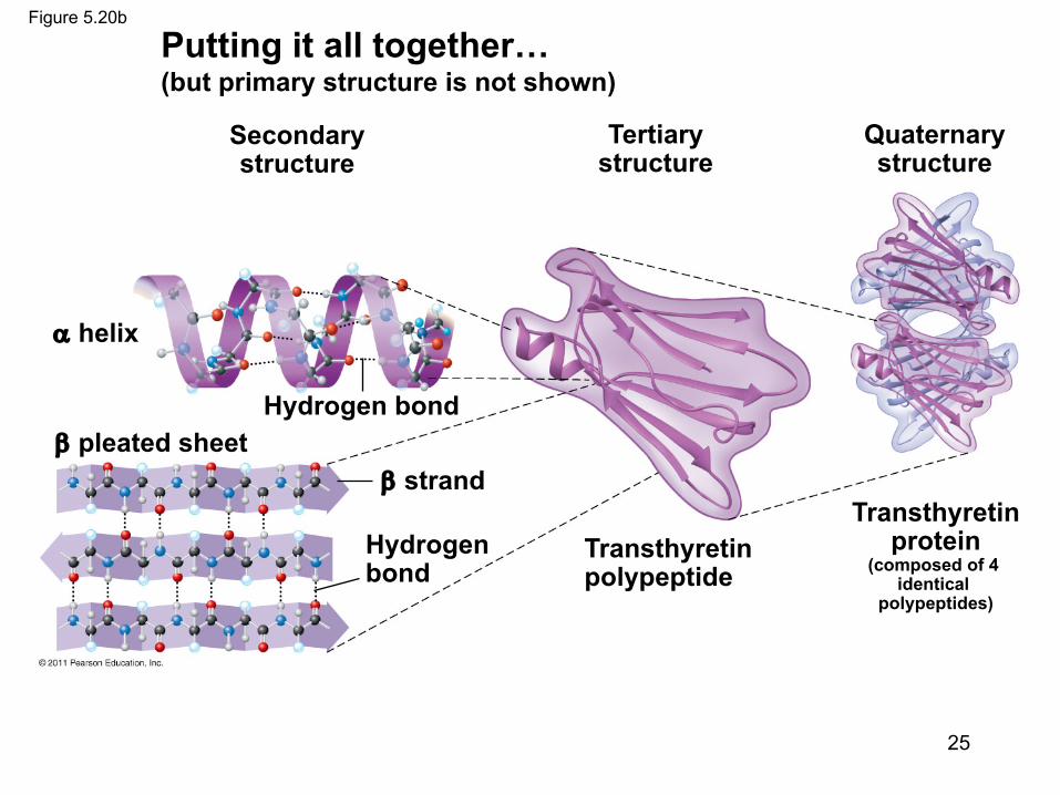
Figure 5.20b
Secondarystructure
Tertiarystructure
Quaternarystructure
Hydrogen bond
a helix
b pleated sheetb strand
Hydrogenbond
Transthyretinpolypeptide
Transthyretinprotein
(composed of 4 identical
polypeptides)
25
Putting it all together…(but primary structure is not shown)

Sickle-Cell Disease: A Change in Primary Structure
• A slight change in primary structure can affect a protein’s structure and ability to function – How does the primary structure change?
• Sickle-cell disease, an inherited blood disorder, results from a single amino acid substitution in the protein hemoglobin
• Genpept:– https://www.ncbi.nlm.nih.gov/protein/4504349
© 2011 Pearson Education, Inc.
26
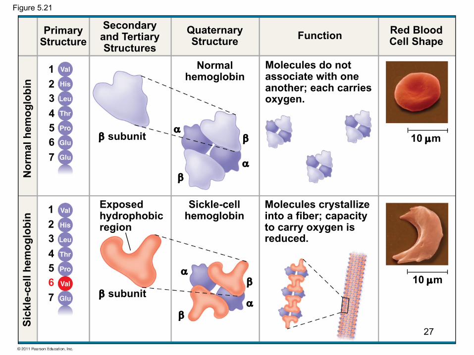
Figure 5.21
PrimaryStructure
Secondaryand TertiaryStructures
QuaternaryStructure Function Red Blood
Cell Shape
b subunit
b subunitb
b
a
a
Exposedhydrophobicregion
Molecules do notassociate with oneanother; each carriesoxygen.
Molecules crystallizeinto a fiber; capacityto carry oxygen isreduced.
Sickle-cellhemoglobin
Normalhemoglobin
10 µm
10 µm
Sick
le-c
ell h
emog
lobi
nN
orm
al h
emog
lobi
n
1234567
1234567
b
ba
a
27