jaundice in children
DESCRIPTION
jaundice in older children wide spectrum of differential diagnosishere's the approachTRANSCRIPT


Jaundice in Children
Abdulwahab TelmesaniFRCPC,FFAP
Faculty of Medicine and Medical Science
Umm Al-Qura University

An Approach to a
Child With Direct Hyperbilirubinemi
a
Classic Approach
• Proper detailed history
• Proper physical examination
• Formalize an impression of prioritized DDx
• Appropriate investigations

Identify
• Acute
• Chronic (more than 6 months)

In Children
•Acute
• Chronic (more than 6 months)

Identify
• Hepatocellular
• Chlestatic

In Children
•Hepatocellular (ALT/AST more than twice of ALP)
• Cholestatic (ALT/AST less than twice of ALP)
Remember
The prognostic value of • Albumin• Coagulation profile

Etiology
• Infection• Drugs• Specific Entities• Vascular

Etiology
•Infection• Drugs• Specific Entities• Vascular

Infections
• Viral• Bacterial• Parasitic

Viral Hepatitis
• Hepatotropic Virus’s (replicate in the liver and causes hepatitis)
• Others
Hepatotropic Viruses
• HBV (10-20% Chronic active hepatitis)
• HCV (70-80% Chronic active hepatitis)

Hepatotropic Viruses
Non B / C Viral Hepatitis
• HAV
• HEV
• HFV
• HGV
• TTV
• SEN

Others
• EBV
• CMV
• Herpes
• Other

Hepatitis A Virus
Most common cause of community acquired hepatitis through out the
world

Hepatitis A Virus
• RNA Picorna Virus (Rhinovirus, Enterovirus, Cocxackievirus)
• Feco - oral transmission (Food – borne +/- Water – borne)
• Day care centers account for 10% of cases

Hepatitis A Virus
Transmission in 50% of contacts

Hepatitis A Virus
Liver injury in HAV is secondary to immune response not to cytopathy

Hepatitis A Virus
Presentation• Incubation period 4 weeks• Prodrome 1 week• Jaundice 1 – 3 weeks• Hepatomegaly• Liver enzymes 20 – 100 time upper
normal• Spontaneous resolution

Hepatitis A Virus
Presentation• Sporadic
• Epidemic
• Endemic
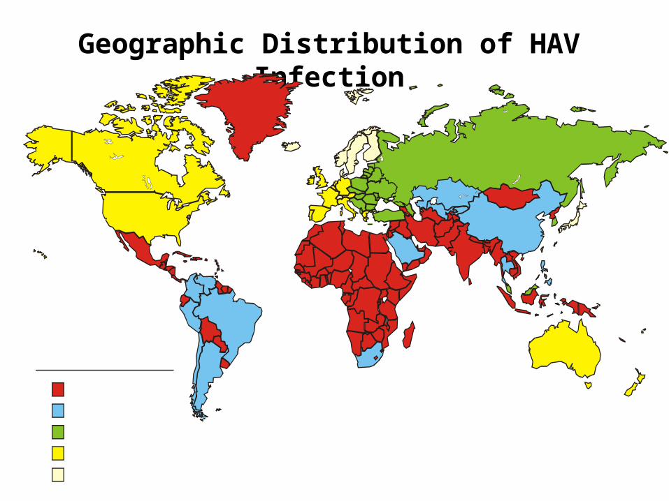
Geographic Distribution of HAV Infection

Hepatitis A Virus
Clinical Presentation in Endemic areas
• 10 % of children below 6 years• 40 % of children 6 – 14 years• 70 % of subjects older than 14 years• 70 – 100 % of children have been
infected
Hepatitis A Virus
Epidemic• Tend to seasonal• Symptoms as in sporadic cases

Hepatitis A Virus
No Chronic Sequelae

Hepatitis A Virus
Variants • Relapsing course up to 1 year
• Cholestatic up to 2 years
• Immune-complex features ( vasculitis, arthritis…)
Hepatitis A Virus
Fatalities• Secondary to acute hepatic failure• Less than 2 %• More in older children and adults• When on top of chronic hepatitis

Hepatitis A Virus
In Shanghais HVA epidemic, mortality was 5 times higher among patients with chronic hepatitis B
Hepatitis A Virus
Prevention• Immunoglobulin
• Vaccination ( 2 doses 6 months apart above 1
year of age)

Hepatitis A Virus
? Atopy protect against enteric infection
including HAV
P N Black Allergy 2005

Hepatitis B Virus
Vaccination decreased the incidence of hepatic
carcinoma in children (in adults in future)

Hepatitis C Virus
• Perinatal transmission about 6%
• Elective C/S might lower the risk
• No evidence of risk of breast feeding

Hepatitis E Virus
• Single Strand RNA• Feco – oral transmission• Endemic in Tropical and
Subtropical countries• Mortalities 0.2 % but as high as 4
% in pregnant women

Hepatitis E Virus
• Incubation period 2 – 9 weeks• Presentation similar to Hepatitis A • Diagnosed by Anti HEV IGM
serology• No chronic sequelae reported• It worsens chronic hepatitis• No vaccine available yet

Hepatitis G Virus
• Enveloped RNA virus• Parental transmission• Detected by PCR• 2-39% of non A-E hepatitis• 16-43% of Fulminant hepatitis• ? Hepatotropic• No established serology

TTV
• Single strand DNA• Isolated from patients post transfusion
(100 %)• Isolated from patients with non A-E
Hepatitis• Presents in health individuals 1 – 13%
(89 %)• ? Feco – oral transmission• ? Normal human viral flora

SEN Virus
• Single strand DNA virus• Most recent cause of non A- E
Hepatitis• Found in Blood donors 1- 13%• In 70% of transfused patients• ? Hepatotropic• ? Feco – oral transmission.
Etiology
• Infection
•Drugs• Specific Entities• Vascular

Paracetamol
• Commonest cause of acute liver failure in USA
• We all have it at home
• Toxic dose is more than 150 mg /Kg

Paracetamol
• Need repeated serum drug level • Follow Rumack-Matthew nomogram• A point of irreversible liver damage
(end stage liver disease)• N-cetylcysteine is the anti-dote
(oral/intravenous)• Liver transplant when end stage liver
disease

Etiology
• Infection• Drugs
•Specific Entities• Vascular

Specific Entities• Wilson’s Disease• A1 Antitrypsin deficiency• IBD Hepatitis• Auto-immune Hepatitis• Syndromatic Diseases• Metabolic• Progressive Familial Intrahepatic
Cholestasis

Wilson’s Disease
• Autosomal Recessive Disease• Low cerulplasmin• Copper deposition in; liver, brain, kidneys, eyes, heart, Hemolysis

Wilson’s DiseasePresents in any of the following;
• Acute liver disease• Chronic liver disease• Minimal neurological manifestations• Sever neurological manifestations• Psychiatric symptoms• Renal tubular acidosis• Bony deformities• Hemolytic anemia

Wilson’s Disease
An 18 years old male and 19 years female reported with Schizophrenic symptoms;
• No Kayser -Fleischer ring• Normal physical examination• Low cerulplasmin, high serum copper and
high 24 HR urine copper• Symptoms improved on D – Penicillamine Patrick Stiller J
Psych. Neurosci 2002

Wilson’s Disease
Liver biopsy and determination of hepatic copper is the golden standard for diagnosis of Wilson’s Disease

Wilson’s Disease
Diagnosis can be made based on at least two
of the following;
• Low serum Cerulplasmin• High 24 HR urine copper• K.F Ring Ashish Bavdekar
J Gastr & Hepat 2004
Wilson’s Disease
Treatment;
• D- Penicillamine• Trientine• Zinc
Etiology
• Infection• Drugs• Specific Entities
•Vascular

Vascular
• Sickle cell Disease• Budd - Chiari Syndrome• Constrictive Pericarditis• Veno - occlusive disease seen
with chemotherapy
