jaw region endometriosis: case report
TRANSCRIPT

Revista de la Facultad de Medicina Humana Revista de la Facultad de Medicina Humana
Volume 21 Issue 4 Article 25
2021
Jaw region endometriosis: case report Jaw region endometriosis: case report
Katherine L. Contreras Gala Hospital General María Auxiliadora, Lima-Perú., [email protected]
Mónica Villafuerte Rozas
Follow this and additional works at: http://inicib.urp.edu.pe/rfmh
Recommended Citation Recommended Citation Contreras Gala, Katherine L. and Villafuerte Rozas, Mónica (2021) "Jaw region endometriosis: case report," Revista de la Facultad de Medicina Humana: Vol. 21 : Iss. 4 , Article 25. Available at: http://inicib.urp.edu.pe/rfmh/vol21/iss4/25
This Article is brought to you for free and open access by INICIB-URP. It has been accepted for inclusion in Revista de la Facultad de Medicina Humana by an authorized editor of INICIB-URP.

Pág. 867
JAW REGION ENDOMETRIOSIS: CASE REPORT
Katherine Luisa Contreras Gala1,a, Mónica Villafuerte Rozas1,b
ENDOMETRIOSIS DE REGIÓN MANDIBULAR: REPORTE DE CASO
Facultad de Medicina Humana URPISSN Versión Online: 2308-0531Rev. Fac. Med. Hum. October 2021;21(4):867-873.
DOI 10.25176/RFMH.v21i4.4033
1 Hospital General María Auxiliadora, Lima-Perú. a Médico Residente de Anatomía Patológica.b Médico Asistente de Anatomía Patológica.
Cite as: Katherine Luisa Contreras Gala, Mónica Villafuerte Rozas. JAw region endometriosis: case report. Rev. Fac. Med. Hum. October 2021; 21(4):867-873. DOI 10.25176/RFMH.v21i4.4033
ABSTRACTIntroduction: Endometriosis is a chronic gynecological disease, which refers to the presence of endometrial glands and stroma outside the uterine endometrium. This entity has a frequency of 10 to 15% in women of reproductive age, and its most common site of presentation is the ovary. At the extrapelvic level, the most common location is the gastrointestinal and genitourinary location. However, it can occur in any location. Presentation: The case of a 40-year-old female patient with a face tumor in the mandibular region is described, presenting with a disease time of 2 years. Among the important antecedents, two months before the appearance of the tumor region, the patient underwent endodontic surgery of a lower molar tooth, contiguous to the mandibular. Likewise, one month before the appearance of the tumor, the patient underwent an exploratory laparoscopy for electrofulguration of endometriotic foci in the uterine myometrium. The patient underwent a surgical resection of the tumor in the mandibular region described; and after reviewing the histological and immunohistochemical slides at the institution, the diagnosis of endometriosis was established. Conclusion: Extrapelvic endometriosis is rare in our country, and its diagnosis requires experience and visual training in the recognition of normal endometrial tissue. The presentation of this case was considered important because it would be the first case reported in Peru and in the world, of an endometriosis located in the mandibular region.
Key words: Endometriosis; Mandibular; Extrapelvic endometriosis (source: MeSH NLM).
RESUMENIntroducción: La endometriosis es una enfermedad ginecológica crónica, que alude a la presencia de glándulas y estroma de tipo endometrial fuera del endometrio uterino. Esta entidad tiene una frecuencia de 10 a 15% en mujeres en edad reproductiva, y su localización más común de presentación es el ovario. A nivel extrapélvico, la localización más común es la localización gastrointestinal y genitourinaria. Sin embargo, puede presentarse en cualquier localización. Presentación: Se describe el caso de una paciente mujer, de 40 años de edad con una tumoración en rostro en región mandibular, que se presenta con un tiempo de enfermedad de 2 años. Dentro de los antecedentes de importancia, dos meses antes de la aparición de la tumoración la paciente tuvo una cirugía de endodoncia de una pieza dentaria molar inferior, contigua a región mandibular. Asimismo, un mes antes de la aparición de tumoración, la paciente fue sometida quirúrgicamente a una laparoscopia exploratoria para electrofulguración de focos endometriósicos en miometrio uterino. La paciente fue sometida a una resección quirúrgica de la tumoración de región mandibular descrita; y tras la revisión de las láminas histológicas y de inmunohistoquímica en la institución, se estableció el diagnóstico de Endometriosis. Conclusión: La endometriosis extrapélvica es infrecuente en nuestro país, y su diagnóstico requiere de experiencia y entrenamiento visual en el reconocimiento del tejido endometrial normal. Se consideró importante la presentación de este caso debido a que sería el primer caso reportado en el Perú y en el mundo, de una endometriosis localizada en región mandibular.
Palabras clave: Endometriosis; Mandibular; Endometriosis extrapélvica (fuente: DeCS BIREME).
Journal home page: http://revistas.urp.edu.pe/index.php/RFMH
Article published by the Magazine of the Faculty of Human Medicine of the Ricardo Palma University. It is an open access article, distributed under the terms of the Creative Commons License: Creative Commons Attribution 4.0 International, CC BY 4.0 (https://creativecommons.org/licenses/by/4.0/), that allows non-commercial use, distribution and reproduction in any medium, provided that the original work is duly cited. For commercial use, please contact [email protected]
CLINICAL CASE
CLI
NIC
AL
CA
SE
1
Contreras Gala and Villafuerte Rozas: Jaw region endometriosis: case report
Published by INICIB-URP, 2021
Pág. 868
CLI
NIC
AL
CA
SE
INTRODUCTIONEndometriosis is a chronic gynecological disease, defined as the presence of normal endometrial tissue, abnormally located in locations other than the uterine endometrium. It is a relatively common entity, whose frequency ranges from 10 to 15% of women at reproductive age, with a peak incidence between 30 and 45 years. Clinically it can manifest with dyspareunia, dysmenorrhea and infertility, although it can also be asymptomatic(1,2,3,4).
In general terms, endometriosis can be divided into pelvic and extrapelvic(3,5). Regarding pelvic location, the most common location is the ovary in 67%. Other locations that follow in frequency are the fallopian tube, Douglas pouch and pelvic peritoneum(1,2).
Extrapelvic endometriosis has been described in many organs, the most frequent locations being the gastrointestinal tract and the genitourinary tract. However, it could be found in virtually any location5. In Peru and worldwide, there are case reports of extrapelvic endometriosis; however, there are no reports of endometriosis in the jaw region. Therefore, it was decided to publish the present clinical case.
CASE PRESENTATIONA case is presented of a 40-year-old female patient with a background of severe episodes of dysmenorrhea due to endometriosis in the uterine myometrium. As surgical background, two months before the appearance of the tumor, the patient had undergone endodontic surgery of a lower right molar tooth, adjacent to the jaw region. One month before the appearance of the tumor, the patient underwent an exploratory laparoscopy for electrofulguration of endometriotic foci located in the uterine myometrium.
Approximately two years ago, the patient noticed in
the right jaw region, a small tumor, approximately 0.5 cm on clinical palpation, indurated, which grew progressively until it measured approximately 3 cm. The patient presented intermittent pain in this tumor and the pain intensified mainly with acupressure. The patient also reported more intense symptoms during her menstrual period.
Due to the size and growth of the tumor, the head and neck surgeon decided to perform surgery for its surgical resection, raising as a diagnostic presumption a facial fibroma. Subsequently, the surgical specimen was sent to the anatomic pathology service.
The patient did not undergo any imaging study, since the head and neck surgeon chose to perform the surgery as soon as possible. No serum dosage of any tumor marker was performed either.
On macroscopic evaluation of the surgical specimen, a fragment of tissue with an irregular surface measuring 3x 2.5 x 2.5 x 1.8 cm, light brown in color and firm in consistency was observed. Multiple cuts revealed heterogeneous tissue of whitish brown color alternating with yellowish areas and other areas of dark brown color.
Microscopy showed fibroadipose tissue with presence of glandular structures covered by pseudostratified cylindrical epithelium, some of them dilated and with presence of hemosiderophages in the lumen, surrounded by cellular stroma. With the aforementioned histological findings, Endometriosis was proposed as a probable diagnosis.
Due to the atypical location and for diagnostic support, immunohistochemical studies for Estrogen and Progesterone Receptor (ER/PR) were requested, confirming the suspicion, both immunohistochemical markers were positive for stromal cells and cells of the glandular epithelium, concluding with certainty the diagnosis of Endometriosis.
Rev. Fac. Med. Hum. 2021;21(4):867-873. Contreras K et al
2
Revista de la Facultad de Medicina Humana, Vol. 21 [2021], Iss. 4, Art. 25
http://inicib.urp.edu.pe/rfmh/vol21/iss4/25

Pág. 869
CLIN
ICA
L CA
SE
Subsequently, the patient was reevaluated one week after surgery, and reported great clinical improvement. Therefore, it was decided not to prescribe any additional treatment. She did not require any hormonal therapy either.
The patient was clinically stable with no symptoms at 6 months of reevaluation, currently she no longer presents episodes of dysmenorrhea, nor evidence of other tumors in the jaw region.
DISCUSSIONEndometriosis is an estrogen-dependent entity, and is considered to be the most common cause of chronic pelvic pain in women of childbearing age. Because of its estrogen-dependence, it is extremely rare after menopause(3). The patient in the present case is 40 years old, which is in accordance with the most frequent age of presentation of this pathology.
Extrapelvic endometriosis is usually a rare condition and is generally associated with delays in diagnosis and significant morbidity(5).
Within the extrapelvic locations we find the following, summarized in the table below(6):
Figure 1. A) Glands with pseudostratified columnar epithelium, surrounded by cellular stroma [Hematoxylin-Eosin] (10x). B) Dilated glands with presence of hemosiderophages in their lumen [Hematoxylin-Eosin] (10x). C) Estrogen receptor (ER) positive in stromal cells and glandular epithelium (10x). D) Progesterone Receptor (PR) Positive in stromal cells and glandular epithelium (10x).
A B
C D
Rev. Fac. Med. Hum. 2021;21(4):867-873. Jaw region endometriosis
3
Contreras Gala and Villafuerte Rozas: Jaw region endometriosis: case report
Published by INICIB-URP, 2021

Pág. 870
CLI
NIC
AL
CA
SE
Endometriosis has a multifactorial etiology. Traditionally, there are three theories that attempt
to explain the origin of endometriosis, which are summarized in the following table(3,4):
Figure 2. Reported extrapelvic sites of endometriosis [In parentheses, number of cases reported to date].
Table 1. Theories about the etiology of endometriosis.
Theory of coelomic metaplasia (or peritoneal metaplasia)
• Proposed by Meyer in 1903.
• The development of the disease is the result of a continuous process of differentiation of mesothelial cells of the peritoneal or coelomic tissue, under the effect of inflammatory or hormonal factors, resulting in the formation of groups of cells that will gradually go to a remodeling into endometrial glands and stroma.
• This theory explains the development of endometriosis in women with Müllerian agenesis, and the development of endometriosis in distant organs.
Dispersion theory and endometrial "transplantation"
• The dispersion of endometrial cells through lymphatic vessels was proposed by Halban in 1924.
• The most accepted theory to date is that of hematogenous spread of endometrial cells, described by Sampson in 1925, who mentioned that retrograde spread of live endometrial cells can occur in the peritoneal cavity and ovaries, during menstruation.
• In 1952, Javert demonstrated histologically the presence of endometrial tissue in the pelvic veins.
• This theory attempts to explain the extrapelvic locations of endometriosis.
Induction theory • Combination of the two previous theories.
• According to this theory, unknown substances are released from the endometrium and cause transformation of undifferentiated mesenchymal cells to endometrial tissue cells.
Source: Machairiotis N.; Stylianaki A.; Dryllis G. et al.
Source: Andrés MP; Arcoverde FV; Souza CC; Fernandes LFC; Abrão MS and Kho RM.
Rev. Fac. Med. Hum. 2021;21(4):867-873. Contreras K et al
4
Revista de la Facultad de Medicina Humana, Vol. 21 [2021], Iss. 4, Art. 25
http://inicib.urp.edu.pe/rfmh/vol21/iss4/25
Pág. 871
CLIN
ICA
L CA
SE
In general terms, the most widely accepted theory for the pathogenesis of endometriosis is the theory of ectopic endometrial implantation through retrograde menstruation. Likewise, extrapelvic endometriosis, as in the case of the patient in the present clinical picture, can be explained by migration of endometrial cells through lymphatic and blood vessels(3,4).
In this particular case, two important antecedents were found: an electrofulguration surgery of endometriotic foci, and an odontological surgical procedure in a dental piece, close to the area of appearance of the tumor. The odontological procedure stands out mainly because this procedure could have contributed considerably, according to the dispersion theory, to the migration of endometrial cells by hematogenous route to the jaw region.
There are also certain factors that are still under investigation today, and which also seem to be involved in the origin of endometriosis, which are presented below:
• Genetics: Daughters or sisters of patients with endometriosis have been observed to have an increased risk of developing the disease. An approximately 10-fold increase in the incidence of the disease has been described in women with an affected first-degree relative. Likewise, studies
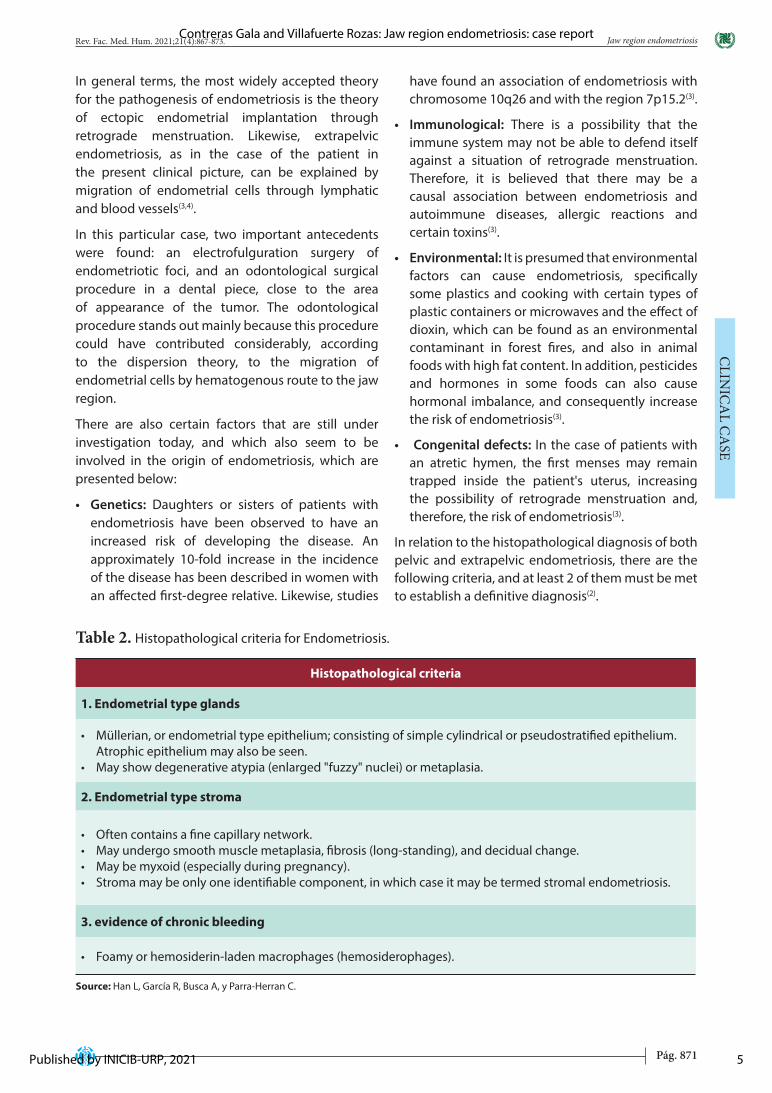
Table 2. Histopathological criteria for Endometriosis.
have found an association of endometriosis with chromosome 10q26 and with the region 7p15.2(3).
• Immunological: There is a possibility that the immune system may not be able to defend itself against a situation of retrograde menstruation. Therefore, it is believed that there may be a causal association between endometriosis and autoimmune diseases, allergic reactions and certain toxins(3).
• Environmental: It is presumed that environmental factors can cause endometriosis, specifically some plastics and cooking with certain types of plastic containers or microwaves and the effect of dioxin, which can be found as an environmental contaminant in forest fires, and also in animal foods with high fat content. In addition, pesticides and hormones in some foods can also cause hormonal imbalance, and consequently increase the risk of endometriosis(3).
• Congenital defects: In the case of patients with an atretic hymen, the first menses may remain trapped inside the patient's uterus, increasing the possibility of retrograde menstruation and, therefore, the risk of endometriosis(3).
In relation to the histopathological diagnosis of both pelvic and extrapelvic endometriosis, there are the following criteria, and at least 2 of them must be met to establish a definitive diagnosis(2).
Histopathological criteria
1. Endometrial type glands
• Müllerian, or endometrial type epithelium; consisting of simple cylindrical or pseudostratified epithelium. Atrophic epithelium may also be seen.
• May show degenerative atypia (enlarged "fuzzy" nuclei) or metaplasia.
2. Endometrial type stroma
• Often contains a fine capillary network.• May undergo smooth muscle metaplasia, fibrosis (long-standing), and decidual change.• May be myxoid (especially during pregnancy).• Stroma may be only one identifiable component, in which case it may be termed stromal endometriosis.
3. evidence of chronic bleeding
• Foamy or hemosiderin-laden macrophages (hemosiderophages).
Source: Han L, García R, Busca A, y Parra-Herran C.
Rev. Fac. Med. Hum. 2021;21(4):867-873. Jaw region endometriosis
5
Contreras Gala and Villafuerte Rozas: Jaw region endometriosis: case report
Published by INICIB-URP, 2021

Pág. 872
CLI
NIC
AL
CA
SE
Endometriosis can be diagnosed with well-defined histopathological criteria, as presented in Table 2(2). In this case report, all these criteria were evidenced, so the final diagnosis of endometriosis was made, in this case, of jaw location.
The immunohistochemical study can be of help in doubtful cases, since the glandular structures express cytokeratin 7 (CK7), estrogen and progesterone receptors (ER/PR), the same immunomarkers that are expressed in normal endometrium(7).
Also, in the case of suspected stromal endometriosis, or in the situation where we wish to verify that the stroma we are looking at microscopically corresponds to endometrial stroma, we can use immunohistochemistry for CD10, as well as estrogen receptor (ER), both of which can be useful for marking stromal cells. However, it is important to keep in mind that CD10 is a rather nonspecific marker and that other stromal cells in the female genital tract may also be ER positive(1).
Clinically, both pelvic and extrapelvic endometriosis can be confused with any proliferative tumor, benign or malignant. The key to suspecting the diagnosis of endometriosis is to inquire about the patient's clinical history, looking for related symptoms such as dysmenorrhea, dyspareunia and infertility(5). In the patient's case, we found dysmenorrhea to be an important symptom.
Taking into account the nature of endometriotic disease and the associated chronic pain, treatment is often a challenge. Endometriosis, being multifactorial and having no defined cause, has a basically empirical treatment(8).
Regarding medical treatment, contraceptive pills can be used, since they cause a reduction in the amount of bleeding and thus allow for the reduction of pain during the menstrual period(3).
In addition, gonadotropin-releasing hormone (GnRH) agonists may also be used because they block the production of ovarian stimulating hormones, which reduces estrogen levels, and consequently causes endometrial tissue to shrink. GnRH agonists can force endometriosis into remission during the time of treatment and sometimes for months or years afterwards(3).
However, medical alternatives that include hormonal suppression have proven to be of limited effectiveness. For this reason, the ideal drug does not yet exist(8).
For this reason, medical treatment is usually associated with conservative or non-conservative surgical treatment, depending on the patient's desire to preserve or not her reproductive capacity.
Conservative surgical treatment can include electrofulguration, laser evaporation and thermal coagulation, and can be performed laparoscopically or laparotomy (open surgery)(3). Regardless of the surgical technique used, it is important that all endometriotic lesions are adequately removed and all adhesions are carefully cleaned in order to significantly reduce pelvic pain and the risk of infertility in women who wish to preserve their reproductive capacity(3).
Non-conservative surgical treatment includes the performance of a total hysterectomy, specifically for those women who already have their parity satisfied(8).
Surgical treatment is recommended only when pain cannot be managed medically and quality of life is affected. Surgical treatment is not free of complications, so a series of conditions are required to ensure that the surgery fulfills its main purpose: pain relief(8).
In relation to treatment, it is relevant to mention that the patient did not receive hormonal treatment with contraceptives or GnRh analogs at any time, since after the surgery for electrofulguration of endometriotic foci, the episodes of dysmenorrhea decreased considerably.
Likewise, after surgical resection of the tumor in the jaw region, the patient presented significant clinical improvement; therefore, it was decided not to prescribe any complementary treatment.
CONCLUSIONExtrapelvic endometriosis is infrequent in our country, and the thought of such diagnosis requires experience and visual training in the recognition of normal endometrial tissue.
This case report is intended to motivate all anatomopathologists to consider the diagnosis of endometriosis whenever we are faced with a patient of reproductive age with a tumor in any location of the body.
Likewise, it is always recommended to make an adequate clinical-pathological correlation in order to establish a properly supported diagnosis, so that a treatment directed to the patient can be offered afterwards.
Rev. Fac. Med. Hum. 2021;21(4):867-873. Contreras K et al
6
Revista de la Facultad de Medicina Humana, Vol. 21 [2021], Iss. 4, Art. 25
http://inicib.urp.edu.pe/rfmh/vol21/iss4/25

Pág. 873
CLIN
ICA
L CA
SE
Correspondence: Katherine Luisa Contreras GalaAddress: Jirón Trinidad Morán 275. Urbanización El Retablo- I Etapa, Lima-Perú.Telephone: +51 936114230Email: [email protected]
Authorship contributions: The authors were involved in the genesis of the idea and design of the project, collected and interpreted the data, analyzed the results and developed the manuscript of this case report.
Funding sources: Self-funded.
Conflict of interest: The authors declare that they have no conflict of interest.
Received: July 7, 2021
Approved: August 9, 2021
BIBLIOGRAPHIC REFERENCES
1. McCluggage, GT. Endometriosis-related pathology: a discussion of selected uncommon benign, premalignant and malignant lesions. Histopathology 2020, 76, 76–92. DOI: 10.1111/his.13970
2. Han L, García R, Busca A, Parra-Herran C. Endometriosis. PathologyOutlines.com website.https://www.pathologyoutlines.com/topic/ovarynontumorendometriosis.html. Accessed April 15th, 2021.
3. Machairiotis, N., Stylianaki, A., Dryllis, G. et al. Extrapelvic endometriosis: a rare entity or an under diagnosed condition? Diagn Pathol 2013, 8: 194. https://doi.org/10.1186/1746-1596-8-194
4. Villena Galarza, MV. Silvers Godoy JP. Silvers Espinosa, JE. Endometrioma subcutáneo en la pared abdominal. A propósito de un caso. Rev Metro Ciencia 2017; 25(1): 20-23. Disponible en: https://docs.bvsalud.org/biblioref/2019/03/986604/metro-junio-out-2017-1-18-21.pdf
5. Mignemi G, Facchini C, Raimondo D, Montanari G, Ferrini G, Seracchioli R. A case report of nasal endometriosis in a patient affected by
Behcet's disease. J Minim Invasive Gynecol 2012. Jul-Aug; 19(4):514-6. doi: 10.1016/j.jmig.2012.03.005. PMID: 22748956.
6. Andrés, MP, Arcoverde, FV, Souza, CC, Fernandes, LFC, Abrão, MS y Kho, RM. Endometriosis extrapélvica: una revisión sistemática. Revista de Ginecología Mínimamente Invasiva, 2019. doi: 10.1016 / j. jmig.2019.10.004
7. Maira González, N., Vidal Santana, F., Castro Sánchez, A., & Pérez Quintela, B. V. Endometriosis cutánea: a propósito de un caso. Progresos de Obstetricia y Ginecología 2013, 56(3), 156–158. doi: 10.1016/j.pog.2012.07.007
8. Negrón Rodríguez José. Manejo del dolor pélvico crónico en pacientes con endometriosis. Rev. peru. ginecol. obstet. [Internet]. 2016 Ene [citado 2021 Jul 18] ; 62( 1 ): 69-76. Disponible en: http://www.scielo.org.pe/scielo.php?script=sci_arttext&pid=S2304-51322016000100007&lng=es.
Rev. Fac. Med. Hum. 2021;21(4):867-873. Jaw region endometriosis
Vol.21 N°4Octubre-Diciembre 2021
�����������������������������������������
Indexed in:
https://doaj.org/ http://lilacs.bvsalud.org/es/2017/07/10/revis-tas-indizadas-en-lilacs/
http://www.scielo.org.pe/scielo.php?script=sci_serial&pid=
2308-0531&lng=es&nrm=iso
https://network.bepress.com/
7
Contreras Gala and Villafuerte Rozas: Jaw region endometriosis: case report
Published by INICIB-URP, 2021