jmbt-02-118 jmbt-10-31 - · pdf fileverdezyne, usa charles e. turick, savannah river national...
TRANSCRIPT
http://www.omicsonline.org/jmbthome.php
John Alverdy,University of Chicago,
USA
Halil Hasar,Firat University, Turkey
Sumner S,RTI International,
USA
VassilisS.Kodogiannis,University of
Westminster, UK
James V. Rogers,Battelle Memorial Institute,
USA
Hongwei wu,Georgia Institute of Technology, USA
Suryanarayana Vulimiri, National Center
for Environmental Assessment, USA
Barakat S. M. Mahmoud,
Mississippi State University, USA
Sergey Shikanov,University of Chicago,
USA
Abhijeet P. Borole,Oak Ridge National
Laboratory, USA
Dike O. Ukuku,FSIT-ERRC-ARS-
USDA, USA
StephenPicataggio,Verdezyne, USA
Charles E. Turick,Savannah River
National Laboratory, USA
Arnold L. Demain,DrewUniversity,
USA
Yan Xiao,National Institute of Standards and Technology, USA
Zonglin Lewis Liu,National Center for
Agricultural Utilization Research, USA
Liu Y,Agricultural Research
Service, USA
Qiong Cheng,DuPont Central Research, USA
Liming Ying,National Heart and
Lung Institute,London
Chongxuan Liu,Pacifi c Northwest
National Laboratory,USA
Parag A. Vaishampayan,Jet Propulsion
Laboratory, USA
Nitin A. Gawande,The Pennsylvania State
University, USA
Nicolini C,University of Genova,
Italy
Domenico Schillaci,Università degli Studi
di Palermo, Italy
Justin C. Biffi nger,Naval Research Laboratory,
USA
Microbes are the fi rst inhabitants of the earth. They are censoriously
important in sustaining life on our planet. Microbial communities are excellent models for understanding biological interactions and has much to teach us about ecology, evolution, and about our understanding of Life using various biochemical technologies.
Journal of Microbial & Biochemical Technology under Open Access gives a worldwide audience in the fi eld of Microbial & Biochemical Technology. The journal sheds new light on key microbiological principles, fundamental microbial processes .Top quality original research papers and critical reviews from around the world are accepted. It covers basic and applied research, which aims and depends on practical and scientifi c aspects of disease diagnosis and treatment, and covers all aspects of microbiology and biochemical technologies.
Journal of Microbial & Biochemical Technology- Open Access using online manuscript submission, review and tracking systems of Editorial Manager® for quality and quick review processing. Submit your manuscript athttp://www.editorialmanager.com/jmbt/
ISSN: 1948-5948
OM
ICS Publishing Group
OMICS Publishing Group5716 Corsa Ave., Suite 110, Westlake, Los Angeles, CA 91362-7354, USA, Phone: +1- 650-268-9744, Fax: +1-650-618-1414, Toll free: +1-800-216-6499
OM
ICS Publishing GroupJ Microbial Biochem Technol
ISSN:1948-5948 JMBT, an open access journalVolume 2(5): 118-123 (2010) - 118
Journal of Microbial & Biochemical Technology - Open Access
www.omicsonline.org
Research ArticleOPEN ACCESS Freely available online
doi:10.4172/1948-5948.1000035
JMBT/Vol.2 Issue 5
In vitro, Evaluation of Medicinal Activity of Egyptian Honey from Different Floral Sources as Anticancer and Antimycotic Infective AgentsMervat M.A. El-Gendy
Department of Chemistry of Natural and Microbial Products, National Research Centre, Dokki, Giza, Egypt
Keywords: Cassia; Citrus; Ziziphus; Honey; Dermatophytes; HCT-116; HTB-26 and HepG2 cell line
IntroductionColon, breast and liver cancer are the leading causes of cancer
related deaths and illnesses in developed and developing country (Jaganathan and Mandal, 2009a & b) According to the American Cancer Society in the year 2008; 49,960; 40,480 and 41,480 estimated deaths for colon, breast (females) and liver carcinoma respectively are indicating not much reduction in the incidence or mortality rates. The National Cancer Institute in Egypt (NCI) reported that, each year about 21,000 human have primary liver cancer, colon cancer is the third most dangerous type in men and it ranks fifth in females and breast cancer is the first deadly type of cancer for women in Egypt (Abou-Zeid et al., 2002). On the other hand, common dermatomycosis and dermatophytosis including cutaneous infections in cancer patient caused by dermatophytes is continuing to be a major threaten problem assuming greater significance due to advent of immunosuppressive disease and multidrug resistant (Chandra, 1996; Hay, 2003).
With dietary and environmental factors playing critical roles in the cause and progression of these cancers and fungal disease, it is necessary to explore natural alternatives that are inexpensive and can be available in bulk (Nobili et al., 2009). Honey is a naturally available food with a long history of traditional use as an active medicinal compound in a large number of cultures. It can act as inexpensive alternatives to expensive therapeutic modalities toward the treatment strategies for dermatophytosis and cancer either as separate entities or in synergism to slow down the progression of these vital diseases (Maeda et al., 2005; Jaganathan and Mandal, 2009a).
However, little information is available on the anticancer and antifungal activities of honey from floral sources common in Arabic region that might be responsible for its botanical origin. The objective of this study was therefore, to evaluate and compare the anticancer and antifungal activities of honey from different floral sources common in Arabic region as Egypt (Cassia javanica, Citrus reticulata and Ziziphus spina-Christi) to improve our knowledge about honey as
an inexpensive source of pharmaceutical agents which could protect the human from invasive fungi and deadly cancer diseases. Herein we report the evaluation in vitro study that provides a primary evidence for further in vivo studies.
Materials and MethodsHoney samples
Honey materials used in this study were purchased from apiaries in different geographical regions in Egypt, Cassia honey from Assiut (Upper Egypt); Citrus honey from Behera (Nile valley region); Ziziphus honey from Saint Cathrerine (Sinai). The study was done in the National Research Center of Egypt. The botanical origins of these types of honey were Cassia javanica, Citrus reticulata and Ziziphus spina-Christi. Sugar analogue (artificial honey) was prepared by dissolving 1.5 g of sucrose, 7.5 g of maltose, 40.5 g of fructose and 33.5 g of glucose in 17 ml of deionized water (Piljac-Žegarac et al., 2010).
Honey extraction
For in vitro evaluation, extraction processes were made by twice extraction of 50 g of each honey type with 200 ml of each organic solvent (ethyl acetate, acetone, methanol and chloroform, separately) under mechanical stirring for 12 h at room temperature (25ºC). After filtering the contents using Whatmann No 1 filter paper, the extracts
*Corresponding author: Mervat M.A. El-Gendy, Department of Chemistry of Natural and Microbial Products, National Research Centre, Dokki, Giza, Egypt, E-mail: [email protected]
Received June 21, 2010; Accepted September 06, 2010; Published September 06, 2010
Citation: El-Gendy MMA (2010) In vitro, Evaluation of Medicinal Activity of Egyptian Honey from Different Floral Sources as Anticancer and Antimycotic Infective Agents. J Microbial Biochem Technol 2: 118-123. doi:10.4172/1948-5948.1000035
Copyright: © 2010 El-Gendy MMA. This is an open-access article distributed under the terms of the Creative Commons Attribution License, which permits unrestricted use, distribution, and reproduction in any medium, provided the original author and source are credited.
AbstractThe anticancer and antimycotic activity of crude and extracted honey samples of three different Egyptian fl oral
sources (Cassia javanica, Citrus reticulata and Ziziphus spina-Christi) against colon, breast and liver tumor cell line (HCT-116, HTB-26 and HepG2) and over clinical dermatophytes (Tricophyton, Microsporum and Epidermophyton), which are involved in dermatomycosis and dermatophytosis including cutaneous infections in cancer patient were evaluated in vitro study. Cassia honey showed antifungal activity against Epidermophyton and Microsporum species with inhibition zone ranged from 15 to 28 mm and it showed moderate cytotoxic activity against colon and breast cancer with the weakest cytotoxic activity against liver cancer. Crude Citrus honey exhibited potent antimycotic activity against Tricophyton species with inhibition zone ranged from 22 to 35 mm in diameter and the highest cytotoxic activity against breast cancer with growth inhibition of 99.4 ± 0.4 %. However crude Ziziphus honey provided the largest average inhibition zone diameter against all dermatophytes species which were ranged from 29 to 43 mm as well as it display potent effi ciency against colon, liver and breast cancer with tumor growth suppression of 100 ± 0.1, 99.2 ± 0.4 and 88.14 ± 0.1 %. The Maximum extractive bioactive agents, anticancer and antimycotic substances, were detected with ethyl acetate or acetone extract, while the minimum were detected with methanol or chloroform extract.
Citation: El-Gendy MMA (2010) In vitro, Evaluation of Medicinal Activity of Egyptian Honey from Different Floral Sources as Anticancer and Antimycotic Infective Agents. J Microbial Biochem Technol 2: 118-123. doi:10.4172/1948-5948.1000035
OM
ICS Publishing GroupJ Microbial Biochem Technol
ISSN:1948-5948 JMBT, an open access journalVolume 2(5): 118-123 (2010) - 119
were completely dried in rotary evaporator at 35ºC and subsequently stored in dark at 4ºC.
Determination of total flavonoid, phenolic, and proline contents of honey
The phenolic, flavonoids and proline contents were determined according to (Meda et al., 2005). The total flavonoid, phenolic and proline contents were measured in mg/g from triplicate assays.
Clinical specimens, isolation and identification of dermato-phytes
Samples of hair, nail clipping, burn, ulcers and wound were collected from 150 clinically suspected cases of dermaotmycosis infection and transferred to the lab aseptically in ice boxes. The samples were processed at the Chemistry of Natural and Microbial products Department of the National Research Center, Egypt. Each sample was kept on a slide with equal mixed proportion of 10% Potassium hydroxide (KOH) and 40% Dimethyl Sulfoxide (DMSO)and then examined for the presence of filamentous, septate, branched hyphae with or without arthrospores by direct microscopic examination (Rebell and Taplin, 1970).
For isolation of dermatophytes; sabouraud dextrose agar (SDA), dermatophytes test medium (DTM), enriched dermatophytes medium (EDM) (sigma) supplemented with color indicator were used as selective isolation media, then inoculated into these media in duplicate; one incubated at 30°C and other at 37°C for 3 weeks (Rebell and Taplin, 1970; Yavuzdemir, 1992). Efficiency of the three media for the isolation of dermatophytes was compared. The clinical fungal isolates were identified by standard morphological and physiological studies (Philpot, 1967; Rippon, 1982; Sudman and Schmitt, 1965).
Determination of antifungal activity against dermatophytes
The antimycotic activity of honey samples was assayed using the disc diffusion technique as previously described by (Georgii and Korting, 1991) and the resultant zone of inhibition diameter was measured in mm. Different standard antibiotics were used as reference antimycotic drugs for comparison, clotrimazole (CTZ), itraconazole (ITZ), ravuconazole (RVZ), terbinafine (TF), and voriconazole (VCZ). All tests were performed in triplicate.
Determination of minimum inhibitory concentration (MIC) and minimum fungicidal concentration (MFC) values of honey samples and reference drugs
The MIC values of honey samples and reference antimycotic drugs were determined by broth tube dilution procedure using two-fold dilution in DTB and EDB broth media at 37°C for 96 h, MIC was determined as the lowest concentration of that showed no visible growth (Cappuccino and Sherman 1999). MFC values were determined by sub-culturing 50 ml from tubes not visibly turbid and spot inoculating onto DTA and EDA plates. MFC values were determined as the lowest concentration that prevented growth on subculture (Lavermicocca et al., 2003). The MIC and MFC were expressed in g/ml.
In vitro antitumor evaluation: The crude and extracted honey samples were evaluated for their cytotoxic activity in vitro using three human tumor cell lines representing different cancer types. Human tumor cell lines were HCT-116 (colon tumor cell line), HTB-26 (breast tumor cell line) and HepG2 (liver carcinoma cell line). Colon cancer cell line HCT-116 was grown in McCoy’s 5a medium supplemented with 10% fetal bovine serum (FBS; HyClone, Logan UT) and 100 g / ml
of penicillin and streptomycin (Sigma). Breast cancer cell line (HTB-26) was cultured at 37°C in DMEM (Dulbecco’s Modified Eagles Medium) in the presence of 10% fetal bovine serum, supplemented with 1 mM L-glutamine, sodium pyruvate, 1 mM sodium bicarbonate and 50 μg/mL gentamycin. Hepatoma cell line (HepG2) was grown in RPMI-1640 medium in the presence of 10% fetal calf serum supplemented with 2 mM L-glutamine, 1 mM sodium bicarbonate and 100g/ml of penicillin and streptomycin (Sigma).
For a typical screening experiment, cells were plated in 96-multiwell plate (104 cells / well) for 24 h before treatment with the tested samples to allow attachment of cell to the wall of the plate. After cell inoculation, the plates were incubated at 37°C, 5% CO2, 95% air for 24 h prior to addition of tested honey samples.
Cytotoxicity and cell proliferation assay
Cytotoxic activity was assessed using MTT [3-(4,5-dimethylthiazol-2-yl)-2,5-diphenyltetrazolium bromide] technique as described by (Weichert et al., 1991) brief, 100 l of cell inoculums of each cell line was seeded in their corresponding media per well in 96-well plates. After 72 h, media were removed and replaced by fresh media containing various concentrations of the test honey sample in the range of 50 -500 g / ml for each crude honey and 10-50 g/ml for each honey extract in triplicates. Cell viability was determined after 24 and 48 h. After incubation with the tested honey samples, MTT was added to culture wells in a final concentration of 0.5 mg/ml, incubated for 4 h at 37°C and the colored formazan product was extracted with 200 l DMSO and measured at 570 nm using an ELISA reader. Optical density was a direct measure of cell survival. Sensitivity of the cells to each treatment was determined in triplicate using (MTT) cell viability assay.
Data analysis
One-way ANOVA (performed in Sigma Stat 3.5) was used to determine whether the differences between treatments of honeys from different botanical origin are significant. Differences at p < 0.05 were considered to be significant.
Results and DiscussionIsolation of clinical dermatophytes strain
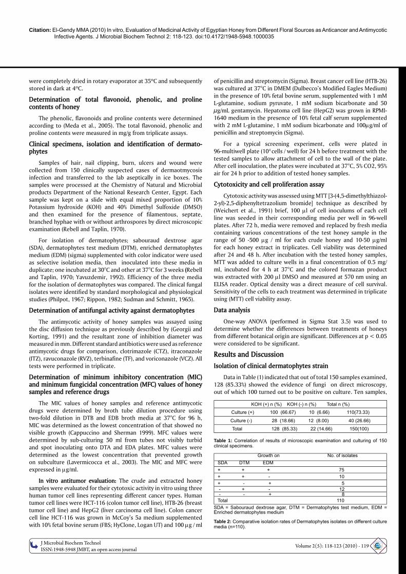
Data in Table (1) indicated that out of total 150 samples examined, 128 (85.33%) showed the evidence of fungi on direct microscopy, out of which 100 turned out to be positive on culture. Ten samples,
KOH (+) n (%) KOH (-) n (%) Total n (%)
Culture (+) 100 (66.67) 10 (6.66) 110(73.33)
Culture (-) 28 (18.66) 12 (8.00) 40 (26.66)
Total 128 (85.33) 22 (14.66) 150(100)
Table 1: Correlation of results of microscopic examination and culturing of 150 clinical specimens.
Growth on No. of isolatesSDA DTM EDM+ + + 75+ + - 10+ - + 5 - + - 12 - - + 8Total 110
SDA = Sabouraud dextrose agar, DTM = Dermatophytes test medium, EDM = Enriched dermatophytes medium
Table 2: Comparative isolation rates of Dermatophytes isolates on different culture media (n=110).
OM
ICS Publishing GroupJ Microbial Biochem Technol
ISSN:1948-5948 JMBT, an open access journalVolume 2(5): 118-123 (2010) - 120
Journal of Microbial & Biochemical Technology - Open Access
www.omicsonline.org
Research ArticleOPEN ACCESS Freely available online
doi:10.4172/1948-5948.1000035
JMBT/Vol.2 Issue 5
which were culture positive, were negative on microscopic examination, making a total of 110 (73.33%) samples culture positive. Dermatophytes were detected in 92% of samples indicating the high incidence of dermatophytosis infections. KOH in Addition to DMSO is a rapid and sensitive method for direct recognition and detection of dermatophytes in clinical specimens; they act as a clearing agent to permit rapid clearing of keratin due to increased transport of chemicals through the stratum corneum (Rebell and Taplin 1970).
Data in Table (2) indicated that while 75 samples were culture positive on all media used, 10 and 8 cultures were isolated only on DTM and EDM, respectively. Fungi were isolated from SDA in 90 (81.81%), DTM in 97 (88.18%) and EDM in 88 (80%) of 110 specimens. While the difference between SDA and EDM was statistically not significant, it was significant between SDA or EDM and DTM medium. Whereas our data are in agreement with that obtained by (Rebell and Taplin, 1970), who found that DTM is a better preparation for the isolation of dermatophytes, (Yavuzdemir, 1992) showed no significant difference in the isolation rates of dermatophytes on SDA, DTM or EDM media. These dermatophytes isolates were identified according to previous studies (Philpot, 1967; Rippon 1982; Sudman and Schmitt, 1965) as Trichophyton rubrum, T. mentagrophytes, T. longfeuseus, T. semmie, T. tonsurance, Microsporum gypseum, M. ajelloi, M. ferrogenium, M. cookie, M. racemosum and Epidermophyton floccosum.
Antifungal and anticancer activity of crude honey
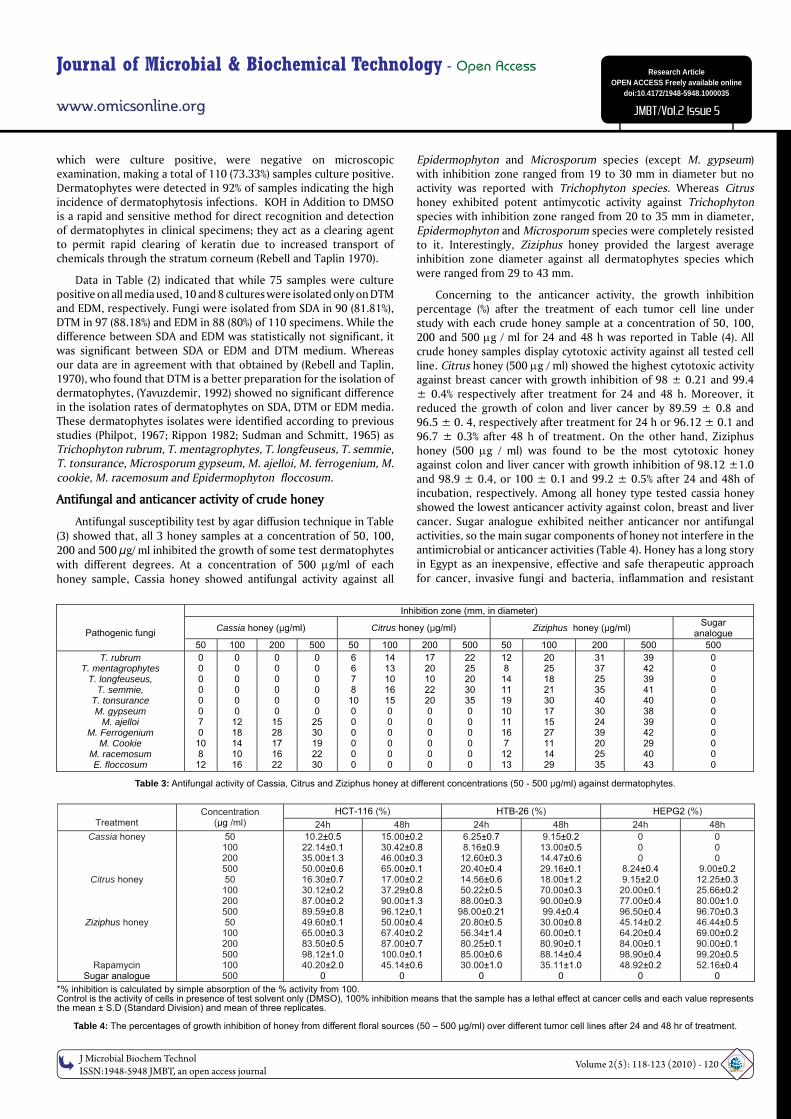
Antifungal susceptibility test by agar diffusion technique in Table (3) showed that, all 3 honey samples at a concentration of 50, 100, 200 and 500 μg/ ml inhibited the growth of some test dermatophytes with different degrees. At a concentration of 500 g/ml of each honey sample, Cassia honey showed antifungal activity against all
Epidermophyton and Microsporum species (except M. gypseum) with inhibition zone ranged from 19 to 30 mm in diameter but no activity was reported with Trichophyton species. Whereas Citrus honey exhibited potent antimycotic activity against Trichophyton species with inhibition zone ranged from 20 to 35 mm in diameter, Epidermophyton and Microsporum species were completely resisted to it. Interestingly, Ziziphus honey provided the largest average inhibition zone diameter against all dermatophytes species which were ranged from 29 to 43 mm.
Concerning to the anticancer activity, the growth inhibition percentage (%) after the treatment of each tumor cell line under study with each crude honey sample at a concentration of 50, 100, 200 and 500 g / ml for 24 and 48 h was reported in Table (4). All crude honey samples display cytotoxic activity against all tested cell line. Citrus honey (500 g / ml) showed the highest cytotoxic activity against breast cancer with growth inhibition of 98 ± 0.21 and 99.4 ± 0.4% respectively after treatment for 24 and 48 h. Moreover, it reduced the growth of colon and liver cancer by 89.59 ± 0.8 and 96.5 ± 0. 4, respectively after treatment for 24 h or 96.12 ± 0.1 and 96.7 ± 0.3% after 48 h of treatment. On the other hand, Ziziphus honey (500 g / ml) was found to be the most cytotoxic honey against colon and liver cancer with growth inhibition of 98.12 ±1.0 and 98.9 ± 0.4, or 100 ± 0.1 and 99.2 ± 0.5% after 24 and 48h of incubation, respectively. Among all honey type tested cassia honey showed the lowest anticancer activity against colon, breast and liver cancer. Sugar analogue exhibited neither anticancer nor antifungal activities, so the main sugar components of honey not interfere in the antimicrobial or anticancer activities (Table 4). Honey has a long story in Egypt as an inexpensive, effective and safe therapeutic approach for cancer, invasive fungi and bacteria, inflammation and resistant
*% inhibition is calculated by simple absorption of the % activity from 100.Control is the activity of cells in presence of test solvent only (DMSO), 100% inhibition means that the sample has a lethal effect at cancer cells and each value represents the mean ± S.D (Standard Division) and mean of three replicates.
Table 4: The percentages of growth inhibition of honey from different fl oral sources (50 – 500 μg/ml) over different tumor cell lines after 24 and 48 hr of treatment.
HCT-116 (%) HTB-26 (%) HEPG2 (%) Treatment
Concentration (μg /ml) 24h 48h 24h 48h 24h 48h
Cassia honey
Citrus honey
Ziziphus honey
Rapamycin Sugar analogue
50 100 200 500 50 100 200 500 50 100 200 500 100 500
10.2±0.5 22.14±0.1 35.00±1.3 50.00±0.6 16.30±0.7 30.12±0.2 87.00±0.2 89.59±0.8 49.60±0.1 65.00±0.3 83.50±0.5 98.12±1.0 40.20±2.0
0
15.00±0.2 30.42±0.8 46.00±0.3 65.00±0.1 17.00±0.2 37.29±0.8 90.00±1.3 96.12±0.1 50.00±0.4 67.40±0.2 87.00±0.7 100.0±0.1 45.14±0.6
0
6.25±0.7 8.16±0.9 12.60±0.3 20.40±0.4 14.56±0.6 50.22±0.5 88.00±0.3
98.00±0.21 20.80±0.5 56.34±1.4 80.25±0.1 85.00±0.6 30.00±1.0
0
9.15±0.2 13.00±0.5 14.47±0.6 29.16±0.1 18.00±1.2 70.00±0.3 90.00±0.9 99.4±0.4 30.00±0.8 60.00±0.1 80.90±0.1 88.14±0.4 35.11±1.0
0
0 0 0
8.24±0.4 9.15±2.0 20.00±0.1 77.00±0.4 96.50±0.4 45.14±0.2 64.20±0.4 84.00±0.1 98.90±0.4 48.92±0.2
0
0 0 0
9.00±0.2 12.25±0.3 25.66±0.2 80.00±1.0 96.70±0.3 46.44±0.5 69.00±0.2 90.00±0.1 99.20±0.5 52.16±0.4
0
Table 3: Antifungal activity of Cassia, Citrus and Ziziphus honey at different concentrations (50 - 500 μg/ml) against dermatophytes.
Inhibition zone (mm, in diameter)
Cassia honey (μg/ml) Citrus honey (μg/ml) Ziziphus honey (μg/ml) Sugar analogue
Pathogenic fungi
50 100 200 500 50 100 200 500 50 100 200 500 500 T. rubrum
T. mentagrophytes T. longfeuseus,
T. semmie, T. tonsurance M. gypseum
M. ajelloi M. Ferrogenium
M. Cookie M. racemosum E. floccosum
0 0 0 0 0 0 7 0
10 8
12
0 0 0 0 0 0
12 18 14 10 16
0 0 0 0 0 0 15 28 17 16 22
0 0 0 0 0 0
25 30 19 22 30
6 6 7 8 10 0 0 0 0 0 0
14 13 10 16 15 0 0 0 0 0 0
17 20 10 22 20 0 0 0 0 0 0
22 25 20 30 35 0 0 0 0 0 0
12 8 14 11 19 10 11 16 7 12 13
20 25 18 21 30 17 15 27 11 14 29
31 37 25 35 40 30 24 39 20 25 35
39 42 39 41 40 38 39 42 29 40 43
0 0 0 0 0 0 0 0 0 0 0
Citation: El-Gendy MMA (2010) In vitro, Evaluation of Medicinal Activity of Egyptian Honey from Different Floral Sources as Anticancer and Antimycotic Infective Agents. J Microbial Biochem Technol 2: 118-123. doi:10.4172/1948-5948.1000035
OM
ICS Publishing GroupJ Microbial Biochem Technol
ISSN:1948-5948 JMBT, an open access journalVolume 2(5): 118-123 (2010) - 121
recurrent intractable Candidiosis by Candida crusei, C. lutsinea, C. albicans, C. parapsilosis and Cryptococcus neoformans with no side effects (Wahdan, 1998; Abdelal and Abdelhafez, 2010).
The inhibitory properties of honey against different diseases, such as cancer and the impressive infections in vitro and in vivo could be attributed to antibacterial and anticancer substances in its plant derivates (Jaganathan and Mandal, 2009 a & b; Dimitrova et al., 2007) The choice of different floral honey samples used in this study was based on the assumption that varying total phenol and flavones content as well as antifungal / anticancer capacity are expected for honeys produced from varying floral sources on different geographic locations in Egypt. Botanical origin of honey in this study; cassia, citrus and ziziphus, have variety of uses in alternative medicine, extract of Citrus reticulate has been reported as antibacterial, antioxidants and anticancer activity against squamous cell carcinoma (Hamed and Hetta, 2005). Many anticancer compounds obtained from Ziziphus sp. such as betulinic acid are selective inhibitor drugs for the growth of human melanoma cell lines by causing apoptosis (Chai and Kinghorn, 2010) Moreover, in the screening of some Egyptian medicinal plants Cassia sp. has been reported to inhibit the toxigenic and food borne moulds (Rasooli et al., 2008) and it exhibited moderate anti-cancer and anti-oxidant activity (Nassr-Allah et al., 2009).
Extraction of bioactive agents in honey
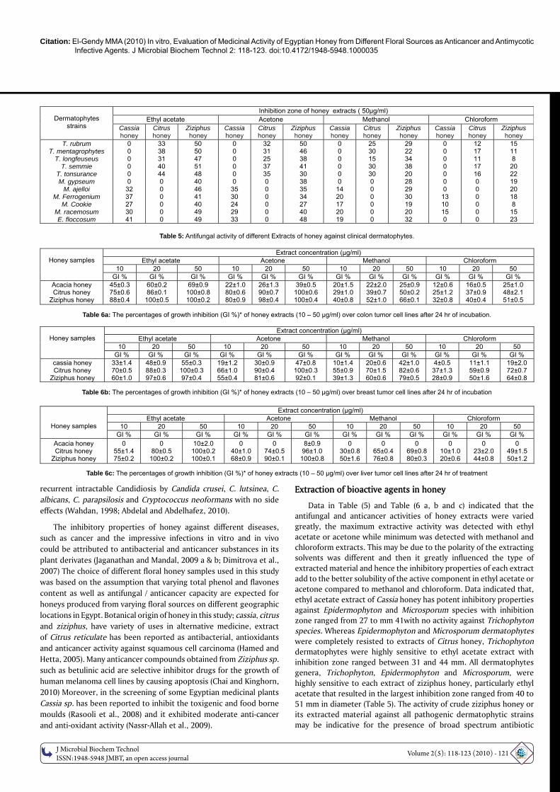
Data in Table (5) and Table (6 a, b and c) indicated that the antifungal and anticancer activities of honey extracts were varied greatly, the maximum extractive activity was detected with ethyl acetate or acetone while minimum was detected with methanol and chloroform extracts. This may be due to the polarity of the extracting solvents was different and then it greatly influenced the type of extracted material and hence the inhibitory properties of each extract add to the better solubility of the active component in ethyl acetate or acetone compared to methanol and chloroform. Data indicated that, ethyl acetate extract of Cassia honey has potent inhibitory properties against Epidermophyton and Microsporum species with inhibition zone ranged from 27 to mm 41with no activity against Trichophyton species. Whereas Epidermophyton and Microsporum dermatophytes were completely resisted to extracts of Citrus honey, Trichophyton dermatophytes were highly sensitive to ethyl acetate extract with inhibition zone ranged between 31 and 44 mm. All dermatophytes genera, Trichophyton, Epidermophyton and Microsporum, were highly sensitive to each extract of ziziphus honey, particularly ethyl acetate that resulted in the largest inhibition zone ranged from 40 to 51 mm in diameter (Table 5). The activity of crude ziziphus honey or its extracted material against all pathogenic dermatophytic strains may be indicative for the presence of broad spectrum antibiotic
Inhibition zone of honey extracts ( 50μg/ml) Ethyl acetate Acetone Methanol Chloroform
Dermatophytes
strains Cassia honey
Citrus honey
Ziziphus honey
Cassia honey
Citrus honey
Ziziphus honey
Cassia honey
Citrus honey
Ziziphus honey
Cassia honey
Citrus honey
Ziziphus honey
T. rubrum T. mentagrophytes
T. longfeuseus T. semmie
T. tonsurance M. gypseum
M. ajelloi M. Ferrogenium
M. Cookie M. racemosum E. floccosum
0 0 0 0 0 0 32 37 27 30 41
33 38 31 40 44 0 0 0 0 0 0
50 50 47 51 48 40 46 41 40 49 49
0 0 0 0 0 0 35 30 24 29 33
32 31 25 37 35 0 0 0 0 0 0
50 46 38 41 30 38 35 34 27 40 48
0 0 0 0 0 0 14 20 17 20 19
25 30 15 30 30 0 0 0 0 0 0
29 22 34 38 20 28 29 30 19 20 32
0 0 0 0 0 0 0 13 10 15 0
12 17 11 17 16 0 0 0 0 0 0
15 11 8
20 22 19 20 18 8
15 23
Table 5: Antifungal activity of different Extracts of honey against clinical dermatophytes.
Extract concentration (μg/ml) Ethyl acetate Acetone Methanol Chloroform
10 20 50 10 20 50 10 20 50 10 20 50
Honey samples
GI % GI % GI % GI % GI % GI % GI % GI % GI % GI % GI % GI % Acacia honey Citrus honey
Ziziphus honey
45±0.3 75±0.6 88±0.4
60±0.2 86±0.1
100±0.5
69±0.9 100±0.8 100±0.2
22±1.0 80±0.6 80±0.9
26±1.3 90±0.7 98±0.4
39±0.5 100±0.6 100±0.4
20±1.5 29±1.0 40±0.8
22±2.0 39±0.7 52±1.0
25±0.9 50±0.2 66±0.1
12±0.6 25±1.2 32±0.8
16±0.5 37±0.9 40±0.4
25±1.0 48±2.1 51±0.5
Table 6a: The percentages of growth inhibition (GI %)* of honey extracts (10 – 50 μg/ml) over colon tumor cell lines after 24 hr of incubation.
Extract concentration (μg/ml) Ethyl acetate Acetone Methanol Chloroform
10 20 50 10 20 50 10 20 50 10 20 50
Honey samples
GI % GI % GI % GI % GI % GI % GI % GI % GI % GI % GI % GI % cassia honey Citrus honey
Ziziphus honey
33±1.4 70±0.5 60±1.0
48±0.9 88±0.3 97±0.6
55±0.3 100±0.3 97±0.4
19±1.2 66±1.0 55±0.4
30±0.9 90±0.4 81±0.6
47±0.8 100±0.3 92±0.1
10±1.4 55±0.9 39±1.3
20±0.6 70±1.5 60±0.6
42±1.0 82±0.6 79±0.5
4±0.5 37±1.3 28±0.9
11±1.1 59±0.9 50±1.6
19±2.0 72±0.7 64±0.8
Table 6b: The percentages of growth inhibition (GI %)* of honey extracts (10 – 50 μg/ml) over breast tumor cell lines after 24 hr of incubation
Extract concentration (μg/ml) Ethyl acetate Acetone Methanol Chloroform
10 20 50 10 20 50 10 20 50 10 20 50
Honey samples GI % GI % GI % GI % GI % GI % GI % GI % GI % GI % GI % GI %
Acacia honey Citrus honey
Ziziphus honey
0 55±1.4 75±0.2
0 80±0.5
100±0.2
10±2.0 100±0.2 100±0.1
0 40±1.0 68±0.9
0 74±0.5 90±0.1
8±0.9 96±1.0
100±0.8
0 30±0.8 50±1.6
0 65±0.4 76±0.8
0 69±0.8 80±0.3
0 10±1.0 20±0.6
0 23±2.0 44±0.8
0 49±1.5 50±1.2
Table 6c: The percentages of growth inhibition (GI %)* of honey extracts (10 – 50 μg/ml) over liver tumor cell lines after 24 hr of treatment
OM
ICS Publishing GroupJ Microbial Biochem Technol
ISSN:1948-5948 JMBT, an open access journalVolume 2(5): 118-123 (2010) - 122
Journal of Microbial & Biochemical Technology - Open Access
www.omicsonline.org
Research ArticleOPEN ACCESS Freely available online
doi:10.4172/1948-5948.1000035
JMBT/Vol.2 Issue 5
compounds or simply general metabolic inhibitors in ziziphus honey towards pathogenic fungi as well as the polarity of these antimycotic substances are in line with the polarity of ethyl acetate.
On the other hand, ethyl acetate extract of Cassia honey showed moderate cytotoxic activity against colon and breast cancer and weak activity with liver carcinoma (growth inhibition was 69 ± 0.9, 55 ±0.3 and 10 ± 2.0%, respectively). Acetone and ethyl acetate extracts of Citrus honey were found to be the most cytotoxic extracts over breast cancer cell line; the growth inhibition of HTB-26 cell line treated with 50 g / ml of both extracts of was 100 ± 0.3%. Whereas the growth of colon cancer was inhibited by 100% after treatment with 20 g / ml of ethyl acetate or 50 g/ml of acetone extract of Citrus honey, the growth inhibition of HepG2 was 100 ± 0.2 and 96 ± 1.0% respectively after treatment with 50 g / ml of ethyl acetate and acetone extracts. Overall, ethyl acetate extract of Ziziphus honey exhibited the potent cytotoxic activity against colon and liver cancer followed by breast cancer at a concentration of 20 g / ml. As a result the growth of HCT-116, HTB-26 and HepG2 was inhibited by100 ± 0.5, 97 ± 0.3 and 100 ± 0.2% respectively. When compared these data with the cytotoxic activity of antitumor drug, rapamycin which achieved 40.2 ± 2.0, 30.0 ± 1.0, 48.92 ± 0.2 or 45.14 ± 0.6, 35.11 ± 1.0, 52.16± 0.4% growth inhibition respectively of HCT-116, HTB-26 and HepG2 after treatment for 24 and 48 hr, honey of these floral sources (Cassia, Citrus and Ziziphus species) seem to be the future drug of choice for the treatment of these deadly diseases.
With the evolution of extraction procedure for various phytochemicals, which had been attributed with anticancerous and antifungal property of honey, researchers concentrated on the polyphenolic and flavonoids compounds extracted from the honey rather than crude honey itself. The highest antifungal and anticancer activities of the ethyl acetate and acetone extracts of Ziziphus honey followed by Citrus honey can be rationalized in terms of the polarity of the compound being extracted by each solvent, in addition to their intrinsic bioactivity, by their ability to dissolve in this solvent or diffuse in the media used in the assay (Parekh and Chanda, 2007). If such active components are present in such honey extract, it could be management ailments caused by these diseases and give impressive results.
Determination of phytochemical components in different extracts of honey
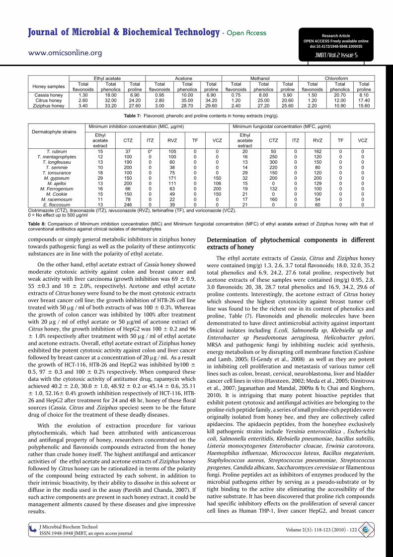
The ethyl acetate extracts of Cassia, Citrus and Ziziphus honey were contained (mg/g) 1.3, 2.6, 3.7 total flavonoids; 18.0, 32.0, 35.2 total phenolics and 6.9, 24.2, 27.6 total proline, respectively but acetone extracts of these samples were contained (mg/g) 0.95, 2.8, 3.0 flavonoids; 20, 38, 28.7 total phenolics and 16.9, 34.2, 29.6 of proline contents. Interestingly, the acetone extract of Citrus honey which showed the highest cytotoxicity against breast tumor cell line was found to be the richest one in its content of phenolics and proline, Table (7). Flavonoids and phenolic molecules have been demonstrated to have direct antimicrobial activity against important clinical isolates including E.coli, Salmonella sp, Klebsiella sp and Enterobacter sp Pseudomonas aeruginosa, Helicobacter pylori, MRSA and pathogenic fungi by inhibiting nucleic acid synthesis, energy metabolism or by disrupting cell membrane function (Cushine and Lamb, 2005; El-Gendy et al., 2008) as well as they are potent in inhibiting cell proliferation and metastasis of various tumor cell lines such as colon, breast, cervical, neuroblastoma, liver and bladder cancer cell lines in vitro (Havsteen, 2002; Meda et al., 2005; Dimitrova et al., 2007; Jaganathan and Mandal, 2009a & b; Chai and Kinghorn, 2010). It is intriguing that many potent bioactive peptides that exhibit potent cytotoxic and antifungal activities are belonging to the proline-rich peptide family, a series of small proline-rich peptides were originally isolated from honey bee, and they are collectively called apidaecins. The apidaecin peptides, from the honeybee exclusively kill pathogenic strains include Yersinia enterocolitica , Escherichia coli, Salmonella enteritidis, Klebsiella pneumoniae, bacillus subtilis, Listeria monocytogenes Enterobacter cloacae, Erwinia carotovora, Haemophilus influenzae, Micrococcus luteus, Bacillus megaterium, Staphylococcus aureus, Streptococcus pneumoniae, Streptococcus pyogenes, Candida albicans, Saccharomyces cerevisiae or filamentous fungi. Proline peptides act as inhibitors of enzymes produced by the microbial pathogens either by serving as a pseudo-substrate or by tight binding to the active site eliminating the accessibility of the native substrate. It has been discovered that proline rich compounds had specific inhibitory effects on the proliferation of several cancer cell lines as Human THP-1, liver cancer HepG2, and breast cancer
Ethyl acetate Acetone Methanol Chloroform Honey samples Total
flavonoids Total
phenolics Total
proline Total
flavonoids Total
phenolics Total
proline Total
flavonoids Total
phenolics Total
proline Total
flavonoids Total
phenolics Total
proline Cassia honey Citrus honey
Ziziphus honey
1.30 2.60 3.40
18.00 32.00 33.20
6.90 24.20 27.60
0.95 2.80 3.00
10.00 35.00 28.70
6.90 34.20 29.60
0.75 1.20 2.40
8.00 25.00 27.20
5.90 20.60 25.60
1.50 1.20 2.20
20.70 12.00 10.90
8.10 17.40 15.60
Table 7: Flavonoid, phenolic and proline contents in honey extracts (mg/g).
Minimum inhibition concentration (MIC, μg/ml) Minimum fungicidal concentration (MFC, μg/ml) Dermatophyte strains
Ethyl acetate extract
CTZ
ITZ
RVZ
TF
VCZ
Ethyl acetate extract
CTZ
ITZ
RVZ
TF
VCZ
T. rubrum T. mentagrophytes
T. longfeuseu T. semmie
T. tonsurance M. gypseum
M. ajelloi M. Ferrogenium
M. Cookie M. racemosum E. floccosum
15 12 13 10 18 29 13 16 15 11 13
37 100 190 200 100 150 200 66
150 78
246
0* 0 0 0 0 0 0 0 0 0 0
105 100 60 38 75
171 111 63 49 22 39
0 0 0 0 0 0 0 0 0 0 0
0 0 0 0 0
150 106 200 150 0 0
20 16 13 14 29 32 15 19 21 17 21
50 250 300 220 150 200 0
132 0
160 0
0 0 0 0 0 0 0 0 0 0 0
162 120 150 80
120 200 129 100 100 54 60
0 0 0 0 0 0 0 0 0 0 0
0 0 0 0 0 0 0 0 0 0 0
Clotrimazole (CTZ), itraconazole (ITZ), ravuconazole (RVZ), terbinafi ne (TF), and voriconazole (VCZ). 0 = No effect up to 500 μg/ml
Table 8: Comparison of Minimum inhibition concentration (MIC) and Minimum fungicidal concentration (MFC) of ethyl acetate extract of Ziziphus honey with that of conventional antibiotics against clinical isolates of dermatophytes
Citation: El-Gendy MMA (2010) In vitro, Evaluation of Medicinal Activity of Egyptian Honey from Different Floral Sources as Anticancer and Antimycotic Infective Agents. J Microbial Biochem Technol 2: 118-123. doi:10.4172/1948-5948.1000035
OM
ICS Publishing GroupJ Microbial Biochem Technol
ISSN:1948-5948 JMBT, an open access journalVolume 2(5): 118-123 (2010) - 123
MCF-7 cells. Moreover, combination of proline or a proline derivative, e.g. cis-4-hydroxy-L-proline, and an anti-cancer ligand, is effective in inhibiting survival and growth of cancer cells in the treatment of a cell proliferative disorder (Gallo and Huttner, 1998; Casteels et al., 1989; Casteels et al., 1990; Moore et al., 1994).
Minimum inhibition concentration (MIC) and minimum fungicidal concentration (MFC) of ethyl acetate extract of ziziphus honey compared with that of conventional antibiotics against clinical dermatophytes
Data in Table (8) showed that the ethyl acetate extract of Ziziphus honey could be has the most significant inhibitory substances of Ziziphus honey against 100% of the investigated fungal strains with MIC and MFC concentrations much lesser than that of the reference antimycotic drugs. The MIC was in the range of 10 to 29, while MFC ranged between 13 and 32 g/ml. whereas dermatophytes strains showed completely resistant to voriconazole, terbinafine and itraconazole, they showed moderate sensitivity against clotrimazole (MIC ranged from 37 to 246 and MFC was in the range of 50 - 300 g/ml) and ravuconazole (MIC ranged from 22 to 171 and MFC was in the range of 54-200 g/ml). The inhibitory activity of honey against dermatophytes is of interest because these organisms are the causative agents of infectious death and morbidity in million of people around the globe annually (Laorpaksa et al., 1992).
ConclusionThis study reveals that the extracts of honey samples studied
proved to be a good source of medicinal agents that might serve to fight against several cancer and infectious diseases. The efficiency of honey against cancer and invasive infections can confirm its place in medicine and will lead to a huge economic and health benefit worldwide. So, more work is recommended for further experiments on isolation and characterization of the bioactive compounds, which present in honey and ascertain their roles in inhibitory effects of honey.
Acknowledgments
The authors are grateful to National Cancer Institute, Cancer Biology Department for anticancer evaluation.
References
1. Abdelal EA, Abdelhafez AT (2010) Egyptian bee honey and propolis for recurrent intractable childhood candidal vulvovaginitis. 2nd International Conference on the medicinal use of honey, Kota Bharu, Malaysia, 13th - 16th January 2010. Aerosol Sci Technol 2: 31-60.
2. Abou-Zeid A, Khafagy W, Marzouk D, Alaa A, Ela-Aboul M (2002) Colorectal Cancer in Egypt. Diseases of the Colon and Rectum 45: 1255 -1260.
3. American Cancer Society (2008) Cancer Facts and Figures 2008.
4. Cappuccino JG, Sherman N (1999) Microbiology:A Laboratory Manual, (4th Edition) Addison Wesley Longman, Inc. Harlow, England.
5. Casteels P, Ampe C, Jacobs F, Vaeck M, Tempst P (1989) Apidaecins: antibacterial peptides from honeybees. EMBO J 8: 2387-2391.
6. Casteels P, Romagnolo J, Castle M, Casteels-Josson K, Erdjument-Bromage H, et al. (1990) Isolation and characterization of abaecin, a major antibacterial response peptide in the honeybee (Apis mellifera). Eur J Biochem 187: 381-386.
7. Chai H, Kinghorn D (2010) The continuing search for antitumor agents from higher plants. Phytochemistry Letters 3: 1-8.
8. Chandra JA (1996) Textbook of Medical Mycology. Mehta Publishers, New Delhi 67-79.
9. Cushine TP, Lamb AJ (2005) Antimicrobial activity of fl avonoids. Int J Antimicrob Agents 26: 343-356.
10. Dimitrova B, Gevrenova R, Anklam E (2007) Analysis of phenolic acids in honeys of different fl oral origin by solid-phase extraction and high-performance liquid chromatography. Phytochem Anal 18: 24-32.
11. El-Gendy MA, Shaaban M, EL-Bondkly AM, Shaaban KA (2008) Bioactive Benzopyrone Derivatives from New Recombinant Fusant of Marine Streptomyces. Appl Biochem Biotechnol 150: 85-96.
12. Gallo RL, Huttner KM (1998) Antimicrobial peptides: an emerging concept in cutaneous biology. J Invest Dermatol 111: 739-743.
13. Georgii A, Korting C (1991) Antifungal susceptibility testing with dermatofi t. Mycoses 34: 193-199.
14. Hamed AM, Hetta HM (2005) Effi cacy of Citrus reticulata and Mirazid in treatment of Schistosoma mansoni. Mem Inst Oswaldo Cruz Rio de Janeiro 100: 771-778.
15. Havsteen BH (2002) The biochemistry and medical signifi cance of the fl avonoids. Pharmacology & Therapeutics 96: 67-202.
16. Hay RJ (2003) Antifungal drugs used for systemic mycoses. Dermatol Clin 21: 577-587.
17. Jaganathan SK, Mandal M (2009 a) Antiproliferative Effects of Honey and of Its Polyphenols Journal of Biomedicine and Biotechnology. J Biomed Biotechnol 2009: Article ID 830616.
18. Jaganathan SK, Mandal M (2009b) Honey constituents and its apoptotic effect in colon cancer cells. Journal of Apiproduct and Apimedical Science 1: 29-36.
19. Laorpaksa A, Virunhaphol S, Sriubolmas N (1992) The Antimicrobial Action of Honey, Antifungal Activity of Honey. J Infect Dis Antimicrob Agents 9: 11-14.
20. Lavermicocca P, Valerio F, Visconti A (2003) Antifungal activity of phenyl lactic acid against molds isolated from bakery products. Appl Environ Microbiol 69: 634-40.
21. Meda A, Lamien CE, Romito M, Millogo J, Nacoulma OG (2005) Determination of the total phenolic, fl avonoid and proline contents in Burkina Fasan honey, as well as their radical scavenging activity. Food Chem 91: 571-577.
22. Moore AJ, Devine DA, Bibby MC (1994) Preliminary experimental anticancer activity of cecropins. Pept Res 7: 265-269.
23. Nassr-Allah A, Aboul-Enein A, Aboul-Enein K, Lightfoot D, Cocchetto A, et al. (2009) Anti-cancer and anti-oxidant activity of some Egyptian medicinal plants. J Med Plant Res 3: 799-808.
24. Nobili S, Lippi D, Witort E, Donnini M, Bausi L, et al. (2009) Natural compounds for cancer treatment and prevention. Pharmacol Res 59: 365-378.
25. Parekh J, Chanda S (2007) In vitro antimicrobial activity of Trapa natans L. fruit rind extracted in different solvents. Afr J Biotechnol 6: 766-770.
26. Philpot C (1967) The differentation of Trichophyton mentagrophytes from T. rubrum by a simple urease test. Sabouraudia 5: 189-193.
27. Piljac-Žegarac J, Stipčević T, Belščak A (2010) Antioxidant properties and phenolic content of different fl oral origin honeys. Journal of ApiProduct and ApiMedical Science 1: 43-50.
28. Rasooli I, Fakoor M, Yadegarinia D, Gachkar L, Allameh A, et al. (2008) Antimycotoxigenic characteristics of Rosmarinus offi cinalis and Trachyspermum copticum L. essential oils. Int J Food Microbiol 122: 135-139.
29. Rebell G, Taplin D (1970) Dermatophytes, their recognition and identifi cation. 2nd ed. Miami University Press, Miami.
30. Rippon JW (1982) Dermatophytosis and dermatomycosis, p. 154-248. In J. W. Rippon (ed.), Medical mycology. W. B. Saunders Company, Philadelphia.
31. Sudman MS, Schmitt JA (1965) Differentiation of Trichophyton rubrum and Trichophyton mentagrophytes by pigment production. Appl Environ Microbiol 13: 290.
32. Wahdan HA (1998) Causes of the antimicrobial activity of honey. Infection 26: 26-31.
33. Weichert H, Blechschmidt I, Schröder S, Ambrosius H (1991) The MTT-assay as a rapid test for cell proliferation and cell killing: application to human peripheral blood lymphocytes (PBL). Allerg Immunol 37: 139-144.
34. Yavuzdemir S (1992) Comparative evaluation of the isolation of the dermatophytes by direct laboratory evidence and MSDA with MDTM culture media. Microbiol Bulletin 26: 367-72.