jnk inhibition reduces apoptosis and neovascularization … · a critical role in the development...
TRANSCRIPT

JNK inhibition reduces apoptosis andneovascularization in a murine model ofage-related macular degenerationHongjun Dua,b,c,1, Xufang Sunb,d,1, Monica Gumae,f,g,1, Jing Luob, Hong Ouyangb, Xiaohui Zhangb,c, Jing Zengb,John Quachb, Duy H. Nguyenb, Peter X. Shawb, Michael Karine,f,2, and Kang Zhangb,c,h,2
aDepartment of Ophthalmology, Xijing Hospital, Fourth Military Medical University, Xi’an 710032, China; bInstitute for Genomic Medicine and Shiley EyeCenter, University of California at San Diego, La Jolla, CA 92093; cMolecular Medicine Research Center and Department of Ophthalmology, State KeyLaboratory of Biotherapy, West China Hospital and Sichuan University, Chengdu 610064, China; dTongji Hospital of Tongji Medical College, HuazhongUniversity of Science and Technology, Wuhan 430030, China; eLaboratory of Gene Regulation and Signal Transduction, and Departments of fPharmacologyand gPathology, School of Medicine, University of California at San Diego, La Jolla, CA 92093; and hVeterans Administration Healthcare System, San Diego,CA, 92161
Contributed by Michael Karin, December 14, 2012 (sent for review August 24, 2012)
Age-related macular degeneration (AMD) is the leading cause ofregistered blindness among the elderly and affects over 30 millionpeople worldwide. It is well established that oxidative stress,inflammation, and apoptosis play critical roles in pathogenesis ofAMD. In advanced wet AMD, although, most of the severe visionloss is due to bleeding and exudation of choroidal neovasculariza-tion (CNV), and it is well known that vascular endothelial growthfactor (VEGF) plays a pivotal role in the growth of the abnormalblood vessels. VEGF suppression therapy improves visual acuity inAMD patients. However, there are unresolved issues, includingsafety and cost. Here we show that mice lacking c-Jun N-terminalkinase 1 (JNK1) exhibit decreased inflammation, reduced CNV,lower levels of choroidal VEGF, and impaired choroidal macro-phage recruitment in a murine model of wet AMD (laser-inducedCNV). Interestingly, we also detected a substantial reduction inchoroidal apoptosis of JNK1-deficient mice. Intravitreal injection ofa pan-caspase inhibitor reduced neovascularization in the laser-induced CNVmodel, suggesting that apoptosis plays a role in laser-induced pathological angiogenesis. Intravitreal injection of a spe-cific JNK inhibitor decreased choroidal VEGF expression andreduced pathological CNV. These results suggest that JNK1 playsa key role in linking oxidative stress, inflammation, macrophagerecruitment apoptosis, and VEGF production in wet AMD andpharmacological JNK inhibition offers a unique and alternativeavenue for prevention and treatment of AMD.
Age-related macular degeneration (AMD) is the leading causeof blindness among elderly patients in developed countries
(1, 2). AMD is a progressive degenerative condition with patho-logical changes in the retinal pigment epithelium (RPE), Bruch’smembrane, and the overlying photoreceptors. Early AMD is char-acterized by the presence of drusen, debris accumulated under-neath the retina, without vision loss. Advanced AMD is associ-ated with vision loss and can be divided to geographic atrophy(GA), which is characterized by regional RPE loss and eventualdegeneration of overlying photoreceptors, or wet AMD, which ischaracterized by growth of blood vessels from the choroidthrough Bruch’s membrane toward the retina (choroidal neo-vascularization, or CNV). These vessels may bleed, resulting inacute loss of central vision. Vascular endothelial growth factor(VEGF) plays a pivotal role in the growth of the abnormal bloodvessels in CNV in wet AMD (3).Although the pathogenesis of AMD is still largely unknown,
inflammation, oxidative damage, and RPE senescence havebeen implicated in this process (4, 5). Oxidative stress is con-sidered by many to be the main initial determinant for variousage-related retinal changes. Retinal photoreceptors containinghigh content of polyunsaturated fatty acids are prone to damageby oxidative stress due to the high oxygen level of the eye and
sunlight exposure. The retinal photoreceptors are highly en-riched with polyunsaturated fatty acid containing phospholipidsand a large amount of oxidized phospholipids (oxPLs) aregenerated due to oxidative stress caused by stimuli such assunlight exposure and high oxygen content. Under oxidativestress, these phospholipids can generate a variety of breakdownproducts to form oxPLs (6), which can be recognized by a nat-ural antibody, TEPC-15 (aka T15). oxPLs on oxidized lowdensity lipoprotein (oxLDL), which can be recognized by T15,have been extensively studied as a marker for systemic in-flammation in atherosclerotic lesion development, a conditionthat shares similarities with drusen development in AMD (7, 8).OxPLs bind to the RPE and macrophages and strongly activatedownstream inflammatory cascades (9). oxPLs and their proteinadducts can also stimulate RPE cells to express chemoattractantmolecules that recruit monocytes into the subretinal space anddifferentiate into tissue resident macrophages. These oxidativelydamaged structures will be further taken up by the macro-phages, which consequently release proinflammatory mediatorsand initiate the inflammation cascade. The accumulation ofoxidatively damaged molecules also leads to retinal apoptosisand inflammation. Apoptotic cells, if not rapidly cleared, un-dergo secondary necrosis and can also stimulate the innateimmune system. Thus, oxidative stress and apoptosis are prob-ably the main initial determinants for retinal inflammatoryresponses (4, 5).The Jun kinases (JNKs) belong to the mitogen-activated
protein kinase (MAPK) family (10). The JNKs, which areencoded by three separate loci, Jnk1-3, are activated in re-sponse to growth factors, proinflammatory cytokines, microbialcomponents, and a variety of stresses, including oxidative stress(11). The JNKs regulate key cellular processes such as cellproliferation, migration, survival, and cytokine production. In-terestingly, it was shown that reactive oxygen species (ROS)cause JNK activation and cell death (12). We also recentlyshowed that JNK1 is a critical factor in hypoxia-induced retinalVEGF production and that it promotes hypoxia-inducedpathological angiogenesis (13). Thus, JNK1 might play a key
Author contributions: M.K. and K.Z. designed research; H.D., X.S., M.G., J.L., X.Z., J.Z., J.Q.,and P.X.S., performed research; H.D., M.G., H.O., P.X.S., M.K., and K.Z. contributed newreagents/analytic tools; H.D., M.G., D.H.N., P.X.S., M.K., and K.Z. analyzed data; and H.D.,X.S., M.G., P.X.S., M.K., and K.Z. wrote the paper.
The authors declare no conflict of interest.1H.D., X.S., and M.G. contributed equally to this work.2To whom correspondence may be addressed. E-mail: [email protected] or [email protected].
This article contains supporting information online at www.pnas.org/lookup/suppl/doi:10.1073/pnas.1221729110/-/DCSupplemental.
www.pnas.org/cgi/doi/10.1073/pnas.1221729110 PNAS | February 5, 2013 | vol. 110 | no. 6 | 2377–2382
PHARM
ACO
LOGY
role in the development of AMD due to its roles in stressresponses and involvement in apoptosis, inflammation, andVEGF production.
Here we show that JNK1 deficiency or JNK inhibition leads toa decrease in apoptosis, VEGFexpression, and reduction of CNV ina murine model of wet AMD. Our results indicate that JNK1 plays
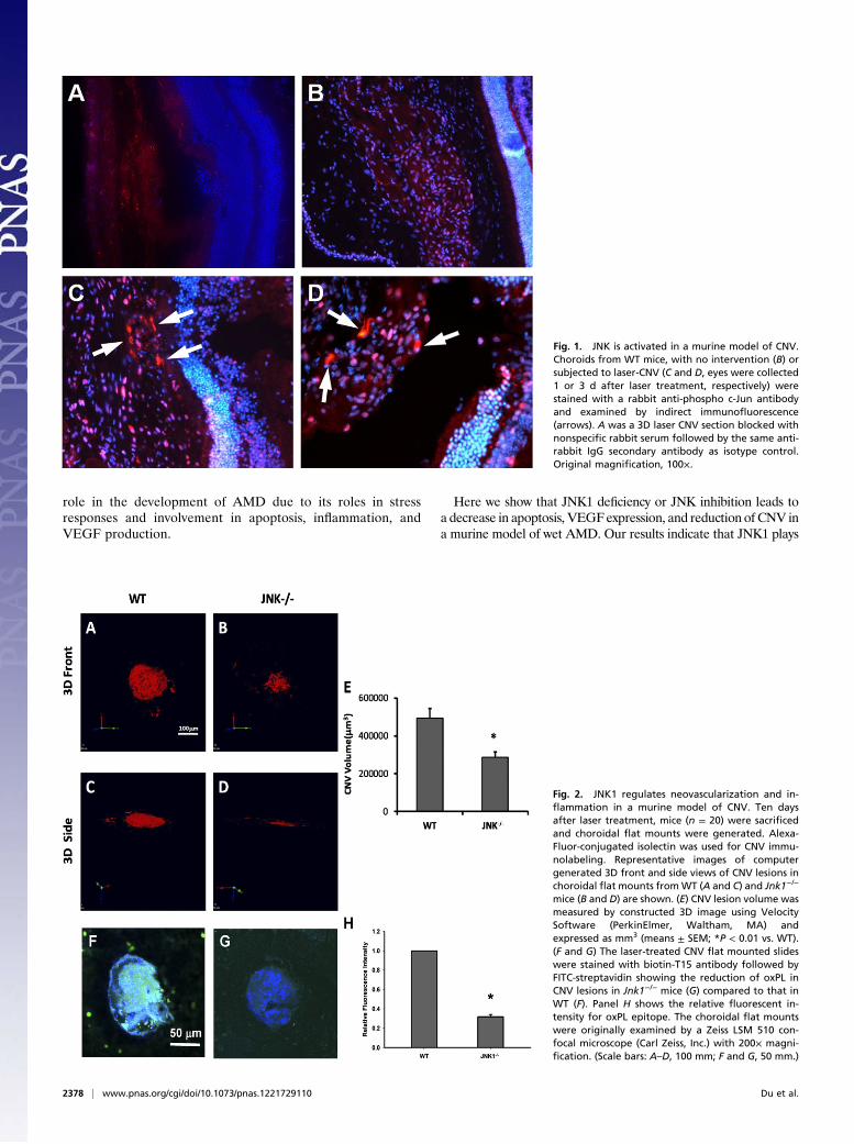
Fig. 1. JNK is activated in a murine model of CNV.Choroids from WT mice, with no intervention (B) orsubjected to laser-CNV (C and D, eyes were collected1 or 3 d after laser treatment, respectively) werestained with a rabbit anti-phospho c-Jun antibodyand examined by indirect immunofluorescence(arrows). A was a 3D laser CNV section blocked withnonspecific rabbit serum followed by the same anti-rabbit IgG secondary antibody as isotype control.Original magnification, 100×.
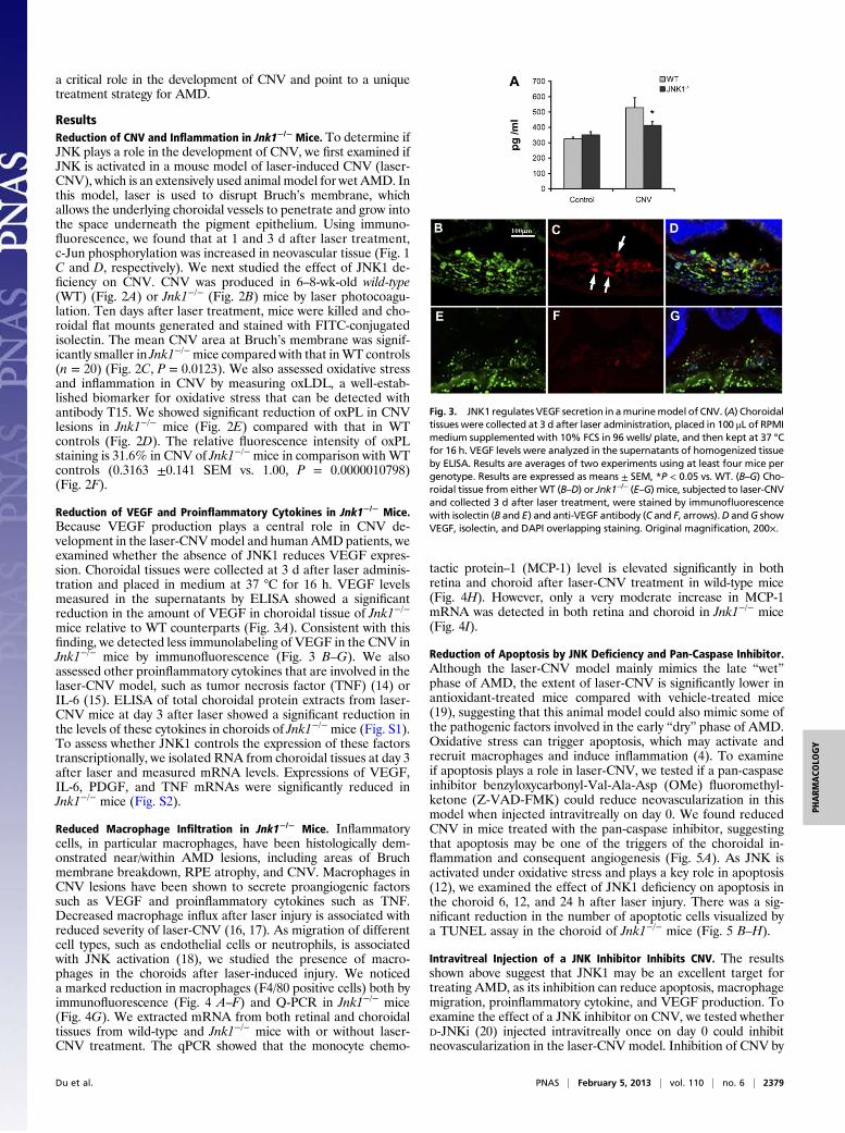
Fig. 2. JNK1 regulates neovascularization and in-flammation in a murine model of CNV. Ten daysafter laser treatment, mice (n = 20) were sacrificedand choroidal flat mounts were generated. Alexa-Fluor-conjugated isolectin was used for CNV immu-nolabeling. Representative images of computergenerated 3D front and side views of CNV lesions inchoroidal flat mounts fromWT (A and C) and Jnk1−/−
mice (B and D) are shown. (E) CNV lesion volume wasmeasured by constructed 3D image using VelocitySoftware (PerkinElmer, Waltham, MA) andexpressed as mm3 (means ± SEM; *P < 0.01 vs. WT).(F and G) The laser-treated CNV flat mounted slideswere stained with biotin-T15 antibody followed byFITC-streptavidin showing the reduction of oxPL inCNV lesions in Jnk1−/− mice (G) compared to that inWT (F). Panel H shows the relative fluorescent in-tensity for oxPL epitope. The choroidal flat mountswere originally examined by a Zeiss LSM 510 con-focal microscope (Carl Zeiss, Inc.) with 200× magni-fication. (Scale bars: A–D, 100 mm; F and G, 50 mm.)
2378 | www.pnas.org/cgi/doi/10.1073/pnas.1221729110 Du et al.
a critical role in the development of CNV and point to a uniquetreatment strategy for AMD.
ResultsReduction of CNV and Inflammation in Jnk1−/− Mice. To determine ifJNK plays a role in the development of CNV, we first examined ifJNK is activated in a mouse model of laser-induced CNV (laser-CNV), which is an extensively used animal model for wet AMD. Inthis model, laser is used to disrupt Bruch’s membrane, whichallows the underlying choroidal vessels to penetrate and grow intothe space underneath the pigment epithelium. Using immuno-fluorescence, we found that at 1 and 3 d after laser treatment,c-Jun phosphorylation was increased in neovascular tissue (Fig. 1C and D, respectively). We next studied the effect of JNK1 de-ficiency on CNV. CNV was produced in 6–8-wk-old wild-type(WT) (Fig. 2A) or Jnk1−/− (Fig. 2B) mice by laser photocoagu-lation. Ten days after laser treatment, mice were killed and cho-roidal flat mounts generated and stained with FITC-conjugatedisolectin. The mean CNV area at Bruch’s membrane was signif-icantly smaller in Jnk1−/−mice compared with that inWT controls(n = 20) (Fig. 2C, P = 0.0123). We also assessed oxidative stressand inflammation in CNV by measuring oxLDL, a well-estab-lished biomarker for oxidative stress that can be detected withantibody T15. We showed significant reduction of oxPL in CNVlesions in Jnk1−/− mice (Fig. 2E) compared with that in WTcontrols (Fig. 2D). The relative fluorescence intensity of oxPLstaining is 31.6% in CNV of Jnk1−/− mice in comparison with WTcontrols (0.3163 ±0.141 SEM vs. 1.00, P = 0.0000010798)(Fig. 2F).
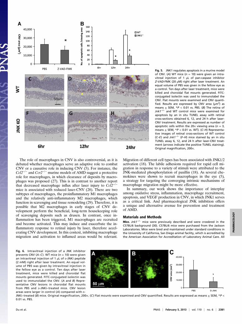
Reduction of VEGF and Proinflammatory Cytokines in Jnk1−/− Mice.Because VEGF production plays a central role in CNV de-velopment in the laser-CNVmodel and human AMDpatients, weexamined whether the absence of JNK1 reduces VEGF expres-sion. Choroidal tissues were collected at 3 d after laser adminis-tration and placed in medium at 37 °C for 16 h. VEGF levelsmeasured in the supernatants by ELISA showed a significantreduction in the amount of VEGF in choroidal tissue of Jnk1−/−
mice relative to WT counterparts (Fig. 3A). Consistent with thisfinding, we detected less immunolabeling of VEGF in the CNV inJnk1−/− mice by immunofluorescence (Fig. 3 B–G). We alsoassessed other proinflammatory cytokines that are involved in thelaser-CNV model, such as tumor necrosis factor (TNF) (14) orIL-6 (15). ELISA of total choroidal protein extracts from laser-CNV mice at day 3 after laser showed a significant reduction inthe levels of these cytokines in choroids of Jnk1−/− mice (Fig. S1).To assess whether JNK1 controls the expression of these factorstranscriptionally, we isolated RNA from choroidal tissues at day 3after laser and measured mRNA levels. Expressions of VEGF,IL-6, PDGF, and TNF mRNAs were significantly reduced inJnk1−/− mice (Fig. S2).
Reduced Macrophage Infiltration in Jnk1−/− Mice. Inflammatorycells, in particular macrophages, have been histologically dem-onstrated near/within AMD lesions, including areas of Bruchmembrane breakdown, RPE atrophy, and CNV. Macrophages inCNV lesions have been shown to secrete proangiogenic factorssuch as VEGF and proinflammatory cytokines such as TNF.Decreased macrophage influx after laser injury is associated withreduced severity of laser-CNV (16, 17). As migration of differentcell types, such as endothelial cells or neutrophils, is associatedwith JNK activation (18), we studied the presence of macro-phages in the choroids after laser-induced injury. We noticeda marked reduction in macrophages (F4/80 positive cells) both byimmunofluorescence (Fig. 4 A–F) and Q-PCR in Jnk1−/− mice(Fig. 4G). We extracted mRNA from both retinal and choroidaltissues from wild-type and Jnk1−/− mice with or without laser-CNV treatment. The qPCR showed that the monocyte chemo-
tactic protein–1 (MCP-1) level is elevated significantly in bothretina and choroid after laser-CNV treatment in wild-type mice(Fig. 4H). However, only a very moderate increase in MCP-1mRNA was detected in both retina and choroid in Jnk1−/− mice(Fig. 4I).
Reduction of Apoptosis by JNK Deficiency and Pan-Caspase Inhibitor.Although the laser-CNV model mainly mimics the late “wet”phase of AMD, the extent of laser-CNV is significantly lower inantioxidant-treated mice compared with vehicle-treated mice(19), suggesting that this animal model could also mimic some ofthe pathogenic factors involved in the early “dry” phase of AMD.Oxidative stress can trigger apoptosis, which may activate andrecruit macrophages and induce inflammation (4). To examineif apoptosis plays a role in laser-CNV, we tested if a pan-caspaseinhibitor benzyloxycarbonyl-Val-Ala-Asp (OMe) fluoromethyl-ketone (Z-VAD-FMK) could reduce neovascularization in thismodel when injected intravitreally on day 0. We found reducedCNV in mice treated with the pan-caspase inhibitor, suggestingthat apoptosis may be one of the triggers of the choroidal in-flammation and consequent angiogenesis (Fig. 5A). As JNK isactivated under oxidative stress and plays a key role in apoptosis(12), we examined the effect of JNK1 deficiency on apoptosis inthe choroid 6, 12, and 24 h after laser injury. There was a sig-nificant reduction in the number of apoptotic cells visualized bya TUNEL assay in the choroid of Jnk1−/− mice (Fig. 5 B–H).
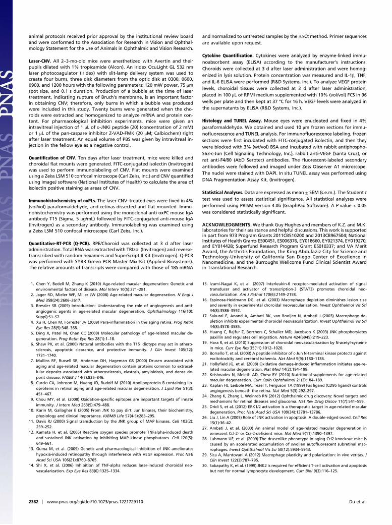
Intravitreal Injection of a JNK Inhibitor Inhibits CNV. The resultsshown above suggest that JNK1 may be an excellent target fortreating AMD, as its inhibition can reduce apoptosis, macrophagemigration, proinflammatory cytokine, and VEGF production. Toexamine the effect of a JNK inhibitor on CNV, we tested whetherD-JNKi (20) injected intravitreally once on day 0 could inhibitneovascularization in the laser-CNV model. Inhibition of CNV by
Fig. 3. JNK1 regulates VEGF secretion in amurinemodel of CNV. (A) Choroidaltissues were collected at 3 d after laser administration, placed in 100 μL of RPMImedium supplemented with 10% FCS in 96 wells/ plate, and then kept at 37 °Cfor 16 h. VEGF levels were analyzed in the supernatants of homogenized tissueby ELISA. Results are averages of two experiments using at least four mice pergenotype. Results are expressed as means ± SEM, *P < 0.05 vs. WT. (B–G) Cho-roidal tissue from eitherWT (B–D) or Jnk1−/− (E–G) mice, subjected to laser-CNVand collected 3 d after laser treatment, were stained by immunofluorescencewith isolectin (B and E) and anti-VEGF antibody (C and F, arrows).D andG showVEGF, isolectin, and DAPI overlapping staining. Original magnification, 200×.
Du et al. PNAS | February 5, 2013 | vol. 110 | no. 6 | 2379
PHARM
ACO
LOGY

D-JNKi (Fig. 6 A and B) was clearly evident 10 d post–lasertreatment, where there was reduction of neovascular areas (Fig.6C, P < 0.01). D-JNKi treatment also reduced the amount ofVEGF and F4/80 positive cells in the neovascular tissue (Figs. S3and S4).
DiscussionIn this report, we show that JNK1 deficiency or total JNK in-hibition decreases VEGF expression and reduces CNV in a mu-rine model of wet AMD. The decrease of VEGF in JNK1-deficient mice was expected, as JNK1 regulates VEGF expressionunder different stimuli such as hypoxia and inflammation (13).However, our data further suggest that JNK1may also be involvedin the initial stages of AMD. Unlike diabetic retinopathy in whichthe initial triggers for retinal pathogenesis are clear, direct incitingfactors of AMD are still largely unknown. Aging, family history,cigarette smoking, systemic inflammation, and genetic predis-position are risk factors forAMD (5).With age, increased oxidativestress exists in retinal/RPE/choroidal tissues, which may result ina number of pathophysiological changes including the formation ofoxidized lipids, protein, and DNA; advanced glycation end-prod-ucts (AGEs); and degeneration of Bruch’s membrane, leading toRPE and photoreceptor death. Several mouse models illustrate thecapacity of oxidative damage to induce AMD-like pathology (21).Furthermore, one of the known interventions that can reduceAMD progression is consumption of antioxidants (22).Under normal physiological conditions, cells that undergo ap-
optosis due to oxidative stress are rapidly cleared by tissue residentphagocytic cells and the proinflammatory response is kept ata minimum. If, however, the apoptotic cells are not rapidly cleared,they undergo secondary necrosis and can generate proin-flammatory debris. Late stage apoptotic cells (or secondary ne-crotic cells) also release damage-associated molecular patterns(DAMP), which can stimulate the innate immune system. In-flammation, such as microglial/macrophage activation, is then
induced to trigger tissue repair and remodelling. In AMD, thebalance between the stress-induced damage and inflammation-related tissue repair and remodelling is disturbed due to eitherincreased damage or decreased/altered repair/remodelling abilityof the immune system (4, 5). Thus, a strategy for targeting apo-ptosis, for instance, by inhibiting JNK, in this context, mightbe beneficial.Apoptosis, however, might play a dual role in the murine laser-
CNV model. In this model, which mainly mimics wet AMD,several reports have demonstrated FasL+ RPE cells in closeproximity to and surrounding Fas+ vascular endothelial cells innew vessels (23). This indicates that RPE cells attempt to controlthe spread of new Fas+ vessels as they penetrate Bruch’s mem-brane and are localized beneath the retina. There is also a sub-stantial increase in the incidence and severity of new vesselgrowth in Fas-deficient and FasL-defective mice, suggesting thatapoptosis may control the growth and development of new sub-retinal vessels that can damage vision (23). However, our datashow that inhibiting apoptosis in the first several hours after lasertreatment reduced neovascularization, suggesting that apoptosismay be one of the triggers of the choroidal inflammation andconsequent angiogenesis in this model. That, together with a re-cent report that showed a reduction of CNV in antioxidant-treated mice (19), suggests that this model may have some char-acteristics of dry AMD and treatments that inhibit apoptosis mayalso be beneficial in some dry AMD patients, in addition to in-hibition of other pathways involved in dry AMD (24, 25). In-terestingly, JNK is not simply a proapoptotic protein kinase (26).It is activated by a variety of extracellular stimuli as well as bygrowth factors, only some of which induce apoptosis. In addition,apoptosis can also be induced independent of caspase activation.Therefore, although we showed that the apoptosis was markedlyreduced after laser treatment in JNK1-deficient mice, furtherexperiments are needed to study the pro- or anti-apoptotic roleof JNK1 in neovascular growth.
Fig. 4. Reduced macrophage infiltration in Jnk1−/−
mice. (A–F) Choroidal tissue from either WT (A–C) orJnk1−/− (D–F) mice, subjected to laser-CNV and col-lected 3 d after laser treatment, were stained byimmunofluorescence with isolectin (A and D) andanti-F4/80 antibody (B and E, arrows). C and F showF4/80, isolectin, and DAPI overlapping staining.Original magnification, 200×. (G) Choroidal RNA wasextracted at day 3 after laser treatment and analyzedby qPCR in triplicates for expression of F4/80 mRNA.mRNA amounts were normalized to 18S rRNA. (Hand I) qPCR showed that the MCP-1 level is elevatedsignificantly in both retina and choroid after laser-CNV treatment in wild-type mice, however, only verymoderate increase in MCP-1 level in both retina andchoroid was seen in Jnk1−/− mice. Results areexpressed as means ± SEM, *P < 0.05 vs. WT.
2380 | www.pnas.org/cgi/doi/10.1073/pnas.1221729110 Du et al.
The role of macrophages in CNV is also controversial, as it isdebated whether macrophages serve an adaptive role to combatCNV or a causative role in inducing CNV (5). For instance, theCcl2−/− and Ccr2−/− murine models of AMD suggest a protectiverole for macrophages, in which clearance of deposits by macro-phages was proposed (27). This is in contrast to another reportthat decreased macrophage influx after laser injury to Ccl2−/−
mice is associated with reduced laser-CNV (28). There are twosubtypes of macrophages, the proinflammatory M1 macrophagesand the relatively anti-inflammatory M2 macrophages, whichfunction in scavenging and tissue remodeling (29). Therefore, it ispossible that M2 macrophages in early stages of CNV de-velopment perform the beneficial, long-term housekeeping roleof scavenging deposits such as drusen. In contrast, once in-flammation has been triggered, M1 macrophages are recruitedand become activated. This may induce and exacerbate the in-flammatory response to retinal injury by laser, therefore accel-erating CNV development. In this context, inhibiting macrophagemigration and activation to inflamed areas would be relevant.
Migration of different cell types has been associated with JNK1/2activation (18). The labile adhesions required for rapid cell mi-gration in response to a variety of stimuli were attributed to theJNK-mediated phosphorylation of paxillin (18). As several che-mokines were shown to recruit macrophages in the eye (5),a strategy for targeting the converging intrinsic mechanisms ofmacrophage migration might be more effective.In summary, our work shows the importance of interplay
among oxidative stress, inflammation, macrophage recruitment,apoptosis, and VEGF production in CNV, in which JNK1 servesas a critical link. And pharmacological JNK inhibition offersa unique and alternative avenue for prevention and treatmentof AMD.
Materials and MethodsMice. Jnk1−/− mice were previously described and were crossbred in theC57BL/6 background (30). C57BL/6 mice were purchased from the JacksonLaboratories. Mice were bred and maintained under standard conditions inthe University of California, San Diego animal facility, which is accredited bythe American Association for Accreditation of Laboratory Animal Care. All
Fig. 5. JNK1 regulates apoptosis in a murine modelof CNV. (A) WT mice (n = 10) were given an intra-vitreal injection of 1 μL of pan-caspase inhibitorZ-VAD-FMK (20 μM) right after laser treatment. Anequal volume of PBS was given in the fellow eye asa control. Ten days after laser treatment, mice werekilled and choroidal flat mounts generated. FITC-conjugated isolectin was used to immunolabel theCNV. Flat mounts were examined and CNV quanti-fied. Results are expressed by CNV area (μm2) asmeans ± SEM, *P < 0.01 vs. PBS. (B) The retina ofJnk1−/− and WT control mice were examined forapoptosis by an in situ TUNEL assay with retinalcross-sections obtained 6, 12, and 24 h after laser-CNV treatment. Results are expressed as number ofapoptotic cells within the 20× viewing area (n = 3,means ± SEM, *P < 0.01 vs. WT). (C–H) Representa-tive images of retinal cross-sections of WT control(C–E) and Jnk1−/− (F–H) mice stained by an in situTUNEL assay 6, 12, and 24 h after laser-CNV treat-ment (arrows indicate the positive TUNEL staining).Original magnification, 200×.
Fig. 6. Intravitreal injection of a JNK inhibitorprevents CNV (A–C ). WT mice (n = 10) were givenan intravitreal injection of 1 μL of D-JNKi peptide(2 mM) right after laser treatment. An equal vol-ume of PBS was given by intravitreal injection inthe fellow eye as a control. Ten days after lasertreatment, mice were killed and choroidal flatmounts generated. FITC-conjugated isolectin wasused to immunolabel the CNV. (A and B) Repre-sentative CNV lesions in choroidal flat mountsfrom PBS and D-JNKi–treated mice. CNV lesionareas were larger in control (A) compared with D-JNKi–treated (B) mice. Original magnification, 200×. (C ) Flat mounts were examined and CNV quantified. Results are expressed as means ± SEM, *P <0.01 vs. PBS.
Du et al. PNAS | February 5, 2013 | vol. 110 | no. 6 | 2381
PHARM
ACO
LOGY
animal protocols received prior approval by the institutional review boardand were conformed to the Association for Research in Vision and Ophthal-mology Statement for the Use of Animals in Ophthalmic and Vision Research.
Laser-CNV. All 2–3-mo-old mice were anesthetized with Avertin and theirpupils dilated with 1% tropicamide (Alcon). An Iridex OcuLight GL 532 nmlaser photocoagulator (Iridex) with slit-lamp delivery system was used tocreate four burns, three disk diameters from the optic disk at 0300, 0600,0900, and 1200 hours with the following parameters: 120 mW power, 75 μmspot size, and 0.1 s duration. Production of a bubble at the time of lasertreatment, indicating rupture of Bruch’s membrane, is an important factorin obtaining CNV; therefore, only burns in which a bubble was producedwere included in this study. Twenty burns were generated when the cho-roids were extracted and homogenized to analyze mRNA and protein con-tent. For pharmacological inhibition experiments, mice were given anintravitreal injection of 1 μL of D-JNKi peptide (20) (concentration of 2 mM)or 1 μL of the pan-caspase inhibitor Z-VAD-FMK (20 μM; Calbiochem) rightafter laser treatment. An equal volume of PBS was given by intravitreal in-jection in the fellow eye as a negative control.
Quantification of CNV. Ten days after laser treatment, mice were killed andchoroidal flat mounts were generated. FITC-conjugated isolectin (Invitrogen)was used to perform immunolabeling of CNV. Flat mounts were examinedusing a Zeiss LSM 510 confocal microscope (Carl Zeiss, Inc.) and CNV quantifiedusing ImageJ software (National Institutes of Health) to calculate the area ofisolectin positive staining as areas of CNV.
Immunohistochemistry of oxPLs. The laser-CNV–treated eyes were fixed in 4%(vol/vol) paraformaldehyde, and retinas dissected and flat mounted. Immu-nohistochemistry was performed using the monoclonal anti oxPC mouse IgAantibody T15 (Sigma, 5 μg/mL) followed by FITC-conjugated anti-mouse IgA(Invitrogen) as a secondary antibody. Immunolabeling was examined usinga Zeiss LSM 510 confocal microscope (Carl Zeiss, Inc.).
Quantitative-RT-PCR (Q-PCR). RPE/Choroid was collected at 3 d after laseradministration. Total RNAwas extracted with TRIzol (Invitrogen) and reverse-transcribed with random hexamers and SuperScript II Kit (Invitrogen). Q-PCRwas performed with SYBR Green PCR Master Mix Kit (Applied Biosystems).The relative amounts of transcripts were compared with those of 18S mRNA
and normalized to untreated samples by the ΔΔCt method. Primer sequencesare available upon request.
Cytokine Quantification. Cytokines were analyzed by enzyme-linked immu-noabsorbent assay (ELISA) according to the manufacturer’s instructions.Choroids were collected at 3 d after laser administration and were homog-enized in lysis solution. Protein concentration was measured and IL-1β, TNF,and IL-6 ELISA were performed (R&D Systems, Inc.). To analyze VEGF proteinlevels, choroidal tissues were collected at 3 d after laser administration,placed in 100 μL of RPMI medium supplemented with 10% (vol/vol) FCS in 96wells per plate and then kept at 37 °C for 16 h. VEGF levels were analyzed inthe supernatants by ELISA (R&D Systems, Inc.).
Histology and TUNEL Assay. Mouse eyes were enucleated and fixed in 4%paraformaldehyde. We obtained and used 10 μm frozen sections for immu-nofluorescence and TUNEL analysis. For immunofluorescence labeling, frozensections were first incubated with FITC-conjugated isolectin, and then theywere blocked with 3% (wt/vol) BSA and incubated with rabbit antiphospho-S63-c-Jun (Cell Signaling Technology, Inc.), rabbit anti-VEGF (Santa Cruz), orrat anti-F4/80 (AbD Serotec) antibodies. The fluorescent-labeled secondaryantibodies were followed and imaged under Zess Observer A1 microscope.The nuclei were stained with DAPI. In situ TUNEL assay was performed usingDNA Fragmentation Assay Kit, (Invitrogen).
Statistical Analyses. Data are expressed as mean ± SEM (s.e.m.). The Student ttest was used to assess statistical significance. All statistical analyses wereperformed using PRISM version 4.0b (GraphPad Software). A P value < 0.05was considered statistically significant.
ACKNOWLEDGMENTS.We thank Guy Hughes and members of K.Z. and M.K.laboratories for their assistance and helpful discussions. This work is supportedin part from 973 Program Grants 2011CB510200 and 2013CB967504; NationalInstitutes of Health Grants ES00451, ES006376, EY018660, EY021374, EY019270,and EY014428; Superfund Research Program Grant ES010337; and VA MeritAward, the Arthritis Foundation, the King Abdulaziz City for Science andTechnology-University of California San Diego Center of Excellence inNanomedicine, and the Burroughs Wellcome Fund Clinical Scientist Awardin Translational Research.
1. Chen Y, Bedell M, Zhang K (2010) Age-related macular degeneration: Genetic andenvironmental factors of disease. Mol Interv 10(5):271–281.
2. Jager RD, Mieler WF, Miller JW (2008) Age-related macular degeneration. N Engl JMed 358(24):2606–2617.
3. Bressler SB (2009) Introduction: Understanding the role of angiogenesis and anti-angiogenic agents in age-related macular degeneration. Ophthalmology 116(10):Suppl):S1–S7.
4. Xu H, Chen M, Forrester JV (2009) Para-inflammation in the aging retina. Prog RetinEye Res 28(5):348–368.
5. Ding X, Patel M, Chan CC (2009) Molecular pathology of age-related macular de-generation. Prog Retin Eye Res 28(1):1–18.
6. Shaw PX, et al. (2000) Natural antibodies with the T15 idiotype may act in athero-sclerosis, apoptotic clearance, and protective immunity. J Clin Invest 105(12):1731–1740.
7. Mullins RF, Russell SR, Anderson DH, Hageman GS (2000) Drusen associated withaging and age-related macular degeneration contain proteins common to extracel-lular deposits associated with atherosclerosis, elastosis, amyloidosis, and dense de-posit disease. FASEB J 14(7):835–846.
8. Curcio CA, Johnson M, Huang JD, Rudolf M (2010) Apolipoprotein B-containing lip-oproteins in retinal aging and age-related macular degeneration. J Lipid Res 51(3):451–467.
9. Chou MY, et al. (2008) Oxidation-specific epitopes are important targets of innateimmunity. J Intern Med 263(5):479–488.
10. Karin M, Gallagher E (2005) From JNK to pay dirt: Jun kinases, their biochemistry,physiology and clinical importance. IUBMB Life 57(4-5):283–295.
11. Davis RJ (2000) Signal transduction by the JNK group of MAP kinases. Cell 103(2):239–252.
12. Kamata H, et al. (2005) Reactive oxygen species promote TNFalpha-induced deathand sustained JNK activation by inhibiting MAP kinase phosphatases. Cell 120(5):649–661.
13. Guma M, et al. (2009) Genetic and pharmacological inhibition of JNK ameliorateshypoxia-induced retinopathy through interference with VEGF expression. Proc NatlAcad Sci USA 106(21):8760–8765.
14. Shi X, et al. (2006) Inhibition of TNF-alpha reduces laser-induced choroidal neo-vascularization. Exp Eye Res 83(6):1325–1334.
15. Izumi-Nagai K, et al. (2007) Interleukin-6 receptor-mediated activation of signaltransducer and activator of transcription-3 (STAT3) promotes choroidal neo-vascularization. Am J Pathol 170(6):2149–2158.
16. Espinosa-Heidmann DG, et al. (2003) Macrophage depletion diminishes lesion sizeand severity in experimental choroidal neovascularization. Invest Ophthalmol Vis Sci44(8):3586–3592.
17. Sakurai E, Anand A, Ambati BK, van Rooijen N, Ambati J (2003) Macrophage de-pletion inhibits experimental choroidal neovascularization. Invest Ophthalmol Vis Sci44(8):3578–3585.
18. Huang C, Rajfur Z, Borchers C, Schaller MD, Jacobson K (2003) JNK phosphorylatespaxillin and regulates cell migration. Nature 424(6945):219–223.
19. Hara R, et al. (2010) Suppression of choroidal neovascularization by N-acetyl-cysteinein mice. Curr Eye Res 35(11):1012–1020.
20. Borsello T, et al. (2003) A peptide inhibitor of c-Jun N-terminal kinase protects againstexcitotoxicity and cerebral ischemia. Nat Med 9(9):1180–1186.
21. Hollyfield JG, et al. (2008) Oxidative damage-induced inflammation initiates age-re-lated macular degeneration. Nat Med 14(2):194–198.
22. Krishnadev N, Meleth AD, Chew EY (2010) Nutritional supplements for age-relatedmacular degeneration. Curr Opin Ophthalmol 21(3):184–189.
23. Kaplan HJ, Leibole MA, Tezel T, Ferguson TA (1999) Fas ligand (CD95 ligand) controlsangiogenesis beneath the retina. Nat Med 5(3):292–297.
24. Zhang K, Zhang L, Weinreb RN (2012) Ophthalmic drug discovery: Novel targets andmechanisms for retinal diseases and glaucoma. Nat Rev Drug Discov 11(7):541–559.
25. Dridi S, et al. (2012) ERK1/2 activation is a therapeutic target in age-related maculardegeneration. Proc Natl Acad Sci USA 109(34):13781–13786.
26. Liu J, Lin A (2005) Role of JNK activation in apoptosis: A double-edged sword. Cell Res15(1):36–42.
27. Ambati J, et al. (2003) An animal model of age-related macular degeneration insenescent Ccl-2- or Ccr-2-deficient mice. Nat Med 9(11):1390–1397.
28. Luhmann UF, et al. (2009) The drusenlike phenotype in aging Ccl2-knockout mice iscaused by an accelerated accumulation of swollen autofluorescent subretinal mac-rophages. Invest Ophthalmol Vis Sci 50(12):5934–5943.
29. Sica A, Mantovani A (2012) Macrophage plasticity and polarization: in vivo veritas. JClin Invest 122(3):787–795.
30. Sabapathy K, et al. (1999) JNK2 is required for efficient T-cell activation and apoptosisbut not for normal lymphocyte development. Curr Biol 9(3):116–125.
2382 | www.pnas.org/cgi/doi/10.1073/pnas.1221729110 Du et al.