joint trauma system clinical practice guideline (jts cpg) chemical, biological ... ·...
TRANSCRIPT

JOINT TRAUMA SYSTEM CLINICAL PRACTICE GUIDELINE (JTS CPG)
Chemical, Biological, Radiological and Nuclear (CBRN) Injury Response Part 2: Medical Management of Chemical Agent Exposure (CPG ID:69) This guideline is intended for use in conjunction with Tactical Combat Casualty Care (TCCC) Guidelines as an organized approach to the care of CBRN casualties.
Contributors
LTC George Barbee
SFC Devin DeFeo
MAJ Chris Gonzalez
LtCol Jill Harvilchuck
MSG Carl Hoover
COL (ret) James Madsen
MAJ Rodney Saunders
LTC Brock Benedict
COL Melissa Givens
MAJ Louis Haase
SFC David Hodge
LTC Darrell E. Jones
HMC John Martinez
Col John Wightman
Col Stacy Shackelford
Publication Date: 23 Jan 2019
TABLE OF CONTENTS
INTRODUCTION.................................................................................................................................................. 3
CYANIDE EXPOSURE ........................................................................................................................................... 4
Background ............................................................................................................................................................... 4
Signs and symptoms .................................................................................................................................................. 4
Decontamination ....................................................................................................................................................... 4
Diagnostics ................................................................................................................................................................ 4
Treatment.................................................................................................................................................................. 6
NERVE AGENT EXPOSURE ................................................................................................................................... 7
Background ............................................................................................................................................................... 7
Signs and Symptoms ................................................................................................................................................. 8
PULMONARY AGENT EXPOSURE ........................................................................................................................ 13
Background ............................................................................................................................................................. 13
Pulmonary Agent Signs and Symptoms ................................................................................................................... 13
Pulmonary Agent Decontamination ........................................................................................................................ 14
Pulmonary Agent Diagnostics ................................................................................................................................. 14
Pulmonary Agent Treatment ................................................................................................................................... 16
VESICANT OR BLISTER AGENTS .......................................................................................................................... 17
Background ............................................................................................................................................................. 17
Signs and Symptoms of Vesicant or Blister Agents ................................................................................................. 17

Chemical, Biological, Radiological & Nuclear Injury Response Part 2: Medical Management CPG ID: 69
Guideline Only/Not a Substitute for Clinical Judgment 2
Decontamination of Vesicant or Blister Agents ...................................................................................................... 18
Vesicant or Blister Agents Diagnostics .................................................................................................................... 18
Treatment of Vesicant or Blister Agents ................................................................................................................. 19
INCAPACITATING AGENTS ................................................................................................................................. 21
Background ............................................................................................................................................................. 21
Signs and Symptoms of Incapacitating Agents ........................................................................................................ 23
General Management ............................................................................................................................................. 23
Anticholinergic Agents ............................................................................................................................................ 23
Sedating Agents ....................................................................................................................................................... 24
Riot Control Agents ................................................................................................................................................. 24
Incapacitating Agent Diagnostics ............................................................................................................................ 24
Incapacitating Agent Treatment.............................................................................................................................. 24
ACRONYM DEFINITIONS .................................................................................................................................... 25
REFERENCES ...................................................................................................................................................... 26
APPENDIX A: Rapid Reference Cards .................................................................................................................. 30
APPENDIX B: Atropine/Scopolamine Protocol ..................................................................................................... 33
APPENDIX C: Pralidoxime (2-PAM) Drip Protocol................................................................................................. 34
APPENDIX D: Pulmonary Agents Inhalation Injury Treatment Protocol ................................................................. 34
APPENDIX E: Eye Injury Treatment Protocol ....................................................................................................... 35
APPENDIX F: Severe Vesicant Inhalation Protocol ............................................................................................... 35
APPENDIX G: Additional Information Regarding Off-label Uses in CPGs ................................................................ 36
Legend of Figures & Tables
Figure 1. Toxidrome-Based Rapid Identification Of Chemical-Warfare Agent Classes ........................................... 3
Table 1. Blood Agents ............................................................................................................................................ 5
Table 2. Other Treatment Regimens ..................................................................................................................... 6
Table 3. Nerve Agents ........................................................................................................................................... 9
Table 4. Nerve Agent Treatment/Intervention ................................................................................................... 12
Table 5. Pulmonary Agents .................................................................................................................................. 15
Table 6. Pulmonary Agent Intervention .............................................................................................................. 16
Table 7. Vesicant Or Blister Agent Intervention .................................................................................................. 19
Table 8. Vesicant Or Blister Agents ..................................................................................................................... 20
Table 9. Incapacitating Agents ............................................................................................................................ 22
Table 10. Interventions For Incapacitating Agents ................................................................................................ 24

Chemical, Biological, Radiological & Nuclear Injury Response Part 2: Medical Management CPG ID: 69
Guideline Only/Not a Substitute for Clinical Judgment 3
INTRODUCTION
The following is a review of the medical management of specific chemical agents in the continuum of care from point of injury (POI) through Role 3. More detailed information is available in the Field Management of Chemical and Biological Casualties Handbook.1 Rapid reference cards are included for each category of agent. Discussion of the recommendations included in the rapid reference cards are in the text below. Additionally, the below flow chart can be referenced to rapidly triage for chemical warfare agents based on clinical presentation. Diagnostic testing and/or treatment decisions will be made based on resource availability and the tactical situation at each phase of care. Tactical combat casualty care guidelines should be incorporated into the care of the chemically exposed casualty using (MARCHE)2 as described in the JTS Chemical, Biological, Radiological and Nuclear Injury Response Part I: Initial Response Clinical Practice Guideline. 2
Figure 1. Toxidrome-based Rapid Identification of Chemical-Warfare Agent Classes
Source: Toxidrome Recognition in Chemical-Weapons Attacks, Ciottone GR. 26 Apr 2018. N Engl J Med 2018; 378:1611-1620.

Chemical, Biological, Radiological & Nuclear Injury Response Part 2: Medical Management CPG ID: 69
Guideline Only/Not a Substitute for Clinical Judgment 4
CYANIDE EXPOSURE
BACKGROUND
Cyanide has historically been an uncommon warfare agent; however, its lethality and availability worldwide make it a realistic and potential agent of terrorism. Cyanide was found in the Tokyo subway after the Sarin attacks3,4 and reported to have possibly been a contaminant in the explosives of the World Trade Center bombing in 1993.5,6 Cyanide is most likely to be employed in the volatile, water soluble, and liquid forms of hydrogen cyanide (AC) and cyanogen chloride (CK). The reactive salt forms of cyanide (potassium, sodium, and calcium cyanide) are also used in industry and produce hydrogen cyanide (HCN) gas when mixed with an acid. Cyanide is highly volatile and readily transforms from liquid to the potent gas form (HCN). HCN is released by pyrolysis of synthetic polymers such as burning plastics and may be released in structural fires. Because it is lighter than air, it disperses quickly in open spaces. Cyanogen chloride, however, is heavier than air and thus is a more persistent agent.
SIGNS AND SYMPTOMS
Cyanide can cause symptoms within seconds to minutes of inhalational exposure. While it is classified as a blood agent, the early symptoms are cardiotoxicity and central nervous system (CNS) effects. The initial symptoms can be non-specific and transient. Dizziness, headache, weakness, diaphoresis, and dyspnea are all possible and leave a broad differential diagnosis. The well-described odor of bitter almonds is not a reliable sign as many cannot detect the odor. The hallmark clinical presentation that leads to diagnosis is tissue hypoxia without cyanosis (pulse oximetry may be normal) with the finding of metabolic acidosis. Without rapid treatment, the patient is likely to rapidly progress to coma, hemodynamic compromise, arrhythmias, seizures, secondary cardiac arrest, and eventually death.
DECONTAMINATION
Victims exposed to cyanide should be rapidly removed from the location of exposure while ensuring that the rescuer is protected. For gas exposure, evacuating from the location and then removing all clothing addresses the majority of decontamination. Further decontamination with irrigation solutions can be done, but treatment with antidote is the first priority and should not be delayed.
DIAGNOSTICS
Prompt diagnosis to facilitate treatment is essential. However, in a tactical environment, the availability of diagnostic adjuncts is limited. Cyanide levels are not rapidly available and not useful in the acute phase of care. The clinical picture coupled with arterial blood gas demonstrating metabolic acidosis and very high lactate levels should alert the clinician to possible cyanide exposure.7 This can be an extremely challenging diagnosis in the setting of trauma with massive hemorrhage and hypovolemic shock. Thus a high index of suspicion and awareness of intelligence indicating risk of exposure are key to early diagnosis. Drawing an arterial and venous blood gas at the same time and comparing the oxygen content can be helpful. “Arterialization of venous blood” (similar color/PaO2 of samples) can help round out the clinical picture. From a practical standpoint, it is unlikely that the provider will have diagnostic support in the hot or warm zone, so the decision to treat will be entirely clinical. Fortunately, with the availability of hydroxocobalamin (discussed in detail below), the side effects of empiric administration of the antidote are minimized when compared to the previously used cyanide antidote kit.

Chemical, Biological, Radiological & Nuclear Injury Response Part 2: Medical Management CPG ID: 69
Guideline Only/Not a Substitute for Clinical Judgment 5
Table 1. Blood Agents
Terms: PPE: personal protective equipment; AP-PPE: all-purpose personal protective ensemble; JLIST: joint lightweight integrated suit technology; UIPE: undergarment integrated protective ensemble; LCt50: lethal concentration, 50%; LD50: lethal dose, 50%; CRESS: Consciousness, Respirations, Eyes, Secretions, Skin; ATNAA: antidote treatment nerve agent auto-injector; CANA: convulsive antidote nerve agent; RSDL: reactive skin decontaminant lotion; HPMK: hypothermia management kit; TBI: traumatic brain injury; ICP: intracerebral pressure; MACE: military acute concussion evaluation; PFC: prolonged field care; USAMRICD: US Army Medical Research Institute of Chemical Defense
Blood Agents - Cyanide Treatment
Agent Properties Hydrogen cyanide, Cyanogen chloride Volatile water-soluble liquid M8 Paper: does not detect Odor: Bitter almonds
PPE Requirements Mask and AP-PPE, JLIST, or UIPE Lethality LCt50 HCN 500mg min/m3 LCt50 CK 1.1g min/m3
CRESS Symptomatic Presentation C: altered or unconscious R: normal to apneic E: normal unless vapor irritant S: none S: may be flushed (50% of the time)
POI Hot Zone
Immediate Action: Self Aid, Buddy Aid, Move (Upwind, Upstream, Uphill) Away from Threat M2: Massive hemorrhage / Mask check A2: Airway (assess) / Antidote (Cyanokit) R2: Respiration (assess) / Rapid Spot Decon E: Extraction (egress away from threat)
High concentrations of cyanide can result in death within seconds to minutes. Early symptoms may include dizziness, headache, weakness, diaphoresis, and dyspnea/hyperpnea. CNS and cardiotoxicity occur due to intracellular hypoxia. Consider amyl nitrite (0.3mL ampule)
Dirty CCP Warm Zone
M2 A2 R2 Reassessment, clear airway, O2 as needed, maintain filtered air Decon & Cutout: Remove and bag equipment, PPE, and clothing Evacuation from exposure + clothing removal is adequate decon Can further decontaminate skin with irritation solution, but priority is antidote C2: Circulation (assess vitals, resuscitate) / Countermeasures (Cyanokit) H2: Hypothermia (prevent) / Head wounds (assess mental status--altered due to agent or TBI?) E: Evacuation
Cyanokit 5gm IV over 5 min in 200mL NS Decision to give in hot or warm zone is based on clinical presentation. Unlikely to have diagnostic adjuncts (lactate, arterial/venous samples) prior to cold zone. If Cyanokit (hydroxocobalamin) antidote is not available, aggressive supportive care may be sufficient treatment.
Cold Zone
(MARCHE)2 Reassessment C2: Circulation (Assess vitals, resuscitate) / Countermeasures Supplemental O2 (even with normal SpO2) Cyanokit 5gm IV over 5 min in 200mL NS (May give second dose if first not effective) H2: Hypothermia (HPMK, fluid warmer) / Head wounds (treat elevated ICP, Neuro exam, MACE)
PFC: Supportive care Anticipate hemodynamic compromise, seizures, cardiac arrhythmias Bedside diagnosis: draw venous and arterial samples, similar color of samples indicates arteriolization of venous blood Lactate elevated, cyanide levels not useful When Cyanokit (hydroxocobalamin) antidote is not available, cyanide antidote kit may be used; side effect methemoglobinemia and vasodilation are problematic if concomitant trauma
Reachback: USAMRICD (410) 436-2230 DSN: (312) 584-2230
Chemical, Biological, Radiological & Nuclear Injury Response Part 2: Medical Management CPG ID: 69
Guideline Only/Not a Substitute for Clinical Judgment 6
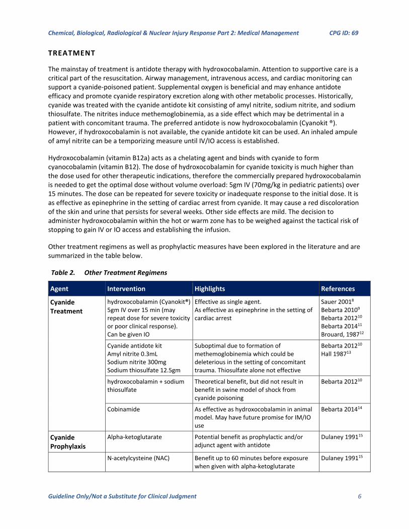
TREATMENT
The mainstay of treatment is antidote therapy with hydroxocobalamin. Attention to supportive care is a critical part of the resuscitation. Airway management, intravenous access, and cardiac monitoring can support a cyanide-poisoned patient. Supplemental oxygen is beneficial and may enhance antidote efficacy and promote cyanide respiratory excretion along with other metabolic processes. Historically, cyanide was treated with the cyanide antidote kit consisting of amyl nitrite, sodium nitrite, and sodium thiosulfate. The nitrites induce methemoglobinemia, as a side effect which may be detrimental in a patient with concomitant trauma. The preferred antidote is now hydroxocobalamin (Cyanokit ®). However, if hydroxocobalamin is not available, the cyanide antidote kit can be used. An inhaled ampule of amyl nitrite can be a temporizing measure until IV/IO access is established.
Hydroxocobalamin (vitamin B12a) acts as a chelating agent and binds with cyanide to form cyanocobalamin (vitamin B12). The dose of hydroxocobalamin for cyanide toxicity is much higher than the dose used for other therapeutic indications, therefore the commercially prepared hydroxocobalamin is needed to get the optimal dose without volume overload: 5gm IV (70mg/kg in pediatric patients) over 15 minutes. The dose can be repeated for severe toxicity or inadequate response to the initial dose. It is as effective as epinephrine in the setting of cardiac arrest from cyanide. It may cause a red discoloration of the skin and urine that persists for several weeks. Other side effects are mild. The decision to administer hydroxocobalamin within the hot or warm zone has to be weighed against the tactical risk of stopping to gain IV or IO access and establishing the infusion.
Other treatment regimens as well as prophylactic measures have been explored in the literature and are summarized in the table below.
Table 2. Other Treatment Regimens
Agent Intervention Highlights References
Cyanide Treatment
hydroxocobalamin (Cyanokit®) 5gm IV over 15 min (may repeat dose for severe toxicity or poor clinical response). Can be given IO
Effective as single agent. As effective as epinephrine in the setting of cardiac arrest
Sauer 20018 Bebarta 20109 Bebarta 201210 Bebarta 201411
Brouard, 198712
Cyanide antidote kit Amyl nitrite 0.3mL Sodium nitrite 300mg Sodium thiosulfate 12.5gm
Suboptimal due to formation of methemoglobinemia which could be deleterious in the setting of concomitant trauma. Thiosulfate alone not effective
Bebarta 201210
Hall 198713
hydroxocobalamin + sodium thiosulfate
Theoretical benefit, but did not result in benefit in swine model of shock from cyanide poisoning
Bebarta 201210
Cobinamide As effective as hydroxocobalamin in animal model. May have future promise for IM/IO use
Bebarta 201414
Cyanide Prophylaxis
Alpha-ketoglutarate Potential benefit as prophylactic and/or adjunct agent with antidote
Dulaney 199115
N-acetylcysteine (NAC) Benefit up to 60 minutes before exposure when given with alpha-ketoglutarate
Dulaney 199115

Chemical, Biological, Radiological & Nuclear Injury Response Part 2: Medical Management CPG ID: 69
Guideline Only/Not a Substitute for Clinical Judgment 7
Agent Intervention Highlights References
Dihydroxyacetone (DHA) Benefit in animals (oral and IV) Niknahad 200216
Hydrogen Sulfide (acts as simple asphyxiant/ irritant but included as blood agent due to HS cytochrome oxidase inhibition)
Sodium nitrite 300mg IV over 5-7 minutes
Anecdotal evidence if given shortly after exposure. Supportive care is often sufficient treatment if patient survives exposure
Hoffman 201517
NERVE AGENT EXPOSURE
BACKGROUND
Nerve agents, or acetylcholinesterase inhibitors, are some of the most lethal substances ever to be weaponized. These agents exist in multiple forms from thick viscous liquids to highly dissolvable gases. They are chemically similar to organophosphates used for pesticides, and the syndromes they cause are much more common in farming communities than they are on the battlefield. The most lethal forms only require 1/1000th of an ounce to obtain a lethal dose in 50% of exposed population (LD50).
Nerve agents consist of mainly two classes, V agents and G agents. V agents are viscous in nature and can be spread in numerous ways. They are extremely dangerous if touched or ingested but can also pose a vapor hazard in close proximity. G agents are liquids at room temperature and are extremely effective as chemical weapons due to the ability to quickly expose a large number of people to lethal inhaled doses by vapor exposure.
Physiologically, these agents bind to acetylcholinesterase, thus inhibiting breakdown of acetylcholine. The two main types of cholinergic receptors where nerve agents interact are muscarinic and nicotinic. Muscarinic receptors are located in the smooth muscles and the glands. Symptoms caused by over-stimulation of muscarinic receptors can be recalled using the DUMBBELS mnemonic (Diarrhea, Urination, Miosis, Bronchorrhea/Bronchoconstriction, Bradycardia, Emesis, Lacrimation, Salivation). These symptoms can be countered by atropine (discussed later in the treatment section). Nicotinic receptors located in skeletal muscle and nerve ganglia are also affected by nerve agents. Symptoms caused by over-stimulation of nicotinic receptors can be remembered by using the first letter of the days of the week as a memory assist (Mydriasis, Tachycardia, Weakness, Hypertension, Fasciculations). Administration of pralidoxime (2PAM) restores cholinesterase activity which typically results in improvement of the nicotinic symptoms.

Chemical, Biological, Radiological & Nuclear Injury Response Part 2: Medical Management CPG ID: 69
Guideline Only/Not a Substitute for Clinical Judgment 8
SIGNS AND SYMPTOMS
Nerve agent poisoning can range from mild to severe; a severe exposure may quickly lead to death if not reversed. Rapid antidote treatment is extremely important since some nerve agents can irreversibly bind to acetylcholinesterase (for example, the half-life for irreversible binding, termed aging half-life, for soman/GD is two minutes).
For mildly affected individuals not wearing eye protection, miosis is commonly seen. Other obvious muscarinic effects include severe lacrimation and profuse sweating, followed by nausea and vomiting, along with dyspnea and shortness of breath due to bronchorrhea and bronchoconstriction. More severely affected patients will have all of these signs and symptoms as well as profound weakness, fasciculations, seizures, loss of consciousness, apnea and death.
The speed of symptom onset depends on the route of exposure and dose of the agent. Inhalational exposure tends to result in faster onset of symptoms and can quickly cause death due to rapid systemic distribution. Dermal exposures, such as exposures with V agents, can cause delayed onset of symptoms.

Chemical, Biological, Radiological & Nuclear Injury Response Part 2: Medical Management CPG ID: 69
Guideline Only/Not a Substitute for Clinical Judgment 9
Table 3. Nerve Agents
Nerve Agent (GA, GB, GD, GF, VX) and Organophosphate Treatment
Agent Properties
Vapor or liquid. Variable aging (GD 2min)
G non-persistent, V persistent
M8 Paper: G - yellow, V- green
PPE Requirements
Mask and AP-PPE, JLIST, or UIPE
LETHALITY (VX most potent)
LCt50 15-70mg min/m3
LD50 3-4000mg
CRESS Symptomatic Presentation
C: altered, unconscious, seizures
R: tachypnea, wheezing, respiratory distress
E: miosis (may not be present with some organophosphates)
S: copious secretions (salivation, lacrimation, bronchorrhea)
S: diaphoresis
POI HOT
ZONE
Immediate Action: Self Aid, Buddy Aid, Move (Upwind, Upstream, Uphill) Away from Threat
M2: Massive hemorrhage / Mask check
A2: Airway (assess) / Antidote (ATNAAx3, CANAx1)
R2: Respiration (assess) / Rapid Spot Decon (RSDL, M295, Fibertect)
E: Extraction (egress away from threat)
Exposure to vapor causes almost immediate symptoms while liquid exposure may have minutes to hours delay in symptoms
Muscarinic symptoms (Diarrhea, Urination, Miosis, Bronchorrhea, Bradycardia, Emesis, Lacrimation, Salivation)
Nicotinic symptoms: (Mydriasis, Tachycardia, Weakness, Hypertension, Fasciculations) and seizures
Dirty CCP
WARM ZONE
M2 A2 R2 Reassessment, clear airway, O2 as needed, maintain filtered air Decon & Cutout:
Remove and bag equipment, PPE, and clothing Wipe away gross contamination, RSDL cut line, Cut out RSDL residual contamination on skin (>2min contact time, then wipe away) Remove and replace contaminated treatments (tourniquets, chest seals, etc)
C2: Circulation (assess vitals, resuscitate) / Countermeasures (atropine, pralidoxime, benzodiazepines) H2: Hypothermia (prevent) / Head wounds (assess mental status--altered due to agent,
antidote, or TBI?) E: Evacuation
Atropine given mistakenly to an individual not exposed to nerve agent may cause tachycardia, dry mouth, and anhydrosis and predispose to heat injury. When uncertain, favor treatment.
COLD ZONE
(MARCHE)2 Reassessment
C2: Circulation (Assess vitals, resuscitate) / Countermeasures
Use the following as symptoms dictate:
Atropine 20mg in 250mL NS IV/IO, titrate to dry respiratory secretions
Pralidoxime (1-2gm in 250mL NS) IV/IO over 15-30min
Benzodiazepines (midazolam preferred) 2mg IV/IO/IM, titrate to effect
Scopolamine 0.8mg IV/IM after ATNAA x3 (as an adjunct to atropine)
H2: Hypothermia (HPMK, fluid warmer) / Head wounds (treat elevated ICP, Neuro exam, MACE)
PFC: Supportive care:
Reassess frequently, follow protocols for respiratory or cardiac compromise.
Patients with ONLY miosis or mild rhinorrhea do not require treatment.
Acetylcholinesterase levels NOT diagnostic but may be trended.
Chest X-ray useful to monitor for pulmonary edema and secondary pneumonia.
Benzodiazepines can help control muscle fasciculations and seizures.
Reachback: USAMRICD (410) 436-2230 DSN: 584-2230

Chemical, Biological, Radiological & Nuclear Injury Response Part 2: Medical Management CPG ID: 69
Guideline Only/Not a Substitute for Clinical Judgment 10
DECONTAMINATION
As with most chemicals, removing the exposed patient from the contaminated area to prevent further exposure and damage is the most important step. In vapor exposures, this means removing the casualty from the area and quickly removing any article of clothing or piece of equipment with possible agent contamination.
Reactive skin decontamination lotion (RSDL) is largely accepted as the most effective decontaminate for nerve agent dermal exposure. Covering all exposed skin with RSDL as quickly as possible can be a life-saving measure. If RSDL is not available, 0.5% hypochlorite solution or soap and water are alternatives. Decontamination should not be delayed if RSDL is not available. For casualties exhibiting moderate to severe symptoms (respiratory distress, seizures, altered consciousness), antidotes should be administered immediately while initiating decontamination.
NERVE AGENT DIAGNOSTICS
Diagnosis of nerve agent exposure is based on rapid identification of the clinical symptoms and identification of the agent through detection methods. Laboratory measurement of serum acetylcholinesterase levels is not useful for nerve agent exposure since the value does not correlate with signs and symptoms or prognosis. There is no utility in baseline ACHE levels due to intra-individual variability.
NERVE AGENT TREATMENT
Nerve agent antidotes include 2PAM, atropine, and benzodiazepines. Autoinjectors to treat nerve agent toxicity are available as Autoinjector Nerve Agent Antidote (ATNAA) which contains atropine and 2PAM, and Convulsant Antidote Nerve Agent (CANA) which contains the benzodiazepine diazepam.
2PAM
Pralidoxime (ATNAA autoinjector or IV/IO) reverses the bond between the acetylcholinesterase and the nerve agent and thus prevents irreversible binding to acetylcholinesterase, termed aging. 2PAM is available both in autoinjector form as well as in an IV form. 2PAM is known to work synergistically with atropine (some 30x greater). The are other oximes with varying effects depending on the agent, but pralidoxime is the available agent in DoD inventory.
Atropine
Atropine (ATNAA autoinjector or IV/IO) is used to treat the muscarinic effects. Atropine will help dry secretions (bronchorrhea) and counter the effects of the bronchoconstriction caused by the nerve agent. The amount of nerve agent and the degree of symptoms a patient is experiencing will determine the amount of atropine required to control symptoms. Large doses of atropine may be required to counter the effects of some nerve agent exposures, in particular organophosphates. Tachycardia is NOT a contraindication for atropine administration, as tachycardia may be secondary to respiratory distress. Therefore, atropine treatment should be titrated to achieve reversal of life-threatening bronchorrhea and bronchoconstriction even in the setting of tachycardia.

Chemical, Biological, Radiological & Nuclear Injury Response Part 2: Medical Management CPG ID: 69
Guideline Only/Not a Substitute for Clinical Judgment 11
Benzodiazepines
Benzodiazepines, such as diazepam (CANA) or midazolam are the mainstay of seizure treatment. Benzodiazepines may also help counter nicotinic effects, particularly muscle fasciculations. Current literature recognizes midazolam as the most effective seizure reversal agent, based on animal studies. Midazolam has the fastest bioavailability when given intramuscularly (IM). Diazepam is the benzodiazepine in autoinjectors and is efficacious, but it is entirely appropriate to use another benzodiazepine when IV access has been established or when autoinjectors have been depleted.
Nerve agent antidotes may be dosed based on the severity of symptoms. However, in a field environment when the amount and type of nerve agent exposure are unknown, give 3 ATNAAs and 1 CANA for any symptomatic patient (other than isolated miosis) with suspected nerve agent exposure. If symptoms persist beyond this treatment, consider an atropine drip (see atropine/scopolamine protocol in Appendix B). For severe poisoning, additional 2PAM can be given after delivering 3 ATNAAs. There is a paucity of literature to guide dosing but current subject matter expert consensus is to dose an additional 500mg IV/IO over 5 minutes and then an infusion of 10mg/kg/hr until clinical improvement is stable which may require infusion for more than 24 hours. (See Appendix C.)
Prophylaxis
The planning considerations and the risk:benefit analysis in the decision to use pyridostigmine bromide (PB) prophylaxis is outside the scope of this CPG. However, it is important for providers to understand the clinical effects experienced by those on pyridostigmine prophylaxis and how pre-treatment can affect clinical presentation and response to treatment. Pyridostigmine (30mg tablet) is approved by the FDA for use as a pretreatment to exposure to soman, based on efficacy in reducing soman lethality when used in conjunction with 2PAM and atropine treatment in animals. (See FDA Pyridostigmine Bromide Package Insert.)18 There are no human studies.
Pyridostigmine acts by inhibiting a portion (20-40%) of peripheral acetylcholinesterase. It does not readily cross the blood brain barrier so it does not cause central inhibition. Thus side effects of pyridostigmine prophylaxis are typically mild cholinergic or nicotinic symptoms (diarrhea, abdominal pain, and dysmenorrhea were the most common side effects in volunteers). Pyridostigmine and mefloquine (for malaria prophylaxis) taken together may have an additive effect on the gastrointestinal tract with increased diarrhea. Opioid-associated bradycardia may be worsened when pyridostigmine is combined with opioids. Pyridostigmine may enhance the activity of depolarizing neuromuscular blocking agents (succinylcholine) but may require a higher dose of non-depolarizing neuromuscular blockers. Treatment for nerve agent toxicity is the same for patients on pyridostigmine prophylaxis.

Chemical, Biological, Radiological & Nuclear Injury Response Part 2: Medical Management CPG ID: 69
Guideline Only/Not a Substitute for Clinical Judgment 12
Table 4. Nerve Agent Treatment/Intervention
Agent Intervention Summary of evidence References
Nerve agents
GA
GB
GD
GF
VX
Atropine 2.1 mg in each ATNAA autoinjector and 2 mg in each separate atropine autoinjector; initial dose 6.1 mg (3 ATNAAs); then titrate in 2-mg increments
(anticipate 10-20 mg in the first several hours)
Atropine well established as anticholinergic. Helps control seizure activity.
McDonough 200119
Sidell 199720
Sidell 197421
Shih 199922
Taylor 198523
Ward 196224
Scopolamine* Dosing not well established
Penetrates the blood-brain barrier and controls CNS effects of nerve agents; enhanced survival in animals when used as adjunct with 2-PAM Cl and atropine; a dose of up to 1 mg may result in a marked decrease in total atropine required
Harris 199425
Koplovitz 201026
McDonough 200119
Pralidoxime chloride (2-PAM Cl)^ 600mg by autoinjector (3x)
1-2 gm IV/IO over 20-30 minutes
Most studied and only oxime in U.S. inventory. 2-PAM effectiveness is agent dependent. In guinea pigs, 2-PAM Cl is effective against sarin and VX.
Clement 198927
Dawson 199428
Gunnarson 200029
Kassa 200230
Snider 201631
Wilhelm 201432
Obidoxime**^ Obidoxime offered protection against tabun, sarin, VX, and pesticide oxons in guinea pigs.
Dawson 199428
Kassa 200230
Wilhelm 201432
Worek 200733
HI-6**^ HI-6 is considered a more broad-spectrum oxime and offered protection against tabun, sarin, cyclosarin, VX, and paraoxon in guinea pigs.
Dawson 199428
Hamilton 198934
Kassa 200230
Wilhelm 201432
Benzodiazepines*** Diazepam - autoinjector
Standard IV/IO dose for seizures 10-20 mg (up to 40 mg may be needed)
Midazolam - off label dosing recommendations for status epilepticus is 10 mg IM, typical IV/IO doses for seizure control start at 1-2 mg and titrate
Two cases in Tokyo sarin attack. One patient received diazepam 35 mg (for seizures) and the other 30 mg (for fasciculations) along with atropine and 2PAM. Both recovered without sequelae.
Some data to suggest that benzodiazepines can be useful for anxiety and restlessness that may precede seizures.
Midazolam shown to be effective at a lower dose and to be more rapidly absorbed IM than diazepam in animal models
Marrs 200735
McDonough 200119
Shih 200236
*While scopolamine is an FDA-approved drug, its use for nerve-agent poisoning is off-label **Obidoxime and HI-6 are not FDA approved or available in the U.S. ***While benzodiazepines are routinely used for termination of seizures, this is an off-label use of this class of medications. ^ Efficacy of oximes is species dependent, so it is difficult to extrapolate effectiveness in humans from existing animal studies.

Chemical, Biological, Radiological & Nuclear Injury Response Part 2: Medical Management CPG ID: 69
Guideline Only/Not a Substitute for Clinical Judgment 13
PULMONARY AGENT EXPOSURE
BACKGROUND
Pulmonary agents (also referred to as toxic industrial chemicals and choking agents) hold historical significance as the forerunners of modern chemical warfare and still hold relevance today as likely chemical culprits given their availability. Chlorine and phosgene are produced and stored in large quantities worldwide, and could have devastating effects when vaporized. There are other agents which can produce chemical lung injury such as ammonia, hydrogen sulfide, zinc oxide, phosphorus smokes, and perfluoroisobutylene (by-product of Teflon). These agents are irritating to the lungs, but are less likely to be used in a chemical attack.
Chlorine becomes a gas at -34 ° C, and is therefore stored as a compressed liquid. Phosgene becomes a toxic gas at 47 °F. The extent of injury caused by either gas is a function of the duration and concentration of exposure. Other variables that impact toxicity include respiratory rate and depth (minute ventilation) and possibly body position.
Exposures to 30ppm of chlorine will cause coughing; more serious damage to the lungs occurs at levels above 40 to 60ppm for more than 30 minutes. Phosgene is more surreptitious and toxicity may occur below its odor threshold of 0.4ppm with prolonged exposure. Additionally olfactory fatigue can occur so an individual may only transiently notice the warning odor. Phosgene IDHL (Immediately Dangerous to Life or Health) is 2ppm whereas chlorine IDHL is 100ppm.
Both gases react with moisture in the respiratory system and undergo hydrolysis. Chlorine causes lung damage through reactions to form hydrochloric and hypochlorous acids, which in turn react with sulfhydryl groups of cysteine and cause enzyme inhibition. In addition to this reaction, hydrolysis of chlorine results in free radical generation that can lead to direct cell injury and death. Phosgene also reacts with water to form carbon dioxide and hydrogen chloride. However, the major toxicity of phosgene is believed to occur through acylation in which phosgene interacts with sulfhydryl, amine, and hydroxyl groups causing protein and lipid denaturation, disruption of membrane structure and interference with enzyme function. Phosgene also disrupts the pulmonary surfactant layer.
PULMONARY AGENT SIGNS AND SYMPTOMS
Both gases have the ability to cause asphyxia due to displacement of oxygen if released in a confined space. More commonly, the gases act as irritants and cause damage to the respiratory tract through the mechanisms described above. Lastly, the gases can cause a systemic inflammatory response.
Chlorine has an unpleasant odor and is highly irritating. Because chlorine undergoes more rapid hydrolysis when contacting mucous membranes, it causes more immediate symptoms in the moist areas of the eyes, mouth, and upper airways. Eye pain, blepharospasm, and lacrimation are common. Other symptoms may include headache, salivation, dyspnea, cough, hemoptysis, chest burning, and vomiting.
Physical examination may reveal tachycardia, tachypnea, and possibly cyanosis. If eye irritation is present, evaluation for corneal burns/abrasions should be done with fluorescein staining of the eye. In the presence of oropharyngeal erythema, there may be more significant distal injury requiring careful

Chemical, Biological, Radiological & Nuclear Injury Response Part 2: Medical Management CPG ID: 69
Guideline Only/Not a Substitute for Clinical Judgment 14
assessment of the airway. Stridor, hoarseness, or aphonia may indicate laryngeal edema or laryngospasm. Oropharyngeal secretions may be copious.
Phosgene smells of fresh mown hay. It has more insidious effects, and early symptoms may be mild or absent. Typical onset of phosgene-induced symptoms occurs 2 to 6 hours after exposure and delayed symptoms have been described up to 15 hours post exposure. The major effects of phosgene are on peripheral airways, therefore dyspnea, chest tightness or pain, and cough are common symptoms. Development of hypoxia and pulmonary edema may occur hours after the onset of symptoms. Fluid shifts secondary to pulmonary edema may result in hypovolemia. Early onset of pulmonary edema portends a grave prognosis.
PULMONARY AGENT DECONTAMINATION
Safe removal from the toxic gas is the priority in chlorine or phosgene exposure. Respiratory protection for rescuers and providers in a potential exposure area is critical. Once the casualty is removed from the exposure area, decontamination should be continued with removal of all clothing. For toxic gas exposure, this removes the majority of risk from the gases. Soap and water is adequate to complete decontamination.
PULMONARY AGENT DIAGNOSTICS
There are no readily available diagnostic tests to confirm or quantify pulmonary agent toxicity. Standard tests such as arterial blood gases and chest x-rays (CXR) should be used to guide supportive care as needed. Arterial blood gases can be useful when needed to follow oxygenation, but may be normal in the early phases of phosgene exposure. PCO2 may be elevated in patients with obstructive pathophysiology and indicate a need for bronchodilators or corticosteroids.
Much like ABGs, CXR performed shortly after exposure may be normal but the patient may progress to frank pulmonary edema within a few hours. Fortunately the CXR can reveal pulmonary edema before clinical exam findings. A baseline CXR may be useful for comparison when trying to detect subtle early findings of pulmonary edema. If the CXR is normal at 8 hours, it is unlikely the patient will develop pulmonary edema.37
Chemical, Biological, Radiological & Nuclear Injury Response Part 2: Medical Management CPG ID: 69
Guideline Only/Not a Substitute for Clinical Judgment 15
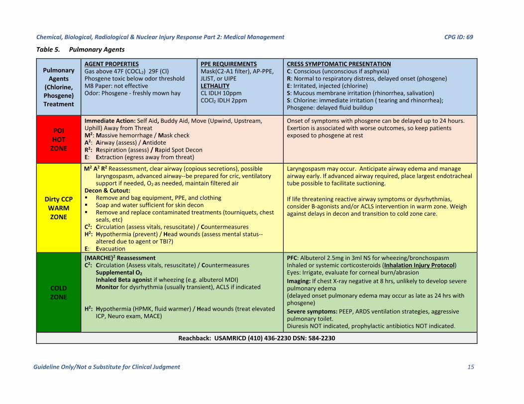
Table 5. Pulmonary Agents
Pulmonary Agents
(Chlorine, Phosgene) Treatment
AGENT PROPERTIES Gas above 47F (COCL2) 29F (Cl) Phosgene toxic below odor threshold M8 Paper: not effective Odor: Phosgene - freshly mown hay
PPE REQUIREMENTS Mask(C2-A1 filter), AP-PPE, JLIST, or UIPE LETHALITY CL IDLH 10ppm COCl2 IDLH 2ppm
CRESS SYMPTOMATIC PRESENTATION C: Conscious (unconscious if asphyxia) R: Normal to respiratory distress, delayed onset (phosgene) E: Irritated, injected (chlorine) S: Mucous membrane irritation (rhinorrhea, salivation) S: Chlorine: immediate irritation ( tearing and rhinorrhea); Phosgene: delayed fluid buildup
POI HOT
ZONE
Immediate Action: Self Aid, Buddy Aid, Move (Upwind, Upstream, Uphill) Away from Threat M2: Massive hemorrhage / Mask check A2: Airway (assess) / Antidote R2: Respiration (assess) / Rapid Spot Decon E: Extraction (egress away from threat)
Onset of symptoms with phosgene can be delayed up to 24 hours. Exertion is associated with worse outcomes, so keep patients exposed to phosgene at rest
Dirty CCP WARM ZONE
M2 A2 R2 Reassessment, clear airway (copious secretions), possible laryngospasm, advanced airway--be prepared for cric, ventilatory support if needed, O2 as needed, maintain filtered air
Decon & Cutout: Remove and bag equipment, PPE, and clothing Soap and water sufficient for skin decon Remove and replace contaminated treatments (tourniquets, chest
seals, etc) C2: Circulation (assess vitals, resuscitate) / Countermeasures H2: Hypothermia (prevent) / Head wounds (assess mental status--
altered due to agent or TBI?) E: Evacuation
Laryngospasm may occur. Anticipate airway edema and manage airway early. If advanced airway required, place largest endotracheal tube possible to facilitate suctioning.
If life threatening reactive airway symptoms or dysrhythmias, consider B-agonists and/or ACLS intervention in warm zone. Weigh against delays in decon and transition to cold zone care.
COLD ZONE
(MARCHE)2 Reassessment C2: Circulation (Assess vitals, resuscitate) / Countermeasures Supplemental O2 Inhaled Beta agonist if wheezing (e.g. albuterol MDI) Monitor for dysrhythmia (usually transient), ACLS if indicated H2: Hypothermia (HPMK, fluid warmer) / Head wounds (treat elevated ICP, Neuro exam, MACE)
PFC: Albuterol 2.5mg in 3ml NS for wheezing/bronchospasm Inhaled or systemic corticosteroids (Inhalation Injury Protocol) Eyes: Irrigate, evaluate for corneal burn/abrasion
Imaging: If chest X-ray negative at 8 hrs, unlikely to develop severe pulmonary edema (delayed onset pulmonary edema may occur as late as 24 hrs with phosgene)
Severe symptoms: PEEP, ARDS ventilation strategies, aggressive pulmonary toilet. Diuresis NOT indicated, prophylactic antibiotics NOT indicated.
Reachback: USAMRICD (410) 436-2230 DSN: 584-2230

Chemical, Biological, Radiological & Nuclear Injury Response Part 2: Medical Management CPG ID: 69
Guideline Only/Not a Substitute for Clinical Judgment 16
PULMONARY AGENT TREATMENT
Chlorine exposures may lead to copious secretions and laryngospasm shortly following exposure, therefore providers should be prepared for airway management and possibly emergent surgical airway control. It is important to remember that phosgene-exposed patients, despite being asymptomatic, need to be treated as casualties. Such casualties should be kept at rest, as exertion is associated with pulmonary edema and worse outcomes in phosgene-exposed patients. If an advanced airway is placed, a large-bore endotracheal tube will facilitate pulmonary toilet as toxic gas exposures can cause sloughing of mucosa and clogging of the airway with debris.
Intravenous fluids may be necessary in the setting of volume depletion, but should not be given empirically. Fluid overload can contribute to pulmonary edema and should be avoided. Laryngoscopy and/or bronchoscopy may be necessary, and preparations for advanced airway management must be in place should airway compromise occur. Portable ventilators with simplified automated setting (e.g. SAVe ventilators) may not be adequate for ventilation management in these patients. Because of the associated pulmonary edema, bronchospasm, and risk of ARDS, the ability to manipulate ventilator settings is crucial. Additionally, suction is a key component of maintaining patent airways, bulb suction is unlikely to be adequate and mechanical suction with the ability to do inline suction is preferred.
Advanced interventions and the supporting evidence is described in the table below. Much of the available evidence is based upon animal studies and human data is limited.
Table 6. Pulmonary Agent Intervention
Agent Intervention Highlights References
Chlorine Beta agonists (albuterol MDI) (typical bronchodilator doses)
May be useful in the setting of bronchospasm and obstructive airway disease
Nelson 2015 38
Corticosteroids (typical doses given for reactive airway disease) Inhaled: fluticasone 200mcg BID budesonide 0.5-2.0mg BID
Possible benefit shown in animal studies using inhaled steroids, even without reactive airways (no human data)
Gunnarson 200029 Wang 2004 39
Nebulized sodium bicarbonate (dilute 8.4% 1:1 with sterile water to make 4.2%)
Theoretical benefit, studies limited. Bosse 1994 40 Vinsel 1990 41
Phosgene Corticosteroids (typical doses for reactive airway disease, IV or inhaled)
Possible benefit shown in animal studies, even without reactive airways (no human data)
Guo 199042 Frosolono197843
Medications that increase cAMP (aminophylline, dibutyryl adenosine 3,5 cyclic monophosphate [DbcAMP], Beta adrenergic agents)
Theoretical benefit when given early after exposure (no human data)
Sciuto 199744 Kennedy 198945
Ibuprofen Reduction of pulmonary edema in rats (with pre and post exposure treatment)
Sciuto 199646
N-acetylcysteine Reduction of pulmonary edema, lipid peroxidation, and leukotriene production in rabbits
Sciuto 199547

Chemical, Biological, Radiological & Nuclear Injury Response Part 2: Medical Management CPG ID: 69
Guideline Only/Not a Substitute for Clinical Judgment 17
VESICANT OR BLISTER AGENTS
BACKGROUND
Blister agents were developed and used during the First World War as chemical warfare agents.48,49 These agents can be used in multiple forms to include liquid, solid or gas. Blister agents are generally broken down into sulfur mustard, nitrogen mustard, and Lewisite. Although used in different concentrations and in different forms, currently sulfur mustard is the most common blister agent used on the battlefield.
Sulfur mustard has three different main forms: HD, which is a distilled product and is close to 100% pure; H, which is undistilled sulfur mustard; and HT which is a mixture of HD and T (a thickener which can be added to sulfur mustard). HD or pure mustard is clear and smells of garlic. H sulfur mustard can be clear, yellow, red brown or black depending on the chemical mixture. In recent conflicts, to include the conflict with ISIS, H sulfur mustard is the chemical seen on the battlefield. This substance, made in crude chemistry labs, is a thick oily black substance which degrades quickly, only lasting 2-3 weeks in storage before degrading beyond utility.
Nitrogen mustard is much less commonly used as a chemical warfare agent. It is separated into 3 forms: N1, N2, and N3. Nitrogen mustard can have different smells with N1 smelling more like fish, N2 like fruit, and N3 like bitter almonds. Nitrogen mustards are clear to yellow oily substances that evaporate slowly and can harm first responders by contact or off-gassing from injured victims. Used for medicinal purposes such as chemotherapy or wart removal, these agents have so far never been used on the battlefield.
Lewisite is the last vesicant in this family of chemical agents. Related to arsenic, Lewisite is a clear liquid in pure form and amber or black in impure forms. Like the other vesicants, it is extremely irritating to the skin, eyes and respiratory tract. Lewisite can be mixed with HD for a more potent chemical warfare agent with properties from both substances. Unlike the other vesicants, Lewisite is the only vesicant that has an antidote to counter its systemic effects.
SIGNS AND SYMPTOMS OF VESICANT OR BLISTER AGENTS
The most common route of exposure of all blister agents is via the skin. Sulfur mustard agents will cause chemical burns associated with blisters within a few hours of contact with the skin or mucous membranes. Effects are not seen immediately on contact, but sulfur mustard is absorbed within minutes of contact on the skin or eye membranes. Second and third degree burns develop over 2-10 hours to the eyes and skin, causing intense pain, corneal perforations, erosions of the eyes, and blistering of all exposed skin. HD had a 2-3% mortality rate during WWI, but burns in excess of 25% body surface can be fatal. Nitrogen mustard has similar effects on the skin and eyes as sulfur mustard. However, Lewisite has immediate effects on the skin and eyes causing immediate pain and irritation and blistering much sooner than HD or nitrogen mustard.
Inhalation or ingestion of these vesicants causes similar problems, damaging the mucosa of both the respiratory and digestive systems, causing severe burns. Liquid agents, which are more concentrated, cause more severe damage than vapors which might be inhaled. Much like skin and eye exposure, inhalation of HD has a delayed effect on the respiratory tract, causing wheezing and swelling of the bronchioles several hours after exposure. As with skin symptoms, Lewisite causes respiratory symptoms

Chemical, Biological, Radiological & Nuclear Injury Response Part 2: Medical Management CPG ID: 69
Guideline Only/Not a Substitute for Clinical Judgment 18
much faster, usually within seconds to minutes, which then rapidly progresses to pulmonary edema following exposure.
Several late effects can occur from all blistering agents depending on the dose and the route of exposure of the agent. Bone marrow suppression can occur with increased likelihood of infection, and nitrogen mustard will cause anemia. High doses of sulfur mustard can cause convulsions and hyper-excitability. High doses of Lewisite can lead to hepatic necrosis, acute renal failure, and shock via capillary leaking, referred to as “Lewisite Shock.” There are many long-term effects suspected such as malignancies, corneal scarring, chronic respiratory disease, and dermal scarring.
DECONTAMINATION OF VESICANT OR BLISTER AGENTS
Safety of the rescuers and healthcare providers is the most important initial step when handling mustard casualties. First responders should have respiratory and skin protection during initial treatment of mustard casualties. Butyl rubber is the recommended level of protection for the hands; however, double layers of nitrile gloves will protect against exposure as well. Remember, contaminated patients may appear innocuous due to delayed onset of symptoms, however providers can still be exposed to significant injury to the lungs, eyes, and skin if not properly protected. The casualty must be decontaminated and all clothing and equipment removed; vesicants enter the body within minutes, but can stay on equipment or clothing for days after exposure.
Removal of the agent must occur within three to five minutes in order to reduce absorption. Removal with a dry cloth is the first step to clear the chemical from the skin, followed by RSDL. There is no antidote for vesicants like there is for nerve agents, so initial treatment is focused on rapid decontamination. Exposure to the eye causes faster absorption than skin and should be washed out immediately with water in order to minimize the effects. Eye wash kits with Morgan Lens can facilitate eye decontamination. It is important not to induce vomiting if there is any concern for ingestion, and activated charcoal has not been shown to be effective in these situations. After decontamination, standard burn care treatment is recommended for all dermal injuries. Fluid replacement may not follow the thermal burn estimates; however, urine output remains a good marker of adequate resuscitation and fluids should be titrated to target a urine output of 30-50 ml/hour.
Wounds which are chemically contaminated should be aggressively flushed and treated as if there is heavy contamination. Mustard enters the body systemically almost immediately after exposure to open wounds or mucus membranes. After initial decontamination, the patient should be transported to a hospital and observed for both systemic and local effects of the contaminated wound. Surgical debridement of open wounds will almost always be required for contaminated wounds.
VESICANT OR BLISTER AGENTS DIAGNOSTICS
There is no readily available test to confirm vesicant exposure. Leukocytosis is anticipated on the first day and will rise with the amount of associated injury. Subsequently, bone marrow suppression occurs and leukocyte count will fall around day 3 to 5 with a nadir seen around day 9. Counts less than 500 indicate a poor prognosis. Chest X-ray can be used to monitor for pneumonitis, which typical appears in the first 2 to 3 days.

Chemical, Biological, Radiological & Nuclear Injury Response Part 2: Medical Management CPG ID: 69
Guideline Only/Not a Substitute for Clinical Judgment 19
TREATMENT OF VESICANT OR BLISTER AGENTS
For asymptomatic patients exposed to sulfur mustard and nitrogen mustard, the effects may be delayed for skin, eye, and lungs; therefore, observation of potential exposures for 6-10 hours is recommended.
Patients with eye exposure may benefit from regular application of an anticholinergic ophthalmic ointment to prevent synechiae formation. A topical antibiotic/steroid ointment should be applied every 1-2 hours with rapid referral to an ophthalmologist. Ointment application to the lids prevents them from sticking together and can help prevent adhesions while allowing drainage of any underlying infection or pus. Blepharospasm can be treated with topical anesthetics to facilitate the eye exam, and systemic analgesics should be given for ongoing eye pain.
Table 7. Vesicant or Blister Agent Intervention
Agent Intervention Highlights References
Sulfur Mustard50 Nitrogen Mustard51, 52
Corticosteroids Limited animal studies, immunosuppression may be detrimental
Wigenstam 2012 53
Antioxidants and scavengers (Vitamin E, N-acetyl cysteine [NAC], glutathione, sodium thiosulfate)
Limited animal studies Vojvodic 1985 54 Anderson 2000 55
Filgrastim/pegfilgrastim Burns >25% Body Surface Area(BSA) with bone marrow suppression
Anderson 2006 56
Amifostine May provide protection prophylactically (animal studies)
Vijayaraghavan 200157,58
Lewisite British Anti Lewisite (BAL)/Dimercaprol
Chelation agent with significant side effects
Vilensky 2003 59, 60
If patients have respiratory symptoms hours after the exposure, they should be treated as chemical pneumonitis; albuterol should aggressively be utilized, and invasive airway management should be considered early in the treatment plan if the patient is not responding to albuterol. Systemic steroids have been recommended if albuterol is not effective, but further immune suppression may not be advisable. Inhaled sodium bicarbonate has been suggested as a possible treatment as well, but there is not robust evidence to support its use. Other antioxidants and scavengers such as sodium thiosulfate have shown some benefit in animal studies, but there is no human data to support their use. These therapies should only be considered in patients refractory to supportive care when the benefit of unproven therapy outweighs potential risks.
Bone marrow suppression usually peaks around 9-10 days. Granulocyte colony stimulating factor analogues may be administered. Severe bone marrow suppression may be an indication for bone marrow transplant.
The antidote for Lewisite is British Anti-Lewisite (BAL), also known as Dimercaprol. BAL should only be used in a hospital setting and is given as an IM injection. BAL is a chelating agent, but due to the possibility of severe acute renal failure and other side effects, BAL is only recommended for patients who have severe respiratory symptoms or Lewisite shock.

Chemical, Biological, Radiological & Nuclear Injury Response Part 2: Medical Management CPG ID: 69
Guideline Only/Not a Substitute for Clinical Judgment 20
Table 8. Vesicant or Blister Agents
Vesicant Lewisite (L) Mustard-Lewisite Mixture (HL)
Immediate Acting Agent!
AGENT PROPERTIES Oily Liquid Persistent, Freezing pt: 0.4°F/13°F M8 Paper: Red to Pink LCD: Red or Orange H AP4C: AS, L, SA / S, H, HL Odor: Geraniums
PPE REQUIREMENTS MOPP 4 (Mask w/ AP-PPE, JLIST, or UIPE) LETHALITY LCt50: 1,200 mg-min/m³ LD50: 30-50 mg/kg (skin)
CRESS SYMPTOMATIC PRESENTATION C: Conscious R: Immediate Irritation, Distress E: Immediate Severe Pain, Blepharospasm, Edema S: Normal to Increased S: Immediate Pain, Erythema, Blisters hours later Other: Systemic Effects - Distributive Shock
POI HOT
ZONE
Immediate Action: Self Aid, Buddy Aid, Move (Upwind, Upstream, Uphill) Away from Threat M2: Massive hemorrhage / Mask check A2: Airway (assess) / Antidote (no antidote for vesicants in hot zone) R2: Respiration (assess) / Rapid Spot Decon (RSDL, M295, Fibertect) E: Extraction (egress away from threat)
Lewisite binds to tissues and absorbs systemically within two minutes of contact. Symptoms begin to manifest immediately upon exposure and worsen over time. Control of massive hemorrhage and rapid spot decon are top priorties.
Extraction to the Dirty CCP [For Small Spills (< 2 kg) move away 100m day/300m night] [Large Spills (< 25kg) = 500m day/1000m night]
Dirty CCP
WARM ZONE
M² A² R² Reassessment, O2 as needed, maintain filtered air, clear airway, albuterol inhaler, invasive airway if not responding to albuterol, pain management Decon & Cutout: Remove and bag equipment, PPE, and clothing Wipe away gross contamination, RSDL cut line, Cut out RSDL residual contamination on skin (>2min contact time, then wipe away) Remove and replace contaminated treatments (tourniquets, chest seals, etc)
C2: Circulation (assess vitals, resuscitate) / Countermeasures (rapid decon, irrigate eyes and wounds with water) H2: Hypothermia (prevent) / Head wounds (assess mental status--altered due to agent, or TBI?) E: Evacuation
Casualties with palm-size exposure without rapid decon, >5% BSA burn, pulmonary edema, or shock symptoms with rapid onset require chelation. Early pain control may be required to ensure casualty cooperation. Administration of BAL within 5 minutes of exposure to skin and eyes can neutralize agent. TIC/TIM Eye Injury Tx Protocol may be required.
Dirty Evacuation to Decontamination MSS or Evacuate Directly to Medical Treatment Facility if Field Decontamination is Sufficient
COLD ZONE
(MARCHE)² Reassessment M²: Convert tourniquets & bandage wounds A²: In case of severe inhalation symptoms upgrade airway adjunct & RSI R²: Vesicant Inhalation Tx SOP, Ventilator, O2 , PEEP, Suction, Bronchoscopy C²: Trend Vitals, TXA, FDP, FWB, Fluid Challenge if Req'd / severe exposures will present with distributive shock requiring chelation therapy with Dimercaprol aka British Anti-Lewisite (BAL) in order to resolve H2: Hypothermia (HPMK, fluid warmer) / Head wounds (treat elevated ICP, Neuro exam, MACE)
Dimercaprol (BAL) Administration Initial Dose: 3 mg/kg deep IM repeat every 4 hours for two days Then: Every 12 hours for 7-10 days Severe & Life Threating Exposure: consider 5 mg/kg Side Effects: Increased BP, Tachycardia, Nausea/vomiting, Headache, Anxiety, Injection Necrosis Contraindications: Nut Allergy. Alternate Drug: DMSA PFC: Pain Management, Expect SIRS and ARDS in cases of severe exposures. Skin: Burns-apply Silvadene & bandage QID (burn fluid resuscitation not necessary) Blister fluid may contain Arsenic, unroof >2cm, irrigate, calamine or steroidal cream Eyes: Petroleum based ophthalmic ointment, possible iristis, ophthalmology consult
Reachback: USAMRICD (410) 436-2230 DSN: 584-2230

Chemical, Biological, Radiological & Nuclear Injury Response Part 2: Medical Management CPG ID: 69
Guideline Only/Not a Substitute for Clinical Judgment 21
INCAPACITATING AGENTS
BACKGROUND
Incapacitating agents produce temporary physical and/or mental effects that result in the inability of the affected individual to continue in their current duties or activity. Often they are described as non-lethal agents, but if administered in high enough doses, incapacitating agents can results in death or serious morbidity. There are three general categories of incapacitating agents:61
1. Anticholinergics
BZ (3-quinuclidinyl benzilate) is the prototypical anticholinergic agent but other anticholinergic agents may be developed for warfare. The British reportedly have a similar agent known as Agent 15. BZ is a centrally acting anticholinergic agent that was originally developed as a gastrointestinal antispasmodic but due to severe central nervous system effects was abandoned as a pharmaceutical. As an incapacitating agent, it has a very high safety profile due to the relatively lower peripheral antimuscarinic effects when compared to atropine. An effective incapacitating dose is 0.5 mg, and BZ is typically dispersed as an aerosol or in smoke-producing munitions.
2. Sedating agents
Opioids and volatile anesthetics are both potential sedative or calming agents. Fentanyl is a synthetic opioid with 100 times the potency of morphine. Newer derivatives of fentanyl are continuously in development. One such example is carfentanil with a potency 100 times fentanyl. While these agents are highly effective in individuals, aerosolizing them to use as an incapacitating agent can be problematic. It is likely that absorption in a crowd would be variable.
3. Riot control agents
Riot control agents, also known as harassing agents, cause irritation and discomfort often in the form lacrimation, mucous membrane irritation, violent coughing, or vomiting. Typical agents include chloroacetophenone (CN) commonly known as Mace, chlorobenzylidene malononitrile (CS), oleoresin capsicum (OC) called pepper spray, and diphenylaminochloroarsine (DM) or Adamsite. CN is more harmful than the other agents with a lower LCt50 and DM tends to cause vomiting.

Chemical, Biological, Radiological & Nuclear Injury Response Part 2: Medical Management CPG ID: 69
Guideline Only/Not a Substitute for Clinical Judgment 22
Table 9. Incapacitating Agents
Incapacitating Agents (Anticholinergics, Opioids, Riot control agents)
AGENT PROPERTIES Variable, aerosol, smoke or liquid M8 Paper: not useful
PPE REQUIREMENTS Mask and AP-PPE, JLIST, or UIPE LETHALITY Variable (fentanyl derivatives extremely potent)
CRESS SYMPTOMATIC PRESENTATION C: varies with agent, see below R: E: S: S:
POI HOT
ZONE
Immediate Action: Self Aid, Buddy Aid, Move (Upwind, Upstream, Uphill) Away from Threat M2: Massive hemorrhage / Mask check A2: Airway (assess) / Antidote (Cyanokit) R2: Respiration (assess) / Rapid Spot Decon E: Extraction (egress away from threat)
Recognize CRESS symptoms: Anticholinergics-Delirium, agitation; red, hot and dry skin, mydriasis, tachycardia Opioids - Sedation; decreased respirations, miosis, normal skin and secretions Riot control agents - mucous membrane irritation (salivation, rhinorrhea and lacrimation, intact mentation)
Dirty CCP
WARM ZONE
M2 A2 R2 Reassessment, O2 as needed, maintain filtered air
Decon & Cutout:
Remove and bag equipment, PPE, and clothing Wipe away gross contamination, RSDL cut line, Cut out RSDL residual contamination on skin (>2min contact time, then wipe away) Remove and replace contaminated treatments (tourniquets, chest seals, etc.)
C2: Circulation/Countermeasures/Drips (benzodiazepines, naloxone)
Hypothermia (prevent) / Head wounds (assess mental status--altered due to agent, or TBI?)
Anticholinergics – titrate benzodiazepines to control severe agitation, delirium
Opioids - Support respiration if needed. Naloxone (2-4 mg) if respiratory depression. May require escalating doses for some synthetic opioids
COLD ZONE
(MARCHE)2 Reassessment C2: Circulation (Vitals/Fluid Challenge) Countermeasures, Chelators, Drips Anticholinergics – titrate benzodiazepines 2-4 mg IV/IO/IM Opioids - Naloxone 2-4 mg IV/IO, titrate to respiratory effort (May require naloxone drip (2/3 response dose/hour) H2: Hypothermia (HPMK, fluid warmer / Head wounds (treat elevated ICP, Neuro exam, MACE)
PFC: supportive care Most agents are self-limited and symptoms will wear off, often as
decontamination is performed. Look for reactive airway disease. Half-life of sedating agents may be longer than naloxone, so may require re-
dosing or a drip Physostigmine can be used as diagnostic agent for anticholinergic delirium.
Requires EKG with normal QRS prior to administration Benzos effective for seizures, agitation, and autonomic activity Sodium metabisulfite can neutralize CS
Reachback: USAMRICD (410) 436-2230 DSN: (312) 584-2230

Chemical, Biological, Radiological & Nuclear Injury Response Part 2: Medical Management CPG ID: 69
Guideline Only/Not a Substitute for Clinical Judgment 23
SIGNS AND SYMPTOMS OF INCAPACITATING AGENTS
Anticholinergics
BZ is intended to target CNS effects, therefore anticholinergic delirium will be the predominant symptom with fewer peripheral effects. Often the patient cannot give a lucid history due to delirium. The delirium may be labile and can range from mild impairment to coma. Hallucinations, severe agitation, and even seizures may occur. The classic peripheral effects often described a “dry as a bone, hot as a hare, red as a beet, and blind as a bat” may be variable or delayed.
Sedating agents
Sedating agents which are opioid derivatives can be expected to present with the classic opioid toxidrome of miosis, CNS depression, and respiratory depression. Bradycardia, hypotension, and hypothermia may occur as secondary sequelae as a result of dose-related progression to opioid coma.
Riot control agents
Riot control agents can be expected to affect the skin, respiratory system and eyes. Ophthalmologic symptoms include pain, tearing, and blepharospasm. The respiratory tract can be variably affected and symptoms may range from mild mucous membrane irritation to severe dyspnea, coughing and chest tightness. Bronchospasm is common and may be severe in those with underlying reactive airway disease. Copious rhinorrhea and salivation may occur and clinical scrutiny is necessary to exclude the possibility of nerve agent exposure. Dermatologic effects typically involve skin pain and burning, but blistering may occur at higher doses.
GENERAL MANAGEMENT
Safe removal from exposure is the priority. Respiratory protection for providers in a potential exposure area is critical. Once the casualty is removed from the exposure area, decontamination can be continued with removal of all clothing and personal effects. Simple soap and copious water are adequate for through decontamination. Recognize that improvement in symptoms caused by riot control agents may be transient with decontamination.
Medical management of all incapacitating agents is predominantly supportive with attention to symptoms and tailoring treatment to the patient presentation.
ANTICHOLINERGIC AGENTS
Patients with anticholinergic toxicity can present with dry mouth and tachycardia leading the provider to believe dehydration is present when the patient is euvolemic. However, psychomotor agitation and hyperthermia are common so careful monitoring of core temperature, volume status and urine output is important. Cooling should be undertaken promptly when hyperthermia is present.
Pharmacologic managements include control of delirium and agitation. Agitation can be safely controlled with a benzodiazepine titrated to effect. Often controlling the agitation will also improve tachycardia and hyperthermia. Physostigmine is also an option to manage the delirium. This is a tertiary amine which crosses the blood-brain barrier. Before using physostigmine, it is critical to exclude the

Chemical, Biological, Radiological & Nuclear Injury Response Part 2: Medical Management CPG ID: 69
Guideline Only/Not a Substitute for Clinical Judgment 24
presence of other sodium channel blocking agents such as tricyclic antidepressants. An EKG should be done to ensure a normal QRS interval <100msec prior to physostigmine use, which limits its utility when EKG is not immediately available. Additionally, atropine should be ready in case there is a cholinergic response that affects the airway. In most cases, benzodiazepines are considered the primary treatment option since they can be administered safely to almost any patient.
SEDATING AGENTS
Support of the respiratory system is the primary focus for treatment of opioid toxicity associated with sedating agents. Naloxone is the antidote of choice and should be titrated to reverse respiratory suppression. Nasal naloxone can be rapidly administered without IV access. Naloxone should be titrated to effect. A starting dose of 2-4 mg is appropriate but much higher doses may be required to reverse the effects of synthetic opioids. The half-life of naloxone may be shorter than the half-life of the agent and repeat dosing or a naloxone drip may be necessary.
RIOT CONTROL AGENTS
Most riot control agents are short acting and supportive care is usually adequate until symptoms subside.
INCAPACITATING AGENT DIAGNOSTICS
Laboratory values are of little diagnostic utility. Opioids may be detected on routine toxin screen.
INCAPACITATING AGENT TREATMENT
Advanced interventions and the supporting evidence are described in the table below.
Table 10. Interventions for Incapacitating Agents
Agent Intervention Clinical Notes References
Anticholinergic agents, 3-quinuclidinyl benzilate (BZ)
Benzodiazepines Control agitation and autonomic activity, seizure prevention/treatment
Burns 200062
Physostigmine Useful to reverse anticholinergic delirium, must have normal QRS on EKG, need atropine on hand if cholinergic crisis ensues
Burns 200062
Wetherall 200263
Opioids Naloxone 2-4 mg, escalating dose to 10 mg Recognize half-life of naloxone may be shorter than some agents so may need re-dosing
Extrapolated from pharmaceutical opioid experience
Boyer 2012 64
Riot control agents Sodium metabisulfite Neutralizes malononitrile (CS) Schep 2015 65

Chemical, Biological, Radiological & Nuclear Injury Response Part 2: Medical Management CPG ID: 69
Guideline Only/Not a Substitute for Clinical Judgment 25
ACRONYM DEFINITIONS
ACLS: Advanced Cardiac Life Support
AP4C: Handheld Chemical Decontamination Device
AP-PPE: All-purpose Personal Protective Ensemble
ARDS: Acute Respiratory Distress Syndrome
CANA: Convulsive Antidote Nerve Agent
CCP: Casualty Collection Point
Cl: Chlorine
COCL2: Phosgene
CRESS: Consciousness, Respirations, Eyes, Secretions, Skin
CWA: Chemical Warfare Agents
DMSA: Dimercaptosuccinic Acid
FDP: Freeze-dried Plasma
FWB: Fresh Whole Blood
HPMK: Hypothermia Management Kit
ICP: Intracerebral Pressure
IDLH: Immediately Dangerous to Life or Health
JLIST: Joint Lightweight Integrated Suit Technology
LCD: Lightweight Chemical Detector
LCt50: Lethal Concentration, 50%;
LD50: Lethal Dose, 50%
MDI: Metered Dose Inhaler
MACE: Military Acute Concussion Evaluation
MOPP: Mission Oriented Protective Posture
MSS: Mission Support Site
NS: Normal Saline
PEEP: Positive End Expiratory Pressure
PFC: Prolonged Field Care
PPE: Personal Protective Equipment
RSDL: Reactive Skin Decontaminant Lotion
SIRS: Systemic Inflammatory Response Syndrome
TBI: Traumatic Brain Injury
TIC: Toxic Industrial Chemical
TIM: Toxic Industrial Material
TXA: Tranexamic Acid
UIPE: Undergarment Integrated Protective Ensemble
USAMRICD: US Army Medical Research Institute of Chemical Defense

Chemical, Biological, Radiological & Nuclear Injury Response Part 2: Medical Management CPG ID: 69
Guideline Only/Not a Substitute for Clinical Judgment 26
REFERENCES
1. Field Management of Chemical and Biological Casualties Handbook. Garr JH, ed. 2016, Borden Instutute, Office of the Surgeon General, Falls Church, VA. Available at: https://www.cs.amedd.army.mil/borden/bookDetail.aspx?ID=d5d5a800-066c-441c-a827-c10cfb53159d&pageTitle=Field%20Management%20of%20Chemical%20and%20Biological%20Casualties%20Handbook. Accessed Dec 2018.
2. Joint Trauma System, Chemical, Biological, Radiological and Nuclear Injury Response Part I: Initial Response Clinical Practice Guideline, 01 May 2018. https://jts.amedd.army.mil/assets/docs/cpgs/JTS_Clinical_Practice_Guidelines_(CPGs)/Chemical_Biological_Radiological_Nuclear_(CBRN)_Injury_Part_I_Initial_Response_01_May_2018_ID69.pdf. Accessed Jan 2019.
3. Suzuki T, Morita H, Ono K,.et al. Sarin poisoning in Tokyo subway. The Lancet. 1995;345(8962) 1446 – 1447.
4. Okumura T, Taskasu N, Ishimatsu S, et al. Report on 640 victims of the Tokyo subway sarin attack. Annals of Emergency Medicine. 1996;28(2): 129-35.
5. Baskin SL, Kelly JB, Maliner BL, et al. Cyanide poisoning. In: Medical aspects of chemical and biological warfare. Washington D.C.: Office of the Surgeon General; 2008: 371-410.
6. Parachini JV. The World Trade Center bombers (1993). In: Tucker JB, ed. Toxic Terror: Assessing Terrorist Use of Chemical and Biological Weapons. Cambridge, MA: MIT Press; 2000: 185-206.
7. Baud FJ, Borron SW, Mégarbane B, et al. Value of lactic acidosis in the assessment of the severity of acute cyanide poisoning. Crit Care Med. 2002 Sep;30(9):2044-50.
8. Sauer SW, Keim ME. Hydroxocobalamin: improved public health readiness for cyanide disasters. Annals of Emergency Medicine. 2001;37(6): 155-68.
9. Bebarta VS, Tanen DA, Lairet J, et al. Hydroxocobalamin and sodium thiosulfate versus sodium nitrite and sodium thiosulfate in the treatment of acute cyanide toxicity in a swine (Sus scrofa) model. Annals of Emergency Medicine. 2010;55: 345–51.
10. Bebarta VS, Pitotti RL, Dixon P, et al. Hydroxocobalamin and epinephrine both improve survival in a swine model of cyanide-induced cardiac arrest. Annals of Emergency Medicine. 2012;60(4): 415-22.
11. Bebarta VS, Pitotti RL, Boudreau S, et al. Intraosseous versus intravenous infusion of hydroxocobalamin for the treatment of acute severe cyanide toxicity in the swine model. Academic Emergency Medicine. 2014;21:1203-1211.
12. Brouard A, Blaisot B, Bismuth. Hydroxocobalamin in cyanide poisoning. Journal de Toxicologie Clinique et Experimentale. 1987;7(3): 155-68.
13. Hall AH, Rumack BH. Hydroxycobalamin/sodium thiosulfate as a cyanide antidote. Journal of Emergency Medicine. 1987;5(2): 115-21
14. Bebarta VS, Tanen DA, Boudreau S, et al. Intravenous cobinamide versus hydroxocobalamin for acute treatment of severe cyanide poisoning in a swine (Sus scrofa) model. Annals of emergency medicine. 2014;64(6): 612-19.
15. Dulaney Jr MD, Brumley M, Willis JT, Hume AS. Protection against cyanide toxicity by oral alpha-ketoglutaric acid. Veterinary and Human Toxicology. 1991;33:571-5.
16. Niknahad H1, Ghelichkhani E. Antagonism of cyanide poisoning by dihydroxyacetone. Toxicol Lett. 2002 Jun 14;132(2):95-100.

Chemical, Biological, Radiological & Nuclear Injury Response Part 2: Medical Management CPG ID: 69
Guideline Only/Not a Substitute for Clinical Judgment 27
17. Hoffman RS, Howland MA, Lewin NA, et al. Toxicologic Emergencies. 10th Ed. New York, NY: McGraw-Hill Education; 2015.
18. Food and Drug Administration. Pyrodistigmine bromide package insert. https://www.fda.gov/downloads/drugs/emergencypreparedness/bioterrorismanddrugpreparedness/ucm133154.pdf. Accessed Jan 2019.
19. McDonough JH. Midazolam: An improved anticonvulsant treatment for nerve agent-induced seizures. Paper presented at: ADA409494 Proceedings of the 2001 Edgewood Chemical Biological Center Scientific Conference on Chemical and Biological Defense Research; Mar 2001; Hunt Valley, MD.
20. Sidell FR. Nerve Agents. In Medical Aspects of Chemical and Biological Warfare, Textbook of Military Medicine, Part I Warfare, Weaponry and the Casualty (Sidell FR, Takafuji ET, and Franz DR eds). The Office of the Surgeon General. Washington DC. 1997: 129-179.
21. Sidell FR. Soman and Sarin: clinical manifestations and treatment of accidental poisoning by organophosphates. Clinical Toxicology. 1974;7(1): 1-17.
22. Shih T, McDonough JH. Organophosphorus nerve agents-induced seizures and efficacy of atropine sulfate as anticonvulsant treatment. Pharmacology, biochemistry and behavior. 1999;64:147-153.
23. Taylor P. Anticholinesterase agents (Chapter 10) in The pharmacological basis of therapeutics (6th Ed), Gilman AG, Goodman LS, Rall TW, Murad F. Macmillan, New York.1985: 110-129.
24. Ward JR. Case report: exposure to nerve gas. In: Whittenberger JL, ed. Artificial Respiration: Theory and Application. New York, NY: Harper and Row; 1962: 258-65.
25. Harris LW, Gennings C, Carter WH, et al. Efficacy comparison of scopolamine (SCP) and diazepam (DZ) against soman-induced lethality in guinea pigs. Drug and Chemical Toxicology. 1994;17(1): 35-50.
26. Koplovitz I, Schulz S. Perspectives on the use of scopolamine as an adjunct treatment to enhance survival following organophosphorus nerve agent poisoning. Military Medicine. 2010;175(11): 878-82.
27. Clement JG. Survivors of soman poisoning: recovery of the soman LD50 to control value in the presence of extensive acetylcholinesterase inhibition. Archives of Toxicology. 1989;63(2): 150-54.
28. Dawson, R. M. Review of oximes available for treatment of nerve agent poisoning. J. Appl. Toxicol. 1994;14: 317–331. doi:10.1002/jat.2550140502
29. Gunnarson M WS, Seidal T, Lennquist S. Effects of inhalation of corticosteroids immediately after experimental chlorine injury. Journal of Trauma and Acute Care Surgery. 2000;48(1): 101.
30. Kassa J. Review of Oximes in the Antidotal Treatment of Poisoning by Organophosphorus Nerve Agents. Journal of Toxicology. 2002;40(6): 803-16.
31. Snider TH, Yeung DT, Jett, DA, Babin M. Toxicity and median effective doses of oxime therapies against percutaneous organophosphorus pesticide and nerve agent challenges in the Hartley guinea pig. J Toxicol Sci, Jul 2016. ;41(4):511-21.
32. Wilhelm CM, Snider TH, Babin MC, et al. A comprehensive evaluation of the efficacy of leading oxime therapies in guinea pigs exposed to organophosphorus chemical warfare agents or pesticides. Toxicology and Applied Pharmacy. 2014;281(3):254-65.
33. Worek F, Aurbek N, Thiermann H. Reactivation of organophosphate-inhibited human AchE by combinations of obidoxime and HI 6 in vitro. Journal of Apllied Toxicology. 2007;27(6):582-588.
34. Hamilton MG, Lundy PM. HI-6 therapy of soman and tabun poisoning in primates and rodents. Archives of Toxicology. 1989;63(2): 144-49.
Chemical, Biological, Radiological & Nuclear Injury Response Part 2: Medical Management CPG ID: 69
Guideline Only/Not a Substitute for Clinical Judgment 28
35. Marrs TC, Maynard RL, Sidell FR. The use of benzodiazepines in organophosphorus nerve agent intoxication. In: Chichester, UK: John Wiley & Sons, Ltd; 2007:331-342
36. Shih TM, Duniho SM, and McDonough JH. Control of Nerve Agent-Induced Seizures is Critical for Neuroprotection and Survival. U.S. Army Medical Research Institute of Chemical Defense Report. Jan 2002. www.dtic.mil/dtic/tr/fulltext/u2/a436059.pdf. Accessed Aug 2018.
37. Diller WF. Early diagnosis of phosgene overexposure. Toxicology and Industrial Health. 1985;1(2): 73-80.
38. Nelson LS, Odujebe OA. Simple Asphyxiants and Pulmonary Irritants. In Goldfrank’s Toxicologic Emergencies 10th ed. McGraw Hill. 2015.
39. Wang J, Zhang L, Walther SM. Administration of aerosolized terbutaline and budesonide reduces chlorine gas-induced acute lung injury. J Trauma. Apr 2004;56(4):850-862.
40. Bosse GM. Nebulized sodium bicarbonate in the treatment of chlorine gas inhalation. Journal of Toxicology – Clinical Toxicology. 1994;32:233-241.
41. Vinsel PJ. Treatment of acute chlorine gas inhalation with nebulized sodium bicarbonate. J Emerg Med. 1990 May-Jun 1990;8(3):327-329.
42. Guo YL, Kennedy TP, Michael R, et al. Mechanism of phosgene induced lung injury: role of arachidonate mediators. Journal of Applied Physiology. 1990;69(5): 1615-22
43. Frosolono MF, Scarpelli EM, Holzman BH, et al. Effect of aminophylline, hydrocortisone, and prostaglandin E1 on survival time and percent lung water in rabbits after exposure to phosgene. American Review of Respiratory Disease. 1978;117(suppl): 233-34.
44. Sciuto AM, Strickland PT, Kennedy TP, Gurtner GH. Postexposure treatment with aminophylline protects against phosgene induced acute lung injury. Experimental Lung Research. 1997;23(4): 317-32.
45. Kennedy TP, Michael JR, Hoidal JR, et al. Dibutyryl cAMP, aminophylline, and beta-adrenergic agonists protect against pulmonary edema caused by phosgene. Journal of Applied Physiology. 1989;67(6): 2542-52.
46. Sciuto AM, Stotts RR, Hurt HH. Efficacy of ibuprofen and pentoxifylline in the treatment of phosgene-induced acute lung injury. Journal of Applied Toxicology. 1996;16(5): 381-84.
47. Sciuto AM, Strickland PT, Kennedy TP, Gurtner GH. Protective effects of N-acetylcysteine treatment after phosgene exposure in rabbits. American Journal of Respiratory and Critical Care Medicine. 1995;151(3 Pt 1): 68-72
48. Fitzgerald GJ. Chemical warfare and medical response during World War I. American Journal of Public Health. 2008;98(4): 611-25
49. Le HQ, Knudsen SJ. Exposure to a First World War blistering agent, Emergency Medicine Journal. 2006;23: 296–99.
50. Medical management guidelines for blister agents: sulfur mustard agent H/HD, sulfur mustard agent HT. US Centers for Disease Control and Prevention, Agency for Toxic Substances and Disease Registry Web site. http://www.atsdr.cdc.gov/MMG/MMG.asp?id=924&tid=191. Accessed Dec 2018.
51. Medical management guidelines for blister agents: HN-1HN-2HN-3 nitrogen mustards. US Centers for Disease Control and Prevention, Agency for Toxic Substances and Disease Registry Web site. http://www.atsdr.cdc.gov/mmg/mmg.asp?id=920&tid=189. Accessed Dec 2018.
52. Johns Hopkins Center for Health Security, Sulfur mustard (blister agent) Fact Sheet, 25 Mar 2013. http://www.centerforhealthsecurity.org/resources/fact-sheets/pdfs/sulfur-mustard.pdf. Accessed Jan 2019
Chemical, Biological, Radiological & Nuclear Injury Response Part 2: Medical Management CPG ID: 69
Guideline Only/Not a Substitute for Clinical Judgment 29
53. Wigenstam E, Jonasson S, Koch B, Bucht A. Corticosteroids treatment inhibits airway hyperresponsiveness and lung injury in a murine model of chemical-induced airway inflammation. Toxicology. 2012;301(103):66-71.
54. Vojvodić V, Milosavljević Z, Bosković B, Bojanić N. The protective effect of different drugs in rats poisoned by sulfur and nitrogen mustards. Fundamental and Applied Toxicology. 1985;5(6 Pt 2): S160-8.
55. Anderson DR, Byers SL, Vesely KR. Treatment of sulfur mustard (HD)-induced lung injury. Journal of Applied Toxicology. 2000;20(supp 1): S129-32.
56. Anderson DR, Holmes WW, Lee RB, et al. Sulfur mustard-induced neutropenia: treatment with granulocyte colony-stimulating factor. Military Medicine. 2006;171: 448-53.
57. Vijayaraghavan R, Kumar P, Joshi U, et al. Prophylactic wfficacy of amifostine and its analogues against sulfur musturd toxicity. Toxicology. 2001;163(2-3):83-91.
58. Bhattacharya IR, Rao PVL, Pant SC, et al. Effects of Amifostine and Its Analogues on Sulfur Mustard Toxicity in Vitro and in Vivo. Toxicology and Applied Pharmacology. 2001;176(1): 24-33.
59. Vilensky JA, Redman K. British anti-Lewisite (dimercaprol): an amazing history. Annals of Emergency Medicine. 2003;41(3): 378-83.
60. Medical management guidelines for blister agents: lewisite (L), mustard-lewisite mixture (HL). US Centers for Disease Control and Prevention, Agency for Toxic Substances and Disease Registry Web site. http://www.atsdr.cdc.gov/mmg/mmg.asp?id=922&tid=190. Accessed Dec 2018.
61. Ketchum JS, Sidell, FR. Incapacitating Agents. In: Borden Institute. Textbook of Military Medicine: Medical Aspects of Chemical Warfare. United States Department of Defense. 2009. https://ke.army.mil/bordeninstitute/published_volumes/chemwarfare/ch12_pgs411-440.pdf. Accessed Jan 2019.
62. Burns MJ, Linden CH, Graduins A, et al. A comparison of physostigmine and benzodiazepines for the treatment of anticholinergic poisoning. Annals of Emergency Medicine. 2000;35(4):374-381.
63. Wetherall J, Hall T, Passingham S. Physostigmine and hyoscamine improves protection against the lethal and incapacitating effects of nerve agent poisoning in the guinea pig. Neurotoxicology. 2002;23:341-349.
64. Boyer E. Management of opioid analgesic overdose. The New England Journal of Medicine. 2012;367(2): 146-55.
65. Schep LJ, Slaughter RJ, McBride DI. Riot control agents: the tear gases CN, CS and OC-a medical review. Journal of the Royal Army Medical Corps. 2015;161(2): 94-9.

Chemical, Biological, Radiological & Nuclear Injury Response Part 2: Medical Management CPG ID: 69
Guideline Only/Not a Substitute for Clinical Judgment 30
APPENDIX A: RAPID REFERENCE CARDS
Blood Agent Reference Card

Chemical, Biological, Radiological & Nuclear Injury Response Part 2: Medical Management CPG ID: 69
Guideline Only/Not a Substitute for Clinical Judgment 31
Nerve Agent Reference Card
Pulmonary Agents Reference Card

Chemical, Biological, Radiological & Nuclear Injury Response Part 2: Medical Management CPG ID: 69
Guideline Only/Not a Substitute for Clinical Judgment 32
Vesicant or Blister Agent Reference Card
Incapacitating Agent Reference Card

Chemical, Biological, Radiological & Nuclear Injury Response Part 2: Medical Management CPG ID: 69
Guideline Only/Not a Substitute for Clinical Judgment 33
APPENDIX B: ATROPINE/SCOPOLAMINE PROTOCOL
Atropine/Scopolamine Protocol
ITEMS NEEDED FOR ONE CASUALTY
(2) Vials 0.4mg/mL Scopolamine
(1) 3mL syringe
(1) 18ga hard needle
(1)250 mL Bag of NS.
(3) Vials 8mg/20mL Atropine
(1) Micro drip dial-a-flow.
(1) Pressure Infuser
(1) FAST One IO device w/ Flush or IV access kit
PREPARATION
If anticipating Nerve/Organophosphate/Carbamate exposed patients:
Prep Scopolamine as follows: 1. Draw 0.8mg (2mL) of scopolamine from the 0.4 mg vials into one 3mL syringe. 2. Leave syringe complete with a capped 18 ga needle so that it is ready for rapid administration. 3. Label syringe conspicuously with “SCOPE 0.8mg/2mL”
Prep Atropine drip bag as follows: 1. Add 50 mL of 8mg/20mL Atropine to 250 mL bag of NS. 2. Bag is now 20 mg/300mL. Ensure no air is in the bag as the extra 50 mL may barely fit. 3. Conspicuously mark bag w/ “ATROPINE 20mg/300mL approx. 1mg/3min”
ADMINISTRATION
After the administration of 3 ATNAA and 1 CANA IM assess patient and if not improved: 1. Administer 0.8mg Scopolamine via IV/IO and flush. IM as a secondary. 2. Spike bag and flush air from the line. Attach to IV/IO site after Saline flush. 3. Set Drip rate to 300 mL/hr this will administer approximately 1mg of atropine every 3 min. 4. Monitor Patient for signs and symptoms of atropinization, (drying up of secretions) 5. Once clinical improvement is achieved restrict drip to 10-20% of original dose (approx. 30-60 mL/hr)
and monitor for recurrence. Note: This protocol is intended to use as a starting point for providers who do not routinely use these medications. The protocol should be reviewed by physicians supervising non-licensed providers. This protocol does not supersede the clinical judgement of the responsible provider.

Chemical, Biological, Radiological & Nuclear Injury Response Part 2: Medical Management CPG ID: 69
Guideline Only/Not a Substitute for Clinical Judgment 34
APPENDIX C: PRALIDOXIME (2-PAM) DRIP PROTOCOL
APPENDIX D: PULMONARY AGENTS INHALATION INJURY TREATMENT PROTOCOL
Pulmonary Agents Inhalation Injury Treatment Protocol Symptom relief for cough and wheezing/reactive airways
Albuterol nebulize 2.5mg in 3mL Can combine with 3mL 4% or 2% lidocaine (for cough / pain suppression prn) **only use lidocaine in alert casaulty**
Note: The following treatments do not have robust evidence to support use but may be considered in patients with moderate to severe symptoms that are refractory to supportive care. Nebulized Bicarbonate + N-acetylcysteine (NAC) 3mL 4.2% Bicarb (1.5 mL 8.4% diluted in 1.5mL Sterile Water) combine w/ 3mL NAC (for acidic inhalation, do not mix with other drugs or use if non-acidic inhalation) Albuterol + NAC Albuterol 2.5mg in 3mL combined with 3mL NAC (may repeat q 4-6 hrs) Corticosteroids Inhaled (can nebulize or use inhalant device) :
Budesonide 0.5mg-2.0mg BID or Fluticasone 200mcg BID
Administration via IV/IO:
Dexamethasone 8mg IV/IO q6hrs or Solu-medrol 125mg IV/IM q6hrs
Pralidoxime (2-PAM) Drip Protocol *given after 1800mg IM Injection (3 ATNAAs) if symptoms persist.
1. Reconstitute 1 gram 2-PAM with 20mL of sterile water
2. Add 10mL 2Pam (500mg) to 100 mL Bag of NS and infuse over 5 minutes
3. Prepare continuous infusion by mixing 1 gram (20mL reconstituted) in 100mL bag of NS which will provide a 10mg/mL solution. Can also prepare 10 grams in 1L NS for a 10mg/mL solution.
4. Begin continuous infusion at 10mg/kg/hr.

Chemical, Biological, Radiological & Nuclear Injury Response Part 2: Medical Management CPG ID: 69
Guideline Only/Not a Substitute for Clinical Judgment 35
APPENDIX E: EYE INJURY TREATMENT PROTOCOL
Toxic Industrial Chemical/Toxic Industrial Material/Vesicant Eye Injury Treatment Protocol
Tetracaine Eye Drops for Pain
1. 20 min Normal saline Flush with Morgan's Lens (LR is appropriate for Acids)
2. Ophthalmic antibiotic ointment to prevent eyelids from sticking shut
3. Allow Eyes to Drain. Avoid Tight Bandaging.
APPENDIX F: SEVERE VESICANT INHALATION PROTOCOL
Severe Vesicant Inhalation Protocol
Mustard symptoms will be delayed, if severe airway complications arise it will most likely be during PFC. Lewisite injury will be immediate.
Deep Tracheal Suctioning, Bronchoscopy, RSI, and Mechanical Ventilation also may be required.
IV/IO 8mg Dexamethasone (preferred) or 125 mg Solu-Medrol
Albuterol + NAC + Lidocaine
Combine in a nebulizer q 4-6 hrs:
(1) Albuterol bullet, 2.5mg in 3mL ,
(2) NAC 20%, 3mL
(2) Lidocaine 4% or 2%, 1 mL
Complications & Adverse Effects: Lidocaine can create an airway protection issue if casualty has ALOC.
There is a potential for pneumonitis to develop after vesicant lung injury. Antibiotics (azithromycin, levofloxacin or moxifloxacin), should be used in the setting of suspected infection and not prophylactically

Chemical, Biological, Radiological & Nuclear Injury Response Part 2: Medical Management CPG ID: 69
Guideline Only/Not a Substitute for Clinical Judgment 36
APPENDIX G: ADDITIONAL INFORMATION REGARDING OFF-LABEL USES IN CPGS
PURPOSE
The purpose of this Appendix is to ensure an understanding of DoD policy and practice regarding inclusion in CPGs of “off-label” uses of U.S. Food and Drug Administration (FDA)–approved products. This applies to off-label uses with patients who are armed forces members.
BACKGROUND
Unapproved (i.e. “off-label”) uses of FDA-approved products are extremely common in American medicine and are usually not subject to any special regulations. However, under Federal law, in some circumstances, unapproved uses of approved drugs are subject to FDA regulations governing “investigational new drugs.” These circumstances include such uses as part of clinical trials, and in the military context, command required, unapproved uses. Some command requested unapproved uses may also be subject to special regulations.
ADDITIONAL INFORMATION REGARDING OFF-LABEL USES IN CPGS
The inclusion in CPGs of off-label uses is not a clinical trial, nor is it a command request or requirement. Further, it does not imply that the Military Health System requires that use by DoD health care practitioners or considers it to be the “standard of care.” Rather, the inclusion in CPGs of off-label uses is to inform the clinical judgment of the responsible health care practitioner by providing information regarding potential risks and benefits of treatment alternatives. The decision is for the clinical judgment of the responsible health care practitioner within the practitioner-patient relationship.
ADDITIONAL PROCEDURES
Balanced Discussion
Consistent with this purpose, CPG discussions of off-label uses specifically state that they are uses not approved by the FDA. Further, such discussions are balanced in the presentation of appropriate clinical study data, including any such data that suggest caution in the use of the product and specifically including any FDA-issued warnings.
Quality Assurance Monitoring
With respect to such off-label uses, DoD procedure is to maintain a regular system of quality assurance monitoring of outcomes and known potential adverse events. For this reason, the importance of accurate clinical records is underscored.
Information to Patients
Good clinical practice includes the provision of appropriate information to patients. Each CPG discussing an unusual off-label use will address the issue of information to patients. When practicable, consideration will be given to including in an appendix an appropriate information sheet for distribution to patients, whether before or after use of the product. Information to patients should address in plain language: a) that the use is not approved by the FDA; b) the reasons why a DoD health care practitioner would decide to use the product for this purpose; and c) the potential risks associated with such use.