journal club - wesley ob/gyn infection.pdf · journal club rachel wykes, pgy4. in utero treatment...
TRANSCRIPT

Journal ClubRachel Wykes, PGY4

In utero treatment of
congenital CMV infection
with valacyclovir in a
multicenter, open-label,
phase II studyMarianne Leurez-Ville, MD,PhD; Idir Ghout, MSc;
Laurence Bussieres, PhD; Julien Stimermamn, MD et
al

Background: CMV
Herpes virus
Mono-like sxs
up to 80% of population
infected by age 40
1-4% women will get
primary infection during
pregnancy
Incubation ~40 days
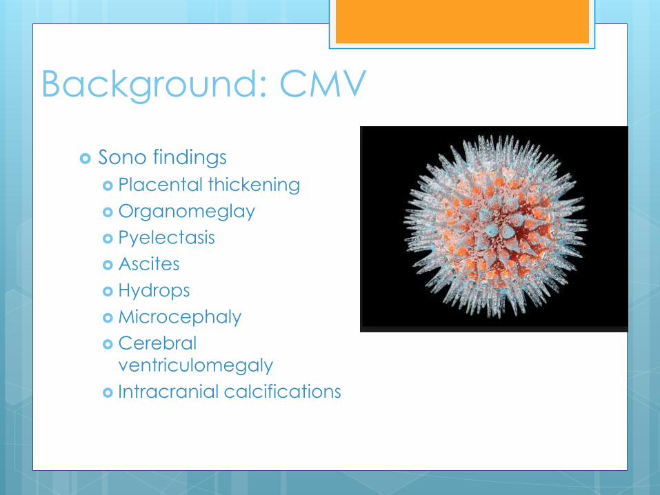
Background: CMV
Sono findings
Placental thickening
Organomeglay
Pyelectasis
Ascites
Hydrops
Microcephaly
Cerebral
ventriculomegaly
Intracranial calcifications
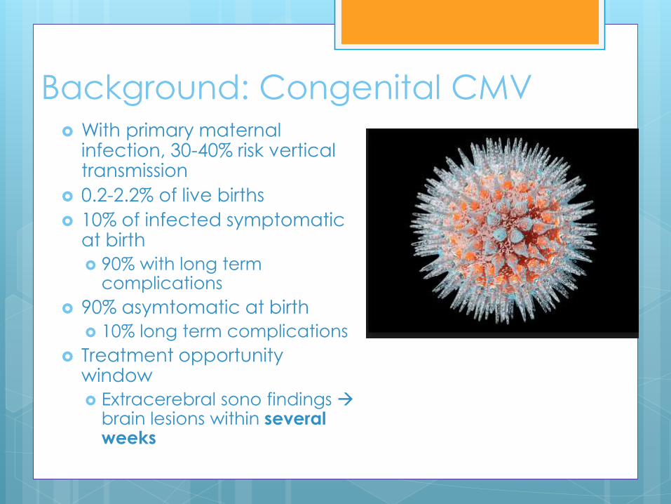
Background: Congenital CMV With primary maternal
infection, 30-40% risk vertical transmission
0.2-2.2% of live births
10% of infected symptomatic at birth
90% with long term complications
90% asymtomatic at birth
10% long term complications
Treatment opportunity window
Extracerebral sono findings brain lesions within several weeks

Background: Congenital CMV
No vaccination
Current treatment options
CMV-Ig to prevent transmission motherfetus
Neonatal antiviral
treatment with ganciclovir
and valganciclovir
improves outcomes
Highly genotoxic, not
approved in pregnancy
Valacylcovir less effective
in vitro, but proven safety

Background: Valacyclovir
Direct inhibition of CMV
replication
In renal transplants and
HIV infection, high dose
regimens (8g/day) can
prevent disease and
suppress viremia.
Currently use 1g/day for
HSV suppression

Methods: Study Design
CMV infected pregnancy with at least
one sign of fetal infection
Multi center, Open-label
Phase II
One arm
(all participants received drug)
Failed to enroll sufficient for RCT

Methods: Study Design
2g Oral Valcyclovir 4 times a day
Treatment from diagnosis through delivery
Or 24 completed weeks
Clinical visits q 2 weeks
Monthly lab eval (LFTs, Cr)
Neonatal eval day 4 and 7 of life

Methods: Study Endpoint
Proportion of asymptomatic neonates
born to women with valacyclovir
Birthweight >10th%
Normal: cerebral imaging
Normal auditory testing
Normal funduscopic exam
Normal labs and clinical exam

Secondary Endpoints
Medication tolerance/adherance
CMV DNA levels and platelet counts pre
and post treatment
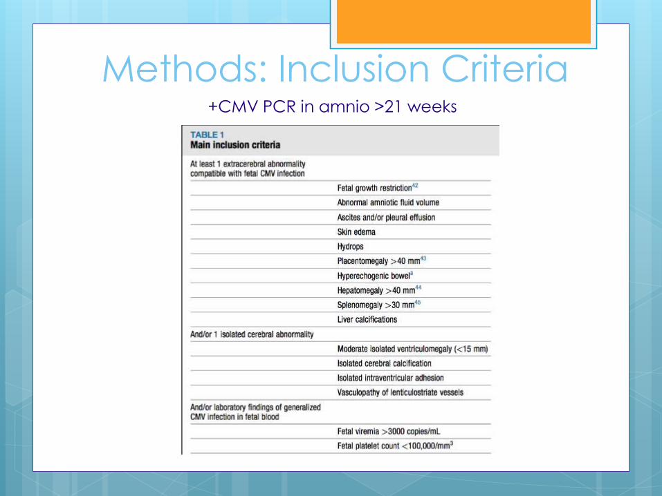
Methods: Inclusion Criteria+CMV PCR in amnio >21 weeks
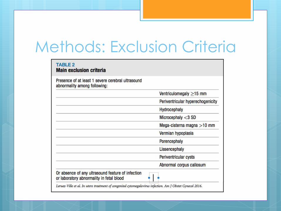
Methods: Exclusion Criteria

Methods: “Historical
Comparator Group”
Lit review
Limited to English, detailed descriptors or
ultrasound and neonatal outcomes
724 pregnancies
217 infected fetuses
47 with matching ultrasonographic inclusion
criteria
42.5% born asymptomatic

Methods: Statistical analysis
Simon 2-stage design
Clinical relevance if >80% asymptomatic
Type I error fixed at .05
Power 80%
Stage 1: 11 infected fetuses, if 8 with good outcome proceed to stage 2 (32
additional)
Analysis: all fetuses (ITT), 2 sided T-tests

Enrollment41 pregnant women enrolled
40 completed treatment
41 newborns + terminated
fetuses evaluated

Results - descriptive
41 women
Avg age 31
97.6% with primary infection
Avg GA of infection = 10
53% of fetuses had >1 symptom on sono
Most commonly hyperechoic bowel (58%),
splenomegaly (21%), placentomegaly
(30%)

Results
Primary Endpoint
82% of neonates were asymptomatic vs43% in historical
Secondary Endpoint
>90% Adherence to medication, well tolerated
No difference in DNA viral load bwsymptomatic and asymptomatic neonates
Neonatal platelet count significantly lower in symptomatic neonates

Results
5 year follow-up
Not yet complete
Range 3-36 months
Asymptomatic
Remained asymptomatic
Symptomatic
None progressed to worsening sxs

Conclusions
Significant increase of asymptomatic neonates with treatment
Safe/well tolerated medication (16 tabs/day)
Only studied neonates already showing signs of infection
Viral load did not correlate with disease progression
Long term follow-up not complete

Strengths
Single lab
Intention to treat with low drop out rate
(>90% adherence)
First ever to study valacyclovir as
treatment for CMV
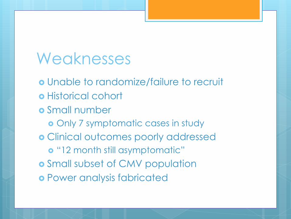
Weaknesses
Unable to randomize/failure to recruit
Historical cohort
Small number
Only 7 symptomatic cases in study
Clinical outcomes poorly addressed
“12 month still asymptomatic”
Small subset of CMV population
Power analysis fabricated

Next steps: cmRCT
“Cohort Multiple
Randomized
Control Trial”

Will it change my practice?
I will continue to consult MFM for my congenital CMV expertise

Now recruiting…
RCT of wine cork vs pacifier to
increase maternal sleep

Original Research ajog.org
OBSTETRICS
In utero treatment of congenital cytomegalovirusinfection with valacyclovir in a multicenter,open-label, phase II study
Marianne Leruez-Ville, MD, PhD; Idir Ghout, MSc; Laurence Bussières, PhD;Julien Stirnemann, MD, PhD; Jean-François Magny, MD; Sophie Couderc, MD;Laurent J. Salomon, MD, PhD; Tiffany Guilleminot, BA; Philippe Aegerter, MD, PhD;Guillaume Benoist, MD, PhD; Norbert Winer, MD; Olivier Picone, MD, PhD;François Jacquemard, MD; Yves Ville, MD, FRCOGBACKGROUND: Congenital infection with human cytomegalovirus RESULTS: At the interim analysis, 8 of 11 women delivered an
is a major cause of morbidity and mortality. A randomized controlled
trial showed that high-dosage valacyclovir prevents cytomegalovirus
disease in transplant recipients. Fetuses showing ultrasound features
of infection are at high risk of being symptomatic at or before birth.
In a pilot study, oral administration of high-dosage valacyclovir to
mothers significantly decreased viral load and produced therapeutic
concentrations in the blood of infected fetuses. A randomized
controlled trial comparing prenatal treatment with valacyclovir against
placebo in infected fetuses failed to recruit because women declined
randomization. Randomized controlled trials in fetal medicine have
often proven unacceptable by women who decline termination of
pregnancy and are not prepared to resign themselves to the odds of
the natural history of the disease.
OBJECTIVE:We evaluated the efficacy of oral valacyclovir, 8 g daily, forpregnant women carrying a symptomatic cytomegalovirus-infected fetus,
targeting a high-risk group for developing both neurosensory and neuro-
logical impairment.
STUDY DESIGN: We designed a multicenter, open-label, phase II
study with 1 arm, using one of Simon’s optimal 2-stage designs. Symp-
tomatic fetuses were defined by the presence of measurable extracerebral
or mild cerebral ultrasound symptoms. They were treated in utero from
prenatal diagnosis at a median of 25.9 weeks’ gestation until delivery or
termination of pregnancy. Fetuses with severe brain anomalies on ultra-
sound were not included as were cases completely asymptomatic at
presentation, because treatment was unlikely to modify either outcome.
The primary endpoint was the proportion of asymptomatic neonates born
to treated mothers.
Cite this article as: Leruez-Ville M, Ghout I, Bussieres L,et al. In utero treatment of congenital cytomegalovirus
infection with valacyclovir in a multicenter, open-label,
phase II study. Am J Obstet Gynecol 2016;215:
462.e1-10.
0002-9378ª 2016 The Authors. Published by Elsevier Inc. This is an
open access article under the CC BY-NC-ND license (http://
creativecommons.org/licenses/by-nc-nd/4.0/).http://dx.doi.org/10.1016/j.ajog.2016.04.003
462.e1 American Journal of Obstetrics & Gynecology OCTOBER 2016
asymptomatic neonate (required:�7). In step 2, 32 additional cases were
included for a total of 43; the final number of asymptomatic neonates was
34, more than the 31 required to indicate efficacy according to the Simon
2-stage design. They remained asymptomatic at 12 months. High-dosage
valacyclovir given for a median of 89 days to pregnant women carrying a
moderately infected fetus was efficient at giving birth to asymptomatic
neonates. Fetal blood viral loads decreased and platelet counts increased,
both significantly (P¼ .01 and P< .001, respectively), between treatment
initiation and birth after treatment completion, regardless of duration of
fetal infection. Compared with a historical cohort obtained by a meta-
analysis of the literature, the use of valacyclovir (8 g daily) significantly
increased the proportion of asymptomatic neonates from 43% without
treatment to 82% with treatment. Although the pill burden was high
(16 pills a day) adherence to treatment was>90%. Finally, valacyclovir at
this high dosage was extremely well tolerated.
CONCLUSION: Our results indicate that high-dosage valacyclovir
given in pregnancy is effective for improving the outcome of moderately
symptomatic infected fetuses. Although this study is not a randomized
controlled trial, this is the first study reporting the efficacy of an antiviral
drug to treat cytomegalovirus-infected fetuses. Moreover, this first study
will allow new trials to be conducted, using valacyclovir as a baseline safe
and effective treatment in pregnancy, to be compared to the new emerging
and more potent anticytomegalovirus drugs that have not currently been
tested in pregnancy.
Key words: congenital infection, cytomegalovirus, fetal therapy, fetus,symptomatic, valaciclovir
IntroductionCongenital cytomegalovirus (CMV)infection affects 0.7% of live births and isthe leading cause of congenital neuro-logical disease of infectious origin.1
Among all neonates positive for infec-tion on screening, 20% eventually haveneurodevelopmental impairment withpermanent sequelae.2 Around 10% ofinfected neonates are symptomatic atbirth; their risk of sequelae reaches 58%,including sensorineural hearing loss or
cognitive or motor defects. The risk ofsequelae in newborns who were asymp-tomatic at birth is around 13%, mainlydue to progressive hearing loss.2
An infected fetus’s risk of symptomsat birth is assessed by interpreting theresults of both prenatal imaging andlaboratory tests.3-8 Fetal CMV disease isprogressive: early symptoms of systemicinfection can be expressed as extracere-bral findings at prenatal ultrasound; fetalbrain involvement usually does not show

TABLE 1Main inclusion criteria
At least 1 extracerebral abnormalitycompatible with fetal CMV infection
Fetal growth restriction42
Abnormal amniotic fluid volume
Ascites and/or pleural effusion
Skin edema
Hydrops
Placentomegaly >40 mm43
Hyperechogenic bowela
Hepatomegaly >40 mm44
Splenomegaly >30 mm45
Liver calcifications
And/or 1 isolated cerebral abnormality
Moderate isolated ventriculomegaly (<15 mm)
Isolated cerebral calcification
Isolated intraventricular adhesion
Vasculopathy of lenticulostriate vessels
And/or laboratory findings of generalizedCMV infection in fetal blood
Fetal viremia >3000 copies/mL
Fetal platelet count <100,000/mm3
All ultrasound examinations leading to inclusion in study were reviewed by principal investigator at each center. Measurementsof spleen, liver, and placenta were standardized according to literature.43-45
CMV, cytomegalovirus.
a Since diagnosis of hyperechogenic bowel can be subjective and associated with high interoberver and intraobservervariability, diagnosis of hyperechogenic bowel was only considered for grade-2 or grade-3 hyperechogenic bowel46; thissemiquantitative analysis was chosen to limit subjectivity sometimes associated with ultrasound findings.
Leruez-Ville et al. In utero treatment of congenital cytomegalovirus infection. Am J Obstet Gynecol 2016.
ajog.org OBSTETRICS Original Research
until several weeks later.9 Severe brainlesions seen on prenatal ultrasoundpredict a dismal outcome.4 This leaves awindow of opportunity for treatmentof symptomatic fetuses without braininvolvement.10
Vaccination is not available11 andno prenatal treatment of congenitalCMV has yet been validated. The use ofCMV-specific hyperimmune globulinto prevent transmission from motherto fetus has produced conflicting re-sults.12,13 Neonatal antiviral treatmentwith either ganciclovir or valganciclovirimproves auditory and neurologicaloutcomes in symptomatic newborns,14
but these drugs, highly genotoxicin vitro, are not approved in pregnancy.Although valacyclovir is less effective
than ganciclovir against CMV in vitro,15
high-dosage valacyclovir has provenclinically efficient to prevent CMVdisease in transplant recipients.16 Themechanism of acyclovir’s anti-CMVactivity in clinical settings remains un-explained. Valacyclovir also has the bestsafety profile of the anti-CMV drugs.Neither cell transformation norincreased risk of neoplasia has been re-ported in vitro, and no increased risk ofbirth defects has been detected inthe offspring of thousands of womenexposed during pregnancy.17,18 Finally,valacyclovir is well tolerated with rareside effects. In a pilot study, we foundthat oral administration of high-dosagevalacyclovir to mothers significantlydecreased viral load and produced
OCTOBER 2016 Ameri
therapeutic concentrations in fetal bloodwith a mean fetal blood plasma con-centration of >17 mmol/L. These resultssuggested the value of a clinical trialto investigate this therapeutic optionfurther.10 We failed to complete a ran-domized controlled trial comparingprenatal treatment with valacycloviragainst placebo in moderately symp-tomatic infected fetuses due to failure torecruit (Cymeval NCT01037712). In thisopen-label phase II trial with 1 arm weshow that high-dosage valacyclovir givenin pregnancy is safe and appears effectivefor improving the outcome of moder-ately symptomatic fetuses.
Materials and MethodsPatientsEligible women were pregnant with aninfected fetus identified by a positiveCMV polymerase chain reaction assay inamnioticfluid, sampled by amniocentesis>21 weeks,19-22 together with the pres-ence of �1 extracerebral ultrasoundfeatures compatible with CMV infectionand/or 1 isolated cerebral abnormalityand/or 1 of the following laboratoryfindings in fetal blood: fetal platelet count<100,000/mm3 or CMV DNA viral load>3000 copies/mL (Table 1). The presenceof severe ultrasound brain abnormalities(Table 2) and the absence of any ultra-sound feature of infection or laboratoryabnormality in fetal blood were exclusioncriteria. Thedetailed eligibility criteria arelisted in the supplementary Appendix.Fetal blood sampling by cordocentesisunder ultrasound guidance was offered toall participants to evaluate fetal plateletsand viral DNA load to help refine thefetal prognosis.4 Cordocentesis was not,however, required for study eligibility, asthis invasive procedure was not the stan-dard of care in all participating centers.
ProceduresStudy designThe trial was a multicenter, open-label,phase II study with 1 arm, based on 1 ofSimon’s23 optimal 2-stage designs. Allparticipants received oral valacyclovir(2 g, 4 times a day, therefore 8 g daily).The medication was continued untildelivery or 24 treatment weeks, which-ever was sooner. The study drug was
can Journal of Obstetrics & Gynecology 462.e2

TABLE 2Main exclusion criteria
Presence of at least 1 severe cerebral ultrasoundabnormality among following:
Ventriculomegaly �15 mm
Periventricular hyperechogenicity
Hydrocephaly
Microcephaly <3 SD
Mega-cisterna magna >10 mm
Vermian hypoplasia
Porencephaly
Lissencephaly
Periventricular cysts
Abnormal corpus callosum
Or absence of any ultrasound feature of infectionor laboratory abnormality in fetal blood
Leruez-Ville et al. In utero treatment of congenital cytomegalovirus infection. Am J Obstet Gynecol 2016.
Original Research OBSTETRICS ajog.org
purchased fromGlaxoSmithKline (MarlyLe Roi, France), which had no other rolein the study. Visits for clinical and ultra-sound examinations, questions aboutclinical side effects (headache, nausea,neurologic effects listed in the productmonograph), and adherence assessmentby pill count were scheduled every 2weeks until delivery. Maternal plasmalevels of aspartate aminotransferase,alanine aminotransferase, and creatininewere assessed once a month duringtreatment. All newborns were examinedbetween days 4 and 7 of life by a trainedpediatrician according to a standardizedclinical evaluation. Auditory brainstemresponses, cranial ultrasound, fundo-scopy, and laboratory tests (see criteriafor primary endpoint in supplementarydata) were also performed.
The ethics committee of Poissy-SaintGermain Hospital approved the study(2011-001610-34). Participants gavewritten informed consent before inclu-sion. Study oversight was provided by anindependent data and safety monitoringboard (Clinical Research Unit, Cochin-Necker, Paris).
Study endpointsThe primary study endpoint was theproportion of asymptomatic neonatesborn to women treated with valacyclovir.An asymptomatic neonate was a neonatewithout growth restriction (that is, withbirthweight �10th percentile), normalclinical examination, normal laboratoryfindings, no severe features of infectionon cerebral imaging, normal fundu-scopic examination, and normal audi-ology findings (see the supplementaryAppendix for details).
The secondary endpoints includedadverse events related to the study medi-cation and adherence to treatment. CMVDNA levels and platelet counts werecompared in pretreatment fetal bloodwhen available and cord blood at birthand in symptomatic and asymptomaticneonates. The effect of the duration ofmaternal treatment was also assessed.
Historical comparator groupOur systematic review on PubMedusing 3 key words (“cytomegalovirus,”“congenital,” “ultrasound”) yielded 216
462.e3 American Journal of Obstetrics & Gynecol
articles, but only 3 were written inEnglish and included detailed tables thatdescribed both prenatal ultrasoundfindings and neonatal outcome and alsoincluded postmortem findings in termi-nation of pregnancy cases.10,24,25 These 3suitable studies included 724 pregnancieswith a maternal CMV primary infection,217 with an infected fetus. The reviewof the ultrasound symptoms of these217 fetuses showed no ultrasound ab-normalities in 142 of them and severecerebral ultrasound abnormalities in 28.Ultrasound abnormalities matching ourinclusion criteria were therefore seen in47 cases, which formed the historicalcomparator group. Among them 20(42.55%) neonates were born asymp-tomatic. Twenty fetuses were terminatedand all underwent postmortem exami-nation showing both macroscopic andmicroscopic evidence of brain damagein all cases. We therefore assumed thatthese fetuses would have been bornsymptomatic and classified then into thesymptomatic group. See Table S1 fordetails in the supplementary Appendix.
Laboratory assaysCMV serology and viral load quantifi-cation were centralized and analyzed atthe Necker Hospital virology laboratory.Maternal CMV primary infection was
ogy OCTOBER 2016
diagnosed by seroconversion or theconcomitant presence of CMV-specificIgM antibodies and low IgG avidity.22
After extracting DNA from fetal wholeblood, cord whole blood, and neonatalurine with the MagnaPure LC platform(Roche Diagnostic, Meylan, France), weperformed quantitative polymerasechain reaction for CMVwith the CMV-Rgene kit (Argene, BioMerieux, Marcy-L’Etoile, France). The limit of detectionwas 446 copies (or 178 IU)/mL.
Statistical analysisTo estimate the sample size accordingto Simon23 optimal 2-stage design, weassumed that a proportion of asymp-tomatic neonates of <60% was notclinically relevant in relation to valacy-clovir efficacy, while a proportion of>80% was deemed acceptable. With atype I error fixed at 0.05 and a power of80%, we needed 11 infected fetuses forthe first stage: if at least 8 cases in stage 1had a good outcome (were asymptom-atic), then 32 additional infected fetuseswould be included in stage 2 for a totalof 43. If at least 31 of the 43 caseswere asymptomatic at birth, valacyclovirwould be judged to have a positive effect.
Analyses of efficacy data wereperformed for all included fetuses(intention to treat). The proportion of
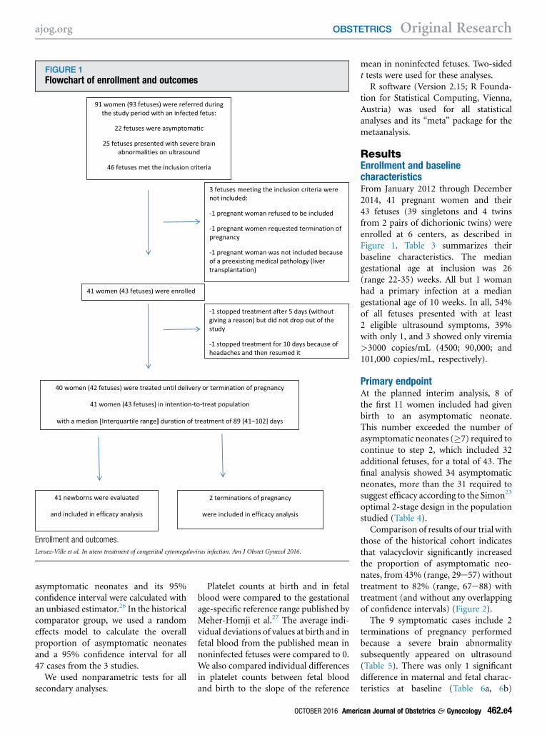
FIGURE 1Flowchart of enrollment and outcomes
Enrollment and outcomes.
Leruez-Ville et al. In utero treatment of congenital cytomegalovirus infection. Am J Obstet Gynecol 2016.
ajog.org OBSTETRICS Original Research
asymptomatic neonates and its 95%confidence interval were calculated withan unbiased estimator.26 In the historicalcomparator group, we used a randomeffects model to calculate the overallproportion of asymptomatic neonatesand a 95% confidence interval for all47 cases from the 3 studies.
We used nonparametric tests for allsecondary analyses.
Platelet counts at birth and in fetalblood were compared to the gestationalage-specific reference range published byMeher-Homji et al.27 The average indi-vidual deviations of values at birth and infetal blood from the published mean innoninfected fetuses were compared to 0.We also compared individual differencesin platelet counts between fetal bloodand birth to the slope of the reference
OCTOBER 2016 Ameri
mean in noninfected fetuses. Two-sidedt tests were used for these analyses.
R software (Version 2.15; R Founda-tion for Statistical Computing, Vienna,Austria) was used for all statisticalanalyses and its “meta” package for themetaanalysis.
ResultsEnrollment and baselinecharacteristicsFrom January 2012 through December2014, 41 pregnant women and their43 fetuses (39 singletons and 4 twinsfrom 2 pairs of dichorionic twins) wereenrolled at 6 centers, as described inFigure 1. Table 3 summarizes theirbaseline characteristics. The mediangestational age at inclusion was 26(range 22-35) weeks. All but 1 womanhad a primary infection at a mediangestational age of 10 weeks. In all, 54%of all fetuses presented with at least2 eligible ultrasound symptoms, 39%with only 1, and 3 showed only viremia>3000 copies/mL (4500; 90,000; and101,000 copies/mL, respectively).
Primary endpointAt the planned interim analysis, 8 ofthe first 11 women included had givenbirth to an asymptomatic neonate.This number exceeded the number ofasymptomatic neonates (�7) required tocontinue to step 2, which included 32additional fetuses, for a total of 43. Thefinal analysis showed 34 asymptomaticneonates, more than the 31 required tosuggest efficacy according to the Simon23
optimal 2-stage design in the populationstudied (Table 4).
Comparison of results of our trial withthose of the historical cohort indicatesthat valacyclovir significantly increasedthe proportion of asymptomatic neo-nates, from 43% (range, 29e57) withouttreatment to 82% (range, 67e88) withtreatment (and without any overlappingof confidence intervals) (Figure 2).
The 9 symptomatic cases include 2terminations of pregnancy performedbecause a severe brain abnormalitysubsequently appeared on ultrasound(Table 5). There was only 1 significantdifference in maternal and fetal charac-teristics at baseline (Table 6a, 6b)
can Journal of Obstetrics & Gynecology 462.e4

TABLE 4Primary endpoint and efficacy
First step Second step
Outcome no.
Asymptomatic neonates 8 34
Symptomatic neonates or termination of pregnancy 3 9
Total 11 43
Leruez-Ville et al. In utero treatment of congenital cytomegalovirus infection. Am J Obstet Gynecol 2016.
TABLE 3Characteristics of population at baseline
CharacteristicsMedian [interquartilerange] or n (%)
Women (N ¼ 41)
Age at inclusion, y 31.2 [28.6e33.9]
Body mass index before pregnancy 21.6 [19.8e23]
Parity
0 11 (26.8)
�1 30 (73.2)
No. of pregnancies 2 [2e3]
Primary infectiona 40 (97.6)
Gestational age at primary infection, wk 10 [7.8e16.2]
Gestational age at inclusion, wk 25.9 [24.1e31.7]
Interval between primary infection and inclusion, wk 16 [12.3e18.6]
Fetuses (N ¼ 43)
Only 1 symptom at ultrasound 17 (39.5)
>1 Symptom at ultrasound 23 (53.5)
Fetal blood CMV DNA load >3000 copies/mL 3 (7)
Fetal growth restriction 3 (7)
Abnormal amount of amniotic fluid 3 (7)
Ascites and/or pleural effusion 1 (2.3)
Placentomegaly 13 (30.2)
Hyperechogenic bowel 25 (58.1)
Hepatomegaly 6 (14)
Splenomegaly 9 (20.9)
Liver calcification 1 (2.3)
Moderate cerebral abnormality 5 (11.6)
CMV, cytomegalovirus.
a Forty women had primary infection and 1 woman was diagnosed with secondary infection.
Leruez-Ville et al. In utero treatment of congenital cytomegalovirus infection. Am J Obstet Gynecol 2016.
Original Research OBSTETRICS ajog.org
between the cases with good (asymp-tomatic neonates) and poor (symptom-atic neonates or termination ofpregnancy) outcomes: fetal plateletcount at inclusion, which was signifi-cantly lower in the poor outcome group(P < .007) (Table 6b). The durationof maternal treatment did notdiffer significantly between the 2 groups(P ¼ .236) (Table 6a).
Table S2 reports all details on preg-nancy outcome (gestational age atdelivery, birthweight, clinical examina-tion, imaging, and laboratory data).
Secondary endpointsAdverse eventsValacyclovir was well tolerated. Only2 women reported headaches, and treat-ment was suspended for 10 days in only1. Although maternal alanine amino-transferase and aspartate aminotrans-ferase levels increased after 3 months oftreatment, this increase was not clinicallyrelevant; all values were <40 IU/L, andcreatinine levels did not changethroughout treatment (Figure S1).Adherence in the subgroup of 27 womenevaluated for it was >90% (Table S3).
Viral loads and platelet countsAlthough the duration of maternaltreatment was not correlated with CMVDNA levels in either cord blood (P¼.65)or neonatal urine (P ¼ .24), longerduration was associated with a higherplatelet count at birth (P ¼ .018)(Figure S2 and Table S4). Neither viralloads nor platelet counts at birth werecorrelated with the time of maternalinfection in pregnancy and thereforewith the duration of fetal infection(Table S4). Symptomatic and asymp-tomatic neonates did not differ signi-ficantly for viral DNA load levelsin either neonatal cord blood (P ¼ .391)or neonatal urine (P ¼ .081), but theneonatal platelet count was significantlylower in symptomatic neonates (P <.001) (Table S5). Blood viral loaddecreased and platelet count increased,both significantly, between the fetalblood obtained in utero before maternaltreatment began and the cord bloodsampled at birth (P ¼ .01 and P < .001,respectively) (Table 7). The mean
462.e5 American Journal of Obstetrics & Gynecol
increase in platelet counts from thebeginning to the end of valacyclovirtreatment was significantly higher than
ogy OCTOBER 2016
the expected corresponding increasein noninfected fetuses (P ¼ .008)(Figure S3).
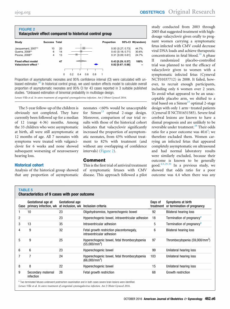
FIGURE 2Valacyclovir effect compared to historical control group
Study
Fixed effect model Valaciclovir effect *
Jacquemard, 200710
Guerra, 200824 Picone, 200825
Success
10 6 4
Total
47
20 14 13
0 0.2 0.4 0.6 0.8 1
Proportion
0.43 0.82
0.50 0.43 0.31
95%-CI
[0.29; 0.57] [0.67; 0.88]
[0.27; 0.73] [0.18; 0.71] [0.09; 0.61]
W(random)
100% --
44.7% 30.6% 24.7%
Proportion of asymptomatic neonates and 95% confidence interval (CI) were calculated with un-biased estimator.26 In historical control group, we used random effects model to calculate overallproportion of asymptomatic neonates and 95% CI for 43 cases reported in 3 suitable publishedstudies. *Unbiased estimation of binomial probability in multistage design.
Leruez-Ville et al. In utero treatment of congenital cytomegalovirus infection. Am J Obstet Gynecol 2016.
ajog.org OBSTETRICS Original Research
The 5-year follow-up of the children isobviously not completed. They havecurrently been followed up for a medianof 12 (range 4-36) months. Amongthe 33 children who were asymptomaticat birth, all were still asymptomatic at12 months of age. All 7 neonates withsymptoms were treated with valganci-clovir for 6 weeks and none showedsubsequent worsening of neurosensoryhearing loss.
Historical cohortAnalysis of the historical group showedthat any proportion of asymptomatic
TABLE 5Characteristics of 9 cases with poor ou
CaseGestational age atprimary infection, wk
Gestational ageat inclusion, wk
1 10 23
2 23
3 13 35
4 9 32
5 9 25
6 6 23
7 7 24
8 8 22
9 Secondary maternalinfection
26
a Two terminated fetuses underwent postmortem examination a
Leruez-Ville et al. In utero treatment of congenital cytomega
neonates <60% would be unacceptablefor Simon23 optimal 2-stage design.Moreover, comparison of our trial re-sults with those of the historical cohortindicates that valacyclovir significantlyincreased the proportion of asymptom-atic neonates, from 43% without treat-ment to 82% with treatment (andwithout any overlapping of confidenceintervals) (Figure 2).
CommentThis is the first trial of antiviral treatmentof symptomatic fetuses with CMVdisease. This approach followed a pilot
tcome
Inclusion criteriaDt
Oligohydramnios, hyperechogenic bowel
Hyperechogenic bowel, intraventricular adhesion
Intraventricular adhesion
Fetal growth restriction placentomegaly,intraventricular adhesion
Hyperechogenic bowel, fetal thrombocytopenia(55,000/mm3)
Hyperechogenic bowel
Hyperechogenic bowel, fetal thrombocytopenia(66,000/mm3)
1
Hyperechogenic bowel
Fetal growth restriction
nd in both cases severe brain lesions were identified.
lovirus infection. Am J Obstet Gynecol 2016.
OCTOBER 2016 Ameri
study conducted from 2003 through2005 that suggested treatment with high-dosage valacyclovir given orally to preg-nant women carrying a symptomaticfetus infected with CMV could decreaseviral DNA loads and achieve therapeuticconcentrations in fetal blood.10 A phaseII randomized placebo-controlledtrial was planned to test the efficacy ofvalacyclovir given to women with asymptomatic infected fetus (CymevalNCT01037712) in 2008. It failed, how-ever, to recruit enough participants,including only 6 women over 2 years.To avoid what appeared to be an unac-ceptable placebo arm, we shifted to atrial based on a Simon23 optimal 2-stagedesign with only 1 armetreated patients(Cymeval II NCT01651585). Severe fetalcerebral lesions are known to have adismal prognosis and are unlikely to bereversible under treatment.28 Their oddsratio for a poor outcome was 40.64; wetherefore excluded them. Women car-rying an infected fetus that appearedcompletely asymptomatic on ultrasoundand had normal laboratory resultswere similarly excluded, because theiroutcome is known to be generallygood.4,9,29,30 In a previous study, weshowed that odds ratio for a pooroutcome was 4.4 when there was any
ays ofreatment
Symptoms at birthor termination of pregnancy
92 Bilateral hearing loss
18 Termination of pregnancya
5 Termination of pregnancya
6 Bilateral hearing loss
97 Thrombocytopenia (59,000/mm3)
99 Unilateral hearing loss
03 Unilateral hearing loss
15 Unilateral hearing loss
68 Growth restriction
can Journal of Obstetrics & Gynecology 462.e6

TABLE 6Comparison of maternal and fetal characteristics at baseline between cases with good (asymptomatic neonate)and poor (symptomatic neonate or termination of pregnancy) outcomes
Table 6a.Women’s characteristics
Total Good outcome Poor outcome
P
Median [interquartile range] Median [interquartile range] Median [interquartile range]
(N ¼ 41) (N ¼ 33) (N ¼ 8)
Maternal age at inclusion, y 31.2 [28.6e33.9] 31.5 [28.8e33.9] 30.2 [28.1e33.5] .717
Body mass index before pregnancy 21.6 [19.8e23] 20.4 [19.4e22.7] 22.9 [21.8e23.9] .058
Parity, n (%)
0 11 (26.8) 8 (24.2) 3 (37.5) .744
1 20 (48.8) 17 (51.5) 3 (37.5)
2 9 (22) 7 (21.2) 2 (25)
3 1 (2.4) 1 (3) 0 (0)
No. of pregnancies 2 [2e3] 2 [2e3] 2 [1.8e2.5] .73
Primary infection, n (%) 40 (97.6) 33 (100) 7 (87.5) .195
Gestational age at primary infection, wk 10 [7.8e16.2] 11 [8e17] 9 [7.5e9.5] .199
Gestational age at inclusion, wk 25.9 [24.1e31.7] 27 [24.6e31.7] 24.6 [23.8e27.6] .411
Interval between primary infectionand inclusion, wk
16 [12.3e18.6] 15.9 [12.1e18.6] 17.1 [15.9e20] .182
Treatment interruption, n (%) 2/39 (5.1) 1/33 (3) 1/6 (16.7) .287
Duration of treatment, d 89 [41e102] 89 [43e102] 80 [15e97.5] .236
Table 6b.Fetal characteristics
Total Good outcome Poor outcome
P
Median [interquartile range] Median [interquartile range] Median [interquartile range]
(N ¼ 43) (N ¼ 34) (N ¼ 9)
Inclusion criteria, n (%)
Only 1 symptom at ultrasound 17 (39.5) 15 (44.1) 2 (22.2) .332
Fetal blood DNA load >3000 copies/mL 3 (7) 3 (8.8) 0 (0)
>1 Symptom at ultrasound 23 (53.5) 16 (47.1) 7 (77.8)
Fetal growth restriction, n (%) 3 (7) 1 (2.9) 2 (22.2) .106
Abnormal amount of amniotic fluid, n (%) 3 (7) 2 (5.9) 1 (11.1) .515
Ascites and/or pleural effusion, n (%) 1 (2.3) 1 (2.9) 0 (0) 1
Placentomegaly, n (%) 13 (30.2) 11 (32.4) 2 (22.2) .699
Hyperechogenic bowel, n (%) 25 (58.1) 19 (55.9) 6 (66.7) .712
Hepatomegaly, n (%) 6 (14) 5 (14.7) 1 (11.1) 1
Splenomegaly, n (%) 9 (20.9) 7 (20.6) 2 (22.2) 1
Liver calcifications, n (%) 1 (2.3) 0 (0) 1 (11.1) .209
Moderate cerebral anomalies,a n (%) 5 (11.6) 2 (5.9) 3 (33.3) .054
Fetal viremia at inclusion in log10 IU/mL 4.4 [4e5] 4.3 [3.8e4.8] 5.1 [4.4e5.9] .1
Fetal platelet count at inclusion/mm3 174,000 [145,000e208,000] 177,500 [155,250e208,000] 67,000 [61,000e88,000] .006a Moderate isolated ventriculomegaly (<15 mm) or isolated cerebral calcification or isolated intraventricular adhesion or vasculopathy of lenticulostriate vessels.
Leruez-Ville et al. In utero treatment of congenital cytomegalovirus infection. Am J Obstet Gynecol 2016.
Original Research OBSTETRICS ajog.org
462.e7 American Journal of Obstetrics & Gynecology OCTOBER 2016

TABLE 7Correlation of fetal and neonatal laboratory indicators (viral DNA load and platelet count, from cord blood,compared with viral DNA load and fetal platelet count obtained in utero before inclusion)
Fetal blood before beginningmaternal treatment Neonatal cord blood Differencesa P
Viral DNA in blood, log10 IU/mL
Median (interquartile range) 4.0 (3.55e4.6) 3.05 (2.57e3.92) e0.5 (e2.075 to e0.075) .01
N 28 32 24
Platelet count/mm3
Median (interquartile range) 173,000 (141,500e201,500) 245,000 (193,000e274,000) 101,000 (47,500e122,000) <.001
N 27 41 27a Differences between values obtained for same case in utero in fetal blood before maternal treatment and in neonatal cord blood after treatment. Participants with viral DNA load less than thresholdvalue of 178 IU were considered to have viral DNA load of 89 IU (1.94 IU).
Leruez-Ville et al. In utero treatment of congenital cytomegalovirus infection. Am J Obstet Gynecol 2016.
ajog.org OBSTETRICS Original Research
noncerebral ultrasound abnormality and1.13 for each 10,000/mm3 decrease infetal platelet count <100,000/mm3.4
Another observational study showedthat a fetal platelet count <50,000/mm3
had 80% predictive value for pooroutcome.6 We therefore enrolled womenwith either extracerebral or nonseverecerebral ultrasound features, a lowplatelet count, or a high viral load inblood sampled in utero.4,6,31 Thesecriteria were selected specifically totarget a group at high risk for progressivedamage, on the hypothesis that prenataltreatment could lower this risk thatcould lead to neonatal neuro-developmental impairment.
According to Simon23 optimal 2-stagedesign, high-dosage valacyclovir givenorally to pregnant women was effectivein improving the outcome of pregnan-cies with a fetus infected with symp-tomatic CMV, shown by eitherextracerebral or nonsevere cerebral ul-trasound features or abnormal fetal lab-oratory results. Moreover, whencompared with a historical cohort ofsimilar cases collected from the litera-ture,10,24,25,32,33 an unbiased estimationconfirmed the efficacy of valacyclovircompared to no treatment. Amongthe 43 symptomatic fetuses treated inutero, there were only 4 cases with anindisputably poor outcome (2 termina-tions of pregnancy for severe brainabnormalities and 2 cases of bilateralhearing loss), while the other 5 infantssymptomatic at birth showed only mild
disease (3 with unilateral hearing loss,1 with isolated thrombocytopenia, andanother with growth restriction). Theclinical impact of valacyclovir may beexplained by its direct inhibition of CMVreplication. In renal transplantation andin HIV infection, high-dose regimens ofvalacyclovir are effective in preventingCMV disease and suppressing CMVviremia.16,34,35 A sort of placebo effectmay have also participated in the effect ofvalacyclovir. That is, although the par-ticipants were informed that the efficacyof valacyclovir in utero was unknownand that they could request a termina-tion of pregnancy in accordance withFrench law, only 2 terminations wererequested, both after fetuses developedsevere cerebral symptoms visible on ul-trasound. The availability of treatmentmay have alleviated parental anxiety anddissuaded women from requestingtermination of pregnancy by offeringthem something more than helplessanxiety and expectant management.Although the valacyclovir dosage used
in this study was much higher than thatused for treatment of herpes simplexinfection in pregnancy (8 g per day vs 1 gper day), the maternal clinical andlaboratory tolerances were excellent andno adverse effect was observed in theneonates.Moreover, despite the burden of taking
16 tablets throughout the day, cumula-tive adherence to treatment was >90%.Overall, 40% of the women met
the inclusion criterion of 1 ultrasound
OCTOBER 2016 Ameri
abnormality suggestive of CMV, 53%had >1, and 7% had only a high fetalblood viral load (>3000 copies/mL). Thelatter criterion was selected on the basisof previous work by Boppana et al,31 whoreported that no neonates with a viralload <3000 copies developed hearingloss. However, a more recent studyreports that fetal viral load >30,000copies/mL is a predictivemarker for pooroutcome.6 Therefore, the cut-off of 3000copies/mL might have been too low.Nonetheless, only 3 cases were includedbased on this criterion alone, and 2 ofthem had much higher viral loads(110,000 and 90,000 copies/mL). Noneof these 3 patients had a poor outcome.
At birth, viral loads in cord blood weresignificantly lower and platelet countssignificantly higher than in fetal bloodobtained in utero before treatmentbegan. The antiviral effects of valacy-clovir can easily explain these changes.Nonetheless, both differences might alsobe due either to chance or the naturalcourse of these markers since little isknown about the spontaneous kineticsof blood viral loads or platelet countsover time after acute fetal infection. Thesimilarity of blood viral DNA loads re-ported in neonates born to untreatedmothers, regardless of the trimester ofpregnancy of her primary infection,36
suggests that no spontaneous change inviral load is likely to be significant overthe few weeks of intrauterine life. Post-natal decreases in viral load have beenevaluated over a much longer period
can Journal of Obstetrics & Gynecology 462.e8

Original Research OBSTETRICS ajog.org
of time.37 Another reason for the dif-ferences in the prenatal and postnatalcourses evolutions of viremia is theclosed intrauterine circuit across thefetal-placental circulation. Moreover,the 1.0 log reduction we observed inblood viral load between fetal bloodsampled before treatment and cordblood at birth after antiviral treatment issimilar to the 1.3 log reduction reportedin a controlled trial of HIV-infectedpatients also treated with high-dosevalacyclovir.34 We observed no correla-tion between the duration of fetalinfection and neonatal platelet counts orviral loads. Although platelet counts arereported to increase during pregnancy,their increase in our study was correlatedwith the duration of valacyclovir treat-ment and was significantly higher thanexpected in a population of noninfectedfetuses (17). A spontaneous decrease infetal viral loads and an increase inplatelet counts over time, independentlyof treatment, is very unlikely.
Blood viral load levels have beenreported to be significantly higher insymptomatic neonates.31,38,39 In ourstudy viral loads in neonatal blood and inneonatal urine showed only a trend tobeing higher in the symptomatic group.This could be due to the small number ofsymptomatic cases in the study (N ¼ 7),which may not allow reaching statisticalsignificance. One could also speculatethat this gap in viral load levels betweensymptomatic and asymptomatic neo-nates during the natural history of theinfection might have been reduced in apopulation of treated fetuses.
The main limitation of our study isthat it is not randomized. It thereforeremains difficult to assess definitively therespective roles of true antiviral effectand a placebo effect to explain the posi-tive effect of treatment demonstratedin this setting. However, conductinga randomized placebo-controlled trialin pregnant women with a symptomaticCMV-infected fetus proved utterlyimpracticable. This difficulty has previ-ously been encountered for another rarefetal condition carrying a risk of death orsevere handicap.40 This is especiallyrelevant for women who choose not toterminate the pregnancy, at least as long
462.e9 American Journal of Obstetrics & Gynecol
as a potential good outcome may exist.Because a classic randomized controlledtrial proved to be too difficult to achieve,a possibility to strengthen the results ofthe present study could be to follow thedesign described by Relton et al41 of acohort multiple randomized controlledtrial. In such trial, half of the eligiblepatients would be randomly selectedto be treated while the other halfwould receive usual care; this wouldallow keeping comparable arms whileavoiding the unacceptability of classicrandomization. n
AcknowledgmentWe thank all the women who participated in thetrial and Sylvain Goupil, the clinical researchassistant.We are grateful toMrs JoAnnCahn forher help in editing the manuscript.
References
1. Kenneson A, Cannon MJ. Review and meta-analysis of the epidemiology of congenital cyto-megalovirus (CMV) infection. Rev Med Virol2007;17:253-76.2. Dollard SC, Grosse SD, Ross DS. New esti-mates of the prevalence of neurological andsensory sequelae and mortality associated withcongenital cytomegalovirus infection. Rev MedVirol 2007;17:355-63.3. Guerra B, Lazzarotto T, Quarta S, et al.Prenatal diagnosis of symptomatic congenitalcytomegalovirus infection. Am J Obstet Gynecol2000;183:476-82.4. Benoist G, Salomon LJ, Jacquemard F,Daffos F, Ville Y. The prognostic value of ultra-sound abnormalities and biological parametersin blood of fetuses infected with cytomegalo-virus. BJOG 2008;115:823-9.5. Simonazzi G, Guerra B, Bonasoni P, et al.Fetal cerebral periventricular halo at midg-estation: an ultrasound finding suggestive of fetalcytomegalovirus infection. Am J Obstet Gynecol2010;202:599.e1-5.6. Fabbri E, Revello MG, Furione M, et al.Prognostic markers of symptomatic congenitalhuman cytomegalovirus infection in fetal blood.BJOG 2011;118:448-56.7. Feldman B, Yinon Y, Tepperberg Oikawa M,Yoeli R, Schiff E, Lipitz S. Pregestational, peri-conceptional, and gestational primary maternalcytomegalovirus infection: prenatal diagnosis in508 pregnancies. Am J Obstet Gynecol2011;205:342.e1-6.8. Society for Maternal-Fetal Medicine (SMFM),Hughes BL, Gyamfi-Bannerman C. Societyfor Maternal-Fetal Medicine (SMFM) consult no.39: diagnosis and antenatal management ofcongenital cytomegalovirus (CMV) infection. AmJ Obstet Gynecol 2016;214:B5-11.9. Benoist G, Salomon LJ, Mohlo M, Suarez B,Jacquemard F, Ville Y. Cytomegalovirus-related
ogy OCTOBER 2016
fetal brain lesions: comparison between tar-geted ultrasound examination and magneticresonance imaging. Ultrasound Obstet Gynecol2008;32:900-5.10. Jacquemard F, Yamamoto M, Costa J-M,et al. Maternal administration of valacyclovir insymptomatic intrauterine cytomegalovirusinfection. BJOG 2007;114:1113-21.11. Griffiths PD. Burden of disease associatedwith human cytomegalovirus and prospects forelimination by universal immunization. LancetInfect Dis 2012;12:790-8.12. Nigro G, Adler SP, La Torre R, Best AM;Congenital Cytomegalovirus CollaboratingGroup. Passive immunization during pregnancyfor congenital cytomegalovirus infection. N EnglJ Med 2005;353:1350-62.13. Revello MG, Lazzarotto T, Guerra B, et al.A randomized trial of hyperimmune globulinto prevent congenital cytomegalovirus. N Engl JMed 2014;370:1316-26.14. Kimberlin DW, Jester PM, Sánchez PJ, et al.Valganciclovir for symptomatic congenital cyto-megalovirus disease. N Engl J Med 2015;372:933-43.15. Tyms AS, Scamans EM, Naim HM. Thein vitro activity of acyclovir and related com-pounds against cytomegalovirus infections.J Antimicrob Chemother 1981;8:65-72.16. Lowance D, Neumayer HH, Legendre CM,et al. Valacyclovir for the prevention of cyto-megalovirus disease after renal transplantation.International Valacyclovir Cytomegalovirus Pro-phylaxis Transplantation Study Group. N Engl JMed 1999;340:1462-70.17. Stone KM, Reiff-Eldridge R, White AD, et al.Pregnancy outcomes following systemic pre-natal acyclovir exposure: conclusions from theinternational acyclovir pregnancy registry, 1984-1999. Birth Defects Res A Clin Mol Teratol2004;70:201-7.18. Pasternak B, Hviid A. Use of acyclovir,valacyclovir, and famciclovir in the first trimesterof pregnancy and the risk of birth defects. JAMA2010;304:859-66.19. Duff P. A thoughtful algorithm for the accu-rate diagnosis of primary CMV infection inpregnancy. Am J Obstet Gynecol 2007;196:196-7.20. Cahill AG, Odibo AO, Stamilio DM,Macones GA. Screening and treating for primarycytomegalovirus infection in pregnancy: wheredo we stand? A decision-analytic and economicanalysis. Am J Obstet Gynecol 2009;201:466.e1-7.21. Guerra B, Simonazzi G, Banfi A, et al.Impact of diagnostic and confirmatory tests andprenatal counseling on the rate of pregnancytermination among women with positive cyto-megalovirus immunoglobulin M antibody titers.Am J Obstet Gynecol 2007;196:221.e1-6.22. Leruez-Ville M, Sellier Y, Salomon LJ,Stirnemann JJ, Jacquemard F, Ville Y. Predictionof fetal infection in cases with cytomegalovirusimmunoglobulin M in the first trimester of preg-nancy: a retrospective cohort. Clin Infect Dis2013;56:1428-35.

ajog.org OBSTETRICS Original Research
23. Simon R. Optimal two-stage designs forphase II clinical trials. Control Clin Trials 1989;10:1-10.24. Picone O, Simon I, Benachi A, Brunelle F,Sonigo P. Comparison between ultrasound andmagnetic resonance imaging in assessment offetal cytomegalovirus infection. Prenat Diagn2008;28:753-8.25. Guerra B, Simonazzi G, Puccetti C, et al.Ultrasound prediction of symptomatic congen-ital cytomegalovirus infection. Am J ObstetGynecol 2008;198:380.e1-7.26. Jung S-H, Kim KM. On the estimation of thebinomial probability in multistage clinical trials.Stat Med 2004;23:881-96.27. Meher-Homji NJ, Montemagno R,Thilaganathan B, Nicolaides KH. Platelet sizeand glycoprotein Ib and IIIa expression in normalfetal and maternal blood. Am J Obstet Gynecol1994;171:791-6.28. Gaytant MA, Steegers EAP, Semmekrot BA,Merkus HMMW, Galama JMD. Congenitalcytomegalovirus infection: review of the epide-miology and outcome. Obstet Gynecol Surv2002;57:245-56.29. Picone O, Vauloup-Fellous C, Cordier AG,et al. A series of 238 cytomegalovirus primaryinfections during pregnancy: description andoutcome. Prenat Diagn 2013;33:751-8.30. Farkas N, Hoffmann C, Ben-Sira L, et al.Does normal fetal brain ultrasound predictnormal neurodevelopmental outcome incongenital cytomegalovirus infection? PrenatDiagn 2011;31:360-6.31. Boppana SB, Fowler KB, Pass RF, et al.Congenital cytomegalovirus infection: associa-tion between virus burden in infancy and hearingloss. J Pediatr 2005;146:817-23.32. Liesnard C, Donner C, Brancart F,Gosselin F, Delforge ML, Rodesch F. Prenataldiagnosis of congenital cytomegalovirus infec-tion: prospective study of 237 pregnancies atrisk. Obstet Gynecol 2000;95:881-8.33. Lipitz S, Hoffmann C, Feldman B, Tepper-berg-Dikawa M, Schiff E, Weisz B. Value ofprenatal ultrasound and magnetic resonanceimaging in assessment of congenital primarycytomegalovirus infection. Ultrasound ObstetGynecol 2010;36:709-17.34. Griffiths PD, Feinberg JE, Fry J, et al. Theeffect of valacyclovir on cytomegalovirus viremiaand viruria detected by polymerase chain reac-tion in patients with advanced human immuno-deficiency virus disease. AIDS Clinical TrialsGroup Protocol 204/Glaxo Wellcome 123-014
International CMV Prophylaxis Study Group.J Infect Dis 1998;177:57-64.35. Emery VC, Sabin C, Feinberg JE,Grywacz M, Knight S, Griffiths PD. Quantitativeeffects of valacyclovir on the replication of cyto-megalovirus (CMV) in persons with advancedhuman immunodeficiency virus disease: base-line CMV load dictates time to disease and sur-vival. The AIDS Clinical Trials Group 204/GlaxoWellcome 123-014 International CMV Prophy-laxis Study Group. J Infect Dis 1999;180:695-701.36. Zavattoni M, Lombardi G, Rognoni V,et al. Maternal, fetal, and neonatal parame-ters for prognosis and counseling of HCMVcongenital infection. J Med Virol 2014;86:2163-70.37. Forner G, Abate D, Mengoli C, Palù G,Gussetti N. High cytomegalovirus (CMV)DNAemia predicts CMV sequelae in asymp-tomatic congenitally infected newborns born towomen with primary infection during pregnancy.J Infect Dis 2015;212:67-71.38. Vauloup-Fellous C, Ducroux A,Couloigner V, et al. Evaluation of cytomegalo-virus (CMV) DNA quantification in driedblood spots: retrospective study of CMVcongenital infection. J Clin Microbiol 2007;45:3804-6.39.Walter S, Atkinson C, Sharland M, et al.Congenital cytomegalovirus: association be-tween dried blood spot viral load and hearingloss. Arch Dis Child Fetal Neonatal Ed 2008;93:F280-5.40. Morris RK, Malin GL, Quinlan-Jones E, et al.Percutaneous vesicoamniotic shunting versusconservative management for fetal lower urinarytract obstruction (PLUTO): a randomized trial.Lancet 2013;382:1496-506.41. Relton C, Torgerson D, O’Cathain A,Nicholl J. Rethinking pragmatic randomizedcontrolled trials: introducing the “cohort multiplerandomized controlled trial” design. BMJ2010;340:c1066.42. Hadlock FP, Harrist RB, Sharman RS,Deter RL, Park SK. Estimation of fetal weightwith the use of head, body, and femurmeasurementsea prospective study. Am JObstet Gynecol 1985;151:333-7.43. Grannum PA. Ultrasound examination of theplacenta. Clin Obstet Gynaecol 1983;10:459-73.44. Vintzileos AM, Neckles S, Campbell WA,Andreoli JW, Kaplan BM, Nochimson DJ.Fetal liver ultrasound measurements during
OCTOBER 2016 Americ
normal pregnancy. Obstet Gynecol 1985;66:477-80.45. Hata T, Deter RL. A review of fetal organmeasurements obtained with ultrasound:normal growth. J Clin Ultrasound 1992;20:155-74.46. Slotnick RN, Abuhamad AZ. Prognosticimplications of fetal echogenic bowel. Lancet1996;347:85-7.
Author and article informationFrom Equipe d’Accueil 73-28, Universite Paris Descartes,
Sorbonne Paris Cite, Paris (Drs Leruez-Ville, Bussieres,
Stirnemann, Magny, Salomon, and Ville, and Ms Guille-
minot); Laboratoire de Microbiologie Clinique (Dr Leruez-
Ville and Ms Guilleminot), Unite de Recherche Clinique
(Dr Bussieres), Maternite, Unite de Medecine Fœtale
(Drs Stirnemann, Salomon, and Ville), and Reanimation
Neonatale (Dr Magny), Assistance publique-Hopitaux de
Paris, Hopital Necker-Enfants Malades, Paris; Centre
National de Reference Cytomegalovirus-Laboratoire
Associe, Paris (Dr Leruez-Ville and Ms Guilleminot);
Assistance publique-Hopitaux de Paris, Hopital Ambroise
Pare, Unite de Recherche Clinique et Departement de
Sante Publique, Boulogne (Mr Ghout and Dr Aegerter);
Universite Versailles Saint Quentin, Unite Mixte de
Recherche-S 1168, Universite Versailles St-Quentin-en-
Yvelines, Montigny (Mr Ghout and Dr Aegerter); Hopital
Intercommunal de Poissy-Saint Germain, Maternite,
Poissy (Dr Couderc); Hopital Universitaire de Caen,
Maternite, Caen (Dr Benoist); Hopital Universitaire de
Nantes, Departement d’Obstetrique et de Medecine
Fœtale, Nantes (Dr Winer); UMR 1280 Physiologie des
Adaptations Nutritionnelles, Institut National de Recher-
che Agronomique, Universite de Nantes (Dr Winer);
Hopital Foch, Service de Gynecologie-Obstetrique,
Suresnes (Dr Picone); and Hopital Americain de Paris,
Unite de Medecine prenatale, Neuilly Sur Seine
(Dr Jacquemard), France.
Received Feb. 11, 2016; revised March 22, 2016;
accepted April 4, 2016.
This work was funded by the French government
(Direction de la recherche Clinique et Developpement).
There was no confidentiality agreement between the
authors and the sponsor. Cymeval II Clinicaltrial.gov
number, NCT01651585.
M.L-V. declares receiving financial support for
meeting expenses from BioMerieux outside the submitted
work. Y.V. was a clinical advisor for SEQUENOM until
2014 and declares receiving payment for lectures by
General Electric outside the submitted work. The
remaining authors report no conflict of interest.
Corresponding author: Yves Ville, MD. Ville.yves@
gmail.com
an Journal of Obstetrics & Gynecology 462.e10