journal of american science 2012;8(1s) ......the effect of bees honey on msg-induced kidney damage....
TRANSCRIPT

Journal of American Science 2012;8(1s) http://www.jofamericanscience.org
146
Effect of Honey on Monosodium Glutamate Induced Nephrotoxicity (Histological and Electron Microscopic Studies)
Amal A. Afeefy1, Marwa S. Mahmoud2 and Mona A.A. Arafa1
Anatomy Department, Faculty of Medicine, Alazhar university1, Zoology Department, Faculty of Science, Beni-Suef University2
Abstract: Monosodium glutamate (MSG) is frequently used as a flavor enhancer, the fact of which makes it one of the most applied food additives in the modern nutrition all over the world. The aim of this work was to study the effect of bees honey on MSG-induced kidney damage. Sixty adult male albino rats were divided into three groups. Group I animals served as control were given saline for 30 days. Group II animals were given MSG in saline for 30 days. Animals of group III were treated with 6mg/g b.wt MSG and 2mg / rat / day of bees honey for 30 days. Light and transmission electron microscopic examination was used for study the histological changes. Light microscopic examination of the cortex in kidney after administration of MSG, revealed severe dilatation of Bowman's capsule and shrinkage of glomeruli. Loss of brush border of proximal convoluted tubules and dilatation of both proximal and distal convoluted tubules were noticed, in addition to areas of huge vacuole were also observed. The medulla of MSG treated rats showed sever histological changes in collecting tubules and collecting duct showed thin rim of cytoplasm and small deeply stained nuclei. Most of loops of Henle are undifferentiated with the presence of several areas of syncytium. Moreover PAS positive reaction was decreased. Results obtained by electron microscopic examination revealed that there was partial loss of brush border of proximal convoluted tubules with destruction of most cytoplasmic organelles and thickening of basement membrane. In addition, the lumen of several tubules was filled with cell debris. The nuclei of podocytes and endothelial cells frequently appeared irregular with nuclear condensation, moreover thickening of basal lamina were observed also. It is noticed that concurrent administration of bees honey with MSG improved histological changes in kidney by light and electron microscope. It could be concluded that honey via its antioxidant activity has the ability to protect against MSG induced histopathological, and ultrastructural alterations to near normal. [Amal A. Afeefy Marwa S.Mahmoud and Mona A.A. Arafa. Effect of Honey on Monosodium Glutamate Induced Nephrotoxicity (Histological and Electron Microscopic Studies). J Am Sci 2012;8(1s):146-156]. (ISSN: 1545-1003). http://www.americanscience.org. 23 Keywords: kidney, histopathology, ultrastructure, monosodium glutamate, honey.
1. Introduction
Various environmental chemicals, industrial pollutants and food additives have been implicated as causing harmful effects, (Kirk-Othmer, 1992). Monosodium glutamate (MSG) is a common example of one of the chemicals used in our modern Foods. It is the sodium salt of glutamic acids. Glutamate is one of the most common amino acids in nature and is the main component of many proteins and peptides of most tissues. Monosodium glutamate contains 78% glutamic acid, 22% sodium and water (Adrienne, 1999). During intestinal absorption, a large amount of glutamic acid is transaminated and consequently alanine levels in portal blood are elevated. If large amounts of glutamate are ingested, portal glutamate levels will increase, this elevation results in an increase hepatic metabolism of glutamate, leading to release of glucose, lactate, glutamine, and other amino acids into systemic circulation (Stegink, 1984). Glutamic acid is transformed into alanine in intestinal mucosa and lactate in liver (Garattiini, 2000). Glutamic acid is absorbed from gut by active transport system specific
for amino acids. The injection of MSG (4 mg/g b.wt.) to the rats in food resulted in a decrease of the number of Grafian follicles and lowered the thickness of endometrial. MSG induced alterations in metabolic rate of glucose utilization and decreased antioxidant defenses (Miskowiak et al., 1999). MSG caused many pathological effects; it alters the activity and sensitivity of rat hypothalamo- pituitary- adrenocortical axis (Larson et al., 1999). It produces neurotoxicity (Park et al., 2000) and (Singh et al., 2003), obesity and impaired vision (Praputpittaya and Wililak, 2003). Chronic administration of MSG (4mg/g b. wt. and above) induced oxidative stress in experimental animals (Ahluwalia et al., 1996, Singh et al., 2003 and Diniz et al., 2004).
The human body possesses a number of mechanisms to deal with the potentially damaging effects of free radicals and their metabolic products. Nutrients also contribute to defense against oxidative stress and cellular damage.
Honey is a product produced by bees from the nectar and other sugary substances derived from many

Journal of American Science 2012;8(1s) http://www.jofamericanscience.org
147
plants. It has been traditionally used for centuries to promote health and fight disease, but the associated biochemical mechanisms for its possible protective and therapeutic effects are not yet clarified and remain an important challenge in research. It has been reported to contain about 181 active substances (Hassan et al., 2012). Honey is a mixture of carbohydrates like fructose, glucose and sucrose and other carbohydrates. It also contains minerals and proteins, with a water content of about 17.2% (Žegarac et al., 2009). The antioxidant effects of honey were attributed to its constituents like antioxidant trace elements and flavonoids compounds; therefore honey has been suggested to be able to decrease lipid peroxidation (Sathyasurya and Aziz, 2009). Honey contains flavonoids, at low concentrations, acquired due to the contact with pollen which is rich in these secondary metabolites with high antioxidant activity (Almaraz-Abarca et al., 2007 and El-kott et al., 2012), or propolis which is a synthetic compound extracted from bee honey (El Denshary et al., 2011). Recently, (Samarghandian et al. 2011) stated that honey potentiated the antitumor activity of chemotherapeutic drugs, such as 5-fluorouracil and cyclophosphamide. (Gribel and Pashinskii, 1990) indicated that honey possessed moderate antitumor and pronounced antimetastatic effects in 5 different strains of rat and mouse tumors.
Therefore, the present work was designed to evaluate the role of bees honey against the histopathological and ultrastructural changes of kidney induced by MSG in male albino rats. 2. Material and Methods Experimental animals:
Sixty adult male albino rats (2-3 months old) weighing about (200±20g) were used in this study. The rats were obtained from National Research center breeding farm, Cairo, Egypt. The animals maintained under standardized environmental conditions on 12 h light/dark cycle under a constant temperature of 25+1°C with free access to rat chow and tap water. Rats were acclimated to laboratory conditions for one week prior to the experiment.
Monosodium glutamate (MSG) was the purest grades available Sigma (USA). Pure honey was available from the apiary of Faculty of Agriculture at Fayoum University, Egypt. Animal grouping:
The animals were randomly divided into three groups 20 rats for each group Group 1: The control group: each rat was given a daily
oral dose of saline (1cc) for 30 days. Group 2: The MSG treated group: each rat was given a
daily oral dose of MSG (6mg/g b.wt) dissolved in 1 cc of saline for 30 days (Abd El-Mawla and Othman, 2012).
Group 3: animal were given both of MSG (6mg/g b.wt) and Bee honey 2mg / rat / day till the time of sacrifice (Mabrouk et al., 2002).
All groups were sacrificed at the end of the experiment. The kidney was taken from the sacrificed rats in all experimental groups. Specimens from kidney were divided into two groups, first group for light microscopic preparation and second group for electron microscopic examination. Preparation for light microscopic examination:
For light microscopic preparation, Pieces of kidneys tissues from each group were fixed in Bouin’s solution for 48 hrs. Later, they were dehydrated in graded levels in ascending grades of ethyl alcohol (50%, 70%, 96% and absolute alcohol), cleared in xylene, and embedded in paraffin wax for sectioning. The 4-μm thick sections were cut, mounted on glass slides, and stained with hematoxylin and eosin stain for general histopathology, Periodic Acid Schiff's technique of for carbohydrates (Bancroft and Gamble, 2002). Preparation for Transmission Electron Microscopy (TEM) examination:
Small pieces (1 mm) of tissues were cut and fixed in 3% glutaraldehyde (pH7.4) in phosphate buffer and post fixed in 2% osmium tetraoxide in phosphate buffer. Following fixation, tissues were dehydrated in ascending gradient concentrations of ethanol. They were then embedded in araldyteresin. Ultrathin sections were cut using an ultratome. Ultrathin sections were stained by uranyl acetate saturated in 70% ethanol, and lead citrate (Reynolds, 1963). Tissue sections were evaluated using a JEOL transmission electron microscope JEM-1200. Ex, Japan. 3. Results Light Microscopic Observations:
The cortex of control kidney is mostly occupied by renal corpuscles and surrounding proximal and distal convoluted tubules. The renal corpuscle is formed of glomerular tuft of blood capillaries surrounded by capsular space and Bowman's capsule. The proximal convoluted tubules are more or less rounded in shape and formed of single layer of cuboidal cells bearing a brush border in their luminal surfaces. The distal convoluted tubules had large lumen than the proximal and their lumen has no brush border (Fig.1a). The medulla was divisible into inner and outer medulla, the medulla showing normal histological structures of collecting tubules, collecting duct thick limb, and thin limb loops of Henle with presence of interstitial cells and vasa recta. The collecting tubules were present in cortex and medulla. They were lined with rounded cuboidal cells with basophilic nuclei and clear sytoplasm. The thick loop of Henle was lined with cuboidal cells with deeply stained eosinophilic cytoplasm and rounded basophilic nuclei. The thin

Journal of American Science 2012;8(1s) http://www.jofamericanscience.org
148
loops of Henle line with flattened cells. The cells of collecting duct were cuboidal with deeply stained eosinophilic cytoplasm and rounded basophilic nuclei (Fig.1b).
Histological examination of the cortex in kidney after administration of MSG, revealed severe dilatation of Bowman's capsule and shrinkage of glomeruli. Loss of brush border of proximal convoluted tubules and dilatation of both proximal and distal convoluted tubules were noticed (Figs.1c &1d) in addition to areas of huge vacuole were also observed (Fig.1d). The medulla of MSG treated rats showed sever histological changes in the renal tubules. The collecting tubules and collecting duct showed thin rim of cytoplasm and small deeply stained nuclei. Most of loops of Henle were undifferentiated with the presence of several areas of syncytium which were form of cells of ill defined boundaries and cytoplasm containing irregular basophilic, deeply stained nuclei. Interstitial cells and vasa recta were observed (Figs.1e and f).
Administration of both honey and MSG resulted in less renal tubular alteration than MSG treated group (Figs.1g and h). The renal corpuscle and most of the renal tubules approximately return to their normal appearance. The lumen in the most of proximal and distal convoluted tubules appeared normal and the brush borders of proximal convoluted tubules were observed (Fig. 1g). The medulla showed nearly normal histological structures of collecting tubules, collecting duct, thick and thin limb loops of Henle with presence of interstitial cells and vasa recta as a control one (Fig.1h). Histochemical study:
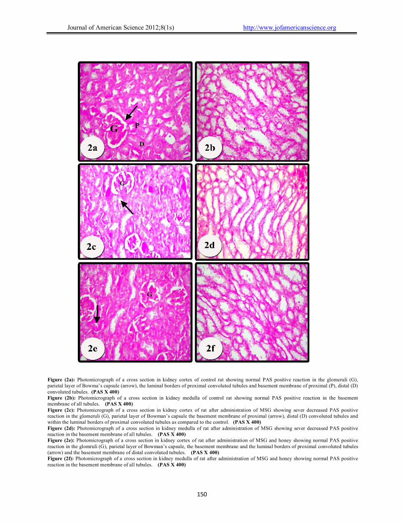
In control group, the glomerulus, the parietal layer of Bowman’s capsule, the basement membrane of all renal tubules, and brush borders of proximal convoluted tubules showed normal PAS positive reaction (Figs.2 a and b). After treatment with MSG the glomerulus, the parietal layer of Bowman’s capsule, the basement membrane of all renal tubules, and brush borders of proximal convoluted tubules showed sever decreased PAS positive reaction (Figs. 2 c and d).
The kidney of animals treated with both MSG and honey showed nearly normal PAS positive reaction in the the glomerulus, the parietal layer of Bowman’s capsule and the basement membrane of all renal tubules (Figs.2 e and f). PAS positive reaction was also observed in the brush border of proximal convoluted tubules (Fig.2e). Electron microscopic examination: I- Proximal convoluted tubules:
Regarding the cells of the proximal convoluted tubule in control rats: The apical cell membrane is occupied by numerous microvilli. The cytoplasm contains numerous mitochondria of various sizes. Few cisternal profiles of rough endoplasmic reticulum and
ribosomes were detected in the cytoplasm. The nuclei are rounded euchromatic with prominent nucleoli. The basement membrane exhibited basal infoldings with elongated mitochondria between them (Fig. 3a ).
In animals treated with MSG the proximal convoluted tubules revealed partial loss of brush border, destruction of most cytoplasmic organelles and vacuolated cytoplasm, and few lysosomes were observed. Several cells showed splitting and thickening of basement membrane. In addition, the lumen of several tubules was filled with cell debris. The nuclei frequently appeared shrinkage and irregular (Figs. 3b, c and d).
After treatment with both MSG and honey, the proximal tubule cells showed a marked reduction of basal lamina thickening and large number of cytoplasmic organells was observed. The nuclei return to their regular appearance and normal size. The apical cell membrane appeared nearly normal; however, the cytoplasmic and nuclear changes were milder than those seen in MSG group (Fig.3e). II- Glomerulus:
The renal corpuscle of control rats appears as dense round tufts of capillaries. The outer layer of Bowman’s capsule is the parietal layer and the inner visceral layer applies closely to the glomerular capillaries. Each capillary loop is lined by endothelial cells and Podocytes surround the capillaries. (Fig. 4a). Each podocyte has a large nucleus and abundant cytoplasm. The pedicels are separated by slit pores with slit membranes. The filtration barrier consists of capillary endothelial inner layer, thin glomerular basement membrane and podocyte layer (Fig. 4b). Podocytes give rise to primary processes which in turn give numerous secondary foot processes or pedicels that rest on a thin basal lamina. The glomerular basal lamina mainly consists of an external and internal laminae rarae and lamina densa in between (Fig.4c).
In animals treated with MSG alone, the renal corpuscle appeared with marked degeneration of most structures of glomerulus. Some glomerular capillaries showed luminal deformity including congestion and infiltrated cells, the endothelial cells were highly active appearing with highly indented nuclei (Fig.4d). The podocytes appeared vacuolated with irregular nucleus (Fig.4e). Marked thickening of the basal lamina and fusion of foot processes were also observed (Fig.4f).
Electron micrograph of proximal tubule of rat treated with both MSG and honey showing nearly normal nucleus, mitochondria, brush border and reduction in thickening of basal lamina (Fig.3e). Electron micrograph of glomerulus showing nearly normal nucleui of endothelium (Fig.4g). The nuclei of podocytes and cytoplasmic organells returned to normal appearance (Fig. 4h). The basal lamina appeared thin compared to MSG group (Fig.4i).

Journal of American Science 2012;8(1s) http://www.jofamericanscience.org
149
Figure (1a): Photomicrograph of a cross section in kidney cortex of control rat showing normal histological structures of Malpighian corpuscles with its glomerulus (G) Bowman’s capsule (arrow) proximal tubules (P) and distal tubule (D). (Hx&E X 400) Figure (1b): Photomicrograph of a cross section in kidney medulla of control rat showing normal histological structures of collecting tubules (Ct), collecting duct (Cd) thick limb (thick arrow),thin limb (thin arrow) of loops of Henle. Notice the presence of interstitial cells (head arrow) and vasa recta (V). (Hx&E X 400) Figure (1c): Photomicrograph of a cross section in kidney cortex of rat after administration of MSG showing dilatation of Bawmn's capsule (arrow), shrinkage glomerulus (G), dilatation of the proximal (P) and distal convoluted tubules (D). Note the loss of brush border of proximal tubules. (Hx&E X 400 ) Figure (1d): Photomicrograph of a cross section in kidney cortex of rat after administration of MSG showing dilatation of Bowman's capsule (arrow) and shrinkage of glomerulus (G). Notice presence of huge cavity with fragmented areas (V). (Hx&E X 400) Figure (1e): Photomicrograph of a cross section in kidney medulla of rat after administration of MSG showing areas of syncytium of ill defined tubules (Sy). (Hx&E X 400) Figure (1f): Photomicrograph of a cross section in kidney medulla of rat after administration of MSG showing the cells of collecting tubules (Ct) has a rim of cytoplasm, deeply stained nuclei and loss of differentiation between the thick and thin loops of Henle. Notice the presence of several areas of cells of ill defined boundaries, containing of irregular basophilic, deeply stained nuclei (Sy). (Hx&E X 400) Figure (1g): Photomicrograph of a cross section in kidney cortex of rat after administration of MSG and honey showing most of the cortex return to normal structure. Renal corpuscle formed of glomeruls (G), Bowman’s capsule (arrow), distal convoluted tubules (D) and proximal convoluted tubules (P). (Hx&E X 400) Figure (1h): Photomicrograph of a cross section in kidney medulla of rat after administration of MSG and honey showing most of the medulla return to normal histological structure. Collecting tubules (Ct), collecting duct (Cd) thick limb (thick arrow), thin limb (thin arrow) of loops of Henle. Some cells of collecting tubules have deeply stained nuclei and thin rim of cytoplasm. (Hx&E X 400)
Journal of American Science 2012;8(1s) http://www.jofamericanscience.org
150
Figure (2a): Photomicrograph of a cross section in kidney cortex of control rat showing normal PAS positive reaction in the glomeruli (G), parietal layer of Bowma’s capsule (arrow), the luminal borders of proximal convoluted tubules and basement membrane of proximal (P), distal (D) convoluted tubules. (PAS X 400) Figure (2b): Photomicrograph of a cross section in kidney medulla of control rat showing normal PAS positive reaction in the basement membrane of all tubules. (PAS X 400) Figure (2c): Photomicrograph of a cross section in kidney cortex of rat after administration of MSG showing sever decreased PAS positive reaction in the glomeruli (G), parietal layer of Bowman’s capsule the basement membrane of proximal (arrow), distal (D) convoluted tubules and within the luminal borders of proximal convoluted tubules as compared to the control. (PAS X 400) Figure (2d): Photomicrograph of a cross section in kidney medulla of rat after administration of MSG showing sever decreased PAS positive reaction in the basement membrane of all tubules. (PAS X 400) Figure (2e): Photomicrograph of a cross section in kidney cortex of rat after administration of MSG and honey showing normal PAS positive reaction in the glomruli (G), parietal layer of Bowman’s capsule, the basement membrane and the luminal borders of proximal convoluted tubules (arrow) and the basement membrane of distal convoluted tubules. (PAS X 400) Figure (2f): Photomicrograph of a cross section in kidney medulla of rat after administration of MSG and honey showing normal PAS positive reaction in the basement membrane of all tubules. (PAS X 400)

Journal of American Science 2012;8(1s) http://www.jofamericanscience.org
151
Figure (3a): Electron micrograph of proximal tubule of control rat showing basal infoldings (arrow) and mitochondria (M) in between. Note the thin basal lamina (Bl), regular nucleus (N) and brush border (Bb) of numerous microvilli. Scale bar = 2µm Figure (3b): Electron micrograph of proximal tubule of rat treated with MSG showing irregular nucleus (N) and thickening basal lamina (Bl). Note cell debris within the tubule lumen (Lu). Scale bar = 2µm Figure (3c): Electron micrograph of proximal tubule of rat treated with MSG showing some vacuolated cytoplasm (V), shrunken nucleus (N) and partial loss of brush border (Bb). Scale bar =2µm Figure (3d): Electron micrograph of proximal tubule of rat treated with MSG showing vacuolated cytoplasm (V) and shrunken nucleus (N) and partial loss of brush border (Bb). Note the presence of lysosomes (L). Scale bar =2µm Figure (3e): Electron micrograph of proximal tubule of rat treated with MSG and honey showing nearly normal nucleus (N), mitochondria (M), brush border (Bb) some cytoplasmic vacuolization (V)) and lysosomes (L). Note the reduction in thickening of basal lamina (Bl). Scale bar =2µm

Journal of American Science 2012;8(1s) http://www.jofamericanscience.org
152
Figure (4a): Electron micrograph of renal corpuscle of control rat showing podocytes (Pd) surrounding the capillary loops (C). The podocyte contains a large nucleus; each capillary loop is lined by endothelial cells (E). Scale bar = 2µm. Figure (4b): Electron micrograph of renal corpuscle of control rat showing podocytes (Pd) resting on basal lamina (Bl). Note the nucleus (N) with dense scattered patches of heterochromatin. Scale bar = 2µm Figure (4c): High magnification of the pervious figure showing the podocyte that give rise to primary process (thin arrow) which in turn gives numerous secondary foot processes (thick arrow). Scale bar = 500 nm Figure (4d): Electron micrograph of renal corpuscle of MSG treated rat showing endothelial cells (E) with highly indented nucleus. The glomerular capillary contains infiltrated cells (arrow). Scale bar = 2µm Figure (4e): Electron micrograph of podocyte of rat treated with MSG showing irregular nuclei (N) and vacuolation in the cytoplasm (V). Scale bar = 500 nm Figure (4f): Electron micrograph of renal corpuscle of MSG treated rat showing thickening of glomerular capillary basement membrane (Bl). Scale bar = 500 nm Figure (4g): Electron micrograph of renal corpuscle of rat treated with MSG and honey showing mild improvement in endothelial cells (E) of the renal corpuscle. Scale bar = 2µm Figure (4h): Electron micrograph of podocyte of rat treated with MSG and honey showing few mitochondria (M) and rough endoplasmic reticulum (arrow). The nucleus appears normal with dark clumps of heterochromatin adjacent to the nuclear membrane. Scale bar = 500 nm Figure (4i): Electron micrograph of renal corpuscle of rat treated with MSG and honey showing reduction in the thickening of basal lamina (Bl). Scale bar = 500 nm

Journal of American Science 2012;8(1s) http://www.jofamericanscience.org
153
4. Discussion Monosodium glutamate (MSG) is frequently
used as a flavor enhancer, the fact of which makes it one of the most applied food additives in the modern nutrition all over the world (Beyreuther et al., 2007).
The present study showed that MSG induced marked histopathological alterations in the kidney tissues of rats in form of shrinkage of glomeruli, dilatation of Bawmn,s capsule, dilatation of proximal and distal convoluted tubules with loss of the brush border of proximal convoluted tubules. Moreover, the medulla showed sever histological changes. The collecting tubules and collecting duct showed thin rim of cytoplasm and small deeply stained nuclei. Most of loops of Henle were undifferentiated with the presence of several areas of ill defined cell boundaries, which form a syncytium of cytoplasm with irregular basophilic and deeply stained nuclei. Similar results have been reported by others (Aughey et al., 1984, kjellstrom, 1986, Mitsumari et al., 1998 and Inkielewicz and Krechniak, 2003). These results also coincides with Eweka, 2007) who examined the kidney sections of animals treated with 6 gm of MSG and recorded marked distortion of cyto-architecture of the renal cortical structures, degenerative and atrophic changes. Damaged tubular cells, in kidney by MSG, was also recorded by (Abass and Abd El-Haleem, 2011) who noticed cytoplasmic, nuclear vacuolations and tubular dilatation. We postulated that the degenerative effect of MSG on the cortex and medulla of kidney was due to the direct effect of toxins on cells of kidney which coincided with (Porter, 1994) who reported that one of the possible mechanism for the tubular lesions was the direct toxic effect on the cell function. (Davies et al., 1995 and Fadel, and Larsen, 1996) attributed damage of the brush border to leakage of alkaline phosphates (ALP) and gammaglutamyle transferase (GGT) enzymes, which are associated with the brush border of the renal tubules, as a result of toxin binding to the brush border, and considered as an early marker of toxic tubular insult.
The present histochemical study revealed a marked decrease in the glycogen content of cortex and medulla in kidney of MSG treated rats. This result is in accordance with (Mohammed and El-Naggar, 1995 and Abd EL-Mawla and Osman, 2011) who reported the decreased PAS in the kidney of rats treated with MSG.
The present ultrastructural examination showed cytoplasmic and nuclear vacuolations of proximal convoluted tubular cells. Several cells revealed partial loss of brush border, destruction of most cytoplasmic organelles, splitting and thickening of basement membrane. The nuclei of podocytes and endothelial cells frequently appeared irregular with chromatin condensation adjacent to the nuclear membrane and
presence of few lysosomes. These results were in agreement with (Abd El-Mawla and Osman, 2011) who reported cytoplasmic and nuclear vacuolations, destruction of cytoplasmic organelles, splitting and thickening of basement membrane in renal tubules after administration of MSG. Our results also make a harmony with (Bopanna et al., 1999) who stated that MSG at higher dose produced sequence of damage in cellular organelles and membranes in renal and hepatic tissues.
The detection of infiltrated cells in the present study which mostly inflammatory cells were confirmed by (Abd El-Mawla and Osman, 2011) and (Abass and Abd El-Haleem, 2011) who found infiltrated cells in the kidney of rat treated with MSG and explained that by production of chronic inflammatory disease after long use of MSG.
In the present study, administration of MSG alone, the renal corpuscle appeared with marked degeneration of almost structures of glomerulus, some glomerular capillaries showed luminal deformity including congestion and infiltrated cells. The endothelial cells were highly active appearing with highly indented nuclei. The podocytes appeared vacuolated with irregular nucleus. Marked thickening of the basal lamina and fusion of foot processes were also observed. Many renal tubules and glomeruli of the rat kidneys showed marked degenerative lesions under the effect of MSG which explained by the sensitivity of tubules to toxic influences that concise with (Tisher and Brenner, 1989) who postulated the degenerative effect of MSG on the kidney due to the effect of toxins on the cells of the kidney through transport mechanisms. Also the tubule came in contact with toxic chemicals during their excretion and elimination by the kidneys. Other mechanisms for the tubular lesions might involve reactive intermediates or oxidative stress, or both (Alden and Frith, 1992and Ebaid and Tag, 2012). This mechanism confirmed by (Farombi and Onyema, 2006), who found that the monosodium glutamate when was administered intraperitoneally at 4 mg/g of body weight dose, increased the formation of malondialdehyde (MDA) which is oxidative material in the rat’s liver and brain. Other mechanism was reported, MSG has a toxic effect on many body organs by altering ionic permeability of neural membrane and induced persistent depolarization (Robinson, 2006).
The mechanisms of MSG-induced damage include the production of free radicals that alter mitochondrial activity and genetic information (Partrick, 2003) and (De Burbure et al., 2006). Therefore, some authors had postulated that antioxidants should be one of the important components of an effective treatment of MSG

Journal of American Science 2012;8(1s) http://www.jofamericanscience.org
154
poisoning (El-Demerdash et al., 2004 and Onyama et al., 2006).
The present histological study showed that supplementation of honey reduced the cellular changes induced by MSG, indicating that honey contributed to the protection against MSG induced kidney toxicity. The present results showed less renal tubular alteration than MSG treated group. The renal corpuscle and most of the renal tubules approximately returned to their normal appearance. The lumen in the most of proximal and distal convoluted tubules appeared normal and the brush borders of proximal convoluted tubules were observed. The medulla showed nearly normal histological structures of collecting tubules, collecting duct, thick and thin limb loops of Henle with presence of interstitial cells and vasa recta as a control one. In the present study, the kidney of treated animals with both MSG and honey showed nearly normal PAS positive reaction in the glomerulus, the parietal layer of Bowman’s capsule, the basement membrane of all renal tubules, and brush borders of proximal convoluted tubules showed PAS positive reaction.
These results were in agreement with (Beretta et al., 2005) who confirmed the role of honey as an antioxidant agent in blood. (Manuela et al., 2007) found that there was a direct link between the honey consumption and the level of polyphenolic antioxidants in the plasma. (El-Khayat et al., 2009) proved the antioxidant effect of honey which ameliorated the toxic effects of ochratoxin A on liver and kidney tissues. Also (Omotayo, 2011a and b) found that combination of honey with metformin or glibenclamide might offer additional antioxidant effect to these drugs. (Abd-Ali and Ismail, 2012) reported that honey decreased nephrotoxicity induced by amikacin through interference with the oxidative stress process.
The present ultrastructure study after supplementation of both honey and MSG revealed a large spherical nucleus, nearly normal mitochondria, in addition to the microvilli forming the brush border with presence of areas of vacuolations and multiple lysosymes in the cells of proximal convoluted tubules. The glomerulus showed nearly normal endothelium and podocytes nuclei which appeared nearly normal and basal lamina appeared thin in the glomeruli compared to MSG group. This result indicated the protective effects of honey against MSG nephrotoxicty that conciseded with (Abdel-Moneim and Ghafeer, 2007) who recorded that the elevation in the free radicals, include lipid peroxidation (LPO), induced by cadmium alone was decreased in the presence of honey. (Halawa et al., 2009) revealed that natural honey could diminish the adverse effects of lead acetate as shown in the histological analysis of rat kidneys. The present study showed sign of recovery after supplementation of honey, the cytoplasm
appeared less vacuolated. Most of mitochondria, rough endoplasmic reticulum acquired their normal appearance. (Erejuwa et al.(2010) reported that by histopathological examinations of the kidneys in the honey-treated diabetic rats showed the mesangial matrix expansion and thickening of glomerular basement membrane were reduced. On hypothesis to explain the beneficial effects of honey in ameliorating histological and ultrastructural changes is that honey may contains flavonoids, ascorbic acid, tocopherols, catalase and phenolic compounds. All of which work together to provide a synergistic antioxidant effect, scavenging and eliminating free radicals (Johnston et al., 2005). Conclusion:
The present study demonstrated that honey administered in combination with MSG minimized its hazards. Honey can protect against oxidative stress induced by MSG, by lowering the free radicals and increased the levels of antioxidants. Further studies are required to recommend the use of honey and it’s therapeutic potential in human. Acknowledgement: My deep gratitude to Prof. Dr Azza Anwar Mohammed for her revision of this manuscript thorough. Corresponding author Amal afeefy Anatomy Department, Faculty of Medicine, Alazhar university1, [email protected] References 1. Abass, M.A. and Abd El-Haleem, M. R. (2011):
Evaluation of Monosodium Glutamate Induced Neurotoxicity and Nephrotoxicity in Adult Male Albino Rats, Journal of American Science, 7(8): 264-76.
2. Abd-Ali, A. R. and Ismail, S. H. (2012): The Protective Effect of Honey against Amikacin-induced Nephrotoxicity in rats. Iraqi J Pharm Sci., 21(2): 85-93.
3. Abd El-Mawla, A. M. and Osman, H. E. H. (2011): HPLC analysis and role of the Saudi Arabian propolis in improving the pathological changes of kidney treated with monosodium glutamate. Spatula DD, 1(3): 119-27.
4. Abdel-Moneim, W. M. and Ghafeer, H. H. (2007): potential protective effect of natural honey against cadmium-induced hepatotoxicity and nephrotoxicity. Mansoura J. Forensic Med. Clin. Toxicol., XV(2) :75-98.
5. Adrienne, S. (1999): The toxicity of MSG, a study in suppression of information. Accountability Res., 6 (4): 259-310.
6. Ahluwalia, P.; Tewari, K. and Choudhary, P. (1996): Studies on the effects of monosodium

Journal of American Science 2012;8(1s) http://www.jofamericanscience.org
155
glutamate (MSG) on oxidative stress in erythrocytes of adult male mice. Toxicol. Lett., 84(3): 161-5.
7. Alden, C. L. and Frith, C. H. (1992): Urinary System. In: Handbook of Toxicologic Pathology. Hashek WM and Rousseaux CG eds., Academic Press, San Diego, CA., pp. 316-79.
8. Almaraz-Abarca, N.; Campos, M. G.; Avila-Reyes, J. A.; Naranjo-Jimenez, N.; Herrera-Corral, J. and Gonzalez-Valdez, L. S. (2007): Antioxidant activity of phenolic extract of monofloral honey-collected pollen from mesquite (Proposis julifloral, leguminosaceae). J. Food Composit. Anal., 20: 119-24.
9. Aughey, E.; Feli, G. S.; Scott, R. and Black, M. (1984): Histopathology of early effects of oral cadmium in the rat kidney, Environment and Health Perspectives, 54: 153-61.
10. Bancroft, J. D. and Gamble, M. (2002): Theory and practice of Histological Techniques. Fifth ed., Churchill Livingstone, London, pp. 109-36.
11. Beretta, G.; Granata, P.; Ferrero, M.; Orioli, M. and Facino, R. (2005): Standardization of antioxidant properties of honey by a combination of spectrophotometric/ fluorimetric assays and chemometrics. Analytica Chimica Acta, 533: 185-91.
12. Beyreuther, K.; Biesalski, H. K.; Fernstrom, J. D.; Grimm, P. and Hammes, W. P. (2007): Consensus meeting: monosodium glutamate - an update. European Journal of Clinical Nutrition, 61: 304–13.
13. Bopanna, K. N.; Balaraman, R. and Nadig, R. S. (1999): Organotropic ultrastructural changes produced by monosodium glutamate in rats on atherogenic diet: effect of S-allyl cysteine sulphoxide. Indian J. of Pharmacology, 31(4): 266-74.
14. Davies, S.; Reichardi, P.; Vaughan D.; Reussell, G. (1995): Differential effect of ischemia-reperfusion injury on anti-oxidant enzyme activity in the rat kidney. Exp. Nephrol., 3: 348-54.
15. De Burbure, C.; Buchet; J. B.; Leroyer, A.; Nisse, C. ; Haguenoer, J. M.; Mutti, A.; Smerhovsky, Z.; Cikrt, M.; Trzcinka-Ochocka, M.; Razniewska, G.; Jakubowski, M. and Bernard, A. (2006): Renal and neurologic effects of cadmium, lead, mercury and arsenic in children: evidence of early effects and multiple interactions at environmental exposure levels. Environ.Health. Perspect., 114: 584-90.
16. Diniz, y. S.; Fernandes, A. A.; Campos, K. E.; Mani, F.; Ribas, B.O. and Novelli, E. L. (2004): Toxicity of hypercaloric diet and monosodium glutamate: oxidative stress and metabolic shifting in hepatic tissue. Food Chem. Toxicol., 42 (2):313-9.
17. Ebaid, H. M. and Tag, H. M. (2012): Monosodium Glutamate Toxic Effect on Spleen Structure and Potentiality of Recovery in Adult Albino Rats .Egypt. Acad. J. Biolog. Sci., 4(1): 1-8.
18. El-Demerdash, F. M.; Yousef, M. I.; Kedwany, F. S. and Baghdadi, H. H. (2004): Cadmium-induced changes in lipid peroxidation, blood hematology, biochemical parameters and semen quality of male rats: protective role of vitamin E and B-carotene. Food Chem. Toxic., 42: 1563-71.
19. El Denshary, E. S.; Al-Gahazali, M. A.; Mannaa, F. A.; Salem, H. A.; Hassan, N. S. and Abdel-Wahhab, M. A. (2011): Dietary honey and ginseng protect against carbon tetrachloride-induced hepatonephrotoxicity in rats. Exp. Toxicol. Pathol. 64 (7-8) :753-60.
20. El-Khayat, Z.; Ahmed R. E.; Mahmoud S. A.; Wafaa, I. R. and Tahany, R. E. (2009): Potential Effects of Bee Honey and Propolis against the Toxicity of Ochratoxin A in Rats. Macedonian Journal of Medical Sciences, 2(4): 311-18.
21. El-kott, A. F.; Kandeel, A. A.; Abed El-Aziz, S. F. and Ribea, H. M. (2012): Anti-tumor Effects of Bee Honey on PCNA and P53 Expression in the Rat Hepatocarcinogenesis. International Journal of Cancer Research, 8: 130-9.
22. Erejuwa, O. O.; Gurtu, S.; Sulaiman, S. A.; Wahab, M. S.; Sirajudeen, K. N. and Salleh, M. S. (2010): Hypoglycemic and antioxidant effects of honey supplementation in streptozotocin-induced diabetic rats Int. J. Vitam. Nutr. Res. 80(1): 74-82.
23. Eweka, A.O. (2007): Histological studies of the effects of monosodium glutamate on the kidney of adult Wistar rats. The Internet Journal of Health, 6 (2): 45-67.
24. Fadel, A. A. and Larsen, H. A. (1996): Gentamicin-induced nephrotoxiois in lambs. Res. Vet. Sci., 61: 181-92.
25. Farmobi, E. O. and Onyema, O. O. (2006): Monomonosodium glutamate-induced oxidative damage and genotoxicity in the rat: modulatory role of vitamin C, vitamin E and quercetin. Toxicol.J. 25(5): 251-59. Garattiini, S. (2000): Glutamic Acid, Twenty Years Later. J. Nutr., 130: 9018-98.
26. Gribel, N.V. and Pashinskii, V.G. (1990): Antitumor properties of honey. Vopr. Onkol., 36: 704-9.
27. Halawa, H. M.; El-Nefiawy, N. E.; Makhlouf, N. A. and Mady, A.A. (2009): Evaluation of honey protective effect on lead induced oxidative stress in rats. JASMR, 4 (2): 197-209.
28. Hassan, M. I.; Mabrouk, G. M.; Shehata, H. H. and Aboelhussein, M. M. (2012): Antineoplastic effects of bee honey and Nigella sativa on hepatocellular carcinoma cells. Integr. Cancer Ther., 11(4): 354-63.
29. Inkielewicz, I. and Krechniak, M. (2003): Fluoride content in soft tissues and urine of rats exposed to sodium fluoride in drinking water. Research Report, 36(4): 263-66.
30. Johnston, J.; Sepe, H.; Miano, C.; Brannan, R. and Alderton, A. (2005): Honey inhibits lipid
Journal of American Science 2012;8(1s) http://www.jofamericanscience.org
156
oxidation in ready-to-eat ground beef patties. Meat Science, 70(4): 627-31.
31. Kirk-Othmer (1992): Encyclopedia of Chemical Technology. 4th Ed., Wiley, USA, pp: 571-9.
32. Kjellstrom, T. (1986): Renal effects. In: cadmium and health: A toxicology and Epidemiological Appraisal, Effects and Response. Friberg LEC, Kjellström T, Nordberg GF, Eds. Boca Raton, FL, CRC Press, Vol. 2, pp. 21-109.
33. Larson, P. J.; Mikkelsen, J. D.; Jessop, D.; Lightman, S. L. and Chwoday, H. S. (1994): Neonatal monosodium glutamate treatment alters both the activity and the sensitivity of the rat hypothalamo-pituitary-adrenocotical axis. J. Endocrino., 141: 497–503.
34. Mabrouk, G. M.; Moselhy, S. S.; Zohny, S. F.; Ali, E. M.; Helal, T. E.; Amin, A. A. and Khalifa, A. A. (2002): Inhibition of methylnitrosourea (MNU) induced oxidative stress and carcinogenesis by orally administered bee honey and Nigella grains in Sprague Dawely rats. J. Exp. Clin. Cancer Res., 21: 341-6. Manuela, B.; Manila, C.; Augusto, A.; Maria, P. and Piacentini, E. P. (2007): Honey flavonoids as protective agents against oxidative damage to humanred blood cells. Food Chemistry, 104(4): 1635-40.
35. Miskowiak, B.; Kesa, B.; Limanowski, A.; Pantayka, M.; Filipiak, B. and Folia, M. (1999): Long-term effect of neonatal (MSG) treatment on reproductive system of the level rats. Folia Morphol (Warsz), 58(2): 105–13.
36. Mitsumari, K.; Shibutani, S.; Sato, S.; Ondoera, H.; Nacagawa, J.; Hayashi, Y.; and Ando, M.; (1998): Relationship between the development of hepatorenal toxicity and cadmium accumulation in rats. Archives of Toxicology, 72(9): 545-52.
37. Mohammed, A. A.; El-Naggar S. M. (1995): Histological and histochemical study on the effect of monosodium glutamate on the albino rat’s kidney. The new Egyptian J of medicine, 12(2): 182-89.
38. Omotayo, O. E.; Siti, A. S.; Mohd, S. A.; Kuttulebbai, N. S.; Sirajudeen, M. d.; Salzihan, M. d.; Salleh, S. G. (2011a): Glibenclamide or Metformin Combined with Honey Improves Glycemic Control in Streptozotocin-Induced Diabetic Rats. Int. J. Biol. Sci., 7(2): 244-52.
39. Omotayo, O.; Siti A. S. , Mohd, S.; Sirajudeen, K.; Mohammed S. and Sunil, G. (2011b): Comparison of Antioxidant Effects of Honey, Glibenclamide, Metformin, and Their Combinations in the Kidneys of Streptozotocin-Induced Diabetic Rats. Int. J. Mol. Sci, 12(1): 829-43.
40. Onyama, O.O.; Farombi, E. O.; Emerole, G. O.; Ukoha, A. I. and Onyeze, G. O. (2006): Effect of vitamin E on monosodium glutamate induced hepatotoxicity and oxidative stress in rats. Indian Journal of biochemistry and Biophyscs, 43(1): 20-4.
41. Park, C. H.; Choi, S. H.; Piao, Y.; Kim, S.; Lee, Y. J.; Kim, H. S.; Jeong, S. J.; Rah, J. C.; Seo, J. H.; Jung, y. J. and Suh, Y. H. (2000): Glutamate and aspartate impair memory retention and damage hypothalamic neurons in adult mice. Toxicol. Lett., 115(2): 117-25.
42. Partrick, L. (2003): Toxic metals and antioxidants: Part II. The role of antioxidants in arsenic and cadmium toxicity. Altern. Med. Rev., 8(2): 106-28.
43. Porter, G. A. (1994): Urinary biomarkers and nephrotoxicity. Miner Electrolyte Metab., 20(4): 181-86.
44. Praputpittaya, C. and Wililak, A. (2003): Visual performance in monosodium glutamate-treated rats. Nutr. Neurosci., 6(5): 301-7.
45. Reynolds, E. A. (1963): The use of PH as an electron opaque stain in electron microscopy. J.Cell Biol.,17: 208-12.
46. Robinson, M.B. (2006): Acute regulation of sodium-dependent glutamate transporters: a focus on constitutive and regulated trafficking. Handb. Exp. Pharmacol., (175):251–75.
47. Samarghandian, S. J. and Afshari, T. and Davoodi, S. (2011): Honey induces apoptosis in renal cell carcinoma. Pharmacogn. Mag., 7(25): 46-52.
48. Sathyasurya, D. R. and Aziz A. I. (2009): Two varieties of honey that are available in Malaysia gave intermediate glycemic index values when tested among healthy individuals. Maced J Med Sci., 153(2): 145-8.
49. Singh, P.; Mann, K. A.; Mangat, H. K. and Kaur, G. (2003): Prolonged glutamate excitotoxicity: effects on mitochondrial antioxidants and antioxidant enzymes. Mol. cell Biochem., 243(1-2): 139-45.
50. Stegink, L.D. (1984): Aspartate and glutamate metabolism of Aspartame. In: physiology and biochemistry, Stegink LD, Filer LJ Jr, eds. New York, Marcel Dekker, pp: 47-76.
51. Tisher, C. C.; Brenner, B. M. (1989): Structure and function of the glomerulus. In: Renal Pathology with Clinical and Functional Correlations, Philadelphia, J.P. Lippincott, pp. 92–110.
52. Žegarac, J. P.; Stipčević, T.; Belščak, A. (2009): Antioxidant properties and phenolic content of different floral origin honeys. J. Api. Prod. Apimed. Sci., 1 (2): 43-50.
11/9/2012