journal of controlled release - heriot-watt university...synthesis, characterization and evaluation...
TRANSCRIPT
�������� ����� ��
Synthesis, characterization and evaluation of in vitro toxicity in hepatocytesof linear polyesters with varied aromatic and aliphatic Co-monomers
Deepak Kakde, Leagh G. Powell, Kuldeep K. Bansal, Steve Howdle,Derek Irvine, Giuseppe Mantovani, Gavin Millar, Lea Ann Dailey, VickiStone, Helinor J. Johnston, Cameron Alexander
PII: S0168-3659(16)30504-1DOI: doi: 10.1016/j.jconrel.2016.08.003Reference: COREL 8415
To appear in: Journal of Controlled Release
Received date: 11 June 2016Revised date: 27 July 2016Accepted date: 3 August 2016
Please cite this article as: Deepak Kakde, Leagh G. Powell, Kuldeep K. Bansal, SteveHowdle, Derek Irvine, Giuseppe Mantovani, Gavin Millar, Lea Ann Dailey, Vicki Stone,Helinor J. Johnston, Cameron Alexander, Synthesis, characterization and evaluation ofin vitro toxicity in hepatocytes of linear polyesters with varied aromatic and aliphaticCo-monomers, Journal of Controlled Release (2016), doi: 10.1016/j.jconrel.2016.08.003
This is a PDF file of an unedited manuscript that has been accepted for publication.As a service to our customers we are providing this early version of the manuscript.The manuscript will undergo copyediting, typesetting, and review of the resulting proofbefore it is published in its final form. Please note that during the production processerrors may be discovered which could affect the content, and all legal disclaimers thatapply to the journal pertain.

ACC
EPTE
D M
ANU
SCR
IPT
ACCEPTED MANUSCRIPT
1
Synthesis, Characterisation and Evaluation of In Vitro Toxicity in Hepatocytes of
Linear Polyesters with Varied Aromatic and Aliphatic Co-monomers
Deepak Kakdea, Leagh G. Powell
b, Kuldeep K Bansal
a, Steve Howdle
c, Derek Irvine
d,
Giuseppe Mantovania, Gavin Millar
b, Lea Ann Dailey
e, Vicki Stone
b, Helinor J Johnston
b*
and Cameron Alexandera*
a School of Pharmacy, University of Nottingham, University Park, Nottingham,
NG72RD, UK. e-mail: [email protected]
b School of Life Sciences, Nano Safety Research Group, Heriot-Watt University,
Edinburgh, EH14 4AS, UK. E-mail: [email protected].
c School of Chemistry, University of Nottingham, University Park, Nottingham
NG72RD, UK
d Department of Chemical and Environmental, Faculty of Engineering, Engineering,
University of Nottingham, University Park, Nottingham, NG7 2RD, UK
e Institute of Pharmaceutical Technology and Biopharmaceutics, Martin-Luther-
University Halle-Wittenberg, Wolfgang-Langenbeck-Str. 4, D - 06120 Halle / Saale

ACC
EPTE
D M
ANU
SCR
IPT
ACCEPTED MANUSCRIPT
2
ABSTRACT
Polyesters are extensively used in drug delivery because of their controllable
biodegradation properties and perceived favourable cytocompatibility. However, new
ester-based materials are continually being sought which can be produced from readily
accessible monomers, which can be tuned for drug encapsulation and which retain
good cellular compatibilities. In this study, 5 polyesters of similar molar mass were
synthesized by reacting 1,10-decanediol with different ratios of succinic
acid/phenylsuccinic acid and the effect of the phenyl side-chain group addition on
polymer properties relevant to drug delivery was investigated. A polymer with a 70/30
ratio of succinic acid and phenylsuccinic acid was selected based on its ability to
encapsulate a model dye in nanoparticle (NP) formulations, and was found to be
slowly degradable in phosphate buffered saline (PBS) but more rapidly degraded in
the presence of a lipase. The compatibility of NP formulations of this polymer either
with or without a Pluronic F68 stabilizing coating was assessed in vitro using the C3A
hepatocyte cell line. Cell viability was assessed, at NP concentrations ranging from
4.68 - 300 μg/mL 24 h post exposure, using the Alamar Blue, CDFA and Neutral Red
assays. C3A cells internalised both coated and uncoated polyester NPs to a similar
extent, with uptake observed to increase over time (10 - 1440 min). Although cell
viability was greater than 80% at the concentrations tested, in all assays, it was found
that a Pluronic F68 coated poly (decanediol-phenylsuccinate-co-succinate) stimulated
significant DNA damage driven by an oxidant mechanism, whereas the non-coated
polyester analogue and the Pluronic F68 alone had no effect. The results obtained
suggest that new polyesters can be synthesised with desirable properties from the

ACC
EPTE
D M
ANU
SCR
IPT
ACCEPTED MANUSCRIPT
3
materials perspective but formulation with additional excipients requires careful
evaluation for drug delivery applications.
Graphical abstract
Keywords: Polyester; polycondensation; nanoparticles; polymer; cytotoxicity;
hepatocyte; in vitro; uptake

ACC
EPTE
D M
ANU
SCR
IPT
ACCEPTED MANUSCRIPT
4
1. Introduction
In recent years, there has been a renewed interest in polyesters as drug delivery devices
due to their favorable biocompatibility and controllable biodegradation profiles [1-4].
Accordingly, polyester materials are regarded as a material of choice for biomedical applications
including drug delivery [5-9], as diagnostic agents [10, 11] and for tissue engineering [12-16].
For these applications, polyesters with a range of physicochemical, biomechanical and biological
properties are needed, along with appropriate biocompatibility, biodegradability and storage
stability [17]. A balance of these properties can be obtained through incorporation of suitable
constituent monomers into the polyester backbone.
For use as therapeutic agents, polyesters need to have strong associative interactions with
active ingredients. An ability to release the drug at a suitable rate for therapy is important to
reduce frequency of treatment [18]. Accordingly, there have been many variations made on the
compositions and co-monomers used for polyesters, with a view to obtaining the most favorable
controlled drug incorporation and release properties. More recently, there has been a strong
economic and ecological drive for polymer precursors to be derived from sustainable building
blocks, and for polymers to be produced with minimal use of solvents [19]. The synthesis of
polyesters for drug delivery has been very thoroughly explored, with many variations around the
use of poly(lactides), poly(caprolactone) and poly(carbonates) [3, 20, 21]. However, these
polyesters by themselves are not easy to formulate into nanoparticles that are stable for storage in
solution or for intravenous injection. Accordingly, methods are needed to enhance the colloidal
properties of polyester nanoparticles, and the most simple of these, and thus the most appealing
from an industrial perspective, is the surface adsorption to the nanoparticles of stabilising layers.
The concept of sterically shielding hydrophobic polyester surfaces by attaching hydrophilic

ACC
EPTE
D M
ANU
SCR
IPT
ACCEPTED MANUSCRIPT
5
polymers is well-established, with an extensive literature on adsorbed amphiphilic block co-
polymers [22-27]. These types of stabilized nanoparticles have long been known to evade uptake
by the mononuclear phagocyte system (MPS),[28] and so for practical drug delivery purposes
there is a strong rationale to develop polyesters which are easy to synthesise from renewable
resources, and which can be easily formulated into drug-loaded nanoparticles.
Here we report the synthesis of linear polyesters from the readily available monomers
succinic acid, phenyl succinic acid and 1, 10-decanediol, with progressive variation with respect
to the phenyl side chain content from the polyester backbone. The polymers were made using
scandium (III) triflate as a catalyst under solvent-free conditions [29-33] and evaluated for drug-
loading via nanoprecipitation in the presence of coumarin-6 dye. The polymer exhibiting the
highest dye loading was tested for colloidal and hydrolytic stability when formulated into
nanoparticles (NPs). In addition, the toxicity of polyester nanoparticles, with and without a
sterically-stabilizing adsorbed Pluronic F68 block co-polymer layer. The toxicity of these NPs
was assessed in vitro, using the human C3A hepatocyte cell line. Hepatocytes were selected as
the liver is the primary site of nanoparticle accumulation following exposure via different routes
(e.g. intratracheal instillation, ingestion, intravenous injection), and existing evidence suggests
that C3A cells respond similarly to primary human hepatocytes [34-37]. The internalization of
coated and uncoated polymer nanoparticles by hepatocytes was assessed over time, and their
effects on cell viability evaluated using three assays; Alamar Blue, Neutral Red, and 5-CFDA-
AM [5-carboxyfluorescein diacetate, acetoxymethyl ester] CFDA-AM. The ability of
nanoparticles to stimulate cytokine production (IL-8) and cause genotoxicity (DNA damage) was
assessed in order to evaluate sub-lethal impacts on cell function. These data showed that while
the new selected polyesters were rapidly internalized with or without the Pluronic F68 coating,

ACC
EPTE
D M
ANU
SCR
IPT
ACCEPTED MANUSCRIPT
6
there were differences in DNA damage induced by the NP formulations which were a
consequence of the combination of the coating and ‘core’ NP, and not a function of the
individual components alone.
2. Experimental Section
2.1. Materials
Succinic acid (ACS reagent, ≥99.0%), Phenylsuccinic acid (98%), 1,10-Decanediol
(98%), Scandium(III) triflate (99%), Coumarin-6 (98%), Pluronic F-68, deuterated chloroform
(CDCl3), 1 mM sodium pyruvate, 1% non- essential amino acids, phosphate buffer saline
solution, CFDA-AM, Neutral Red, acetic acid, 95% ethanol, ammonium chloride, Triton-X100,
DAPI, H2O2, HBSS, low melting point agarose, agarose , lysis buffer base, dimethyl sulfoxide,
HEPES, potassium chloride, Ethylenediaminetetraacetic acid [EDTA], Bovine Serum Albumin,
sodium hydroxide, Tris base and GelRed were purchased from Sigma-Aldrich. All solvents were
purchased from Fischer Scientific UK.
Cell culture reagents including MEM medium, foetal calf serum (FCS), 2 mM L-
glutamine, 100 U/ml penicillin/streptomycin, phenol red free MEM medium, 10 x trypsin (2.5%)
and 0.4% v/v Trypan Blue were purchased from Gibco, Invitrogen. The human hepatocyte cell
line C3A was purchased from ATCC, Manassas, VA, USA.
2.2. Measurements
Nuclear magnetic resonance (NMR) spectra of 1H NMR and
13C NMR were recorded at
400 MHz (1H) and 101 MHz (
13C) using a Bruker DPX400 Ultrashield spectrometer and
deuterated chloroform (CDCl3) as the solvent. Spectral analysis was performed using
MestRENova 6.0.2 software copyright© 2009 Mestrelab Research S.L. All chemical shifts are

ACC
EPTE
D M
ANU
SCR
IPT
ACCEPTED MANUSCRIPT
7
reported in ppm (δ) relative to tetramethylsilane, referenced to the chemical shifts of residual
solvent resonances (CDCl3: δH 7.26, δC 77.16).
Fourier transform infrared (FTIR) spectroscopy was performed on solid or liquid samples
using a Cary 630 FTIR spectrophotometer equipped with a single bounce diamond ATR.
MicroLab software was used for data analysis.
A Polymer Laboratories GPC 50 instrument was used to determine Mn (number-average
molecular weight), Mw (weight average molecular weight) and Ð (polydispersity index, Mw /
Mn). The instrument was fitted with a Polymer Laboratories PLgel guard column (50 × 7.5 mm,
8 µm) followed by a pair of PLgel Mixed-D columns (300 × 7.5 mm, 8 µm) and a refractive
index detector. The flow rate of HPLC grade CHCl3 at 30 °C was 1 mL min-1
. The column
calibration was achieved using narrow molar mass distribution polystyrene standards. Polymer
Laboratories Cirrus 3.0 software was used for data analysis.
The thermal properties of the polymers i.e. Tm (melting temperature) and Tg (glass
transition temperature) were probed using a TA-Q2000 DSC (TA Instruments) under a nitrogen
atmosphere. Typically, the samples (5–10 mg) were exposed to two cooling-heating cycles from
–90 to 150 °C at a rate of 10 °C min-1
.
The particle sizes (z-average diameter) of nanoparticles were measured in HPLC water
determined by dynamic light scattering (DLS) using a NanoZS instrument (Malvern, UK) at 25
°C using 633 nm (4 mW) wavelength laser. The scattered light was detected at an angle of 173˚
and analysis was performed using zetasizer software version 7.03. The zeta potentials of the NPs
were determined in HEPES 10 mM buffer (pH 7.4). The size and surface zeta potentials of NPs
were also measured in complete MEM cell culture medium (MEM medium supplemented with

ACC
EPTE
D M
ANU
SCR
IPT
ACCEPTED MANUSCRIPT
8
10% heat-inactivated foetal calf serum (FCS), 2 mM L-glutamine, 100 U/mL
penicillin/streptomycin, 1 mM sodium pyruvate and 1% non-essential amino acids) containing
125 µg/mL of NPs.
A Tecnai G2 (FEI, Oregon, USA) microscope was utilized for transmission electron
microscopy (TEM). One drop of polymer suspension in HPLC grade water (typically 25–50 μg
mL−1
) was dropped onto a copper grid and allowed to dry in air. The sample was put on a copper
grid and allowed to air dry. The imaging was performed without staining.
A Zeiss LSM880 confocal microscope was used for imaging uptake of labelled polymers
and nanoparticles, and the Zeiss Zen program was used for data analysis.
2.3. Methods
2.3.1. Synthesis of Polyesters
Poly(decamethylene succinate) [PDeMS], poly(decamethylene phenylsuccinate) [PDsMPS] and
their copolymers poly(decamethylene succinate-co-phenylsuccinate) [PDeMS-co-PS] were
synthesized by a solvent-free melt polycondensation method (Scheme 1). The diacid and diol
ratio was maintained at 1:1 in all polycondensation reactions. A scandium (III) triflate: diol ratio
of 5.10-4
(0.05 mol%) was used in all the reactions. Polymer names, abbreviations and codes
along with feed compositions are given in Table 1.
Table 1. Synthesis of polyesters with different feed ratio (mol%) of succinic acid,
phenylsuccinic acid and 1,10- decanediol using scandium (III) triflate as catalyst.
Polymer$ Code
Feed ratio (mol%) Scandium (III)
triflate
(mol% of diol) Succinic acid Phenylsuccinic
acid
1, 10-
Decanediol

ACC
EPTE
D M
ANU
SCR
IPT
ACCEPTED MANUSCRIPT
9
Poly(decamethylene
succinate)
(PDeMS)
P01
100
(0.61 g, 5.15
mmol)
0 100
#
0.05*
Poly(decamethylene
succinate-co-
phenylsuccinate)
(PDeMS-co-PS)
P02
70
(0.42 g, 3.60
mmol)
30
(0.30 g, 1.54
mmol)
100 #
0.05*
P03
50
(0.30 g, 2.57
mmol)
50
(0.50 g, 2.57
mmol)
100 #
0.05*
P04
30
(0.18 g, 1.54
mmol)
70
(0.70 g, 3.60
mmol)
100 #
0.05*
Poly(decamethylene
- phenylsuccinate)
(PDeMPS)
P05
0 100
(1 g, 5.15
mmol)
100 #
0.05*
$Reaction was conducted for 21 h in bulk at 125 °C under reduced pressure.
# For 1, 10-
Decanediol 0.90 g, 5.15 mmol.* For Scandium (III) triflate1.27 mg, 2.58 μmol
2.3.1.1. Poly(decamethylenesuccinate) [PDeMS] polymer (P01)
The polymer was synthesized by mixing succinic acid (0.61 g, 5.2 mmol, 1 eq) and 1, 10-
decanediol (0.90 g, 5.2 mmol, 1 eq) in a reaction vessel at 80 °C for 30 min under stirring for
melting and mixing of the monomers. Scandium (III) triflate (1.27 mg, 2.58 μmol, 5.10-4
eq) was
added followed by purging of the reaction mixture with nitrogen. The reaction was continued for
3 h after increasing the temperature to 125 °C. The pressure was then reduced to 10-2
mbar and
the reaction was further continued for 18 h after which the reaction was stopped, and the mixture
was allowed to cool. The resultant polymer was purified by dissolving it in a small volume of
acetone and precipitated in stirred cold methanol for three times. The precipitate was filtered and
dried under vacuum to obtain a solid white powder in 94% yield.
Mn: 11500 g mol-1
; Ð = 3.7
1H NMR (400 MHz, CDCl3) δ (ppm) 4.07 (CO-O-CH2(C8H18)CH2, bt, 4H), 2.62 (CO-CH2-CH2-
CO, s, 4H), 1.61 (CH2-CH2(C6H12)CH2-CH2, m, 4H), 1.28 (C2H4(C6H12)C2H4, m, 12H).

ACC
EPTE
D M
ANU
SCR
IPT
ACCEPTED MANUSCRIPT
10
13C NMR (101 MHz, CDCl3) δ (ppm) 172.5 (CO-CH2-CH2-CO), 65.0 (CO-O-CH2), 29.6 (CO-
O-CH2-CH2), 29.4 (CO-CH2-CH2-CO), 29.3 (CO-O-CH2CH2-CH2), 28.7 (CO-O-CH2CH2CH2-
CH2), 26.0 (CO-O-CH2CH2CH2CH2-CH2).
FTIR wavenumber (cm-1
): 3446 (O-H, stretching), 2920 (C-H, asymmetric stretching), 2852 (C-
H, symmetric stretching), 1722 (C=O, stretching), 1421 (C-H, bending), 1153 (C-O, stretching).
2.3.1.2. Poly(decamethylene phenylsuccinate) [PDsMPS] polymer (P05)
The polymer was synthesized by the same procedure as described above except instead of
succinic acid, phenyl succinic acid (1.0 g, 5.2 mmol, 1eq) was incorporated as the diacid
component reacting with 1, 10- decanediol (0.90 g, 5.2 mmol, 1 eq) using scandium (III) triflate
(1.3 mg, 2.6 μmol, 5.10-4
eq) as a catalyst. The resultant polymer was dissolved in a small
volume of acetone and purified by precipitation in cold methanol three times followed by drying
under vacuum to obtain a light brown viscous liquid material in 90% yield.
Mn: 11600 g mol-1
; Ð = 3.2
1H NMR (400 MHz, CDCl3) δ (ppm) 7.28 (C6H5, ring protons, m, 5H), 4.06 (CO-O-
CH2(C8H18)CH2 and CO-CH(C6H5), m, 5H), 3.17 (CH(C6H5)-CH2-CO, m, 1H), 2.68 (CH(C6H5)-
CH2-CO, m, 1H), 1.55 (CH2- CH2(C6H12)CH2-CH2, m, 4H), 1.26-1.19 (C2H4(C6H12)C2H4, m,
12H).
13C NMR (101 MHz, CDCl3) δ (ppm) 173.1 (CO-CH(C6H5)-CH2-CO), 171.7 (CO-CH(C6H5)-
CH2-CO), 138.0, 128.9, 127.9, 127.6 (Ring carbon peaks), 65.0 (CH2-CO-O-CH2), 47.4 (O-CO-
CH(C6H5)-CH2-CO-O) , 37.8 (O-CO-CH(C6H5)-CH2-CO-O), 29.5 (CO-O-CH2-CH2), 29.2 (CO-
O-CH2CH2-CH2), 28.6 (CO-O-CH2CH2CH2-CH2), 25.8 (CO-O-CH2CH2CH2CH2-CH2).
ACC
EPTE
D M
ANU
SCR
IPT
ACCEPTED MANUSCRIPT
11
FTIR wavenumber (cm-1
): 3452 (O-H, stretching), 2925 (C-H, asymmetric stretching), 2854 (C-
H, symmetric stretching), 1729 (C=O, stretching), 1612 (aromatic C=C stretching), 1455 (C-H,
bending), 1159 (C-O, stretching).
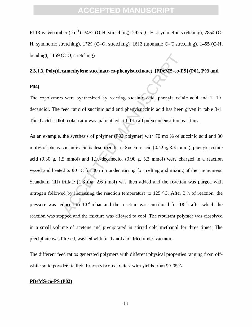
2.3.1.3. Poly(decamethylene succinate-co-phenylsuccinate) [PDeMS-co-PS] (P02, P03 and
P04)
The copolymers were synthesized by reacting succinic acid, phenylsuccinic acid and 1, 10-
decandiol. The feed ratio of succinic acid and phenylsuccinic acid has been given in table 3-1.
The diacids : diol molar ratio was maintained at 1:1 in all polycondensation reactions.
As an example, the synthesis of polymer (P02 polymer) with 70 mol% of succinic acid and 30
mol% of phenylsuccinic acid is described here. Succinic acid (0.42 g, 3.6 mmol), phenylsuccinic
acid (0.30 g, 1.5 mmol) and 1,10-decanediol (0.90 g, 5.2 mmol) were charged in a reaction
vessel and heated to 80 °C for 30 min under stirring for melting and mixing of the monomers.
Scandium (III) triflate (1.3 mg, 2.6 μmol) was then added and the reaction was purged with
nitrogen followed by increasing the reaction temperature to 125 °C. After 3 h of reaction, the
pressure was reduced to 10-2
mbar and the reaction was continued for 18 h after which the
reaction was stopped and the mixture was allowed to cool. The resultant polymer was dissolved
in a small volume of acetone and precipitated in stirred cold methanol for three times. The
precipitate was filtered, washed with methanol and dried under vacuum.
The different feed ratios generated polymers with different physical properties ranging from off-
white solid powders to light brown viscous liquids, with yields from 90-95%.
PDeMS-co-PS (P02)

ACC
EPTE
D M
ANU
SCR
IPT
ACCEPTED MANUSCRIPT
12
Mn: 11500 g mol-1
; Ð = 2.9
1H NMR (400 MHz, CDCl3) δ (ppm) 7.28 (C6H5, ring protons, m, 2.22H), 4.07 (CO-O-
CH2(C8H18)CH2 and CO-CH(C6H5), m, 4.23H), 3.19 (CH(C6H5)-CH2-CO, m, 0.30H and 2.68
(CH(C6H5)-CH2-CO), m, 0.30H), 2.62 (CO-CH2-CH2-CO), s, 2.80H), 1.61 (CH2-
CH2(C6H12)CH2-CH2, m, 4.00H), 1.28 (C2H4(C6H12)C2H4, m, 12.00H).
13C NMR (101 MHz, CDCl3) δ (ppm) 172.9 (CO-CH(C6H5)-CH2-CO), 172.4 (CO-CH2-CH2-
CO), 171.6 (CO-CH(C6H5)-CH2-CO), 137.9, 128.7, 127.7, 127.5 (aromatic), 64.9 (CH2-CO-O-
CH2), 47.3 (O-CO-CH(C6H5)-CH2-CO-O), 37.7 (O-CO-CH(C6H5)-CH2-CO-O), 29.4 (CO-O-
CH2-CH2), 29.2 (CO-CH2-CH2-CO), 29.2 (CO-O-CH2CH2-CH2), 28.6 (CO-O-CH2CH2CH2-
CH2), 25.9 (CO-O-CH2CH2CH2CH2-CH2).
FTIR wavenumber (cm-1
): 3450 (O-H, stretching), 2920 (C-H, asymmetric stretching), 2852 (C-
H, symmetric stretching), 1723 (C=O, stretching), 1609 (aromatic C=C stretching), 1420 (C-H,
bending), 1154 (C-O, stretching).
PDeMS-co-PS (P03)
Mn: 10100 g mol-1
; Ð = 3.4
1H NMR (400 MHz, CDCl3) δ (ppm) 7.28 (C6H5, ring protons, m, 2.92H), 4.07 (CO-O-
CH2(C8H18)CH2 and CO-CH(C6H5), m, 4.45H), 3.19 (CH(C6H5)-CH2-CO, m, 0.50H and 2.68
(CH(C6H5)-CH2-CO), m,0.50H), 2.62 (CO-CH2-CH2-CO), s, 2.00H), 1.61 (CH2-
CH2(C6H12)CH2-CH2, m, 4.00H), 1.27 (C2H4(C6H12)C2H4, m, 12.05H).
13C NMR (101 MHz, CDCl3) δ (ppm) 172.9 (CO-CH(C6H5)-CH2-CO), 172.4 (CO-CH2-CH2-
CO), 171.6 (CO-CH(C6H5)-CH2-CO), 137.9, 128.7, 127.7, 127.5 (aromatic), 64.9 (CH2-CO-O-
CH2), 47.3 (O-CO-CH(C6H5)-CH2-CO-O), 37.7 (O-CO-CH(C6H5)-CH2-CO-O), 29.4 (CO-O-

ACC
EPTE
D M
ANU
SCR
IPT
ACCEPTED MANUSCRIPT
13
CH2-CH2), 29.2 (CO-CH2-CH2-CO), 29.2 (CO-O-CH2CH2-CH2), 28.6 (CO-O-CH2CH2CH2-
CH2), 25.9 (CO-O-CH2CH2CH2CH2-CH2).
FTIR wavenumber (cm-1
): 3453 (O-H, stretching), 2925 (C-H, asymmetric stretching), 2854 (C-
H, symmetric stretching), 1731 (C=O, stretching), 1605 (aromatic C=C stretching), 1423 (C-H,
bending), 1159 (C-O, stretching).
PDeMS-co-PS (P04)
Mn: 11000 g mol-1
; Ð = 3.1
1H NMR (400 MHz, CDCl3) δ (ppm) 7.28 (C6H5, ring protons, m, 4.15H), 4.06 (CO-O-
CH2(C8H18)CH2 and CO-CH(C6H5), m, 4.29H), 3.19 (CH(C6H5)-CH2-CO, m, 0.70H and 2.68
(CH(C6H5)-CH2-CO), m, 0.70H), 2.61 (CO-CH2-CH2-CO), s, 1.30H), 1.55 (CH2-
CH2(C6H12)CH2-CH2, m, 4.00H), 1.26-1.20 (C2H4(C6H12)C2H4, m, 12.00H).
13C NMR (101 MHz, CDCl3) δ (ppm) 172.9 (CO-CH(C6H5)-CH2-CO), 172.4 (CO-CH2-CH2-
CO), 171.6 (CO-CH(C6H5)-CH2-CO), 137.9, 128.7, 127.7, 127.5 (aromatic), 64.9 (CH2-CO-O-
CH2), 47.3 (O-CO-CH(C6H5)-CH2-CO-O), 37.7 (O-CO-CH(C6H5)-CH2-CO-O), 29.4 (CO-O-
CH2-CH2), 29.2 (CO-CH2-CH2-CO), 29.2 (CO-O-CH2CH2-CH2), 28.6 (CO-O-CH2CH2CH2-
CH2), 25.8 (CO-O-CH2CH2CH2CH2-CH2).
FTIR wavenumber (cm-1
): 3447 (O-H, stretching), 2925 (C-H, asymmetric stretching), 2854 (C-
H, symmetric stretching), 1731 (C=O, stretching), 1608 (aromatic C=C stretching), 1428 (C-H,
bending), 1159 (C-O, stretching).
2.3.2. Empty and Dye-loaded NPs formulation

ACC
EPTE
D M
ANU
SCR
IPT
ACCEPTED MANUSCRIPT
14
NPs of synthesized polymers were prepared by a standard precipitation method with some
modification [38]. Briefly, polymer (20 mg) was dissolved in 5 mL of organic solvent (acetone)
to make a homogeneous organic phase. The organic phase was added drop-wise into 10 mL of
HPLC grade water under stirring (1000 rpm) and stirred for 4 h at room temperature. A syringe
pump was used to control the dropping rate (0.5 mL min-1
). The formulation was then left
overnight (open vial) to ensure the complete removal of organic solvent. The NPs suspension
was then passed through a membrane syringe filter (pore size: 220 nm) (Millex-LG, Millipore
Co., USA) to obtain a clear formulation. The sizes and zeta potentials of the NPs formulations
were measured in HPLC grade water and in 10 mM HEPES buffer (pH 7.4), respectively after
appropriate dilutions (100 μg mL-1
). Nanoparticle formulations were stored in a refrigerator and
sizes measured after 1 month using the same dilution (100 μg mL-1
) to determine the stability
against aggregation of the particles upon storage.
Coumarin-6 dye loaded NPs were prepared by a similar method as with blank nanoparticles by
adding dye in the organic phase along with polymer. Briefly, coumarin-6 (1 mg) was dissolved
along with the polymer (20 mg) in acetone (5 mL) and added at a fixed rate (0.5 mL min-1
) to the
HPLC grade water (10 mL). After 4 h of stirring, the formulations were stored overnight (open
vials) to allow acetone to evaporate. The process was performed in the dark to prevent the
degradation of light sensitive coumarin-6 dye. The precipitated polymer and dye was removed by
filtration through a membrane filter (pore size: 220 nm). Coumarin-6 loaded NPs were passed
through PD10 Desalting Column (Sephadex G-25 Medium, GE Healthcare Life Sciences) to
separate the unencapsulated dye from the formulations. The purified NPs solution was used for
further characterization. The Pluronic F-68 coating was achieved by the same method, i.e. where
Pluronic F-68 was dissolved in an aqueous medium (0.2% w/v) to make dye loaded and empty

ACC
EPTE
D M
ANU
SCR
IPT
ACCEPTED MANUSCRIPT
15
nanoparticles. The dye content was determined in freeze dried formulation. The freeze dried
micelle was dissolved in methanol.
The drug content was determined by freeze drying an aliquot of NP suspensions, followed by
dissolution in methanol and quantification by UV-visible spectrophotometry at λmax = 460 nm
(Beckman Coulter DU 800 UV spectrophotometer).
2.3.3. Degradation study of P02 NPs formulation
The degradation profile was assessed for the empty P02 NPs formulation in HPLC water (pH
7.4) and NPs formulation (in HPLC water) with Pseudomonas cepacia lipase (0.2 mg mL-1
). The
NP formulations were incubated at 37 °C. One vial from each set was collected at a
predetermined time (30 days) and freeze-dried. The freeze-dried samples were then dissolved in
chloroform, filtered, and analyzed by GPC to determine the change in molecular weight.
2.3.4. C3A Cell culture
C3A human hepatocellular carcinoma cells were cultured in MEM medium supplemented with
10% heat-inactivated foetal calf serum (FCS), 2 mM L-glutamine, 100 U/mL
penicillin/streptomycin, 1 mM sodium pyruvate and 1% non-essential amino acids (termed
MEM complete medium) and incubated at 37 °C and 5% CO2.
2.3.5. Cell Viability assessment
The cytotoxicity of the coumarin-6 labelled uncoated and Pluronic F68 coated P02 nanoparticles
was determined by a fluorescence-based 3 in 1 assay [39]. This approach allows the
simultaneous assessment of cell viability using the Alamar Blue (for metabolic activity), CFDA-
AM (for cell membrane integrity) and Neutral Red (for lysosomal function) assays. C3A cells
were seeded at a concentration of 1.56 x10⁵
cells/cm2
into a 96 well plate and incubated at 37 °C

ACC
EPTE
D M
ANU
SCR
IPT
ACCEPTED MANUSCRIPT
16
and 5% CO2 for 24 h. Cells were then exposed to MEM complete medium (control), 1% triton
X100 (positive control) or nanoparticles at concentrations of 4.68, 9.37, 18.75, 37.50, 75, 150
and 300 μg mL-1
(100 L/well) and incubated at 37 °C and 5% CO2 for 24 h. Nanoparticles were
prepared via dilution in complete MEM medium and briefly, gently vortexed prior to cell
exposure (nanoparticles were confirmed as intact by DLS). After 24 h the supernatants were
removed (and stored at -80 oC) and the cells were washed twice with phosphate buffered saline
(PBS). A solution of both 1.25% v/v Alamar Blue (TREK Lab Services) and 4 µM CFDA-AM
was prepared in phenol red free MEM medium and added to cells (100 μL) before incubating the
cells in the dark at 37 °C and 5% CO2 for 1 h. Fluorescence was measured on a SpectraMax M5
Microplate Reader (Molecular Devices) at an excitation/emission of 532/590 nm for Alamar
Blue and an excitation/emission of 485/535 nm for CFDA-AM. Next, for the Neutral Red uptake
assay, cells were washed twice using PBS, and Neutral Red (33 µg mL-1
) prepared in phenol red
free MEM medium was added (100 µL) to cells, which were then incubated in the dark at 37 °C
and 5 % CO2 for 1 h. Following incubation, cells were washed 3 times with PBS and then
acidified (1% acetic acid) 50% EtOH (in water) (100 µL). Cells were then incubated in the dark
at room temperature for 20 minutes (with shaking). Fluorescence was measured in a SpectraMax
M5 Microplate Reader at an excitation/emission of 532/645 nm. All experiments were repeated
on different days, on at least 3 separate occasions and data reported as mean % viability
(compared to the control) +/- standard error of the mean (SEM). GraphPad Prism 6 was used to
analyze the statistical significance of the data.

ACC
EPTE
D M
ANU
SCR
IPT
ACCEPTED MANUSCRIPT
17
2.3.6. Cell uptake studies
2.3.6.1. Confocal microscopy
C3A cells were seeded (6.58 x10⁴
cells/cm2) on 12 mm glass cover slips at 37 °C and 5% CO2 in
24 well plates (Falcon) for 24 h. Cells were then exposed to Pluronic F68 coated and uncoated
P02 nanoparticles at a sub lethal concentration (100 µg/mL) or MEM complete medium (control)
for 10, 60, 240 and 1440 minutes at 37 °C and 5% CO2. Following exposure, cells were washed
twice with PBS and fixed in 3% formaldehyde (in PBS, 250 µL) for 30 minutes in the dark at
room temperature. Subsequently, the plates were washed twice with PBS and incubated with 50
mM ammonium chloride (250 µL) for 10 minutes in the dark, at room temperature. Cells were
then washed twice with PBS before permeabilizing with 0.1% Triton-X100 (250 µL) for 20
minutes at room temperature. The cells were washed twice with PBS and then exposed to
primary antibody monoclonal anti α tubulin mouse ascites fluid clone DM1A (1:200 in PBS,
Molecular Probes, 250 µL) for 1 h at room temperature. Cells were then washed twice with PBS
and incubated with secondary antibody Rhodamine Red goat anti mouse IgG (1:100 in PBS,
Molecular Probes, 250 µL) for 1 h at room temperature. The cells were then washed twice with
PBS and incubated for 5 minutes in the dark with 1 µg mL-1
DAPI (250 µL) at room temperature
to stain the nuclei. Finally, cells were washed twice with PBS and coverslips were mounted onto
glass slides with Vector shield (Vector –H1000) and edges sealed with varnish. Cells were
imaged using a Zeiss LSM880 confocal microscope using the Zen program for data analyses.
2.3.6.2. Quantification of cell uptake study
C3A cells were seeded into 96 well plates at a concentration of 1.56 x10⁵ cells/cm
2 and
incubated for 24 h. Cells were exposed to uncoated and coated P02 nanoparticles (4.68-300 µg
mL-1
) or MEM complete medium (control) in duplicate for 10, 60, 240 and 1440 minutes at 37

ACC
EPTE
D M
ANU
SCR
IPT
ACCEPTED MANUSCRIPT
18
°C and 5% CO2. Following treatment cell supernatants were removed and cells were washed
twice using PBS and 0.4% v/v Trypan Blue (50 µL) was added for 10 minutes at room
temperature. Cells were washed twice using PBS and then lysed with 0.2% Triton X100 in the
dark at room temperature for 20 minutes. Fluorescence was measured using a SpectraMax M5
Microplate Reader at an excitation/emission of 488/550 nm. All experiments were repeated on at
least 3 separate occasions.
A standard curve of fluorescence for each NP (4.68-300 µg mL-1
) was generated to enable
uptake to be quantified which was expressed as the percentage (%) of applied dose.
2.3.7. Cytokine Production: measurement of IL-8
Supernatants collected during the cytotoxicity testing were used for cytokine analysis. The level
of IL-8 was quantified for selected NP concentrations (75 and 150 µg mL-1
) 24 h post exposure
using an ELISA kit, carried out following the manufacturer’s instructions (R & D systems).
2.3.8. Genotoxicity: Comet Assay
Oxidative DNA damage and DNA strand breaks were investigated using the
formamidopyrimidine-DNA glycosylase (FPG) modified Comet assay. C3A cells were seeded
at a concentration of 1.66x10⁵ cells/cm
2 into a six well plate and incubated at 37 °C for 24 h.
Cells were then washed with HBSS and exposed to HBSS (control), 60 μM hydrogen peroxide
(H2O2, positive control) or the NPs (dispersed in HBBS) at a concentration of 75 and 150 µg mL-
1. Cells were incubated at 37°C for 4 h and then washed with HBSS. Trypsin (0.25%, 1 mL) was
used to detach the cells and complete MEM medium (3 mL) was then added to each well. The
cell suspension was centrifuged at 850 g for 2 minutes and the cells then re-suspended in HBSS
(1 mL). The cell suspension (20 µL) was added to low melting point agarose (240 µL). The cells
(125 µL) were then pipetted onto microscope slides pre-coated with agarose. After a 10 minute

ACC
EPTE
D M
ANU
SCR
IPT
ACCEPTED MANUSCRIPT
19
period of solidification on ice, slides were placed in Coplin jars containing lysis buffer (66.75
mL lysis buffer base (Sigma), 7.5 mL dimethyl sulfoxide, 750 μL Triton-X100) for 24 h at 4 °C.
Slides were then washed in FPG buffer (400 mM HEPES, 1 M potassium chloride, 5 mM
ethylenediaminetetraacetic acid [EDTA], 2 mg mL-1
Bovine Serum Albumin, pH 8 ) for 15
minutes. The assay was conducted in the presence and absence of the enzyme FPG. FPG buffer
or FPG enzyme in buffer (100 μL) was added to slides, covered with a cover slip and incubated
at 37 °C for 30 minutes. Slides were then transferred to a darkened electrophoresis tank cooled to
4 °C and covered with electrophoresis buffer (300 mM sodium hydroxide, 200 mM EDTA, 2
litres distilled water, pH 13). After a 20 minute period to allow for alkaline unwinding,
electrophoresis was carried out for 20 minutes at 24 V and 270 mA. Slides were then removed
from the tank and placed in neutralisation buffer (48.5 g Tris base, 900 mL distilled water pH
7.5) for 15 minutes at 4 °C. Slides were then dried for 15 minutes before being dipped in 100%
ethanol and stained with GelRed. The tail moment (tail length x total tail intensity) was assessed
using a fluorescence microscope (Zeiss AX10 with Allied Vision Technologies Stingray camera)
connected to image analyzing software (Comet Assay IV: Perceptive Instruments, UK). Fifty cell
measurements were recorded for each slide per experiment.
Data in Figures are means ± standard error of the mean (SEM). Data for the two particle types
were compared to the control values and each other using an ANOVA with Tukey’s multiple
comparison for the tail moment. All statistical tests were performed using Minitab 15. A p value
<0.05 was considered a significant value. For the two particle types the experiment was repeated
a minimum of three times.
ACC
EPTE
D M
ANU
SCR
IPT
ACCEPTED MANUSCRIPT
20
3. Results
3.1. Synthesis and characterization of the polymers
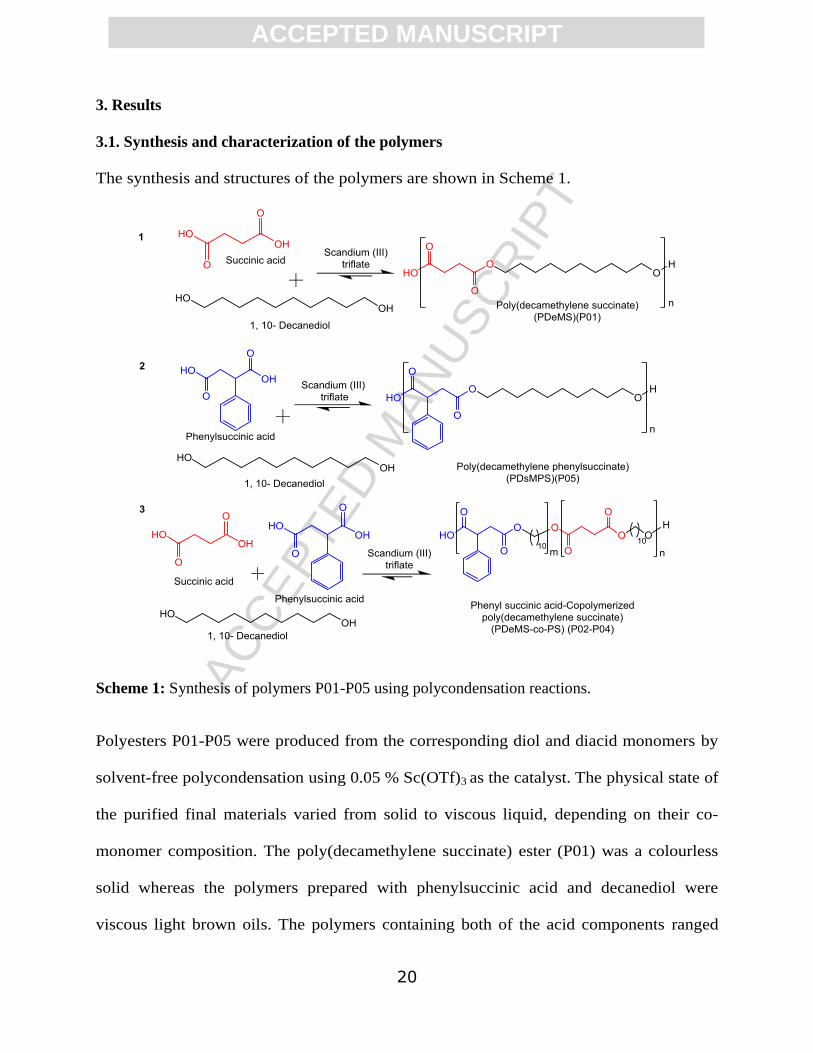
The synthesis and structures of the polymers are shown in Scheme 1.
Scheme 1: Synthesis of polymers P01-P05 using polycondensation reactions.
Polyesters P01-P05 were produced from the corresponding diol and diacid monomers by
solvent-free polycondensation using 0.05 % Sc(OTf)3 as the catalyst. The physical state of
the purified final materials varied from solid to viscous liquid, depending on their co-
monomer composition. The poly(decamethylene succinate) ester (P01) was a colourless
solid whereas the polymers prepared with phenylsuccinic acid and decanediol were
viscous light brown oils. The polymers containing both of the acid components ranged

ACC
EPTE
D M
ANU
SCR
IPT
ACCEPTED MANUSCRIPT
21
from solids to viscous oils according to phenyl content. The isolated yields of the
polymers were typically 90-95%. The characterization data of the synthesis and the
polymers are given in Table 2.
Table 2. Polymers (P01-P05): synthesis, composition (mol % of diacid) and molecular weight (g
mol-1
)
Polymer
Code
Feed
Ratio (mol%)
Product
Ratio (mol %)*
Molecular Weight
(g mol-1
)$ Yield
(%) SA PSA SA PSA Mn Mw
PDI
(Mw/Mn)
P01 100 0 100 0 11500 42200 3.66 94
P02 70 30 70 30 11500 37600 2.87 93
P03 50 50 50 50 10100 34200 3.39 92
P04 30 70 30 70 11000 34400 3.13 91
P05 0 100 0 100 11600 37600 3.24 90 *
Determined by 1H NMR spectroscopy;
$ Determined by gel permeation chromatography (GPC)
using CHCl3 as the mobile phase. SA: Succinic acid, PSA: Phenylsuccinic acid, Mn: Number
average molecular weight, Mw: Weight average molecular weight, PDI: Polydispersity Index.
The polymers showed number average molecular weights (Mn) ranging from 10,100-11,600 g
mol-1
as determined by gel permeation chromatography (GPC). The polydispersity indices were
similar for all the polymers and hence the materials were considered suitable for comparative
studies. The monomer composition of polyesters P01-P05 was determined by 1H NMR
spectroscopy (Figure S1, S3, S5, S7 and S9). The peak assignment was further confirmed by 2D
COSY NMR (Figure S2, S4, S6, S8 and S10) and 13
C NMR spectrum (Figure S 11).
The differential scanning calorimetry (DSC) data showed a pattern for the thermal properties of
the polymers (Table 3). The melting temperature (Tm), crystallization temperature (Tc) and
melting enthalpy (∆H) were found to decrease with increasing phenyl content (Figure S12, Table

ACC
EPTE
D M
ANU
SCR
IPT
ACCEPTED MANUSCRIPT
22
3). For P04 and P05 no Tm were detectable and only glass transition temperatures (Tg) were
observed. The glass transition temperatures for all the polymers were in the -40 to -30 °C range.
The P01 polymer with no phenyl side chains showed a sharp melting temperature, but no glass
transition temperature was detected. Figure S12 shows the trend in the melting temperatures
(Tm), glass transitions temperatures (Tg) and crystallization temperature (Tc) of the synthesized
polyesters P01-P05.
Table 3. Thermal properties and physical properties of the synthesized polyesters with different
phenyl side-chain content.
Polymer Code
Product
Ratio (mol%) Thermal Property
Physical
property SA PSA
Tg
(oC)
Tm
(oC)
Melting
Enthalpy
(∆H) J/g
Tc
(oC)
P01 100 0 ND 71 124 49 Crystalline
P02 70 30 -39 45 44 19 Semicrystalline
P03 50 50 -38 35 32 4 Semicrystalline
P04 30 70 -40 ND ND ND Amorphous
P05 0 100 -34 ND ND ND Amorphous
ND:Not detected; SA:Succinic acid; PSA:Phenylsuccinic acid. Tg: Glass transition temperature,
(Tg) (B) Tm: melting temperature; Tc: crystallization temperature
3.2. Preparation and characterization of nanoparticles
Polymers of similar molar mass but with differences in their phenyl content were investigated to
determine if side-chain content and Tg/Tm variation had any effect on their ability to encapsulate
model drugs. The polymers were mixed with a fluorescent dye, coumarin-6 (C log P ~ 6, as a
model for highly lipophilic drugs) prior to nanoprecipitation from a common solvent (acetone) to
a non-solvent (water). The characteristics of the polymers following nanoprecipitation and dye
loading are given in Table 4.

ACC
EPTE
D M
ANU
SCR
IPT
ACCEPTED MANUSCRIPT
23
Table 4 Characteristics of empty and dye loaded NPs of various polymers.
Polymer
Empty
Nanoparticles£
Dye Loaded
Nanoparticles£
Zeta
potential
(mV)
(±SD)
Drug Content
(%) ± SD
Average
Size (nm) PDI
Average
Size (nm) PDI
P01 144 ± 6 0.09 ± 0.02 146 ± 9 0.10 ± 0.02 -39 ± 9 ND
P02 160 ± 7 0.03 ± 0.01 168 ± 7 0.05 ± 0.01 -40 ± 7 0.28 ± 0.01
P03 163 ± 8 0.06 ± 0.02 166 ± 5 0.07 ± 0.02 -38 ± 5 0.23 ± 0.02
P04 165 ± 6 0.10 ± 0.03 172 ± 6 0.09 ± 0.02 -39 ± 6 0.12 ± 0.01
P05 164 ± 4 0.09 ± 0.02 167 ± 7 0.08 ± 0.02 -41 ± 6 0.05 ± 0.01
P02* 185 ± 5
¥ 0.07 ± 0.02 206 ± 6 0.08 ± 0.02 -21 ± 6
$ 0.23 ± 0.02
*Pluronic F68-coated P02 NPs.
£Determined in HPLC water using Dynamic Light Scattering
(DLS) Technique. ¥Significant difference in size from dye loaded P02* NPs (p < 0.05, unpaired
student’s t test). $Significant difference in zeta potential from P02 NPs (p < 0.05, unpaired
student’s t test).
At ambient temperature P01 polymer was found to be poorly soluble in the organic phase
(acetone) compared to the other polymers, and needed to be dissolved at ~ 40 °C prior to
nanoprecipitation. The method of nanoprecipitation was optimized for solvent to non-solvent and
polymer to coumarin-6 dye ratios. A difference in the color intensity (yellow) was observed
when the nanosuspensions were passed through a 0.22 µm filter to remove large aggregates. No
significant change in dye loading was observed when the formulation was dialyzed for 9 h using
12 kDa membranes, indicating that the dye was loaded only in the nanoparticles and was not
loosely associated with the nanoparticle surfaces. In general, the entrapped dye content was
decreased as the content in phenyl side-chains in the polymer increased. No dye was detected in
the NPs made from P01 polymer. The dye loading was found to be highest with PDeMS-co-PS
(P02) polymer, with 30% phenylsuccinic acid in its diacid repeating units, compared to other

ACC
EPTE
D M
ANU
SCR
IPT
ACCEPTED MANUSCRIPT
24
synthesized polymers, although the overall amounts of incorporated dye were still low. The
amount of incorporated coumarin-6 dye decreased as the proportion of phenyl side chains
increased from 30 to 100% of total diacid content (Figure 1). Nanoparticles made from P02
polymer were also formulated for enhanced suspension stability by the adsorption of Pluronic
F68, which significantly changed the zeta potential from -40 mV to -21 mV in dilute HEPES
buffer. The particle sizes of the NPs following storage for one month were statistically
insignificantly different compared to those of NPs before storage (p > 0.05, unpaired Student’s t
test) indicating good colloidal stability of the formulated NPs. The dye loading was significantly
decreased (P < 0.05, unpaired student’s t test) when uncoated P02 NPs formulations were
compared against the Pluronic F68 coated P02 NPs formulation, although again the overall
loading was low (Figure 1).

ACC
EPTE
D M
ANU
SCR
IPT
ACCEPTED MANUSCRIPT
25
Figure 1: Coumarin 6 loading (wt% to polymer) in copolymers P01-P05. Each point represents
mean dye content (wt% to polymer) ± SD (n=3). ND: Not detected. P02*: Pluronic F68-coated
P02 NPs. A significant difference in dye incorporation (p < 0.05, unpaired Student’s t test) was
observed between P02 and P02* NPs.
There was no significant difference in the sizes observed between empty and dye loaded P02
NPs. The Pluronic F68 coating significantly increased the size of the P02 NPs compared to
uncoated P02 NPs and the dye loaded P02 NPs coated with Pluronic were also of significantly
higher size compared to empty Pluronic coated P02 NPs (Table 4). As expected, the Pluronic
coating stabilized the P02 NPs compared to uncoated P02 NPs when diluted in PBS. Dynamic
light scattering of P02 NPs in HPLC water showed essentially one population group of particles
whereas two distinct populations were observed when the same uncoated P02 NPs were diluted
in PBS. In contrast, Pluronic F68 coated P02 NPs retained a unimodal population distribution
after dilution in both of the media (HPLC water and PBS). Additionally there was a significant
difference between the zeta potential of coated (-40 ± 7) and uncoated (-21 ± 6) P02 NPs when
P 0 1 P 0 2 P 0 3 P 0 4 P 0 5 P 0 2 *
0 .0
0 .1
0 .2
0 .3
0 .4
N a n o p a rtic le s
Dy
e
Co
nte
nt
(wt%
)
N D
*

ACC
EPTE
D M
ANU
SCR
IPT
ACCEPTED MANUSCRIPT
26
dispersed in HEPES 10 mM buffer (pH-7.4). The TEM images of uncoated and Pluronic coated
P02 NPs showed a uniform distribution of spherical nanoparticles (Figure 2).
Figure 2: Size distribution of NPs as determined using DLS of suspensions (100 μg mL-1
) in
HPLC water and PBS. (A) Uncoated P02 nanoparticles in HPLC water, (B) Uncoated P02 NPs
in PBS, (C) Pluronic coated P02 NPs in HPLC water, (D) Pluronic coated P02 NPs in PBS, (E)
TEM of uncoated P02 NPs, and (F) TEM of Pluronic coated P02 NPs.
A
B
C
D
Size/Intensity Size/Volume Size/Number
1000 nm1000 nm
E F

ACC
EPTE
D M
ANU
SCR
IPT
ACCEPTED MANUSCRIPT
27
As previously noted, uncoated (P02) NPs were significantly smaller (140 ± 1 nm) than coated
(P02*) NPs (146 ± 1 nm) when dispersed in MEM complete medium (Table 5). The zeta
potentials of P02 (-12.67 ± 0.37mV) and P02* (-10.68 ± 0.75 mV) NPs (in MEM complete
medium) were negative. The size variations for both NPs when dispersed in complete MEM
medium were low, ranging from polydispersity indices of 0.13 ± 0.01 for uncoated P02 NPs to
0.22 ± 0.01 for coated P02* NPs, therefore indicating that the NPs were stable in media as well
as in HPLC water.
Table 5: Characteristics of nanoparticles in MEM complete medium.
NP Hydrodynamic
Diameter (nm)#
Zeta Potential
(mV)#
Polydispersity Index
(PDI)#
P02 140 ± 1 -12.7 ± 0.4 0.13 ± 0.01
P02* 146 ± 1$ -10.7 ± 0.8 0.22 ± 0.01
$
# P02 and P02* NPs were dispersed in complete MEM medium (125µg/ml) prior to dynamic
light scattering measurements.*Pluronic F68 adsorbed onto the surface of the P02 nanoparticles. $ Significant difference (p<0.05) from uncoated P02 nanoparticles, Data are expressed as average
± SEM (n = 3).
3.3. Degradation of P02 nanoparticle formulations
Polymer degradation profiles are important in polyesters designed for sustained release, as these
data provide valuable information related to in vivo fate and long term formulation storage.
Degradation of the selected P02 formulation was monitored at specific time intervals (30 days) in
the presence and absence of a model hydrolytic enzyme, Pseudomonas cepacia lipase (0.2 mg
mL-1
). The presence of the lipase resulted in a reduction in the polymer Mn to 53% of its original
value after 3 months. Conversely, a drop of only 12% in Mn was observed in the absence of
enzyme (Figure 3).

ACC
EPTE
D M
ANU
SCR
IPT
ACCEPTED MANUSCRIPT
28
Figure 3: Loss in molar mass (Mn) of P02 NPs with time in the presence and absence of
Pseudomonas lipase (0.2 mg mL-1
) at 37 oC.
3.4. Impact of NPs on C3A cell viability
The effects following exposure of C3A hepatocytes to P02 NPs (in the presence and absence of a
Pluronic F68 coating) were assessed using the Alamar Blue (AB), 5-carboxyfluorescein
diacetate, acetoxymethyl ester, (CFDA-AM), and Neutral Red (NR) assays. For all assays, cell
viability (as a proxy for a specific activity) was more than 80% for both coated and uncoated P02
nanoparticles 24 h post exposure, at a concentration range (4.6 to 300 μg mL-1
) relevant to
polymeric nanomedicines administered in vivo (see Supplementary Information for
calculations).. Although dosing at 150 and 300 µg ml-1
for coated P02 (Figure 4) using the NR
assay indicated a statistically significant decrease in cell viability, this was not considered to be
biologically relevant as these were concentrations well above those likely to be used in a clinical
setting and even at these concentrations, the overall viability decrease was no more than 20 %.
0 3 0 6 0 9 0 1 2 0
0
5 0 0 0
1 0 0 0 0
1 5 0 0 0
2 0 0 0 0
2 5 0 0 0
T im e (D a y s )
Mn
(g
/mo
l b
y S
EC
)
P 0 2 N a n o p a rtic le s
(p H 7 .4 )
P 0 2 N a n o p a rtic le s
(w ith e n z y m e )

ACC
EPTE
D M
ANU
SCR
IPT
ACCEPTED MANUSCRIPT
29
Nevertheless, this decrease might be an indicator of effects on the lysosomal function of C3A
cells which could occur after accumulation of these polyester NPs.
Figure 4: Viability of C3A hepatocytes following exposure to Pluronic F68 coated (red) and
uncoated (black) P02 NPs. Cells were exposed to for 24 h with cell viability assessed via Alamar
Blue (A), CFDA (B) and Neutral Red (C) assays. Data are expressed as the average percentage
of cell viability (% of the control (MEM complete medium exposed cells) ± SEM (n = 3
minimum).
3.5. Cellular uptake of NPs by C3A cells
Imaging of the uptake of coated and uncoated P02 nanoparticles by C3A cells suggested that
both types of the nanoparticles were readily taken up by cells within 10 min of exposure. The
0 .0 0 .5 1 .0 1 .5 2 .0 2 .5 3 .0
0
2 0
4 0
6 0
8 0
1 0 0
1 2 0
L o g D o s e ( g /m L )
% V
iab
ilit
y
P 0 2 N P s (U n c o a te d )
P 0 2 N P s (P lu ro n ic c o a te d )
0 .0 0 .5 1 .0 1 .5 2 .0 2 .5 3 .0
0
2 0
4 0
6 0
8 0
1 0 0
1 2 0
L o g D o s e ( g /m L )
% V
iab
ilit
y
P 0 2 N P s (P lu ro n ic c o a te d )
P 0 2 N P s (U n c o a te d )
0 .0 0 .5 1 .0 1 .5 2 .0 2 .5 3 .0
0
2 0
4 0
6 0
8 0
1 0 0
1 2 0
L o g D o s e ( g /m L )
% V
iab
ilit
y
P 0 2 N P s (P lu ro n ic c o a te d )
P 0 2 N P s (U n c o a te d )
A
C
B
ACC
EPTE
D M
ANU
SCR
IPT
ACCEPTED MANUSCRIPT
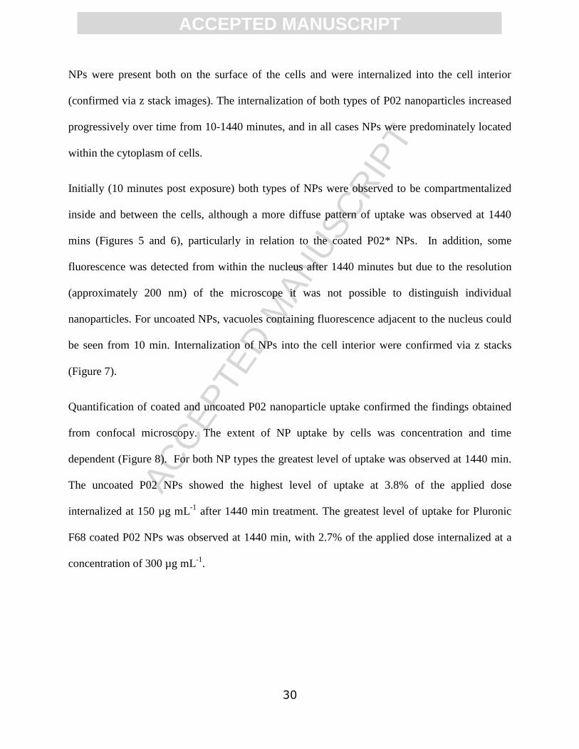
30
NPs were present both on the surface of the cells and were internalized into the cell interior
(confirmed via z stack images). The internalization of both types of P02 nanoparticles increased
progressively over time from 10-1440 minutes, and in all cases NPs were predominately located
within the cytoplasm of cells.
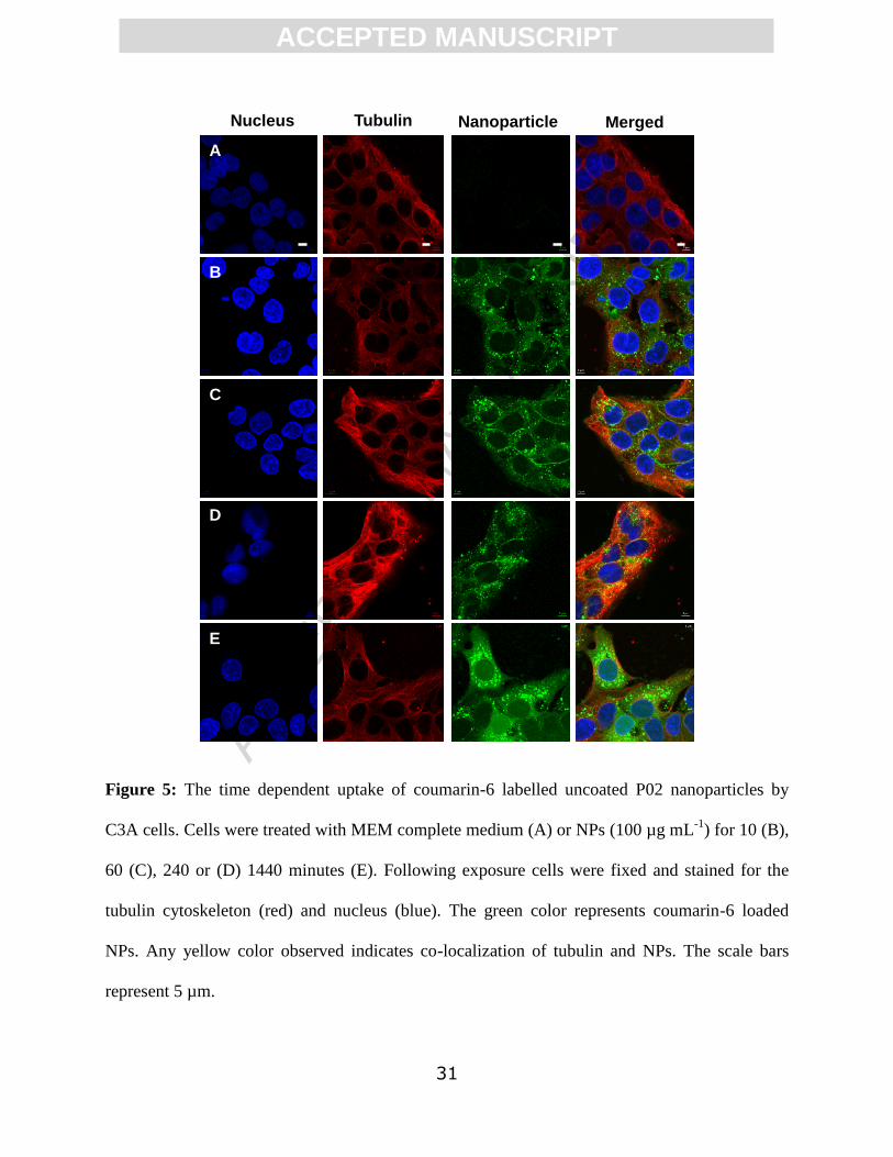
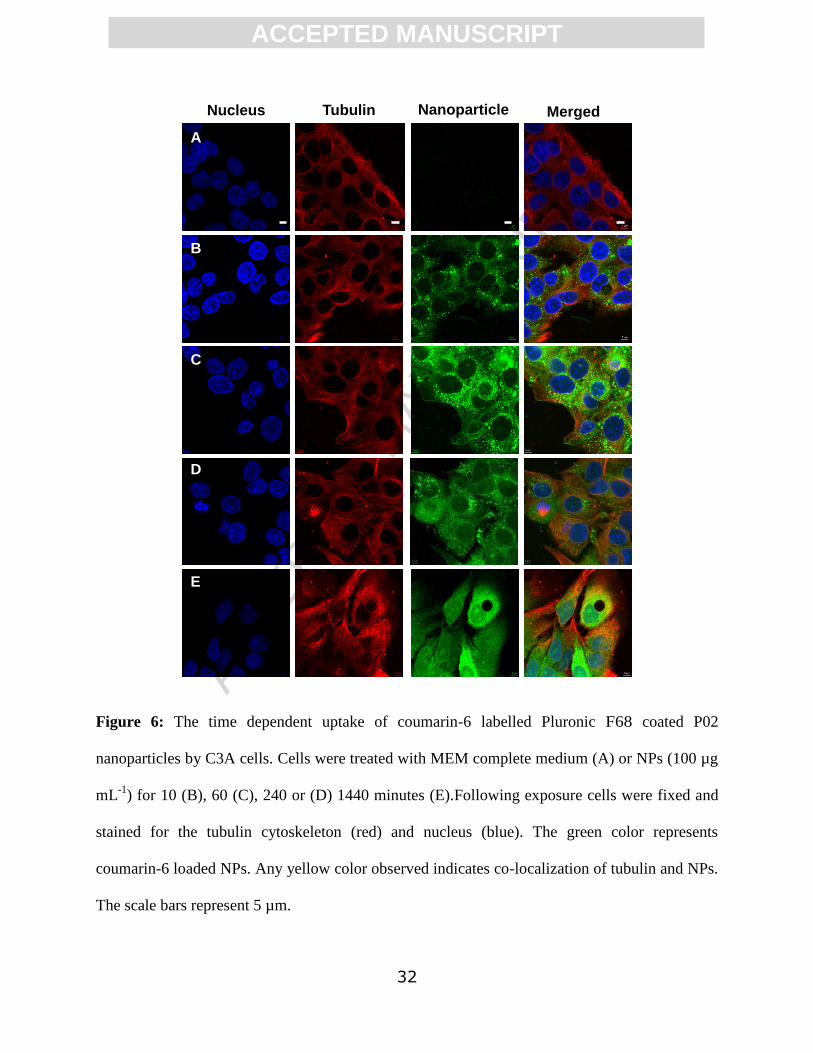
Initially (10 minutes post exposure) both types of NPs were observed to be compartmentalized
inside and between the cells, although a more diffuse pattern of uptake was observed at 1440
mins (Figures 5 and 6), particularly in relation to the coated P02* NPs. In addition, some
fluorescence was detected from within the nucleus after 1440 minutes but due to the resolution
(approximately 200 nm) of the microscope it was not possible to distinguish individual
nanoparticles. For uncoated NPs, vacuoles containing fluorescence adjacent to the nucleus could
be seen from 10 min. Internalization of NPs into the cell interior were confirmed via z stacks
(Figure 7).
Quantification of coated and uncoated P02 nanoparticle uptake confirmed the findings obtained
from confocal microscopy. The extent of NP uptake by cells was concentration and time
dependent (Figure 8). For both NP types the greatest level of uptake was observed at 1440 min.
The uncoated P02 NPs showed the highest level of uptake at 3.8% of the applied dose
internalized at 150 µg mL-1
after 1440 min treatment. The greatest level of uptake for Pluronic
F68 coated P02 NPs was observed at 1440 min, with 2.7% of the applied dose internalized at a
concentration of 300 µg mL-1
.
ACC
EPTE
D M
ANU
SCR
IPT
ACCEPTED MANUSCRIPT
31
Figure 5: The time dependent uptake of coumarin-6 labelled uncoated P02 nanoparticles by
C3A cells. Cells were treated with MEM complete medium (A) or NPs (100 µg mL-1
) for 10 (B),
60 (C), 240 or (D) 1440 minutes (E). Following exposure cells were fixed and stained for the
tubulin cytoskeleton (red) and nucleus (blue). The green color represents coumarin-6 loaded
NPs. Any yellow color observed indicates co-localization of tubulin and NPs. The scale bars
represent 5 µm.
Nucleus Tubulin Nanoparticle Merged
A
B
C
D
E
ACC
EPTE
D M
ANU
SCR
IPT
ACCEPTED MANUSCRIPT
32
Figure 6: The time dependent uptake of coumarin-6 labelled Pluronic F68 coated P02
nanoparticles by C3A cells. Cells were treated with MEM complete medium (A) or NPs (100 µg
mL-1
) for 10 (B), 60 (C), 240 or (D) 1440 minutes (E).Following exposure cells were fixed and
stained for the tubulin cytoskeleton (red) and nucleus (blue). The green color represents
coumarin-6 loaded NPs. Any yellow color observed indicates co-localization of tubulin and NPs.
The scale bars represent 5 µm.
Nucleus Tubulin Nanoparticle Merged
A
B
C
D
E

ACC
EPTE
D M
ANU
SCR
IPT
ACCEPTED MANUSCRIPT
33
Figure 7: Internalization of uncoated P02 and Pluronic F68-coated P02 (P02*) fluorescent NPs
over time by C3A cells. Images represent Z-stacks from which xy and yz micrographs were
generated to confirm internalization of NPs. Cells were treated with 100 µg mL-1
NPs for 10 min
(A), 60 min (B), 240 min (C) and 1440 min (D) and then fixed. Tubulin cytoskeleton is
represented by red, the nucleus by blue and the NPs by green. Co-localization of NPs and tubulin
is represented by yellow.
P02 NPs (Uncoated)
A B C D
P02 NPs (Pluronic coated)
A B C D

ACC
EPTE
D M
ANU
SCR
IPT
ACCEPTED MANUSCRIPT
34
Figure 8: Quantification of the uptake of coated and uncoated P02 NPs in C3A cells over time at
a range of concentrations (4.6-300 µg mL-1
). Cells were treated for 10 min, 60 min, 240 min and
1440 min with uncoated P02 NPs (A) or coated P02* NPs (B). The Percent (%) applied dose was
calculated from the relevant standard curves.
3.6. Cytokine Production
No change in cytokine production (IL-8) by C3A cells was detected at the NP concentrations
tested 24 h post exposure, compared to the control (data not shown).
3.7. Genotoxicity
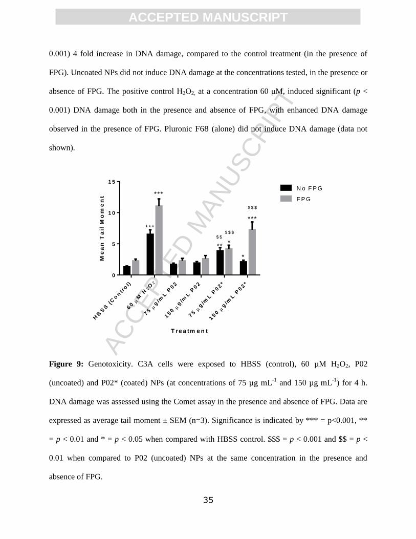
The results in Figure 9 show that at 4 h post exposure, the Pluronic F68 coated P02* NPs
induced DNA damage in C3A cells at both (sub-lethal) concentrations, when tail moment was
used as a measure of genotoxicity. In the absence of FPG, Pluronic coated NPs, at a
concentration of 75 µg mL-1
, induced a significant (p < 0.01) 3 fold increase in DNA damage in
C3A cells, when compared to the control. At a concentration of 150 μg mL-1
, coated NPs
induced a significant (p < 0.05) 1.6 fold increase in DNA damage, compared to the control. In
the presence of FPG, coated NPs, at a concentration of 150 µg mL-1
, induced a significant (p <
ACC
EPTE
D M
ANU
SCR
IPT
ACCEPTED MANUSCRIPT
35
0.001) 4 fold increase in DNA damage, compared to the control treatment (in the presence of
FPG). Uncoated NPs did not induce DNA damage at the concentrations tested, in the presence or
absence of FPG. The positive control H2O2, at a concentration 60 μM, induced significant (p <
0.001) DNA damage both in the presence and absence of FPG, with enhanced DNA damage
observed in the presence of FPG. Pluronic F68 (alone) did not induce DNA damage (data not
shown).
Figure 9: Genotoxicity. C3A cells were exposed to HBSS (control), 60 µM H2O2, P02
(uncoated) and P02* (coated) NPs (at concentrations of 75 µg mL-1
and 150 µg mL-1
) for 4 h.
DNA damage was assessed using the Comet assay in the presence and absence of FPG. Data are
expressed as average tail moment ± SEM (n=3). Significance is indicated by *** = p<0.001, **
= p < 0.01 and * = p < 0.05 when compared with HBSS control. $$$ = p < 0.001 and $$ = p <
0.01 when compared to P02 (uncoated) NPs at the same concentration in the presence and
absence of FPG.
HB
SS
(C
on
tro
l)
60
M H
2O
2
75
g/m
L P
02
150
g/m
L P
02
75
g/m
L P
02*
150
g/m
L P
02*
0
5
1 0
1 5
T re a tm e n t
Me
an
Ta
il M
om
en
t
N o F P G
F P G
**
*
*
***
***
***
$ $
$ $ $
$ $ $

ACC
EPTE
D M
ANU
SCR
IPT
ACCEPTED MANUSCRIPT
36
4. Discussion
This study generated polyesters with varying degrees of phenyl groups in their side chains which
in turn varied with respect to their crystallinity and ability to load a model drug compound. The
use of scandium (III) triflate enabled the reactions to be free of organic solvent and to generate
polyesters of similar molar masses repeatable over several batches of synthesis. Differences in Tg
and Tm in the polymers indicated differences in chain packing of the materials: polymers with
high phenyl content (P04-P05) showed only a Tg while the absence of Tm and Tc indicated that
these polymers were amorphous. Since it is known that aromaticity, glassiness, and crystallinity
in polymers can affect other properties such as drug loading, release and biodegradability, NPs of
the polymers varying in their physical properties were prepared in the presence of a dye
molecule (coumarin-6). The highest loading of coumarin-6 was observed for the P02 polymeric
NPs. This was not expected from first principles as the ring systems of the dye were expected to
interact more favorably with polymers containing high phenyl content through mutual
interactions. In addition, the polymers with higher phenyl content were amorphous hence were
expected to encapsulate more dye due to the higher free volumes in the amorphous core regions.
These initially contradictory results can be explained by considering three factors important
during nanoprecipitation; (1) polymer-polymer interaction (2) dye-dye interaction (3) dye-
polymer interaction. For a higher loading, a favorable dye-polymer interaction is important to
maintain a close proximity of dye and polymer during the late stages of nanoprecipitation.
However, a higher polymer-polymer interaction and dye-dye interaction can cause a reduction in
the dye loading during nanoprecipitation. For the P01 polymer, which contained no phenyl
succinate residues, it is likely that a high polymer-polymer and dye-dye interaction caused the
molecules to precipitate resulting in a low yield of well-defined NPs and with no detectable dye
ACC
EPTE
D M
ANU
SCR
IPT
ACCEPTED MANUSCRIPT
37
content. As the phenyl content was increased for the P02 polymer, a more favorable interaction
between dye and polymer may have resulted hence the higher dye loading. However, upon
further increase of phenyl content in the polymer (P03, P04 and P05), the higher
interactions between polymer molecules, rather than dye-polymer molecules resulted in lower
loading capabilities. Since the P02 polymer (70:30 succinic acid and phenyl succinic acid)
exhibited the highest dye loading capability when formulated into NPs, subsequent assessment of
the degradability of the polymers and its effects on hepatic cells were prioritised. Molar mass
profiles over time revealed that P02 polymer NPs were degradable, although the low degradation
rate of the polymer at pH 7.4 indicated that the polymer was quite resistant to primary hydrolytic
cleavage. The presence of esterolytic enzymes increased the degradation rate suggesting that the
polymer should be degraded in the body more rapidly if in contact with endogenous esterases.
Prior to cytocompatibility experiments, potential factors affecting cell association uptake and
uptake of the polymeric nanoparticles were evaluated. The hydrodynamic diameters of P02
(uncoated) NPs were less than those of P02* (coated) NPs, as expected due to the absorbed
Pluronic coating on P02* NPs but the differences were less than 10% so were unlikely to impact
on endocytic uptake mechanisms.[40] There is evidence that particle charge influences particle-
cell interactions and uptake, and the charge of NPs can also reflect dispersion stability and the
potential for agglomeration. As a consequence zeta potential is often used as an indicator of
charge when characterizing NP properties.[41] The zeta potentials of both P02 and P02* NPs
were negative, ranging from -10.7 to -12.7 mV, and in biologically relevant media the
differences in zeta potential between the coated and uncoated NPs were not statistically
significant, suggesting that NP charge alone was unlikely to influence particle-cell interactions.
ACC
EPTE
D M
ANU
SCR
IPT
ACCEPTED MANUSCRIPT
38
A 3 in 1 assay (Alamar Blue, CFDA-AM and Neutral Red) was used to assess viability of cells
exposed to the polymer NPs. Furthermore, using 3 different assays which assess different cell
responses as indicators of cytotoxicity can provide information on the mechanism of NP toxicity.
All three assays indicated that the coated and uncoated P02 NPs exhibited low acute toxicity in
the experimental conditions tested. A greater sensitivity was observed with the Neutral Red assay
showing statistically significant effects on normal lysosome function at high concentrations of
Pluronic F68 coated P02 NPs, although the small changes in cell viability observed were not
considered to be of potential biological significance.
Assessment of cytotoxicity at 24 h allows for comparison of results obtained in this study to
those obtained in prior literature as the majority of nanotoxicology studies evaluate cytotoxicity
24 h post exposure using the C3A cell line and other cell types (e.g. immune cells, epithelial
cells).[42, 43] A limitation of in vitro studies is that it is more challenging to assess chronic
toxicity. In future studies the development of in vitro assays which enable toxicity to be assessed
after repeated exposures or longer exposure periods would allow for the assessment of recovery
or increased cytotoxicity over time.
In order to evaluate the toxic potency of NPs, the lethal concentration (LC50) can be calculated,
which identifies the concentration of NPs required to kill 50% of cells. No LC50 values could be
calculated for the NPs tested in this study, despite concentrations up to 300 µg mL-1
being tested.
Previous studies which have investigated the response of C3A cells to engineered NPs (e.g.
silver, zinc oxide) at a similar concentration range tested in this study, have calculated LC50
values of approx. 2 µg mL-1
[44], demonstrating the relatively low toxicity of the polymeric NPs
tested in this study.

ACC
EPTE
D M
ANU
SCR
IPT
ACCEPTED MANUSCRIPT
39
Toxicity testing is performed in different phases. By screening the toxicity of the polymeric NPs
in vitro in the first instance, a rapid assessment of the toxicity of NPs of varied physico-chemical
properties is possible. Performing in vitro studies also allowed the mechanism of toxicity to be
probed in a cost and time efficient manner, and ensured that the study was aligned with the 3Rs
principles (reduction, refinement and replacement of animal testing). Although cell lines can lose
functions present in primary cells and therefore have been criticized for their lack of relevancy,
previous studies have demonstrated that the C3A cell line is able to replicate the response of
human and rat primary cells to many different types of NPs [44]. This is particularly important as
it is well established that cells vary widely in their sensitivity when exposed to a range of
nanomaterials [45].
NPs are known to elicit toxicity via mechanisms involving stimulation of inflammation and
oxidative stress [46], which are driven by their physico-chemical characteristics (e.g.
composition, size, charge). The stimulation of inflammatory and oxidant driven responses can
induce a number of downstream consequences such as genotoxicity or cell death. Cytokine
production was used as an indicator of the pro-inflammatory effects of the NPs in this study. No
IL-8 production was stimulated by NPs following exposure of C3A cells. NPs such as silver and
zinc oxide have previously been observed to stimulate IL-8 production [44], oxidative stress [47]
and genotoxicity [47] in C3A cells. Additionally, in vivo studies in mice using cationic NPs for
DNA delivery have shown increased levels of chemokine KC, the homolog of human IL-8.[48]
Accordingly assessment of NP mediated IL-8 production from hepatocytes was prioritised in this
study. No changes in IL-8 production were observed following exposure of cells to P02 and
P02* NPs, and while we cannot rule out production of other cytokines (e.g. TNF-a, IL-6) in
response to stimulation by NPs in these C3A cells, our primary screen based on IL-8 suggested
ACC
EPTE
D M
ANU
SCR
IPT
ACCEPTED MANUSCRIPT
40
no exceptional pro-inflammatory activation. It is also possible that any cytokine proteins
produced by cells may also have adsorbed to the NP surfaces, preventing their detection [49],
although we did not specifically measure for adsorbed proteins.
Pluronic F68-coated NPs were observed to stimulate DNA damage in C3A cells, whilst
uncoated NPs did not induce a response. DNA damage induced by Pluronic coated NPs was
enhanced in the presence of FPG which suggests that the damage is mediated by an oxidant
mechanism. The NPs themselves may have had intrinsic oxidative activity, as well as the ability
to induce production of intracellular reactive oxygen species (ROS) when interacting with the
cells to result in an imbalance between oxidants and antioxidants with in the cell.[50] Data
obtained suggested that the Pluronic F68 coating enhanced the genotoxicity of the NPs in this
study. It has been previously shown that certain Pluronic co-polymers can elicit transcriptional
activation in cell lines under certain conditions, and effects on complement activation have been
known for these polymers for many years [51, 52]. However, it is unlikely that the genotoxicity
observed here derived from the leaching of the amphiphilic co-polymer from the NP surfaces, as
when Pluronic F68 alone was administered to cells, at a concentration equivalent to that
contained in the NPs, no genotoxicity was observed. Nevertheless, since the properties of
different Pluronics change with concentration and aggregation status [53] a possible effect on
key cellular components of Pluronics associated at block co-polymer surfaces cannot be ruled
out. Work by Kabanov et al demonstrated that Pluronic P85 co-polymers were able to cause
energy-depleting effects in multi-drug resistant (MDR) cells, [54] where it was suggested that
depletion occurred partially by membrane permeabilisation and possible release of reactive
oxygen species (ROS). In prior studies of mitochondrial membrane disruption [55], Pluronic P85
was shown to be less potent than Pluronic F68 but more active than a higher molar mass
ACC
EPTE
D M
ANU
SCR
IPT
ACCEPTED MANUSCRIPT
41
Pluronic, L121. These previous data had indicated a ‘hotspot’ of membrane-disruption for the
Pluronic co-polymers with central hydrophobic poly(propylene oxide) block lengths of ~2000
Da, similar to that in Pluronic F68. Specific effects of the F68 coating may thus have accounted
for the observed increase in genotoxicity, and it should be noted that Pluronic F68 has been
shown to increase the production of interferon in Chinese Hamster Ovary cells, [56] albeit at
much higher concentrations than in our assays, through a mechanism that likely involved partial
or temporary membrane modification.
Evaluation of the uptake and intracellular fate of nanoparticles intended for medical use is
important as carrier materials are often required to enter the cell in order to deliver a therapeutic
or identify a disease phenotype [57]. Previous studies have indicated that uptake efficiency and
subcellular localisation can influence material cytotoxicity [58]. Therefore, it is important to
elucidate the uptake pattern of the polyester NPs, particularly in hepatic cell lines, as it is well
established that the liver is a site of accumulation for many nanoparticles following exposure via
various routes (e.g. intravenous injection, ingestion, inhalation/intratracheal instillation) [34, 35,
37]. The uptake of coated and uncoated P02 NPs in C3A cells suggested that the NPs were taken
up by the cells in a time and concentration dependent manner. Previous studies have indicated
that the uptake of polymer NPs by cells increased with time in other cell lines (e.g.
macrophages), although these studies did not look at the impact of NP concentration on uptake
and assessed uptake over a shorter time frame [59, 60]. Different cell types vary with respect to
their efficiency at internalising NPs. Existing studies have investigated the uptake of NPs by
macrophages in vivo and in vitro, due to their prominent role in particle clearance from the body
(e.g. lungs, liver). The predominant mechanism of particle uptake by hepatocytes and thus the
most likely route of uptake in this study is endocytosis, which has a lower efficiency than

ACC
EPTE
D M
ANU
SCR
IPT
ACCEPTED MANUSCRIPT
42
phagocytosis. Interestingly, similar overall levels of uptake compared to those of P02 and P02*
in C3A cells have been noted for carboxymethyl chitosan NPs in the L02 hepatocyte cell
line.[61] In this study the polyester NPs were primarily compartmentalized within and between
C3A cells as early as 10 minutes post exposure. At the later time point of 1440 minutes the
polyester NPs were observed throughout the cytoplasm of the C3A cells. The subcellular fate of
internalized NPs was not investigated in this study, however the pattern of uptake observed
suggests that these particular NPs were initially located within the cell organelles such as
endosomes or lysosomes. P02 NPs were observed to accumulate in lysosomes of the J774
macrophage cell line 1 h post exposure (data not shown) and polystyrene NPs and quantum dots
have also been observed to accumulate in lysosomes and mitochondria of macrophages in vitro
[62]. Current studies are investigating the subcellular localization to better understand the fate of
internalized polyesters from these formulations.
NPs were also seen to accumulate between cells, which may be representative of accumulation in
bile canaliculi. These structures are responsible for the formation and secretion of bile by
hepatocytes and could be indicative of NP removal from the cells. Interestingly, the elimination
of polystyrene NPs (20 nm) in bile has been observed previously in vitro and in vivo [60].
The co-localization of NPs with tubulin was also observed in this study, particularly within the
perinuclear region of the cell. Research has shown that tubulin may be involved with the
directional transport of NPs within the cell and this transport can be targeted towards the nucleus
[63]. NPs in vacuoles in close proximity to the nucleus could also suggest targeted delivery to
the nucleus, although the size of these particular NPs would likely prohibit them crossing an
undamaged nuclear membrane. At 24 h post exposure, coated NPs appeared to become diffuse
throughout the cell, whilst the majority of uncoated NPs remained compartmentalized,

ACC
EPTE
D M
ANU
SCR
IPT
ACCEPTED MANUSCRIPT
43
suggesting that the Pluronic coating may have had an impact on the intracellular fate of NPs.
NPs may thus have been partially degraded within certain cell organelles (e.g. lysosomes) via
hydrolytic enzymes known to degrade polyesters [64], and thus any dye associated near the NP
surfaces may have been released into the cytoplasm. It is also possible that the P02 NPs escaped
the organelles to distribute throughout the cytoplasm. An increase in the number and size of
vacuoles within cells treated with Pluronic coated (P02*) NPs was also apparent which may
indicate damage to the cells had occurred over time.
5. Conclusion
In this study, the synthesis of a new series of polyesters by melt polycondensation under solvent-
free conditions has been described and the effects of co-monomer content on polymer properties
(e.g. crystallinity) and nanoparticle properties (e.g. size, dye loading) relevant to controlled
release applications have been evaluated. The study suggested that side chain phenyl content
changed the loading capability of NPs for a model dye, but the overall level of incorporation
remained very low. The study provided information about the stability, and degradability of a
selected P02 polymer when formulated into Pluronic coated and uncoated nanoparticles. The
cellular studies revealed that P02 NPs displayed low cell toxicity and were effectively taken up
by the cells. However, the Pluronic coating appeared to enhance some aspects relating to the
toxicity of the NPs, and this information should be used to inform the design of formulated NPs
in the future.
Conflict of interest statement
The authors declare that there are no conflicts of interest.

ACC
EPTE
D M
ANU
SCR
IPT
ACCEPTED MANUSCRIPT
44
Acknowledgments
We thank the Indian Government for a PhD Scholarship to DK. We thank Heriot Watt
University for a James Watt PhD scholarship to LGP. We also thank the Engineering and
Physical Sciences Research Council (EPSRC) for financial support (Leadership Fellowship to
CA, Grants EP/H005625/1 and EP/J021180/1) and Christy Grainger-Boultby and Paul Cooling
for technical assistance. We would like to thank Dr Lesley Young (Edinburgh Napier University)
for her assistance in confocal microscopy.
Data access statement
All raw data created during this research are openly available from the corresponding author
([email protected]) and at the University of Nottingham Research Data
Management Repository (https://rdmc.nottingham.ac.uk/) and all analysed data supporting this
study are provided as supplementary information accompanying this paper.
References
[1] A.C. Albertsson, I.K. Varma, Aliphatic polyesters: Synthesis, properties and applications,
Degradable Aliphatic Polyesters, 157 (2002) 1-40.
[2] U. Edlund, A.C. Albertsson, Polyesters based on diacid monomers, Advanced Drug Delivery
Reviews, 55 (2003) 585-609.
[3] H. Seyednejad, A.H. Ghassemi, C.F. van Nostrum, T. Vermonden, W.E. Hennink, Functional
aliphatic polyesters for biomedical and pharmaceutical applications, Journal of Controlled
Release, 152 (2011) 168-176.
[4] T. Ishigaki, W. Sugano, M. Ike, Y. Kawagoshi, I. Fukunaga, M. Fujita, Abundance of
polymers degrading microorganisms in a sea-based solid waste disposal site, Journal of
Basic Microbiology, 40 (2000) 177-186.
[5] H.M. Aliabadi, A. Mahmud, A.D. Sharifabadi, A. Lavasanifar, Micelles of methoxy
poly(ethylene oxide)-b-poly(epsilon-caprolactone) as vehicles for the solubilization and
controlled delivery of Cyclosporine A, Journal of Controlled Release, 104 (2005) 301-311.
[6] M.L. Adams, A. Lavasanifar, G.S. Kwon, Amphiphilic block copolymers for drug delivery,
Journal of Pharmaceutical Sciences, 92 (2003) 1343-1355.

ACC
EPTE
D M
ANU
SCR
IPT
ACCEPTED MANUSCRIPT
45
[7] S. Elhasi, R. Astaneh, A. Lavasanifar, Solubilization of an amphiphilic drug by poly(ethylene
oxide)-block-poly(ester) micelles, European Journal of Pharmaceutics and
Biopharmaceutics, 65 (2007) 406-413.
[8] Z. Ma, A. Haddadi, O. Molavi, A. Lavasanifar, R. Lai, J. Samuel, Micelles of poly(ethylene
oxide)-b-poly(epsilon-caprolactone) as vehicles for the solubilization, stabilization, and
controlled delivery of curcumin, Journal of Biomedical Materials Research Part A, 86A
(2008) 300-310.
[9] L.A. Dailey, M. Wittmar, T. Kissel, The role of branched polyesters and their modifications
in the development of modern drug delivery vehicles, Journal Of Controlled Release, 101
(2005) 137-149.
[10] P. Sharma, S. Brown, G. Walter, S. Santra, B. Moudgil, Nanoparticles for bioimaging,
Advances in Colloid and Interface Science, 123 (2006) 471-485.
[11] J.R. McCarthy, R. Weissleder, Multifunctional magnetic nanoparticles for targeted imaging
and therapy, Advanced Drug Delivery Reviews, 60 (2008) 1241-1251.
[12] M.A. Woodruff, D.W. Hutmacher, The return of a forgotten polymer-Polycaprolactone in
the 21st century, Progress in Polymer Science, 35 (2010) 1217-1256.
[13] Y.D. Wang, G.A. Ameer, B.J. Sheppard, R. Langer, A tough biodegradable elastomer,
Nature Biotechnology, 20 (2002) 602-606.
[14] H. Seyednejad, D. Gawlitta, W.J.A. Dhert, C.F. van Nostrum, T. Vermonden, W.E.
Hennink, Preparation and characterization of a three-dimensional printed scaffold based on a
functionalized polyester for bone tissue engineering applications, Acta Biomaterialia, 7
(2011) 1999-2006.
[15] R.N. Shirazi, W. Ronan, Y. Rochev, P. McHugh, Modelling the degradation and elastic
properties of poly(lactic-co-glycolic acid) films and regular open-cell tissue engineering
scaffolds, Journal of the Mechanical Behavior of Biomedical Materials, 54 (2016) 48-59.
[16] H. Kenar, G.T. Köse, V. Hasirci, Tissue engineering of bone on micropatterned
biodegradable polyester films, Biomaterials, 27 (2006) 885-895.
[17] B.D. Ulery, L.S. Nair, C.T. Laurencin, Biomedical Applications of Biodegradable
Polymers, Journal of Polymer Science Part B-Polymer Physics, 49 (2011) 832-864.
[18] S. Mura, P. Couvreur, Nanotheranostics for personalized medicine, Advanced Drug
Delivery Reviews, 64 (2012) 1394-1416.
[19] J. Thevenot, H. de Oliveira, O. Sandre, L. Pourtau, E. Andres, S. Miraux, E. Thiaudiere, E.
Berra, S. Lecommandoux, Multifunctional polymersomes for cancer theranostics, Journal of
Controlled Release, 172 (2013) E44-E45.
[20] C. Jérôme, P. Lecomte, Recent advances in the synthesis of aliphatic polyesters by ring-
opening polymerization, Advanced Drug Delivery Reviews, 60 (2008) 1056-1076.
[21] H. Tian, Z. Tang, X. Zhuang, X. Chen, X. Jing, Biodegradable synthetic polymers:
Preparation, functionalization and biomedical application, Progress in Polymer Science, 37
(2012) 237-280.
[22] A. Pitto-Barry, N.P.E. Barry, Pluronic (R) block-copolymers in medicine: from chemical
and biological versatility to rationalisation and clinical advances, Polymer Chemistry, 5
(2014) 3291-3297.
[23] L. Li, L. Mu, V. Torchilin, Mixed micelles made of pluronic and PEG-PE for solubilization
of poorly soluble anticancer drug paclitaxel, J. Drug Deliv. Sci. Technol., 17 (2007) 389-
392.

ACC
EPTE
D M
ANU
SCR
IPT
ACCEPTED MANUSCRIPT
46
[24] S.M. Moghimi, A.C. Hunter, Poloxamers and Poloxamines in Nanoparticle Engineering and
Experimental Medicine, Trends in Biotechnology, 18 (2000) 412-420.
[25] R.J. Green, S. Tasker, J. Davies, M.C. Davies, C.J. Roberts, S.J.B. Tendler, Adsorption of
PEO-PPO-PEO triblock copolymers at the solid/liquid interface: A surface plasmon
resonance study, Langmuir, 13 (1997) 6510-6515.
[26] J.C. Neal, S. Stolnik, M.C. Garnett, S.S. Davis, L. Illum, Modification of the copolymers
poloxamer 407 and poloxamine 908 can affect the physical and biological properties of
surface modified nanospheres, Pharmaceutical Research, 15 (1998) 318-324.
[27] S. Stolnik, L. Illum, S.S. Davis, Long Circulating Microparticulate Drug Carriers, Advanced
Drug Delivery Reviews, 16 (1995) 195-214.
[28] L. Illum, S.S. Davis, R.H. Müller, E. Mak, P. West, The organ distribution and circulation
time of intravenously injected colloidal carriers sterically stabilized with a blockcopolymer -
poloxamine 908, Life Sciences, 40 (1987) 367-374.
[29] C. Hahn, S. Wesselbaum, H. Keul, M. Moller, OH-functional polyesters based on malic
acid: Influence of the OH-groups onto the thermal properties, European Polymer Journal, 49
(2013) 217-227.
[30] A. Takasu, Y. Iio, Y. Oishi, Y. Narukawa, T. Hirabayashi, Environmentally benign
polyester synthesis by room temperature direct polycondensation of dicarboxylic acid and
diol, Macromolecules, 38 (2005) 1048-1050.
[31] A. Takasu, Y. Shibata, Y. Narukawa, T. Hirabayashi, Chemoselective dehydration
polycondensations of dicarboxylic acids and diols having pendant hydroxyl groups using the
room temperature polycondensation technique, Macromolecules, 40 (2007) 151-153.
[32] A. Takasu, Y. Oishi, Y. Iio, Y. Inai, T. Hirabayashi, Synthesis of aliphatic polyesters by
direct polyesterification of dicarboxylic acids with diols under mild conditions catalyzed by
reusable rare-earth triflate, Macromolecules, 36 (2003) 1772-1774.
[33] A. Takasu, H. Tsuruta, Y. Narukawa, Y. Shibata, M. Oshimura, T. Hirabayashi, Dual
catalytic system for combination of chain and step polymerizations: Ring-opening
polymerization of epsilon-caprolactone and successive dehydration polycondensation with
dicarboxylic acid using the same catalyst, Macromolecules, 41 (2008) 4688-4693.
[34] C. Schleh, M. Semmler-Behnke, J. Lipka, A. Wenk, S. Hirn, M. Schaeffler, G. Schmid, U.
Simon, W.G. Kreyling, Size and surface charge of gold nanoparticles determine absorption
across intestinal barriers and accumulation in secondary target organs after oral
administration, Nanotoxicology, 6 (2012) 36-46.
[35] S. Hirn, M. Semmler-Behnke, C. Schleh, A. Wenk, J. Lipka, M. Schaeffler, S. Takenaka,
W. Moeller, G. Schmid, U. Simon, W.G. Kreyling, Particle size-dependent and surface
charge-dependent biodistribution of gold nanoparticles after intravenous administration,
European Journal of Pharmaceutics and Biopharmaceutics, 77 (2011) 407-416.
[36] E. Sadauskas, N.R. Jacobsen, G. Danscher, M. Stoltenberg, U. Vogel, A. Larsen, W.
Kreyling, H. Wallin, Biodistribution of gold nanoparticles in mouse lung following
intratracheal instillation, Chemistry Central Journal, 3 (2009).
[37] M. Semmler-Behnke, W.G. Kreyling, J. Lipka, S. Fertsch, A. Wenk, S. Takenaka, G.
Schmid, W. Brandau, Biodistribution of 1.4-and 18-nm Gold Particles in Rats, Small, 4
(2008) 2108-2111.
[38] U. Bilati, E. Allemann, E. Doelker, Development of a nanoprecipitation method intended
for the entrapment of hydrophilic drugs into nanoparticles, European Journal of
Pharmaceutical Sciences, 24 (2005) 67-75.
ACC
EPTE
D M
ANU
SCR
IPT
ACCEPTED MANUSCRIPT
47
[39] M. Connolly, M.-L. Fernandez-Cruz, A. Quesada-Garcia, L. Alte, H. Segner, J.M. Navas,
Comparative Cytotoxicity Study of Silver Nanoparticles (AgNPs) in a Variety of Rainbow
Trout Cell Lines (RTL-W1, RTH-149, RTG-2) and Primary Hepatocytes, International
Journal of Environmental Research and Public Health, 12 (2015) 5386-5405.
[40] C. He, Y. Hu, L. Yin, C. Tang, C. Yin, Effects of particle size and surface charge on cellular
uptake and biodistribution of polymeric nanoparticles, Biomaterials, 31 (2010) 3657-3666.
[41] A.M. Abdelmonem, B. Pelaz, K. Kantner, N.C. Bigall, P. del Pino, W.J. Parak, Charge and
agglomeration dependent in vitro uptake and cytotoxicity of zinc oxide nanoparticles,
Journal of Inorganic Biochemistry, 153 (2015) 334-338.
[42] D.M. Brown, H. Johnston, E. Gubbins, V. Stone, Cytotoxicity and Cytokine Release in Rat
Hepatocytes, C3A Cells and Macrophages Exposed to Gold Nanoparticles-Effect of
Biological Dispersion Media or Corona, Journal of Biomedical Nanotechnology, 10 (2014)
3416-3429.
[43] A. Kermanizadeh, G. Pojana, B.K. Gaiser, R. Birkedal, D. Bilanicova, H. Wallin, K.A.
Jensen, B. Sellergren, G.R. Hutchison, A. Marcomini, V. Stone, In vitro assessment of
engineered nanomaterials using a hepatocyte cell line: cytotoxicity, pro-inflammatory
cytokines and functional markers, Nanotoxicology, 7 (2013) 301-313.
[44] A. Kermanizadeh, B.K. Gaiser, M.B. Ward, V. Stone, Primary human hepatocytes versus
hepatic cell line: assessing their suitability for in vitro nanotoxicology, Nanotoxicology, 7
(2013) 1255-1271.
[45] A. Kermanizadeh, I. Gosens, L. MacCalman, H. Johnston, P.H. Danielsen, N.R. Jacobsen,
A.G. Lenz, T. Fernandes, R.P. Schins, F.R. Cassee, H. Wallin, W. Kreyling, T. Stoeger, S.
Loft, P. Moller, L. Tran, V. Stone, A Multilaboratory Toxicological Assessment of a Panel
of 10 Engineered Nanomaterials to Human Health-ENPRA Project-The Highlights,
Limitations, and Current and Future Challenges, Journal of toxicology and environmental
health. Part B, Critical reviews, 19 (2016) 1-28.
[46] K. Donaldson, V. Stone, P.J. Borm, L.A. Jimenez, P.S. Gilmour, R.P. Schins, A.M.
Knaapen, I. Rahman, S.P. Faux, D.M. Brown, W. MacNee, Oxidative stress and calcium
signaling in the adverse effects of environmental particles (PM10), Free radical biology &
medicine, 34 (2003) 1369-1382.
[47] A. Kermanizadeh, B.K. Gaiser, G.R. Hutchison, V. Stone, An in vitro liver model -
assessing oxidative stress and genotoxicity following exposure of hepatocytes to a panel of
engineered nanomaterials, Particle and Fibre Toxicology, 9 (2012).
[48] M. Elsabahy, K.L. Wooley, Cytokines as biomarkers of nanoparticle immunotoxicity,
Chemical Society Reviews, 42 (2013) 5552-5576.
[49] D.M. Brown, C. Dickson, P. Duncan, F. Al-Attili, V. Stone, Interaction between
nanoparticles and cytokine proteins: impact on protein and particle functionality,
Nanotechnology, 21 (2010) 215104.
[50] K. Unfried, C. Albrecht, L.-O. Klotz, A. Von Mikecz, S. Grether-Beck, R.P.F. Schins,
Cellular responses to nanoparticles: Target structures and mechanisms, Nanotoxicology, 1
(2007) 52-71.
[51] S.M. Moghimi, A.C. Hunter, C.M. Dadswell, S. Savay, C.R. Alving, J. Szebeni, Causative
factors behind poloxamer 188 (Pluronic F68, Flocor™)-induced complement activation in
human sera: A protective role against poloxamer-mediated complement activation by
elevated serum lipoprotein levels, Biochimica et Biophysica Acta (BBA) - Molecular Basis
of Disease, 1689 (2004) 103-113.

ACC
EPTE
D M
ANU
SCR
IPT
ACCEPTED MANUSCRIPT
48
[52] S.M. Moghimi, Modulation of Lymphatic Distribution of Subcutaneously Injected
Poloxamer 407-Coated Nanospheres: the Effect of the Ethylene Oxide Chain Configuration,
Febs Letters, 540 (2003) 241-244.
[53] G. Sahay, E.V. Batrakova, A.V. Kabanov, Different Internalization Pathways of Polymeric
Micelles and Unimers and Their Effects on Vesicular Transport, Bioconjugate Chemistry,
19 (2008) 2023-2029.
[54] E.V. Batrakova, S. Li, A.M. Brynskikh, A.K. Sharma, Y. Li, M. Boska, N. Gong, R.L.
Mosley, V.Y. Alakhov, H.E. Gendelman, A.V. Kabanov, Effects of pluronic and
doxorubicin on drug uptake, cellular metabolism, apoptosis and tumor inhibition in animal
models of MDR cancers, Journal of Controlled Release, 143 (2010) 290-301.
[55] M. Redhead, G. Mantovani, S. Nawaz, P. Carbone, D.C. Gorecki, C. Alexander, C.
Bosquillon, Relationship between the Affinity of PEO-PPO-PEO Block Copolymers for
Biological Membranes and Their Cellular Effects, Pharmaceutical Research, 29 (2012)
1908-1918.
[56] M.F. Clincke, E. Guedon, F.T. Yen, V. Ogier, O. Roitel, J.L. Goergen, Effect of surfactant
pluronic F-68 on CHO cell growth, metabolism, production, and glycosylation of human
recombinant IFN-gamma in mild operating conditions, Biotechnol Prog, 27 (2011) 181-190.
[57] Y. Liu, H. Miyoshi, M. Nakamura, Nanomedicine for drug delivery and imaging: A
promising avenue for cancer therapy and diagnosis using targeted functional nanoparticles,
International Journal of Cancer, 120 (2007) 2527-2537.
[58] J. Lovric, H.S. Bazzi, Y. Cuie, G.R.A. Fortin, F.M. Winnik, D. Maysinger, Differences in
subcellular distribution and toxicity of green and red emitting CdTe quantum dots, Journal
of Molecular Medicine-Jmm, 83 (2005) 377-385.
[59] R. Firdessa, T.A. Oelschlaeger, H. Moll, Identification of multiple cellular uptake pathways
of polystyrene nanoparticles and factors affecting the uptake: Relevance for drug delivery
systems, European Journal of Cell Biology, 93 (2014) 323-337.
[60] H.J. Johnston, M. Semmler-Behnke, D.M. Brown, W. Kreyling, L. Tran, V. Stone,
Evaluating the uptake and intracellular fate of polystyrene nanoparticles by primary and
hepatocyte cell lines in vitro, Toxicology and Applied Pharmacology, 242 (2010) 66-78.
[61] L. Shi, C. Tang, C. Yin, Glycyrrhizin-modified O-carboxymethyl chitosan nanoparticles as
drug vehicles targeting hepatocellular carcinoma, Biomaterials, 33 (2012) 7594-7604.
[62] M.J. Clift, C. Brandenberger, B. Rothen-Rutishauser, D.M. Brown, V. Stone, The uptake
and intracellular fate of a series of different surface coated quantum dots in vitro,
Toxicology, 286 (2011) 58-68.
[63] H. Akita, K. Enoto, H. Tanaka, H. Harashima, Particle Tracking Analysis for the
Intracellular Trafficking of Nanoparticles Modified with African Swine Fever Virus Protein
p54-derived Peptide, Molecular Therapy, 21 (2013) 309-317.
[64] M.S. Shive, J.M. Anderson, Biodegradation and biocompatibility of PLA and PLGA
microspheres, Adv Drug Deliv Rev, 28 (1997) 5-24.