journal of diabetes and metabolism - longdom.org · glycated albumin, the extent of glycation and...
TRANSCRIPT
Research Article Open Access
Maria et al. J Diabetes Metab 2011, S:4 DOI: 10.4172/2155-6156.S4-003
J Diabetes Metab Diabetic Cardiovascular Complications ISSN:2155-6156 JDM, an open access journal
Keywords: Advanced glycation end products; Shear stress;Thrombogenicity; Endothelial cells; Cardiovascular diseases; Diabetes mellitus
IntroductionIt is well known that a diabetic vasculature differs from a normal
vasculature and it promotes cardiovascular disease progression through the actions of advanced glycation end products (AGEs) on endothelial cells and platelets [1-5]. The role of glycation extent on these functions remains unclear. Some claim that conversion to a Schiff base (reversible glycation, 1-2 weeks of glycation) is sufficient to promote disease conditions, whereas others argue that the glycation products must pass through the Amadori rearrangement (irreversible glycation, 6-8 weeks of glycation) to exert an effect [6,7]. Here we aimed to determine the role of glycation extent on endothelial cells exposed to varying magnitudes of shear stress. However, regardless of the extent of glycation, it is clear that AGEs are present during diabetes mellitus, alter cellular functions and may promote cardiovascular disease conditions.
Altered shear stress is a hallmark of many cardiovascular diseases, including high magnitude short duration shear stresses, low magnitude oscillatory shear stresses and low magnitude unidirectional shear stresses [8,9], which has been reviewed extensively [10]. Altered shear has many effects on endothelial cells, including differential expression of eNOS [11], cytoskeletal re-organization [11], enhanced hemostatic potential [12], inflammatory responses [13] and gap junction channel formation [14] and promotion of angiogenesis [15]. Interestingly, nearly all of these effects can lead to pathologies similar to those seen in cardiovascular diseases, if the shear stress stimulus is sufficient.
For instance, the normal mean wall shear stress in the cardiovascular system can be as low as 0.5 Pa, but during cardiovascular diseases, this generally increases to 1-2 Pa, depending on the severity of the disease. Here we stimulate endothelial cells with nominal and pathologically high shear stresses to instigate a cellular response that may mimic disturbed flow due to the short exposure duration.
As we have previously discussed, there has been a significant amount of work on the effects of shear stress or advanced glycation end products on endothelial cell responses which lead to cardiovascular diseases. The major limitation of these studies is that the combined effects of shear stress and glycated albumin may accelerate cardiovascular disease progression through synergistic actions. Our goal in this report was to determine how shear stress, the presence of glycated albumin, the extent of glycation and duration of endothelial
*Corresponding author: David A. Rubenstein, School of Mechanical and Aerospace Engineering, Oklahoma State University, 218 Engineering North, Stillwater, OK 74078-5016, USA, Tel: 405-744-5900; Fax: 405-744-7873; E-mail: [email protected]
Received October 21, 2011; Accepted December 14, 2011; Published December 20, 2011
Citation: Maria Z, Yin W, Rubenstein DA (2011) Glycated Albumin and Pathological Shear Stress Alters Endothelial Cell Thrombogenic Potential, Pro-Inflammatory State and Cytoskeletal Dynamics. J Diabetes Metab S4:003. doi:10.4172/2155-6156.S4-003
Copyright: © 2011 Maria Z, et al. This is an open-access article distributed under the terms of the Creative Commons Attribution License, which permits unrestricted use, distribution, and reproduction in any medium, provided the original author and source are credited.
Glycated Albumin and Pathological Shear Stress Alters Endothelial Cell Thrombogenic Potential, Pro-Inflammatory State and Cytoskeletal DynamicsZahra Maria, Wei Yin and David A. Rubenstein*
School of Mechanical and Aerospace Engineering, Oklahoma State University, Stillwater, OK 74078-5016, USA
AbstractBackground: Diabetes mellitus is a disease that is characterized by a high blood glucose concentration, which
leads to advanced glycation end product (AGE) formation. AGEs have various effects on the vasculature and this may be dependent on the extent and reversibility of glycation. AGEs present in the vasculature can promote cardiovascular diseases through modifications to circulating proteins and endothelial cells. Furthermore, cardiovascular diseases are characterized by altered shear stress, including both high magnitude short duration shear and low oscillatory shear. However, the combined role of high magnitude shear stress and the presence of AGEs on endothelial cell functions have not been elucidated. Our objective was to evaluate changes to endothelial cell responses under these conditions.
Methods: To accomplish this, albumin was glycated for up to 8 weeks and endothelial cells were subjected to glycated albumin for up to 5 days. Endothelial cells were then exposed to shear stress in a cone-and-plate shearing device. Endothelial cell metabolic activity, surface expression of intracellular adhesion molecule-1, thrombomodulin, tissue factor, connexin-43, and caveolin-1, cytoskeletal organization and morphology were investigated.
Results: In general, the combination of pathological shear stress and irreversibly glycated albumin deteriorated endothelial cell culture conditions and cytoskeletal organization, while enhancing pro-inflammatory and pro-thrombotic markers. The expression of connexin-43 and caveolin-1, was independent of shear stress, but was markedly enhanced after exposure to irreversibly glycated albumin.
Conclusions: Our data suggests that the presence of irreversible glycated albumin diminishes endothelial cell culture conditions and that this is exacerbated by the application of high magnitude shear stress. It is therefore possible that the combination of altered shear stress and glycated albumin may accelerate the pathologies seen during diabetes mellitus to promote cardiovascular diseases.
Jour
nal o
f Diabetes & Metabolism
ISSN: 2155-6156Journal of Diabetes and Metabolism

Citation: Maria Z, Yin W, Rubenstein DA (2011) Glycated Albumin and Pathological Shear Stress Alters Endothelial Cell Thrombogenic Potential, Pro-Inflammatory State and Cytoskeletal Dynamics. J Diabetes Metab S4:003. doi:10.4172/2155-6156.S4-003
Page 2 of 9
J Diabetes Metab Diabetic Cardiovascular Complications ISSN:2155-6156 JDM, an open access journal
cell exposure to glycated albumin alters endothelial cells functions promoting cardiovascular diseases. We hypothesized that endothelial cells would be more pro-coagulant, have a heightened inflammatory response and would be less likely to re-organize the cytoskeleton when subjected to pathological shear stresses, after incubation with AGEs glycated for longer durations.
Methods
Advanced glycation end product formation
Bovine serum albumin (BSA) was glycated as previously reported [1,16]. Briefly, 50 mg/mL BSA (≥ 98% pure, Sigma-Aldrich, St. Louis, MO; all materials were purchased from Sigma-Aldrich unless otherwise noted) was incubated with 250 mM D-(+)-glucose in phosphate buffered saline (pH 7.4) at 37°C for 8 weeks. Some albumin samples were incubated without the addition of glucose as a glycation control. Timed samples were removed at 2 weeks (reversible glycation), 6 weeks (irreversible glycation) or 8 weeks (irreversible glycation) and stored at -4°C until use, as described previously [1] (glycation extent was verified as reported prior and these samples matched our previous batches). All samples were insured to be endotoxin free. Samples incubated with glucose are denoted as AGE followed by the week that it was removed and stored. Samples incubated without glucose are denoted as BSA.
Endothelial cell culture conditions
Human umbilical vein endothelial cells (HUVECs) were purchased from ScienCell Research Laboratories (Carlsbad, CA) as passage one cells and were used between passages two and six. Cells were maintained in standard endothelial cell growth media with supplemental fetal bovine serum (5%), endothelial cell growth supplement (1X), penicillin (10 U/mL) and streptomycin (10 μg/mL, all from ScienCell). HUVECs were passaged at confluence with trypsin, at which time, either non-glycated albumin or glycated albumin was added to the samples for up to 5 days (3 days was defined as “Early” culture, which indicates that cells were in the growth phase; 5 days was defined as “Late” culture, which indicates that cells were in the resting phase (e.g. confluent), for our particular conditions), at a final concentration of 2 mg/mL [17]. Some samples had no added albumin, and this was used as an additive control. Fresh culture media (with the appropriate additions) was added on days 1 and 3. For statistical purposes, endothelial cell seeding density was kept constant at ~1000 cells/cm2 for each passage. Note that endothelial cells were not grown on any substrate (e.g. gelatin, collagen, etc.).
Shear stress application
A hemodynamic cell shearing device was used to subject HUVECs to various shear stress conditions. The details of the shearing device components have been described elsewhere [13,18]. Briefly, an ultrahigh molecular weight polyethylene cone, connected to a microstepper motor, is used to apply shear stress to cells in culture. After 3 or 5 days of culture with AGEs, HUVECS were placed in the shearing device, which was maintained at a temperature of 37°C during all experiments. To monitor the effects of physiological and pathological shear magnitudes, HUVECs were exposed to a constant shear stress of 0.4 Pa (pathologically low), 1 Pa (physiological in the adult coronary arteries) or 4 Pa (pathologically high in a stenosed coronary artery) for 60 minutes, which is within the early shear adaptation stages. Each additive was paired during each shear stress exposure and we tried to pair shear stresses during each experiment (e.g. a 0.4Pa and a 1Pa).
Metabolic activity measurements
A standard 3-[4,5-dimethylthiazol-2-yl]-2,5-diphenyl tetrazolium bromide (MTT) assay was conducted to document that metabolic activity via mitochondrial dehydrogenase of endothelial cells [16,19]. After shear stress application, endothelial cells were incubated with reconstituted MTT reagent for 3 hours. Formazan crystals were dissolved in 10% Triton-X and 0.1 M HCl in anhydrous isopropanol. The resulting solution was gently mixed and absorbance at 630nm was quantified using a microplate reader (BioTekElx 800). For statistical analysis, all metabolic activity data was normalized by the metabolic activity of cell exposed to no added albumin at the same shear stress levels.
Enzyme-linked immunosorbent assay/immunofluorescence microscopy
Enzyme-linked immunosorbent assay (ELISA) was used to quantify the surface expression of intracellular adhesion molecule-1 (ICAM-1), thrombomodulin, and tissue factor. HUVECs were washed with PBS and fixed in 0.5% glutaraldehyde for 15 min. Glutaraldehyde was removed with two washes. 100 mM glycine +1% BSA (30 min, room temperature) was used for blocking/neutralizing. Cells were washed and then incubated with 5 μg/ml of the specific primary antibodies (Ancell Corporation, Bayport, MN) for 60 min. Cells were washed and then incubated with 10 μg/ml of an alkaline phosphatase conjugated secondary antibody for 60 min. Binding was detected with pNPP(p-Nitrophenyl Phosphate) treatment, which detects alkaline phosphatase activity. Color development was read at 405 nm in a microplate reader.
Immunofluorescence microscopy was used to quantify the expression and localization of connexin-43 and caveolin-1 and to document the cytoskeletal morphology (F-actin) with phalloidin. For connexin-43 and caveolin-1 dual staining, after shear conditions HUVECs were washed in PBS and fixed in 1.5% glutaraldehyde for 15 min. After washing, PBS + 1% BSA was added for blocking/neutralizing for 30 min. Cells were washed and incubated with 0.6 μg/mL anti-human connexin-43 (Abcam, Cambridge, MA) and 2 μg/mL anti-human caveolin-1 (Invitrogen, Carlsbad, CA) for 60 min (room temperature). Cells were washed and incubated with two fluorescent secondary antibodies, each at a concentration of 5 μg/mL for 60 min (room temperature). Finally, cells were stored in PBS + 1% BSA for imaging.
To quantify changes in cytoskeletal organization, HUVECs were tri-stained with FITC-phalloidin, PE-wheat germ agglutinin and DAPI (all from Invitrogen). After shearing, HUVECs were washed in PBS and fixed in 1.5% glutaraldehyde. 100 mM glycine +0.1% BSA was used for blocking and neutralization. 5 μg/mL wheat germ agglutinin was incubated with cells for 10 min (room temperature, in the dark), followed by a PBS wash. Cells were then permeabilized with 0.2% Triton-X for 5 minutes, which was followed by a PBS wash. 5 U/mL phalloidin was then incubated with cells for 20 min (room temperature, in the dark). Finally, 300 nM DAPI was incubated with cells for 5 min (room temperature, in the dark). After these procedures, cells were washed and stored in PBS for imaging.
All ELISA and immunofluorescent microscopy images were normalized by the paired samples that had no added albumin to the samples. Connexin-43 and caveolin-1 expression was quantified as previously described [16]. Briefly, the intensity of individual color channels was used as a relative measurement of the quantity of proteins expressed on the cell membrane (reported as intensity). Localization of

Citation: Maria Z, Yin W, Rubenstein DA (2011) Glycated Albumin and Pathological Shear Stress Alters Endothelial Cell Thrombogenic Potential, Pro-Inflammatory State and Cytoskeletal Dynamics. J Diabetes Metab S4:003. doi:10.4172/2155-6156.S4-003
Page 3 of 9
J Diabetes Metab Diabetic Cardiovascular Complications ISSN:2155-6156 JDM, an open access journal
connexin-43 and caveolin-1 was manually counted and documented as a measurement of cardiovascular disease potential (reported as localized). Three measurements were taken to quantify cytoskeletal organization; actin structure, actin alignment, and cellular connectivity. Actin structure was defined as the percentage of cells that maintained actin in a fibrous structure versus the total number of cells in the image; thus providing a measurement of cytoskeletal disintegration. Actin alignment was defined as the total number of cells aligned with the shear direction (assumed based on the image) versus the total number of cells in the image; thus providing a measurement of cytoskeletal adaptability towards a shear loading condition. Cellular connectivity was determine based on the number of inter-cellular connections made that involved cytoskeletal reorganization (most likely gap-junctions or adhesion junctions). A value of 5 was assigned to images with 80-100% of cells forming connections; 4 was assigned to images with 60-80% of cells forming connections; 3 was assigned to images with 40-60% cell connections; 2 was assigned to images with 20-40% cell connections; 1 was assigned to images with 5-20% cell connections and 0 was assigned to images with less than 5% of cells forming connections. Representative images of actin data are shown in (Figure 1).
Scanning electron microscopy
Endothelial cell morphology and cytoskeletal structure was verified with scanning electron microscopy. HUVECs were washed in PBS and fixed in 0.5% glutaraldehyde for 15 min at 37°C. Cells were washed and then counter-stained with osmium tetraoxide (Electron Microscopy Sciences, Hatfield, PA) for 60 min at room temperature. Cells were then dried in pure ethanol (Electron Microscopy Sciences) for 15 minutes with three exchanges followed by hexamethyldisilazane (10 min, 2 exchanges). Dried cells were coated with gold (Balzers Union MED 010 Au/Pt Sputter Coater) for 30 seconds (providing a coating of 1nm) and then imaged [16].
Statistics
The means of normalized data from independent experiments were pooled. Pooled data for all experiments was tested for significance using a three-way analysis of variance (ANOVA) approach, with replication. Data was grouped by additive (type of albumin), shear stress (magnitude) and culture duration. Differences in treatments are reported on figures for the applicable pre-determined comparison as calculated with the pairwise differences methods. Data was analyzed with SAS (v 9.2), which also calculates all probabilities for single-, double- and triple-interactions.
ResultsAll data points were normalized to the cells with no added albumin
exposed to the same shear stress and grown for the same duration (this was done for statistical purposes, because not all conditions could be paired; however trends are discussed). During statistical analysis, we looked at the one-way, two-way and three-way interactions to determine if there are some factors that do not affect our response variables. These are discussed in the following sections, as necessary.
Metabolic activity
HUVEC metabolic activity was quantified as a means to determine the culture conditions of cells after incubation with various glycated albumin samples and exposure to varying levels of shear stress (Figure 2). In general, the metabolic activity was not a function of shear exposure for this short duration, remember that the data is normalized in a manner that is dependent on shear stress magnitude so direct
comparisons on the figure between different shear stress exposures cannot be made. However, there is a general increase in metabolic activity with late culture durations as compared with early culture conditions (~40% difference based on raw values, note that this cannot be shown on the figure). Also, the presence of glycated albumin as compared with non-glycated albumin or the no added albumin sample also increased the metabolic activity of HUVECs by 20-40% under most conditions. Furthermore, this appears to be a function of the extent of glycation wherein the endothelial cells that were subjected to irreversibly glycated albumin had an enhanced metabolic activity as compared with reversibly glycate albumin. Three-way ANOVA procedures revealed that the presence of glycated albumin exerted the most effect on metabolic activity as compared with shear stress or extent of glycation.
Expression of inflammatory and thrombotic markers
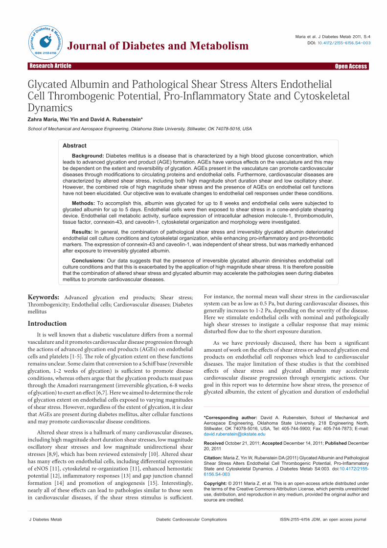
The surface expression of ICAM-1, tissue factor and thrombomodulin were measured as a means to determine if the expression of endothelial cell inflammatory and/or thrombotic markers were altered after exposure to glycated albumin and shear stress. The
Figure 1: Representative images of endothelial cells after exposure to shear stress and imaged for cytoskeletal organization. (A) 25% Actin Structure, (B) 100% Actin Structure, (C) 50% Actin Alignment, (D) 100% Actin Alignment, (E) Connectivity Score of 1, (F) Connectivity Score of 3, (G) Connectivity Score of 4, and (H) Connectivity Score of 5. All scale bars are 100μm.

Citation: Maria Z, Yin W, Rubenstein DA (2011) Glycated Albumin and Pathological Shear Stress Alters Endothelial Cell Thrombogenic Potential, Pro-Inflammatory State and Cytoskeletal Dynamics. J Diabetes Metab S4:003. doi:10.4172/2155-6156.S4-003
Page 4 of 9
J Diabetes Metab Diabetic Cardiovascular Complications ISSN:2155-6156 JDM, an open access journal
expression of these markers was independent of time in culture (three-way ANOVA, P = 0.67, early vs. late, single interaction); therefore, all data has been pooled by culture duration and is shown only as a function of shear stress and additive (Figure 3). ICAM-1 is a known mediator of endothelial cell inflammatory responses and we show that there is a significant increase in ICAM-1 expression for cells that were incubated with any glycated albumin as compared with the paired non-glycated albumin sample and the no added albumin sample (>100% increase). Generally, there was also an increase in ICAM-1 expression with exposure to increased magnitude shear stress (~15-20%, this is not shown on the figure from 0.4 to 4Pa), although it seems that the presence of glycated albumin mediates these changes stronger than shear stress.
The expression of tissue factor was enhanced (>100%) and the expression of thrombomodulin was reduced (~25%) in the presence of glycated albumin as compared with the non-glycated control or the no added albumin samples (Figure 3). It also appears that the expression of tissue factor increase with shear stress magnitude (~35% from 0.4 to 4 Pa) and the expression of thrombomodulin decreased with increased shear stress (~25% from 0.4 to 4Pa). Interestingly, it appears that for thrombotic markers, the presence of reversibly glycated albumin (AGE – 2 week) did not alter the cell surface expression until the shear stress surpassed 1Pa, but the presence of irreversibly glycated albumin altered cell surface expression at all shear stresses. The statistics did show that there is an interaction between shear stress and additive (three-way ANOVA, P< 0.01, shear stress vs. additive, two-way interaction).
Expression of connexin-43 and caveolin-1
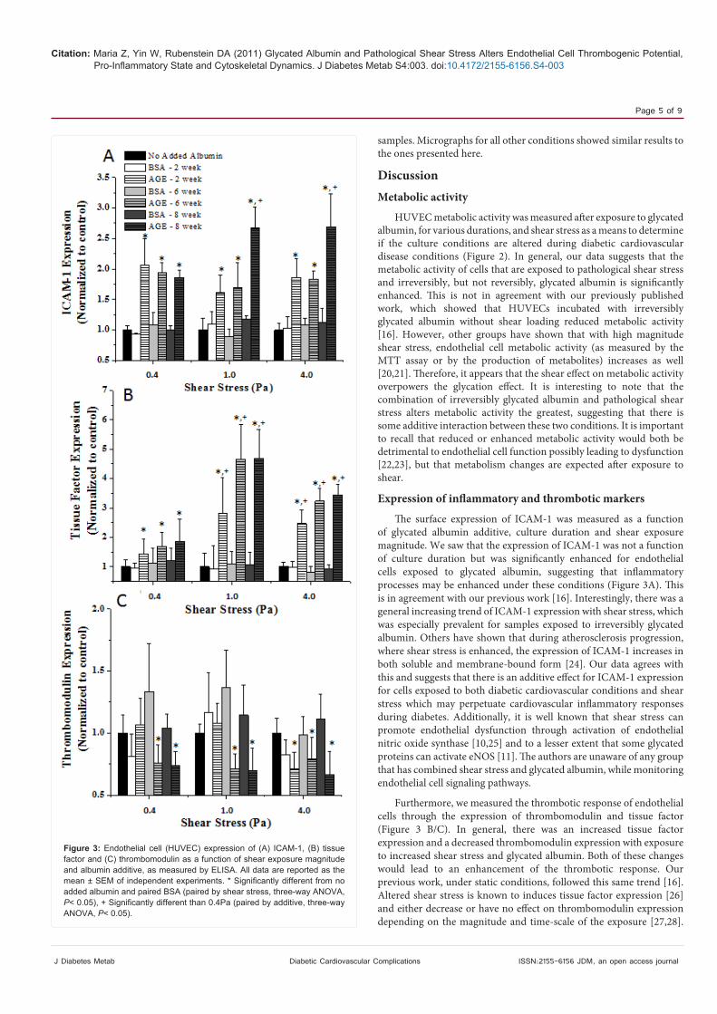
As in the previous section, the expression of connexin-43 and caveolin-1 was independent of the culture duration; therefore we plot the pooled data as a function of shear stress and additive only (Figure 4 A-D). The intensity and localization of connexin-43 was enhanced for HUVECs that were exposed to higher magnitude shear stress and irreversibly glycated albumin, only (~10-20%). At low, possibly pathological, levels of shear stress the expression of connexin-43 was independent of the additive and there were no differences between any of the treatment groups. Caveloin-1 intensity decreased, while the localization of caveolin-1 increased with increased shear stress and the presence of irreversibly glycated albumin. It also appears
that at lower magnitude shear stress levels, caveolin-1 expression was independent of the additive. Quantified data can be visualized from immunofluorescence images of connexin-43 and caveolin-1 (Figure 4 E-H). There is a higher expression (measured by intensity since the exposure time was the same for all conditions) of the proteins after exposure to irreversibly glycated albumin as compared with the no added albumin control. Also, there are more discrete protein clusters (quantified with localization) that can be seen between cells after exposure to irreversibly glycated albumin.
Cytoskeletal organization
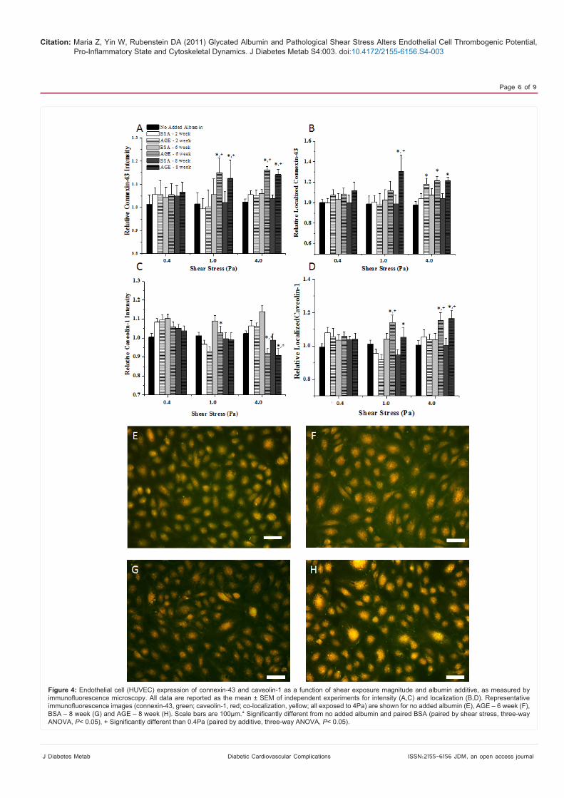
Cytoskeletal organization was quantified as a means to determine the adaptability of the endothelial cells to altered loading conditions, after incubation with advanced glycation end products. Interestingly, the actin structure was dependent on the time in culture (early vs. late), the shear stress and the albumin additive (Figure 5 A/B); with longer culture durations, low shear stress magnitudes and non-glycated samples induced actin to remain in a fibrillar type of structure after exposure to shear stress. However, the disintegration of actin was enhanced at high shear stress magnitudes and with glycated albumin samples. Interestingly, the actin alignment followed a similar trend, where high shear magnitudes, with glycated albumin (especially the irreversible glycated albumin) at the early culture duration induced cells to not align along the primary shear direction (Figure 5 C/D). As for cellular connectivity, mediated by the cytoskeleton, there was a general increasing trend with increased shear stress, a decreasing trend with added glycated albumin and an increasing trend with longer culture durations (Figure 5 E/F), although this was not as consistent as the percent structure and percent alignment data.
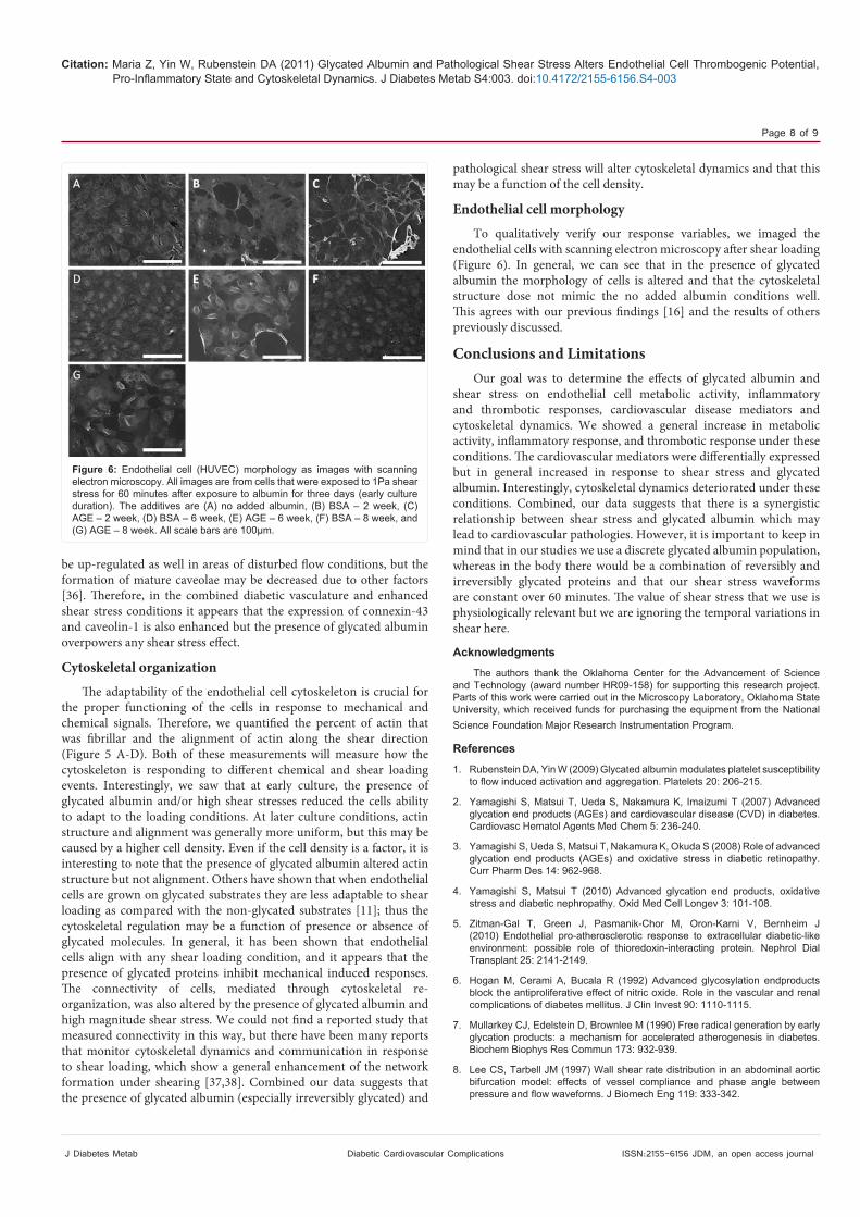
Endothelial cell morphology
Scanning electron micrographs of endothelial cells were taken as a means to qualitatively verify the quantitative data obtained from previous measurement techniques. Micrographs for endothelial cells that were exposed to 1Pa after exposure to particular additives for three days (early culture duration) are presented (Figure 6). We can see that there is a lower density of cells, the cells are not aligned in a particular direction, the cells tend to be more circular (lower actin fiber structure) and overall tend to look like the culture conditions are more deleterious for endothelial cells that are exposed to glycated albumin
Figure 2: Endothelial cell (HUVEC) metabolic activity as a function of shear exposure magnitude, albumin additive and culture duration (A, early; B, late). All data are reported as the mean ± SEM of independent experiments. * Significantly different from no added albumin and paired BSA (paired by shear stress and culture duration, three-way ANOVA, P< 0.05), # Significantly different than early (paired by shear stress and additive, three-way ANOVA, P< 0.05).
Citation: Maria Z, Yin W, Rubenstein DA (2011) Glycated Albumin and Pathological Shear Stress Alters Endothelial Cell Thrombogenic Potential, Pro-Inflammatory State and Cytoskeletal Dynamics. J Diabetes Metab S4:003. doi:10.4172/2155-6156.S4-003
Page 5 of 9
J Diabetes Metab Diabetic Cardiovascular Complications ISSN:2155-6156 JDM, an open access journal
samples. Micrographs for all other conditions showed similar results to the ones presented here.
DiscussionMetabolic activity
HUVEC metabolic activity was measured after exposure to glycated albumin, for various durations, and shear stress as a means to determine if the culture conditions are altered during diabetic cardiovascular disease conditions (Figure 2). In general, our data suggests that the metabolic activity of cells that are exposed to pathological shear stress and irreversibly, but not reversibly, glycated albumin is significantly enhanced. This is not in agreement with our previously published work, which showed that HUVECs incubated with irreversibly glycated albumin without shear loading reduced metabolic activity [16]. However, other groups have shown that with high magnitude shear stress, endothelial cell metabolic activity (as measured by the MTT assay or by the production of metabolites) increases as well [20,21]. Therefore, it appears that the shear effect on metabolic activity overpowers the glycation effect. It is interesting to note that the combination of irreversibly glycated albumin and pathological shear stress alters metabolic activity the greatest, suggesting that there is some additive interaction between these two conditions. It is important to recall that reduced or enhanced metabolic activity would both be detrimental to endothelial cell function possibly leading to dysfunction [22,23], but that metabolism changes are expected after exposure to shear.
Expression of inflammatory and thrombotic markers
The surface expression of ICAM-1 was measured as a function of glycated albumin additive, culture duration and shear exposure magnitude. We saw that the expression of ICAM-1 was not a function of culture duration but was significantly enhanced for endothelial cells exposed to glycated albumin, suggesting that inflammatory processes may be enhanced under these conditions (Figure 3A). This is in agreement with our previous work [16]. Interestingly, there was a general increasing trend of ICAM-1 expression with shear stress, which was especially prevalent for samples exposed to irreversibly glycated albumin. Others have shown that during atherosclerosis progression, where shear stress is enhanced, the expression of ICAM-1 increases in both soluble and membrane-bound form [24]. Our data agrees with this and suggests that there is an additive effect for ICAM-1 expression for cells exposed to both diabetic cardiovascular conditions and shear stress which may perpetuate cardiovascular inflammatory responses during diabetes. Additionally, it is well known that shear stress can promote endothelial dysfunction through activation of endothelial nitric oxide synthase [10,25] and to a lesser extent that some glycated proteins can activate eNOS [11]. The authors are unaware of any group that has combined shear stress and glycated albumin, while monitoring endothelial cell signaling pathways.
Furthermore, we measured the thrombotic response of endothelial cells through the expression of thrombomodulin and tissue factor (Figure 3 B/C). In general, there was an increased tissue factor expression and a decreased thrombomodulin expression with exposure to increased shear stress and glycated albumin. Both of these changes would lead to an enhancement of the thrombotic response. Our previous work, under static conditions, followed this same trend [16]. Altered shear stress is known to induces tissue factor expression [26] and either decrease or have no effect on thrombomodulin expression depending on the magnitude and time-scale of the exposure [27,28].
Figure 3: Endothelial cell (HUVEC) expression of (A) ICAM-1, (B) tissue factor and (C) thrombomodulin as a function of shear exposure magnitude and albumin additive, as measured by ELISA. All data are reported as the mean ± SEM of independent experiments. * Significantly different from no added albumin and paired BSA (paired by shear stress, three-way ANOVA, P< 0.05), + Significantly different than 0.4Pa (paired by additive, three-way ANOVA, P< 0.05).
Citation: Maria Z, Yin W, Rubenstein DA (2011) Glycated Albumin and Pathological Shear Stress Alters Endothelial Cell Thrombogenic Potential, Pro-Inflammatory State and Cytoskeletal Dynamics. J Diabetes Metab S4:003. doi:10.4172/2155-6156.S4-003
Page 6 of 9
J Diabetes Metab Diabetic Cardiovascular Complications ISSN:2155-6156 JDM, an open access journal
Figure 4: Endothelial cell (HUVEC) expression of connexin-43 and caveolin-1 as a function of shear exposure magnitude and albumin additive, as measured by immunofluorescence microscopy. All data are reported as the mean ± SEM of independent experiments for intensity (A,C) and localization (B,D). Representative immunofluorescence images (connexin-43, green; caveolin-1, red; co-localization, yellow; all exposed to 4Pa) are shown for no added albumin (E), AGE – 6 week (F), BSA – 8 week (G) and AGE – 8 week (H). Scale bars are 100μm.* Significantly different from no added albumin and paired BSA (paired by shear stress, three-way ANOVA, P< 0.05), + Significantly different than 0.4Pa (paired by additive, three-way ANOVA, P< 0.05).
Citation: Maria Z, Yin W, Rubenstein DA (2011) Glycated Albumin and Pathological Shear Stress Alters Endothelial Cell Thrombogenic Potential, Pro-Inflammatory State and Cytoskeletal Dynamics. J Diabetes Metab S4:003. doi:10.4172/2155-6156.S4-003
Page 7 of 9
J Diabetes Metab Diabetic Cardiovascular Complications ISSN:2155-6156 JDM, an open access journal
of these markers when HUVECs are exposed to irreversibly glycated albumin [16]. In general, our data from this report agrees with the previous findings and we also found a heightened potential for discrete protein clusters to form after exposure to shear stress and diabetic cardiovascular conditions. There is currently no consensus on the expression of either of these molecules under diabetic conditions and it seems to be highly dependent on the conditions used in the study [29-33]. Interestingly, the effects of shear stress on these molecules are clearer. Others have seen an up-regulation of connexin-43 in the site of altered flow [34,35]; agreeing with our findings. Caveolin-1 appears to
Figure 5: Endothelial cell (HUVEC) cytoskeletal organization as a function of culture duration, shear exposure magnitude and albumin additive, as measured by immunofluorescence microscopy. All data are reported as the mean ± SEM of independent experiments for (A,B) actin structure, (C,D) actin alignment and (E,F) connectivity. * Significantly different from no added albumin and paired BSA (paired by shear stress and culture duration, three-way ANOVA, P< 0.05), + Significantly different than 0.4Pa (paired by additive and culture duration, three-way ANOVA, P< 0.05), # Significantly different than early (paired by shear stress and additive, three-way ANOVA, P< 0.05).
It appears from our data that the shear stress and glycated albumin synergistically alter tissue factor and thrombomodulin expression to promote the thrombotic response possibly leading to disease progression.
Expression of connexin-43 and caveolin-1
Connexin-43 and caveolin-1 expression were measured as a means to monitor cardiovascular disease progression through enhanced mature network formation and angiogenesis potential (Figure 4). Previously, we showed that there was a general increase in expression
Citation: Maria Z, Yin W, Rubenstein DA (2011) Glycated Albumin and Pathological Shear Stress Alters Endothelial Cell Thrombogenic Potential, Pro-Inflammatory State and Cytoskeletal Dynamics. J Diabetes Metab S4:003. doi:10.4172/2155-6156.S4-003
Page 8 of 9
J Diabetes Metab Diabetic Cardiovascular Complications ISSN:2155-6156 JDM, an open access journal
pathological shear stress will alter cytoskeletal dynamics and that this may be a function of the cell density.
Endothelial cell morphology
To qualitatively verify our response variables, we imaged the endothelial cells with scanning electron microscopy after shear loading (Figure 6). In general, we can see that in the presence of glycated albumin the morphology of cells is altered and that the cytoskeletal structure dose not mimic the no added albumin conditions well. This agrees with our previous findings [16] and the results of others previously discussed.
Conclusions and LimitationsOur goal was to determine the effects of glycated albumin and
shear stress on endothelial cell metabolic activity, inflammatory and thrombotic responses, cardiovascular disease mediators and cytoskeletal dynamics. We showed a general increase in metabolic activity, inflammatory response, and thrombotic response under these conditions. The cardiovascular mediators were differentially expressed but in general increased in response to shear stress and glycated albumin. Interestingly, cytoskeletal dynamics deteriorated under these conditions. Combined, our data suggests that there is a synergistic relationship between shear stress and glycated albumin which may lead to cardiovascular pathologies. However, it is important to keep in mind that in our studies we use a discrete glycated albumin population, whereas in the body there would be a combination of reversibly and irreversibly glycated proteins and that our shear stress waveforms are constant over 60 minutes. The value of shear stress that we use is physiologically relevant but we are ignoring the temporal variations in shear here.
Acknowledgments
The authors thank the Oklahoma Center for the Advancement of Science and Technology (award number HR09-158) for supporting this research project. Parts of this work were carried out in the Microscopy Laboratory, Oklahoma State University, which received funds for purchasing the equipment from the National Science Foundation Major Research Instrumentation Program.
References
1. Rubenstein DA, Yin W (2009) Glycated albumin modulates platelet susceptibility to flow induced activation and aggregation. Platelets 20: 206-215.
2. Yamagishi S, Matsui T, Ueda S, Nakamura K, Imaizumi T (2007) Advanced glycation end products (AGEs) and cardiovascular disease (CVD) in diabetes. Cardiovasc Hematol Agents Med Chem 5: 236-240.
3. Yamagishi S, Ueda S, Matsui T, Nakamura K, Okuda S (2008) Role of advanced glycation end products (AGEs) and oxidative stress in diabetic retinopathy. Curr Pharm Des 14: 962-968.
4. Yamagishi S, Matsui T (2010) Advanced glycation end products, oxidative stress and diabetic nephropathy. Oxid Med Cell Longev 3: 101-108.
5. Zitman-Gal T, Green J, Pasmanik-Chor M, Oron-Karni V, Bernheim J (2010) Endothelial pro-atherosclerotic response to extracellular diabetic-like environment: possible role of thioredoxin-interacting protein. Nephrol Dial Transplant 25: 2141-2149.
6. Hogan M, Cerami A, Bucala R (1992) Advanced glycosylation endproducts block the antiproliferative effect of nitric oxide. Role in the vascular and renal complications of diabetes mellitus. J Clin Invest 90: 1110-1115.
7. Mullarkey CJ, Edelstein D, Brownlee M (1990) Free radical generation by early glycation products: a mechanism for accelerated atherogenesis in diabetes. Biochem Biophys Res Commun 173: 932-939.
8. Lee CS, Tarbell JM (1997) Wall shear rate distribution in an abdominal aortic bifurcation model: effects of vessel compliance and phase angle between pressure and flow waveforms. J Biomech Eng 119: 333-342.
Figure 6: Endothelial cell (HUVEC) morphology as images with scanning electron microscopy. All images are from cells that were exposed to 1Pa shear stress for 60 minutes after exposure to albumin for three days (early culture duration). The additives are (A) no added albumin, (B) BSA – 2 week, (C) AGE – 2 week, (D) BSA – 6 week, (E) AGE – 6 week, (F) BSA – 8 week, and (G) AGE – 8 week. All scale bars are 100μm.
be up-regulated as well in areas of disturbed flow conditions, but the formation of mature caveolae may be decreased due to other factors [36]. Therefore, in the combined diabetic vasculature and enhanced shear stress conditions it appears that the expression of connexin-43 and caveolin-1 is also enhanced but the presence of glycated albumin overpowers any shear stress effect.
Cytoskeletal organization
The adaptability of the endothelial cell cytoskeleton is crucial for the proper functioning of the cells in response to mechanical and chemical signals. Therefore, we quantified the percent of actin that was fibrillar and the alignment of actin along the shear direction (Figure 5 A-D). Both of these measurements will measure how the cytoskeleton is responding to different chemical and shear loading events. Interestingly, we saw that at early culture, the presence of glycated albumin and/or high shear stresses reduced the cells ability to adapt to the loading conditions. At later culture conditions, actin structure and alignment was generally more uniform, but this may be caused by a higher cell density. Even if the cell density is a factor, it is interesting to note that the presence of glycated albumin altered actin structure but not alignment. Others have shown that when endothelial cells are grown on glycated substrates they are less adaptable to shear loading as compared with the non-glycated substrates [11]; thus the cytoskeletal regulation may be a function of presence or absence of glycated molecules. In general, it has been shown that endothelial cells align with any shear loading condition, and it appears that the presence of glycated proteins inhibit mechanical induced responses. The connectivity of cells, mediated through cytoskeletal re-organization, was also altered by the presence of glycated albumin and high magnitude shear stress. We could not find a reported study that measured connectivity in this way, but there have been many reports that monitor cytoskeletal dynamics and communication in response to shear loading, which show a general enhancement of the network formation under shearing [37,38]. Combined our data suggests that the presence of glycated albumin (especially irreversibly glycated) and

Citation: Maria Z, Yin W, Rubenstein DA (2011) Glycated Albumin and Pathological Shear Stress Alters Endothelial Cell Thrombogenic Potential, Pro-Inflammatory State and Cytoskeletal Dynamics. J Diabetes Metab S4:003. doi:10.4172/2155-6156.S4-003
Page 9 of 9
J Diabetes Metab Diabetic Cardiovascular Complications ISSN:2155-6156 JDM, an open access journal
9. McMillan DE (1985) Hemorheologic changes in diabetes and their role in increased atherogenesis. Horm Metab Res Suppl 15: 73-79.
10. Davies PF (1995) Flow-mediated endothelial mechanotransduction. Physiol Rev 75: 519-560.
11. Kemeny SF, Figueroa DS, Andrews AM, Barbee KA, Clyne AM (2011) Glycated collagen alters endothelial cell actin alignment and nitric oxide release in response to fluid shear stress. J Biomech 44: 1927-1935.
12. Ensley AE, Nerem RM, Anderson DE, Hanson SR, Hinds MT (2011) Fluid Shear Stress Alters the Coagulation Potential of Endothelial Outgrowth Cells. Tissue Eng Part A
13. Yin W, Shanmugavelayudam SK, Rubenstein DA (2011) The effect of physiologically relevant dynamic shear stress on platelet and endothelial cell activation. Thromb Res 127: 235-241.
14. Johnson TL, Nerem RM (2007) Endothelial connexin 37, connexin 40, and connexin 43 respond uniquely to substrate and shear stress. Endothelium 14: 215-226.
15. Tian J, Hou Y, Lu Q, Wiseman DA, Vasconcelos FF, et al. (2010) A novel role for caveolin-1 in regulating endothelial nitric oxide synthase activation in response to H2O2 and shear stress. Free Radic Biol Med 49: 159-170.
16. Rubenstein DA, Maria Z, Yin W (2011) Glycated albumin modulates endothelial cell thrombogenic and inflammatory responses. J Diabetes Sci Technol 5: 703-713.
17. Rubenstein D, Han D, Goldgraben S, El-Gendi H, Gouma PI, et al. (2007) Bioassay chamber for angiogenesis with perfused explanted arteries and electrospun scaffolding. Microcirculation 14: 723-737.
18. Shanmugavelayudam SK, Rubenstein DA, Yin W (2011) Effects of physiologically relevant dynamic shear stress on platelet complement activation. Platelets 22: 602-610
19. Rubenstein DA, Lu H, Mahadik SS, Leventis N, Yin W (2011) Characterization of the Physical Properties and Biocompatibility of Polybenzoxazine-Based Aerogels for Use as a Novel Hard-Tissue Scaffold. J Biomater Sci Polym Ed.
20. Cebotari S, Mertsching H, Kallenbach K, Kostin S, Repin O, et al. (2002) Construction of autologous human heart valves based on an acellular allograft matrix. Circulation 106: I63-I68.
21. Frangos JA, McIntire LV, Eskin SG (1988) Shear stress induced stimulation of mammalian cell metabolism. Biotechnol Bioeng 32: 1053-1060.
22. Bauer KS, Dixon SC, Figg WD (1998) Inhibition of angiogenesis by thalidomide requires metabolic activation, which is species-dependent. Biochem Pharmacol 55: 1827-1834.
23. Tesfamariam B (1994) Free radicals in diabetic endothelial cell dysfunction. Free Radic Biol Med 16: 383-391.
24. Hwang SJ, Ballantyne CM, Sharrett AR, Smith LC, Davis CE et al. (1997) Circulating adhesion molecules VCAM-1, ICAM-1, and E-selectin in carotid
atherosclerosis and incident coronary heart disease cases: the Atherosclerosis Risk In Communities (ARIC) study. Circulation 96: 4219-4225.
25. Fleming I, Fisslthaler B, Dixit M, Busse R (2005) Role of PECAM-1 in the shear-stress-induced activation of Akt and the endothelial nitric oxide synthase (eNOS) in endothelial cells. J Cell Sci 118: 4103-4111.
26. Lin MC, Almus-Jacobs F, Chen HH, Parry GC, Mackman N, et al. (1997) Shear stress induction of the tissue factor gene. J Clin Invest 99: 737-744.
27. Malek AM, Jackman R, Rosenberg RD, Izumo S (1994) Endothelial expression of thrombomodulin is reversibly regulated by fluid shear stress. Circ Res 74: 852-860.
28. Takahashi M, Ishida T, Traub O, Corson MA, Berk BC (1997) Mechanotransduction in endothelial cells: temporal signaling events in response to shear stress. J Vasc Res 34: 212-219.
29. Abdullah KM, Luthra G, Bilski JJ, Abdullah SA, Reynolds LP, et al. (1999) Cell-to-cell communication and expression of gap junctional proteins in human diabetic and nondiabetic skin fibroblasts: effects of basic fibroblast growth factor. Endocrine 10: 35-41.
30. Gandhi GK, Ball KK, Cruz NF, Dienel GA (2010) Hyperglycaemia and diabetes impair gap junctional communication among astrocytes. ASN Neuro 2: e00030.
31. Makino A, Platoshyn O, Suarez J, Yuan JX, Dillmann WH (2008) Downregulation of connexin40 is associated with coronary endothelial cell dysfunction in streptozotocin-induced diabetic mice. Am J Physiol Cell Physiol 295: C221-C230.
32. Sato T, Haimovici R, Kao R, Li AF, Roy S (2002) Downregulation of connexin 43 expression by high glucose reduces gap junction activity in microvascular endothelial cells. Diabetes 51: 1565-1571.
33. Zhang J, Hill CE (2005) Differential connexin expression in preglomerular and postglomerular vasculature: accentuation during diabetes. Kidney Int 68: 1171-1185.
34. Gabriels JE, Paul DL (1998) Connexin43 is highly localized to sites of disturbed flow in rat aortic endothelium but connexin37 and connexin40 are more uniformly distributed. Circ Res 83: 636-643.
35. Inai T, Mancuso MR, McDonald DM, Kobayashi J, Nakamura K, et al. (2004) Shear stress-induced upregulation of connexin 43 expression in endothelial cells on upstream surfaces of rat cardiac valves. Histochem Cell Biol 122: 477-483.
36. Chen BP, Li YS, Zhao Y, Chen KD, Li S, et al. (2001) DNA microarray analysis of gene expression in endothelial cells in response to 24-h shear stress. Physiol Genomics 7: 55-63.
37. Ali MH, Schumacker PT (2002) Endothelial responses to mechanical stress: where is the mechanosensor? Crit Care Med 30: S198-S206.
38. Zhu C, Bao G, Wang N (2000) Cell mechanics: mechanical response, cell adhesion, and molecular deformation. Annu Rev Biomed Eng 2: 189-226.
Thisarticlewasoriginallypublishedinaspecialissue,Diabetic Cardiovascular Complications handledbyEditor(s).Dr.ZhengyuanXia,UniversityofHongKong,HongKong