journal of feline medicine and surgery - orbi: home pub_thiry.pdf · furthermore, drugs may be...
TRANSCRIPT

http://jfm.sagepub.com/Journal of Feline Medicine and Surgery
http://jfm.sagepub.com/content/15/7/598The online version of this article can be found at:
DOI: 10.1177/1098612X13489222
2013 15: 598Journal of Feline Medicine and SurgeryRadford, Etienne Thiry, Uwe Truyen and Marian C Horzinek
Herman Egberink, Katrin Hartmann, Margaret J Hosie, Albert Lloret, Hans Lutz, Fulvio Marsilio, Karin Möstl, Alan D Tadeusz Frymus, Tim Gruffydd-Jones, Maria Grazia Pennisi, Diane Addie, Sándor Belák, Corine Boucraut-Baralon,
Dermatophytosis in Cats: ABCD guidelines on prevention and management
technique does not amount to an endorsement of its value or quality, or the claims made by its manufacturer.those of the authors and the inclusion in this publication of material relating to a particular product, method or of animals and interpretation of published materials lies with the veterinary practitioner. The opinions expressed arefrom actions or decisions based on information contained in this publication; ultimate responsibility for the treatment
arisingcountry. The authors, editors, owners and publishers do not accept any responsibility for any loss or damage advertising material, it is the responsibility of the reader to check that the product is authorised for use in their ownbear this in mind and be aware of the prescribing laws pertaining to their own country. Likewise, in relation to Furthermore, drugs may be mentioned that are licensed for human use, and not for veterinary use. Readers need toformulations that are not available or licensed in the individual reader's own country.The Journal of Feline Medicine and Surgery is an international journal and authors may discuss products and
Disclaimer
Published by:
International Society of Feline Medicine
American Association of Feline Practitioners
and http://www.sagepublications.com
can be found at:Journal of Feline Medicine and SurgeryAdditional services and information for
http://jfm.sagepub.com/cgi/alertsEmail Alerts:
http://jfm.sagepub.com/subscriptionsSubscriptions:
http://www.sagepub.com/journalsReprints.navReprints:
http://www.sagepub.com/journalsPermissions.navPermissions:
What is This?
- Jun 27, 2013Version of Record >>
at Universite de Liege on September 3, 2013jfm.sagepub.comDownloaded from
Agent properties
In contrast to single-celled yeasts, dermatophytes (‘skin plants’) arecomplex fungi growing as hyphae and forming a mycelium. About 40 species belonging to the genera Microsporum, Trichophyton andEpidermophyton are considered as dermatophytes. Over 90% of felinedermatophytosis cases worldwide are caused by Microsporum canis.1Others are caused by M gypseum, T mentagrophytes, T quinckeanum, T verrucosum or other agents. With the exception of M gypseum, all ofthese agents produce proteolytic and keratolytic enzymes that enablethem to utilise keratin as the sole source of nutrition after colonisationof the dead, keratinised portion of epidermal tissue (mostly stratumcorneum and hairs, sometimes nails).Dermatophytes produce arthro spores, which are highly resistant,
surviving in a dry environment for 12 months or more [EBM gradeIII].2 In a humid environment, however, arthro spores are short-lived.High temperatures (100°C) destroy them quickly. Arthrospores adherevery strongly to keratin.Depending on the source of infection and reservoirs, dermatophyte
species are classified into zoophilic, sylvatic, geophilic and anthro-pophilic fungi.
Epidemiology
Dermatophytosis is worldwide the most common fungal infection ofcats and one of the most important infectious skin diseases in thisspecies. It may be transmitted to other animal species, and is also animportant zoonosis.
M canis is a typical zoophilic dermatophyte.It was generally thought that subclinicalinfections are very common in cats, espe-cially in longhaired animals over 2 years ofage. However, in many groups the preva-lence is relatively low. Therefore, M canis
Journal of Feline Medicine and Surgery (2013) 15, 598–604
C L I N I C A L R E V I E W
DERMATOPHYTOSIS IN CATSABCD guidelines on prevention and management
Tadeusz Frymus, Tim Gruffydd-Jones, Maria Grazia Pennisi, Diane Addie, Sándor Belák, Corine Boucraut-Baralon, Herman Egberink, Katrin Hartmann,Margaret J Hosie, Albert Lloret, Hans Lutz, Fulvio Marsilio, Karin Möstl, Alan D Radford, Etienne Thiry, Uwe Truyen and Marian C Horzinek
598 JFMS CLINICAL PRACTICE
European Advisory Board on Cat Diseases
www.abcd-vets.orgCorresponding author: Tadeusz Frymus
Email: [email protected]
DOI: 10.1177/1098612X13489222© Published by SAGE on behalf of ISFM and AAFP 2013
Overview: Dermatophytosis, usually caused byMicrosporum canis, is the most common fungalinfection in cats worldwide, and one of the mostimportant infectious skin diseases in this species.Many adult cats are asymptomatic carriers. Severe clinical signs are seen mostly in kittens or immunosuppressed adults. Poor hygiene is apredisposing factor, and the disease may beendemic in shelters or catteries. Humans may beeasily infected and develop a similar skin disease.Infection: Infectious arthrospores produced bydermatophytes may survive in the environment forabout a year. They are transmitted through contactwith sick cats or healthy carriers, but also on dustparticles, brushes, clothes and other fomites. Disease signs: Circular alopecia, desquamationand sometimes an erythematous margin aroundcentral healing (‘ringworm’) are typical. In many catsthis is a self-limiting disease with hair loss and scalingonly. In immunosuppressed animals, the outcomemay be a multifocal or generalised skin disease.Diagnosis: Wood’s lamp examination andmicroscopic detection of arthrospores on hairs aresimple methods to confirm M canis infection, buttheir sensitivity is relatively low. The gold standardfor detection is culture on Sabouraud agar of hairsand scales collected from new lesions.Disease management: In shelters and catterieseradication is difficult. Essential is a combination of systemic and topical treatments, maintained forseveral weeks. For systemic therapy itraconazole is the drug of choice, terbinafine an alternative.Recommended topical treatment is repeated bodyrinse with an enilconazole solution or miconazolewith or without chlorhexidine. In catteries/sheltersmedication must be accompanied by intensivedecontamination of the environment.Vaccination: Few efficacy studies on anti-M canisvaccines (prophylactic or therapeutic) for cats havebeen published, and a safe and efficient vaccine isnot available.
ZoonosisM canis is the causative
agent of ringworm.
at Universite de Liege on September 3, 2013jfm.sagepub.comDownloaded from

JFMS CLINICAL PRACTICE 599
should not be considered part of the normalfungal flora of cats and its isolation from ahealthy animal indicates either subclinicalinfection or fomite carriage.1Arthrospores are transmitted through con-
tact with sick or subclinically infected ani-mals, mainly cats, but also dogs or otherspecies. In sick animals, the infected hairshafts are fragile and hair fragments contain-ing arthrospores are very efficient in spread-ing infection. In addition, uninfected cats canpassively transport arthrospores on their hair,thereby acting as a source of infection. Riskfactors include: introducing new animals intoa cattery, cat shows, shelters, mating, etc.Indirect contact is very important too; transmission may occur via contaminated collars, brushes, toys, environments, etc.Arthrospores are easily spread on dust parti-cles, even to rooms without access for cats. Outdoor cats, especially in rural areas, can
be exposed by digging to M gypseum, ageophilic fungus living in soil. Cats may beinfected with T mentagrophytes or T quinck-eanum through contact with small rodents,and with T verrucosum through contact withcattle.
Pathogenesis
Healthy skin acts as an effective barrieragainst fungal invasion. The increased rate ofregeneration of epidermal cells in response to the dermatophyte, with the consequentremoval of fungus from the skin surface, isanother protective mechanism. As dermato-phytes cannot penetrate healthy skin, manycats are merely passive carriers of thearthrospores or remain subclinically infected.Whether such an infection will lead to clinical disease depends on many factors.Predisposing factors to disease include: ayoung age (first 2 years of life), immunosup-pression (including immunosuppressivetreatment), other diseases, nutritional deficits(especially proteins and vitamin A), high tem-perature and high humidity.1Very important for the facilitation of infec-
tion is any kind of skin trauma resulting fromincreased moisture, injury by ectoparasites orscratches due to pruritus, playingor aggressive behaviour, clipping,etc. In general, poor hygiene is apredisposing factor. In over-crowded feline groups, socialstress may play an importantrole. This can make eradicationof ringworm very difficult incatteries or shelters infectedwith M canis.The potential immunosup-
pressive effect of feline
REV IEW / ABCD guidelines on dermatophytosis
immunodeficiency virus (FIV) and felineleukaemia virus (FeLV) on the prevalence offungal infection has been investigated. Thehigher prevalence of M canis in FIV-infectedanimals compared with normal cats reportedin one survey3 was not observed by anothergroup.4 It has been suggested that any associ-ation may be related to differences in the envi-ronment rather than to the retroviral status ofthe cats.5The incubation period of ringworm caused
by M canis is 1–3 weeks. During this time,hyphae grow along the hair shafts through thestratum corneum to the follicles where theyproduce spores that form a thick layer aroundthe hair shafts. As dermatophytes are suscep-tible to high temperatures, they cannotcolonise deeper parts of the skin or the follicleitself. Therefore, the hair grows normally butbreaks easily near the skin surface, resultingin hair loss. Several metabolic products of thefungus may induce an inflammatory responsein the skin, and may be observed mainlyaround the infected area, forming sometimesring-like lesions with central areas of healingand papules on the periphery (‘ringworm’). In many immunocompetent cats living in
hygienic conditions these lesions are limited(eg, to the head) and disappear after severalweeks. In immunosuppressed animals, theoutcome may be a multifocal or generalisedskin disease with secondary bacterial infec-tions. On rare occasions, a marked inflamma-tory reaction to hyphae induces a nodulargranulomatous reaction involving dermis anddraining on the skin surface. These so-calledpseudomycetomas are more often seen inPersian cats, sometimes concurrently withclassical lesions.The pathogenesis of other dermatophyte
infections is similar to that described above.
Immunity
Naturally occurring ringworm is rarely recur-rent, suggesting an effective and long-lastingimmunity. Experimental studies confirm thatanimals express increased resistance to subse-quent challenge by the homologous fungus.Reinfections may occur, but require a much
greater number of spores,and usually these subse-quent infections are clearedmore rapidly.1 It has beensuggested that for the devel-opment of full immunity, theinfection must run its entirenatural course, as in catswhose infection was abortedwith antifungal treatment thedelayed type hypersensitivityreactions were often weaker.6
European Advisory Board on Cat DiseasesThe European Advisory Board on Cat Diseases (ABCD) is a bodyof experts in immunology, vaccinology and clinical feline medicinethat issues guidelines on prevention and management of felineinfectious diseases in Europe, for the benefit of the health andwelfare of cats. The guidelines are based on current scientificknowledge of the diseases and available vaccines concerned.
The latest version of the dermatophytosis in cats guidelines is available at www.abcd-vets.org
Dermatophytes(‘skin plants’)are complexfungi growingas hyphae andforming a
mycelium. Over90% of feline
dermatophytosiscases
worldwide arecaused by
Microsporumcanis.
EBM gradesThe ranking systemfor grading the levelof evidence ofvarious statementswithin this article isdescribed on page 533 of thisSpecial Issue.
at Universite de Liege on September 3, 2013jfm.sagepub.comDownloaded from

REV IEW / ABCD guidelines on dermatophytosis
600 JFMS CLINICAL PRACTICE
Although dermatophyte infection isconfined to the superficial keratinised tissues, a humoral and cellular immuneresponse is induced. Prominent activationof T helper type 2 (Th2) cells and the cor-responding cytokine profile leads to anti-body formation followed by chronicdisease, whereas activation of Th1 cellsstimulates a cell-mediated response char-acterised by interferon-γ, and interleukins12 and 2, and leads to recovery.1,7 Suchcats are protected against reinfection.8 Therole of the humoral response in dermato-phytosis is unclear, although antibodiescould have a fungistatic effect by meansof opsonisation and complement activation.9
Clinical signs
In many cats, dermatophytes cause a mild,self-limiting infection with hair loss and scal-ing (Figure 1). The typical presentation of ringworm in cats
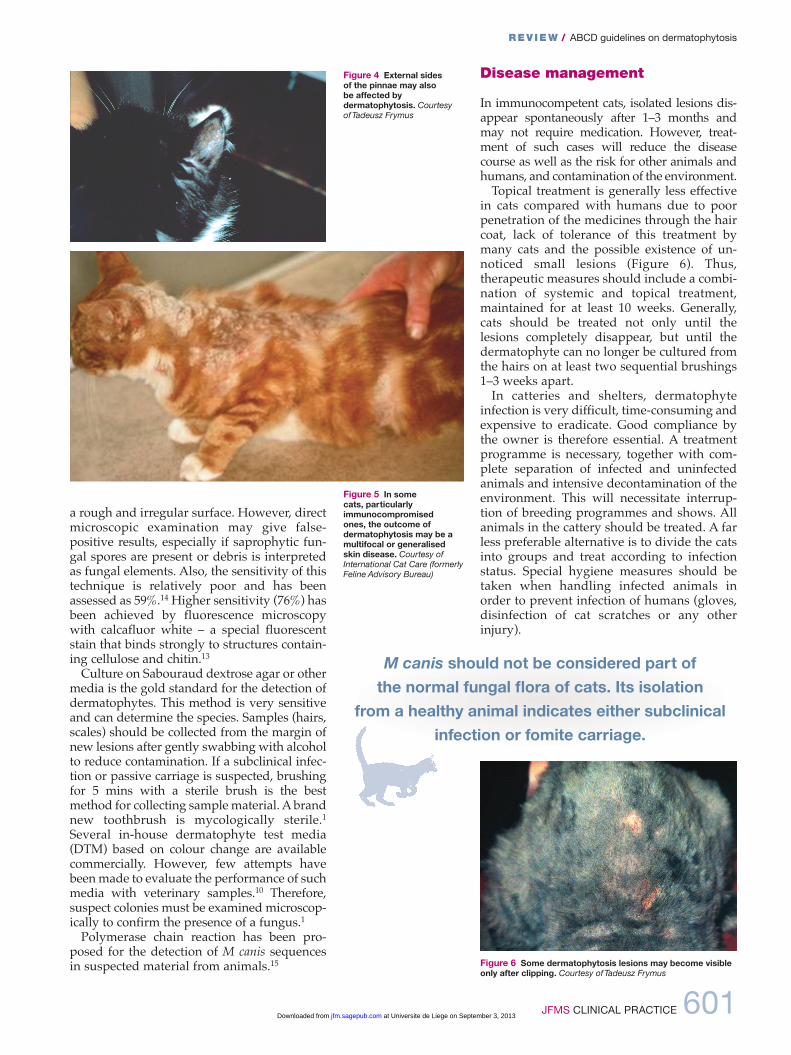
is regular and circular alopecia (Figure 2), withhair breakage, desquamation and sometimesan erythematous margin and central heal-ing.1,10 The lesions are sometimes very small,but occasionally may have a diameter of 4–6cm. Lesions may be single or multiple, and arelocalised mostly on the head (Figure 3) but alsoon any part of the body, including the distalparts of the legs and the tail. Young cats, in par-ticular, display lesions localised at first to thebridge of the nose and then extending to thetemples, the external sides of the pinnae andauricular margins (Figure 4). Multiple lesionsmay coalesce. Pruritus is variable, generallymild to moderate, and usually no fever or lossof appetite is observed.1,10 In some cats, dermatophytosis can present
as a papulocrustous dermatitis (‘miliary der-matitis’) affecting mainly the dorsal trunk.In immunosuppressed cats, extensive lesions
with secondary bacterial involvement are some-
times associated with chronic ringworm.Such patients demonstrate atypical, largealopecic areas, erythema, pruritus, exudationand crusts (Figure 5). At this stage, dermato-phytosis may mimic other dermatologicalconditions. Typical signs may be still visibleat the margins of the lesions.A rare outcome is onyxis and perionyxis
and, exceptionally, nodular granulomatousdermatitis (pseudomycetoma) with singleor multiple cutaneous nodules, firm andnot painful on palpation.11 Fistulisation of these nodules is possible. Pseudo -mycetoma occurring as abdominal massesmay be a rare complication of laparotomy
in animals with cutaneous dermatophytosis.12
Diagnosis
As dermatophytosis can produce lesions simi-lar to many feline skin diseases, it should beconsidered in all cats with any cutaneous condition. If possible, dermatophyte diagnosisshould be undertaken before any treatment.An inexpensive and simple screening tool
for M canis infection is the Wood’s lamp exam-ination. However, it is not very sensitive: onlyabout 50% of M canis strains fluoresce andother dermatophytes do not fluoresce at all.13Furthermore, debris, scale, lint and topicalmedications (eg, tetracycline) can producefalse-positive results. Thus, Wood’s lamp find-ings should be confirmed by other methods.Direct microscopic examination is another
simple and rapid method to detect dermato-phytes on hairs or scales. It is recommended topluck hairs for this purpose under Wood’slamp illumination, or from the edge of a lesion.The sample should be cleared with 10–20%potassium hydroxide solution before examina-tion. There are a number of techniques toimprove the visualisation of fungal elementson the hair shafts.1 Hairs or hair fragmentswith hyphae and arthrospores are thicker, with
Figure 1 In some cats, especially immunocompetent adults,the only sign of dermatophytosis may be scaling. Courtesy ofTadeusz Frymus
Figure 2 Circular alopecia caused by M canis infection.Courtesy of Tadeusz Frymus
Figure 3 On manyoccasions dermatophytosislesions start on the head.Courtesy of International CatCare (formerly Feline AdvisoryBureau)
As dermato-phytosis canproduce
lesions similarto many felineskin diseases,it should beconsidered incats with anycutaneouscondition.
at Universite de Liege on September 3, 2013jfm.sagepub.comDownloaded from
REV IEW / ABCD guidelines on dermatophytosis
JFMS CLINICAL PRACTICE 601
a rough and irregular surface. However, directmicroscopic examination may give false-positive results, especially if saprophytic fun-gal spores are present or debris is interpretedas fungal elements. Also, the sensitivity of thistechnique is relatively poor and has beenassessed as 59%.14 Higher sensitivity (76%) hasbeen achieved by fluorescence microscopywith calcafluor white – a special fluorescentstain that binds strongly to structures contain-ing cellulose and chitin.13Culture on Sabouraud dextrose agar or other
media is the gold standard for the detection ofdermatophytes. This method is very sensitiveand can determine the species. Samples (hairs,scales) should be collected from the margin ofnew lesions after gently swabbing with alcoholto reduce contamination. If a subclinical infec-tion or passive carriage is suspected, brushingfor 5 mins with a sterile brush is the bestmethod for collecting sample material. A brandnew toothbrush is mycologically sterile.1Several in-house dermatophyte test media(DTM) based on colour change are availablecommercially. However, few attempts havebeen made to evaluate the performance of suchmedia with veterinary samples.10 Therefore,suspect colonies must be examined microscop-ically to confirm the presence of a fungus.1Polymerase chain reaction has been pro-
posed for the detection of M canis sequencesin suspected material from animals.15
Disease management
In immunocompetent cats, isolated lesions dis-appear spontaneously after 1–3 months andmay not require medication. However, treat-ment of such cases will reduce the diseasecourse as well as the risk for other animals andhumans, and contamination of the environment. Topical treatment is generally less effective
in cats compared with humans due to poorpenetration of the medicines through the haircoat, lack of tolerance of this treatment bymany cats and the possible existence of un -noticed small lesions (Figure 6). Thus, therapeutic measures should include a combi-nation of systemic and topical treatment,maintained for at least 10 weeks. Generally,cats should be treated not only until thelesions completely disappear, but until thedermatophyte can no longer be cultured fromthe hairs on at least two sequential brushings1–3 weeks apart.In catteries and shelters, dermatophyte
infection is very difficult, time-consuming andexpensive to eradicate. Good compliance bythe owner is therefore essential. A treatmentprogramme is necessary, together with com-plete separation of infected and uninfectedanimals and intensive decontamination of theenvironment. This will necessitate interrup-tion of breeding programmes and shows. Allanimals in the cattery should be treated. A farless preferable alternative is to divide the catsinto groups and treat according to infectionstatus. Special hygiene measures should betaken when handling infected animals inorder to prevent infection of humans (gloves,disinfection of cat scratches or any otherinjury).
Figure 6 Some dermatophytosis lesions may become visibleonly after clipping. Courtesy of Tadeusz Frymus
Figure 4 External sides of the pinnae may also be affected bydermatophytosis. Courtesy of Tadeusz Frymus
Figure 5 In some cats, particularlyimmunocompromised ones, the outcome ofdermatophytosis may be amultifocal or generalisedskin disease. Courtesy ofInternational Cat Care (formerlyFeline Advisory Bureau)
M canis should not be considered part of the normal fungal flora of cats. Its isolation
from a healthy animal indicates either subclinicalinfection or fomite carriage.
at Universite de Liege on September 3, 2013jfm.sagepub.comDownloaded from

602 JFMS CLINICAL PRACTICE
REV IEW / ABCD guidelines on dermatophytosis
Topical therapyIn cats with a limited number of lesions, hairsshould be clipped away from the periphery oflesions incorporating a wide margin. Clippingshould be gentle to avoid spreading the infec-tion due to microtrauma. Spot treatment oflesions may be of limited efficacy; instead,whole body shampooing, dipping or rinsingis recommended. In patients with generaliseddisease, longhaired cats and for cattery decon-tamination, clipping the entire cat is useful tomake topical therapy application easier and toallow for better penetration of the drug. Thisapproach also limits the spread of the sporesinto the environment, to people and to otheranimals. The entire hair coat, includingwhiskers, should be gently clipped and allinfected hairs should be wrapped and disinfected before disposal. Chemical or heatsterilisation of instruments is essential. Catsshould not be clipped in veterinary clinics toavoid environmental contamination. The bestplace for clipping is in the cat’s own house-hold, where the environment is already contaminated.Topical antifungal drugs differ widely in
their efficacy. One of the most effective proce-dures is a whole body treatment with a 0.2%enilconazole solution performed twice week-ly.1 Local or general side effects are very sel-dom reported provided that grooming isprevented (Elizabethan collar) until the cat isdry.16 Very effective is also 2% miconazolewith or without 2% chlorhexidine as a twiceweekly body rinse or shampoo.1 In the USA,lime-sulphur solution is commonly used.
Systemic therapyItraconazoleThough relatively expensive, itraconazole iscurrently the preferred drug in feline der-matophytosis and is licensed for this indica-tion.1 It is comparable (or superior) in efficacyto ketoconazole or griseofulvin and is muchbetter tolerated by cats. The only adverse reac-tion occasionally reported is anorexia. Theembryotoxicity and teratogenicity of itracona-zole also seems to be lower than that of keto-conazole. Nevertheless, its administration inpregnancy is not recommended. Use in kittensas young as 6 weeks is possible. Most veterinary dermatologists will use
itraconazole as so-called pulse therapy, whichis also suggested by the manufacturer. Thisprotocol is effective and also reduces the cost of treatment. A pulse administration of 5 mg/kg/day for 1 week, every 2 weeks for 6 weeks has been suggested.17 Another studydemonstrated that there were sufficient levelsof itraconazole in the plasma and the fur of catswith ringworm that had been given threecycles of treatment consisting of 1 week with
treatment (5 mg/kg) and 1 week without. A25–30% reduction in levels was observed afterthe week without treatment, but the concentra-tions were still high enough even 2 weeks afterthe last administration [EBM grade IV].18 Thesedata illustrate that such a treatment schedule (3 x 7 days of dosing) provides actual coverageof at least 7 weeks.
TerbinafineAn alternative is terbinafine administeredorally 30–40 mg/kg once daily.1,11 It seemsalso suitable for pulse therapy. After adminis-tration lasting 14 days, terbinafine persisted inthe hair of cats at inhibitory concentrations for5.3 weeks [EBM grade III].19 Occasional vom-iting and intensive facial pruritus has beenobserved as side effects.
KetoconazoleKetoconazole has been used orally at 2.5–5mg/kg twice daily. However, cats are relative-ly susceptible to side effects with this drug,which include liver toxicity, anorexia, vomit-ing, diarrhoea and suppression of steroid hormone synthesis. Ketoconazole is also contraindicated in pregnant animals.
GriseofulvinIn some countries, griseofulvin is still used.However, it is not now generally recommend-ed as more safe and effective preparations areavailable. It is administered orally for at least 4–6 weeks at 25–50 mg/kg q12–24h.Griseofulvin is poorly soluble in water andmicronised formulation as well as administra-tion with fatty meals enhance absorption.Adverse reactions include anorexia, vomiting,diarrhoea and bone marrow suppression, particularly in Siamese, Himalayan andAbyssinian cats. The use of griseofulvin is contraindicated in kittens younger than 6weeks of age and in pregnant animals as thecompound is teratogenic, particularly duringthe first weeks of gestation. There are a fewreports suggesting that FIV infection predis-poses cats to griseofulvin-induced bone mar-row suppression. Therefore, cats should betested for this infection prior to therapy. Ifgriseofulvin is chosen, a complete blood countshould be carried out monthly to detect possi-ble bone marrow suppression.
LufenuronLufenuron is a chitin synthesis inhibitor, usedfor the prevention of flea infestations in dogsand cats. As chitin is also a component of thefungal cell wall, some antifungal activity hadbeen anticipated. However, studies in catshave not demonstrated antifungal effect andlufenuron is not recommended for the treat-ment of dermatophytosis.1
Generally, cats should be treated until the
dermatophytecan no longerbe culturedfrom hairs onat least twosequential
brushings 1–3weeks apart.
at Universite de Liege on September 3, 2013jfm.sagepub.comDownloaded from

JFMS CLINICAL PRACTICE 603
REV IEW / ABCD guidelines on dermatophytosis
mals. A trend of improvement in all cats fol-lowing therapeutic vaccination was observed,although this improvement was not signifi-cantly different from that seen in the placebo-treated cats.
Environmental decontamination
Thorough vacuuming and mechanical clean-ing is essential to remove infective material(no visible hairs should be present), especiallyin households with one or a few cats wheredisinfection is impractical and unnecessary.However, in catteries or shelters, disinfectionis very important. Most disinfectants labelledas ‘antifungal’ are fungicidal against mycelial
Other optionsIn cattle and fur-bearing animals, immuno -therapy with anti-dermatophyte vaccines isbelieved to reduce the lesions and to acceler-ate their disappearance. Although M canisvaccines have been marketed for treatment ofaffected cats, controlled studies demonstrat-ing efficacy of this procedure in cats are hardto find. Results of a placebo-controlled double-blind study performed on 55 cats withsevere dermatophytosis caused by M canis orT mentagrophytes have been published.20 Aninactivated vaccine containing antigens of Mcanis, M canis var distortum, M canis var obe-sum, M gypseum and T mentagrophytes wasgiven three times intramuscularly to sick ani-
Vacc i n a t i o n
Very few efficacy studies on anti-M canis vaccines (prophylactic or therapeutic) for cats have beenperformed and published. Although considerable success has been achieved in prophylactic ortherapeutic use of anti-dermatophyte vaccines in cattle and fur-bearing animals, a safe and efficientvaccine for cats is still not available.10,22
A killed M canis cell wall vaccine induced both humoral and cell-mediated immunity in experimentalcats; however, these responses did not protect cats against challenge.23 Similarly, M canis antigenscombined with a live Trichophyton vaccine did not induce protective immunity against topical challengewith M canis.24 A commercial vaccine consisting of killed M canis components in adjuvant was licensedin the USA for treatment of cats rather than prevention. However, in experimental cats, this vaccine did notprevent the establishment of a challenge infection and also did not provide a more rapid cure of an established infection in vaccinatedcats compared with unvaccinated controls.24 The product was withdrawn from the market. Some other studies to developdermatophytosis vaccines have been reviewed elsewhere.22
Notrecommended
The ABCD does notrecommend dermatophytosisvaccination.
< Dermatophytosis, caused usually by M canis, is the most common fungal infection in cats and one of the most important infectious skin diseases in this species.
< M canis produces arthrospores that may remain infective for about a year and are easily transmitted by directcontact or by fomites to cats, other animal species and humans.
< Many cats are infected subclinically or are fomite carriers of the arthrospores.
< Dermatophytosis may be endemic in groups of cats, especially in poor environmental conditions, and theeradication of the disease is very difficult in such cases.
< Circular alopecia, desquamation and sometimes an erythematous margin around central healing (‘ringworm’) aretypical lesions of this chronic skin disease.
< In many cats it is a self-limiting disease with hair loss and scaling only. In young animals and immunosuppressedadults, the outcome may be a multifocal or generalised skin disease.
< The gold standard for the detection of dermatophytes is culture on Sabouraud agar. Wood’s lamp examination and microscopic detection of arthrospores on hairs are much less sensitive.
< In severe cases systemic and topical therapy must be combined and maintained for several weeks. In catteries and shelters, medication must be accompanied by intensive decontamination of the environment.
< For systemic therapy itraconazole is the drug of choice.
< Recommended topical treatment is repeated body rinsing with an enilconazole solution or miconazole with or without chlorhexidine.
< As a safe and efficient vaccine for cats is still not available, the ABCD does not recommend vaccination.
KEY POINTS
at Universite de Liege on September 3, 2013jfm.sagepub.comDownloaded from

604 JFMS CLINICAL PRACTICE
Funding
The authors received no specific grant from any funding agency inthe public, commercial or not-for-profit sectors for the preparationof this article. The ABCD is supported by Merial, but is ascientifically independent body.
Conflict of interest
The authors do not have any potential conflicts of interest todeclare.
References
1 Moriello KA and DeBoer DJ. Dermatophytosis. In: Greene CE(ed). Infectious diseases of the dog and cat. 4th ed. St Louis:Elsevier, 2012, pp 588–602.
2 Sparkes AH, Werrett G, Stokes CR and Gruffyd-Jones TJ.Microsporum canis: inapparent carriage by cats and the via-bility of arthrospores. J Small Anim Pract 1994; 35: 397–401.
3 Mancianti F, Giannelli C, Bendinelli M and Poli A.Mycological findings in feline immunodeficiency virus-infected cats. J Med Vet Mycol 1992; 30: 257–259.
4 Sierra P, Guillot J, Jacob H, Bussiéras S and Chermette R.Fungal flora on cutaneous and mucosal surfaces of catsinfected with feline immunodeficiency virus or felineleukemia virus. Am J Vet Res 2000; 61: 158–161.
5 Mignon BR and Losson B. Prevalence and characterization ofMicrosporum canis carriage in cats. J Med Vet Mycol 1997; 35:249–256.
6 Moriello KA, DeBoer DJ, Greek J, Kukl K and Fintelman M.The prevalence of immediate and delayed-type hypersensi-tivity reactions to Microsporum canis antigens in cats. J FelineMed Surg 2003; 5: 161–166.
7 Sparkes AH, Stokes CR and Gruffydd-Jones TJ. ExperimentalMicrosporum canis infection in cats: correlation betweenimmunological and clinical observations. J Med Vet Mycol1995; 33: 177–184.
8 Sparkes AH, Stokes CR and Gruffydd-Jones TJ. Humoralimmune responses in cats with dermatophytosis. Am J VetRes 1993; 54: 1869–1873.
9 Sparkes AH, Stokes CR and Gruffydd-Jones TJ. SDS-PAGEseparation of dermatophyte antigens, and Westernimmunoblotting in feline dermatophytosis. Mycopathologia1994; 128: 91–98.
10 Chermette R, Ferreiro L and Guillot J. Dermatophytoses inanimals. Mycopathologia 2008; 166: 385–405.
11 Nuttall TJ, German AJ, Holden SL, Hopkinson C and McEwanNA. Successful resolution of dermatophyte mycetoma fol-lowing terbinafine treatment in two cats. Vet Dermatol 2008;19: 405–410.
12 Black SS, Abernethy T, Tyler JW, Thomas MW, Garma-Aviaand Jensen HE. Intra-abdominal dermatophytic pseudo -mycetoma in a Persian cat. J Vet Intern Med 2001; 15: 245–248.
13 Sparkes AH, Werrett G, Stokes CR and Gruffydd-Jones TJ.Improved sensitivity in the diagnosis of dermatophytosis byfluorescence microscopy with calcafluor white. Vet Rec 1994;134: 307–308.
14 Sparkes AH, Gruffydd-Jones TJ, Shaw SE, Wright AI andStokes CR. Epidemiological and diagnostic features of canineand feline dermatophytosis in the United Kingdom from1956 to 1991. Vet Rec 1993; 133: 57–61.
15 Nardoni S, Franceschi A and Mancianti F. Identification ofMicrosporum canis from dermatophytic pseudomycetoma inparaffin-embedded veterinary specimens using a commonPCR protocol. Mycoses 2007; 50: 215–217.
16 Hnilica KA and Medleau L. Evaluation of topically appliedenilconazole for the treatment of dermatophytosis in aPersian cattery. Vet Dermatol 2002; 13: 23–28.
17 Colombo S, Cornegliani L and Vercelli A. Efficacy of itracona-zole as combined continuous/pulse therapy in feline der-matophytosis: preliminary results in nine cases. Vet Dermatol2001; 12: 347–350.
18 Vlaminck KMJA and Engelen MACM. Itraconazole: a treatmentwith pharmacokinetic foundations. Vet Dermatol 2004; 15: 8.
19 Foust AL, Marsella R, Akucewich LH, Kunkle G, Stern A,Moattari S, et al. Evaluation of persistence of terbinafine inthe hair of normal cats after 14 days of daily therapy. VetDermatol 2007; 18: 246–251.
20 Westhoff DK, Kloes M-C, Orveillon FX, Farnow D, Elbers Kand Mueller RS. Treatment of feline dermatophytosis withan inactivated fungal vaccine. Open Mycology J 2010; 4: 10–17.
21 Carlotti DN, Guinot P, Meissonnier E and Germain PA.Eradication of feline dermatophytosis in a shelter: a fieldstudy. Vet Dermatol 2009; 21: 259–266.
22 Lund A and DeBoer DJ. Immunoprophylaxis of dermatophy-tosis in animals. Mycopathologia 2008; 166: 407–424.
23 DeBoer DJ and Moriello KA. The immune response toMicrosporum canis induced by fungal cell wall vaccine. VetDermatol 1994; 5: 47–55.
24 DeBoer DJ, Moriello KA, Blum JL, Volk LM and Bredahl LK.Safety and immunologic effects after inoculation of inacti-vated and combined live-inactivated dermatophytosis vac-cines in cats. Am J Vet Res 2002; 63: 1532–1537.
REV IEW / ABCD guidelines on dermatophytosis
Available online at jfms.com Reprints and permission: sagepub.co.uk/journalsPermissions.nav
forms of the dermatophyte or macroconidiabut not against arthrospores. Most efficientagainst arthrospores are 1:33 lime-sulphur,0.2% enilconazole, and 1:10 to 1:100 house-hold chlorine bleach.1 All surfaces should be cleaned with one of these solutions. An
enilconazole smoke fumigant formulation isavailable in many European countries.Detailed decontamination procedures, as
well as the management of infected catteriesand shelters during treatment, are describedelsewhere.1,21
at Universite de Liege on September 3, 2013jfm.sagepub.comDownloaded from