journal of global infectious diseases - shop.tarjomeplus.com · the clinical specimens were...
TRANSCRIPT
Journal of Global InfectiousDiseasesJ Glob Infect Dis. 8(1): 25-31
Incidence of Multidrug-ResistantPseudomonas Spp. in ICU Patients withSpecial Reference to ESBL, AMPC, MBL andBiofilm ProductionRicha Gupta, Abida Malik, Meher Rizvi, S. Moied Ahmed1
Department of Microbiology, J N Medical College, Aligarh, India1. Department of Anaesthesiology and Critical Care, AMU, Aligarh, India
Address for correspondence: Dr. S Moried Ahmed, E-mail: [email protected]
Copyright: © Journal of Global Infectious DiseasesDOI: 10.4103/0974-777X.176142Published in print: Jan-Mar2016
Abstract
Background:Multidrug-resistant (MDR) Pseudomonas spp. have beenreported to be the important cause of ICU infections. Theappearance of ESBL, AmpC and MBL genes and their spreadamong bacterial pathogens is a matter of great concern. Biofilmproduction also attributes to antimicrobial resistance due toclose cell to cell contact that permits bacteria to moreeffectively transfer plasmids to one another. This study aimedat determining the incidence of ESBL, AmpC, MBL and biofilmproducing Pseudomonas spp. in ICU patients.
Material and Methods:The clinical specimens were collected aseptically from 150 ICUpatients from February 2012 to October 2013. Identification andantimicrobial susceptibility was performed according to Clinicaland Laboratory Standards Institute (CLSI) guidelines. ESBLsand AmpC were detected phenotypically and genotypically.MBL was detected by modified Hodge and imipenem-EDTAdouble-disk synergy test.
Results:Pseudomonas spp. 35(28%) were the most prevalent pathogenin ICU infections. Multidrug resistance and biofilm productionwas observed in 80.1% and 60.4% isolates, respectively.Prevalence of ESBL, AmpC and MBL was 22.9%, 42.8% and14.4%, respectively. The average hospital stay was 25 daysand was associated with 20% mortality.
Conclusions:A regular surveillance is required to detect ESBL, AmpC andMBL producers especially in ICU patients. Carbapenemsshould be judiciously used to prevent their spread. Theeffective antibiotics, such as fluoroquinolones and piperacillin-tazobactum should be used after sensitivity testing.
INTRODUCTIONPseudomonas spp. are one of the most common gram-negative pathogensassociated with infections in ICU patients including bacteremia, urinary tractinfections, and surgical site infections, but they predominate as agents oflower respiratory tract infections.[1]
Pseudomonas spp. shows a high level of intrinsic resistance to antimicrobialdrugs and an ability to become even more drug resistant. These
characteristics are caused by selective pressure of mutations in chromosomalgenes that lead to production of ESBL and AmpC hyper expression,repression or inactivation of oprD, and over expression of efflux pumps.[2] Inaddition, Pseudomonas spp. are able to acquire other drug-resistantdeterminants by horizontal transfer of mobile genetic elements coding forclass B carbapenemases (also called metallo-β-lactamases [MBLs]).[3]Because they can be disseminated horizontally through transfer of resistancedeterminants, MBLs have become a serious concern in hospitals worldwideover the past decade. Such acquired MBLs include the IMP and VIM typesSPM-1, GIM-1, SIM-1, AIM-1, KHM-1, NDM-1, and SID-1.[45] MBLgenes are normally encoded in class 1 integrons along with other resistancedeterminants, such as the aminoglycoside-modifying enzymes. The integronsare frequently located in plasmids or transposons, the dissemination of whichcontributes to the global spread of this resistance mechanism.[67]
Pseudomonas spp. may also acquire resistance to antibiotics due topermeability barrier of the cell surface in the form of biofilm production. Thetendency for bacteria to become surface bound is so ubiquitous in diverseecosystems that it suggests a strong survival strategy and selective advantagefor surface dwellers over their free-ranging counterparts. Virtually anysurface, biotic or abiotic (animal, mineral, or vegetable), is suitable forbacterial colonization and biofilm formation. Biofilm is defined as “astructured community of bacterial cells enclosed in a self-produced polymericmatrix adherent to an inert or living surface.” Biofilm-producing organismsare far more resistant to antimicrobial agents than organisms which do not. Insome extreme cases, the concentrations of antimicrobials required to achievebactericidal activity against adherent organisms can be three- to four-foldhigher than for those bacteria which do not produce biofilm, depending onthe species and drug combination.[8] The versatility and ability of
Pseudomonas spp. to combine different resistance mechanisms has led toemergence of strains that are resistant to multiple antimicrobial drugs, whichseverely limits therapeutic options for treating infections.[9] This emphasizesthe need for the detection of isolates that produce these enzymes to avoidtherapeutic failures and nosocomial outbreaks.
This study was designed to assess the problem of multidrug-resistant (MDR)Pseudomonas spp. prevalent at various infective foci in ICU patients and todetermine the risk factors predisposing to these infections. This study aimedat determining the incidence of ESBL, AmpC, MBL and biofilm producingPseudomonas spp. in ICU patients.
MATERIALS AND METHODSThe present study was carried out in the Department of Microbiology on ICUpatients of J. N. Medical College, Aligarh Muslim University, Aligarh, fromFebruary 2012 to October 2013. A total of 150 patients admitted in the ICUwere recruited in the study.
Complete history was taken from each patient. One or more clinical sampleswere obtained from each patient (tracheobronchial aspirate, blood, pus,urine). All specimens were collected aseptically and were promptly sent tothe Microbiology laboratory. Standard methods for isolation andidentification of aerobic[1011] bacteria were used.
Susceptibility testing of aerobic and anaerobic isolates was performed usingthe disk diffusion method as described by the Clinical and LaboratoryStandards Institute (CLSI).[12] Antimicrobial disk used were imipenem (10μg), cefpodoxime (10 μg), cefotaxime (30 μg), cefepime (30 μg), cefixime (5μg), cefoperazone (75 μg), cefoperazone/sulbactam (75/10 μg), ticarcillin (75

μg), piperacillin (100 μg), piperacillin/tazobactum (100/10 μg), ceftazidime(30 μg), ceftazidime/clavulanic acid (30/10 μg), cefotaxime/clavulanic acid(30/10 μg), ceftriaxone (30 μg), amikacin (30 μg), gentamicin (10 μg),tobramycin (10 μg), ofloxacin (5 μg), levofloxacin (5 μg), polymyxin B (300units) and colistin (10 μg). All discs were obtained from Hi-Media Labs,Mumbai, India.
Phenotypic methods for ESβL detectionPseudomonas isolates were first screened for the production of ESBL by thedisk diffusion method (screening test) using cefotaxime, ceftriaxone,cefepime, and ceftazidime[12] and later on confirmed by thecephalosporin/clavulanate combination disk (disk potentiation test) methodusing ceftazidime, ceftazidime + clavulanic acid, cefotaxime, cefotaxime +clavulanic acid, cefoperazone, cefoperazone + sulbactam, and piperacillin,piperacillin + tazobactum. A difference of 5 mm between the zone diametersof either of the cephalosporin disks and their respectivecephalosporin/clavulanate disk is taken to be phenotypic confirmation ofESBL production.[13]
Double-disk synergy test (DDST)[1314] was also used for phenotypicconfirmation of ESBL producers in which the test strains were adjusted to the0.5 McFarland standard and swabbed on a Muller-Hinton agar plate. Asusceptibility disk containing piperacillin-tazobactam was placed in thecenter of the plate. Disks containing one of the oxyimino-β-lactam antibiotics(cefotaxime, ceftazidime and ceftriaxone) were placed 30 mm (center tocenter) from piperacillin-tazobactam disk. A clear extension of the edge ofthe oxyimino-β-lactam inhibition zone toward the disk containing clavulanatewas interpreted as synergy indicating a positive result. Escherichia coliATCC 25922 (non-ESBL producer) was used as a control strain.
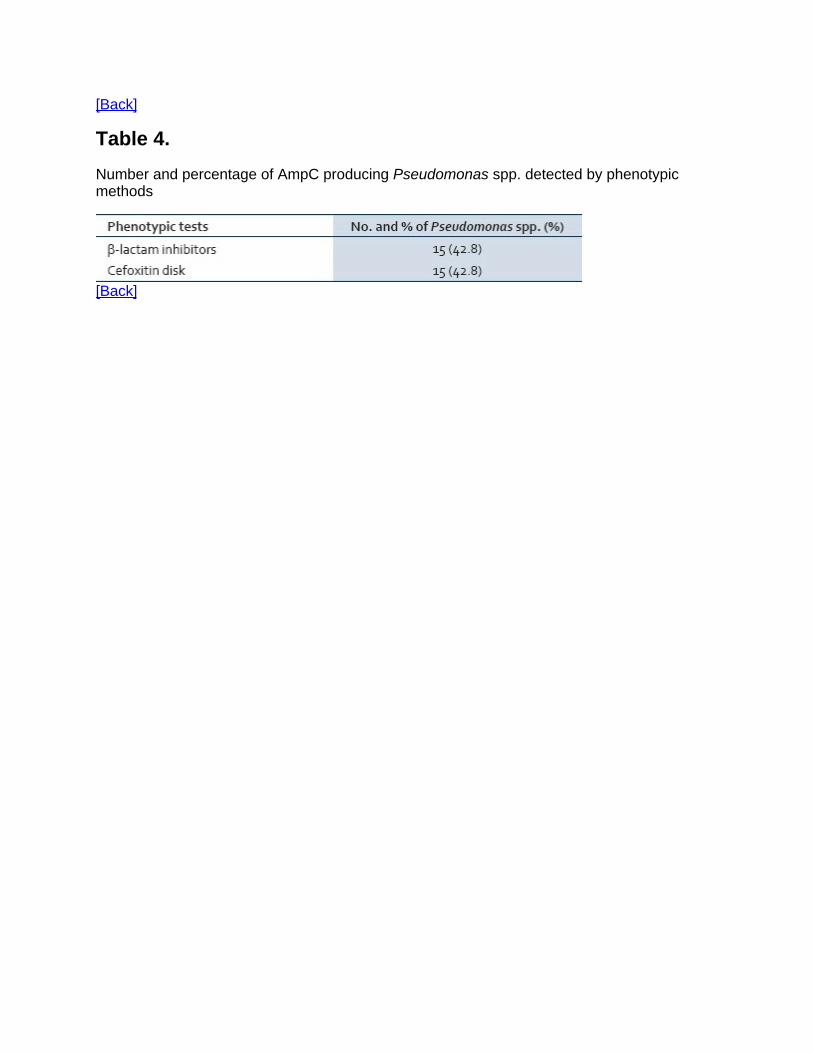
Phenotypic methods for AmpC detectionCefoxitin disks were used to screen AmpC producers by the disk diffusionmethod.[15] Those isolates which were resistant to cefoxitin were consideredas potential AmpC producers.
Phenotypic methods for MBL detectionThe isolates were tested for sensitivity to imipenem (10 μg) using the Kirby-Bauer method as recommended by CLSI.[12] Isolates from various samplesshowing zone of inhibition less than 16 mm or heaped up zone were screenedfor MBL production by the following methods.
Modified hodge test For MBL detection[16]The indicator organism, E. coli ATCC 25922, at a turbidity of 0.5 McFarlandstandard, was used to swab inoculate the surface of a Mueller-Hinton agarplate (Becton Dickinson, Cockeysville, Md.), and the test strain was heavilystreaked from the center to the plate periphery. After the plate was allowed tostand for 15 min at room temperature, a 10 μg IPM disk was placed at thecenter, and the plate was incubated overnight. The presence of distortedinhibition zone (cloverleaf) was interpretated as a positive result forcarbapenem hydrolysis.
The imipenem-EDTA double-disk synergy test[16]The test strains were adjusted to the McFarland 0.5 standard and used toinoculate Muller-Hinton agar plates. Disk containing imipenem 10 μg wasplaced on the plate and a blank filter paper soaked with 10 μl of 0.5 M EDTAsolution was placed at a distance of 10 mm (edge to edge). After overnightincubation, the presence of synergistic inhibition zone was interpreted aspositive.
Genotypic methods for the detection of ESBL and AmpCproductionPreparation of DNA template: Template DNA was prepared from freshlycultured bacterial isolates by suspending bacterial colonies in 50 μL ofmolecular-grade water and then heating at 95ΊC for 5 min and immediatelychilling at 4ΊC.
Detection of bla genes by polymerase chain reaction: Molecular detection ofblaCTX-M, blaTEM, blaSHV, blaAmpC was performed by using polymerase chainreaction (PCR) according to methods described previously with minormodifications.[1718] The primers and cycling conditions for detection of blagenes were the same as those described by Shahid et al.[18] and Feria et al.[19]
Detection of biofilm in Pseudomonas isolatesIn vitro biofilm forming ability of Pseudomonas isolates was tested by thetube method, as described by Hassan et al.[20] and Mathur et al.,[21] withslight modification. 0.5 ml (1.5 × 10⁸ organism/ml) of 48 hour culture salinewashed suspension was inoculated into a polysterene tube containing 4.5 mlof trypticase soy broth (TSB) with 1% glucose.[21] Tubes were incubated at37ΊC for 48 hours without agitation. After 48 hours, the culture broth in thetube was aspirated, and the tubes were washed twice with distilled water. Thewalls of tube were stained with 0.1% crystal violet after media and cells werediscarded. Excess stain was removed and tubes were washed with water.Tubes were than dried in inverted position and observed for biofilmformation.
RESULTS
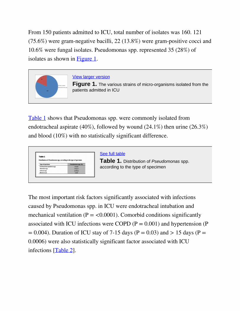
From 150 patients admitted to ICU, total number of isolates was 160. 121(75.6%) were gram-negative bacilli, 22 (13.8%) were gram-positive cocci and10.6% were fungal isolates. Pseudomonas spp. represented 35 (28%) ofisolates as shown in Figure 1.
View larger version
Figure 1. The various strains of micro-organisms isolated from thepatients admitted in ICU
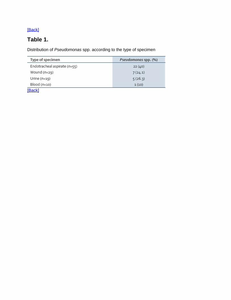
Table 1 shows that Pseudomonas spp. were commonly isolated fromendotracheal aspirate (40%), followed by wound (24.1%) then urine (26.3%)and blood (10%) with no statistically significant difference.
See full table
Table 1. Distribution of Pseudomonas spp.according to the type of specimen
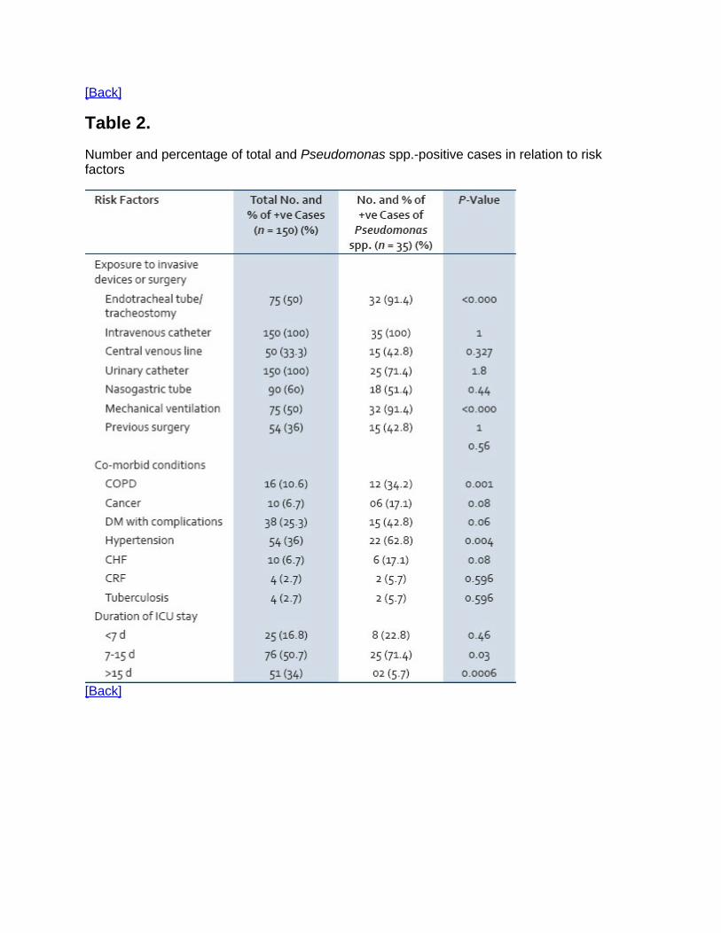
The most important risk factors significantly associated with infectionscaused by Pseudomonas spp. in ICU were endotracheal intubation andmechanical ventilation (P = <0.0001). Comorbid conditions significantlyassociated with ICU infections were COPD (P = 0.001) and hypertension (P= 0.004). Duration of ICU stay of 7-15 days (P = 0.03) and > 15 days (P =0.0006) were also statistically significant factor associated with ICUinfections [Table 2].

See full table
Table 2. Number and percentage of total andPseudomonas spp.-positive cases in relation to riskfactors
Figure 2 shows antibiotic susceptibility pattern of Pseudomonas spp. detectedby the disk diffusion method. Pseudomonas spp. exhibited high degree ofantibiotic resistance against ofloxacin (80.0%), ticarcillin (80.0%),piperacillin (74.2%), gentamycin (71.4%), ceftazidime (68.5%) andcefpodoxime (65.7%). Five isolates (14.3%) of Pseudomonas spp. wereresistant to imipenem. There was no resistance for polymyxin B and colistin.
View larger version
Figure 2. Antibiotic resistance pattern of Pseudomonas spp.detected by the disk diffusion method
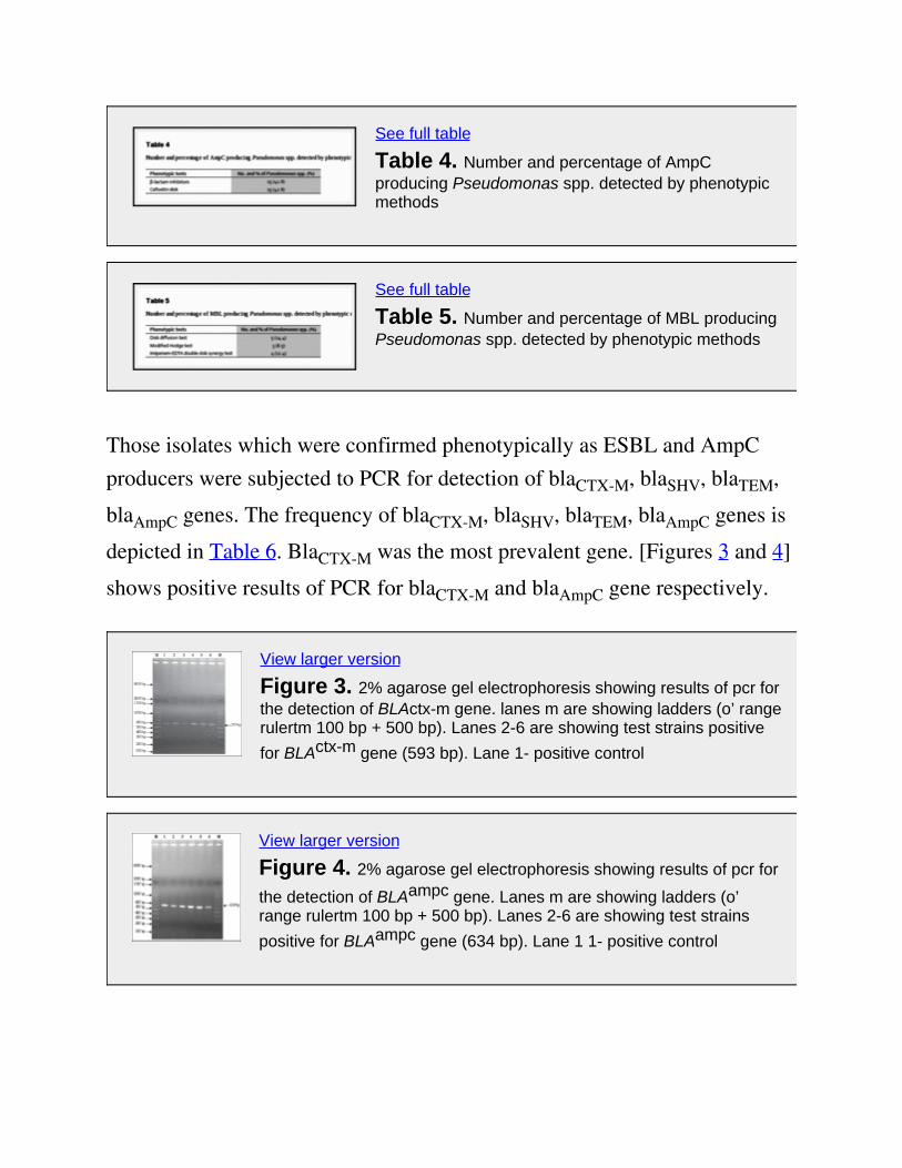
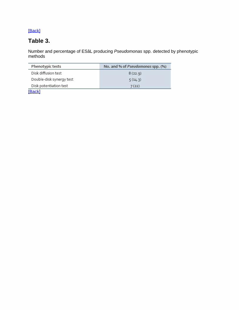
All the Pseudomonas spp. isolates were further screened for the production ofESBL, AmpC and MBL activity first by phenotypic methods. The positivityfor ESBL, AmpC and MBL by phenotypic methods is shown in Tables 3–5,respectively.
See full table
Table 3. Number and percentage of ESâL producingPseudomonas spp. detected by phenotypic methods
See full table
Table 4. Number and percentage of AmpCproducing Pseudomonas spp. detected by phenotypicmethods
See full table
Table 5. Number and percentage of MBL producingPseudomonas spp. detected by phenotypic methods
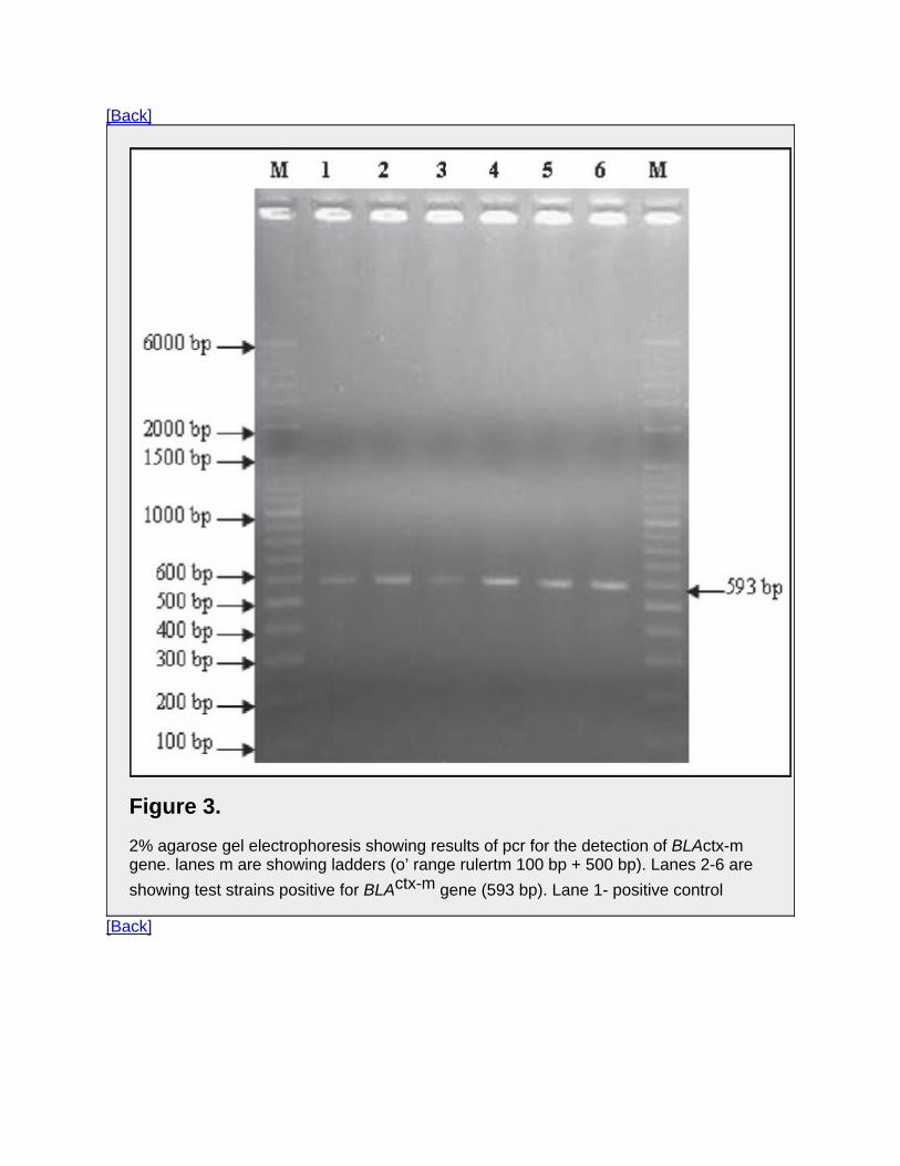
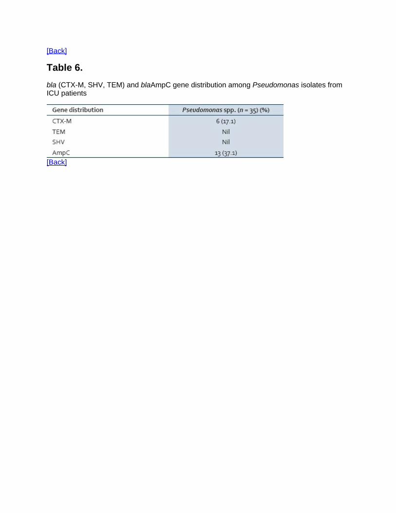
Those isolates which were confirmed phenotypically as ESBL and AmpCproducers were subjected to PCR for detection of blaCTX-M, blaSHV, blaTEM,blaAmpC genes. The frequency of blaCTX-M, blaSHV, blaTEM, blaAmpC genes isdepicted in Table 6. BlaCTX-M was the most prevalent gene. [Figures 3 and 4]shows positive results of PCR for blaCTX-M and blaAmpC gene respectively.
View larger version
Figure 3. 2% agarose gel electrophoresis showing results of pcr forthe detection of BLActx-m gene. lanes m are showing ladders (o’ rangerulertm 100 bp + 500 bp). Lanes 2-6 are showing test strains positivefor BLActx-m gene (593 bp). Lane 1- positive control
View larger version
Figure 4. 2% agarose gel electrophoresis showing results of pcr for
the detection of BLAampc gene. Lanes m are showing ladders (o’range rulertm 100 bp + 500 bp). Lanes 2-6 are showing test strainspositive for BLAampc gene (634 bp). Lane 1 1- positive control
See full table
Table 6. bla (CTX-M, SHV, TEM) and blaAmpCgene distribution among Pseudomonas isolates fromICU patients
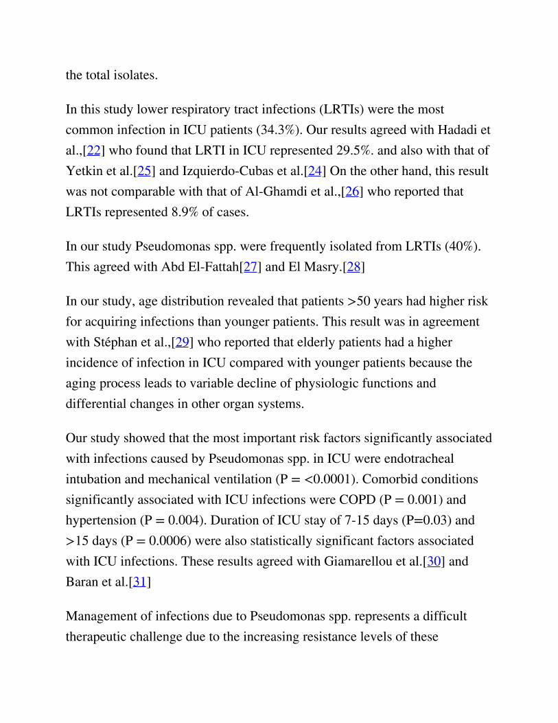
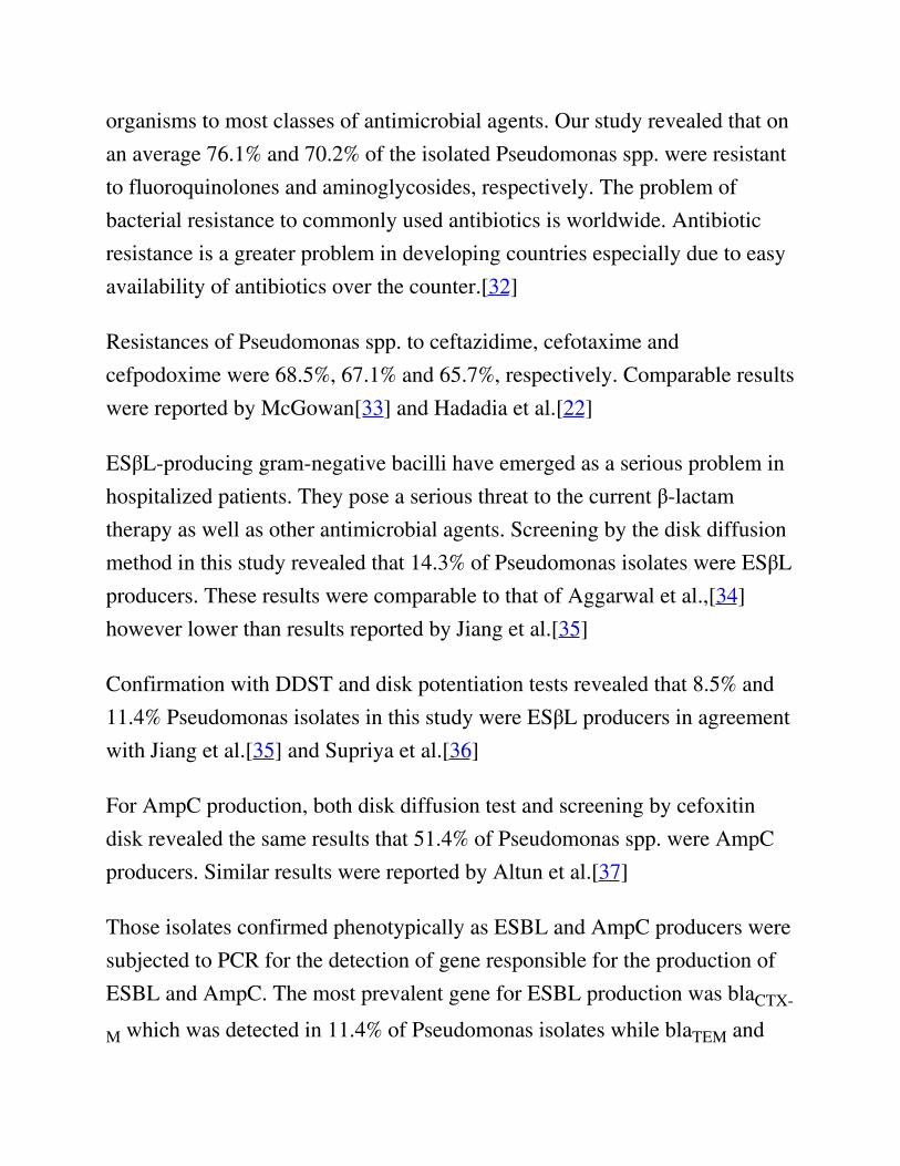
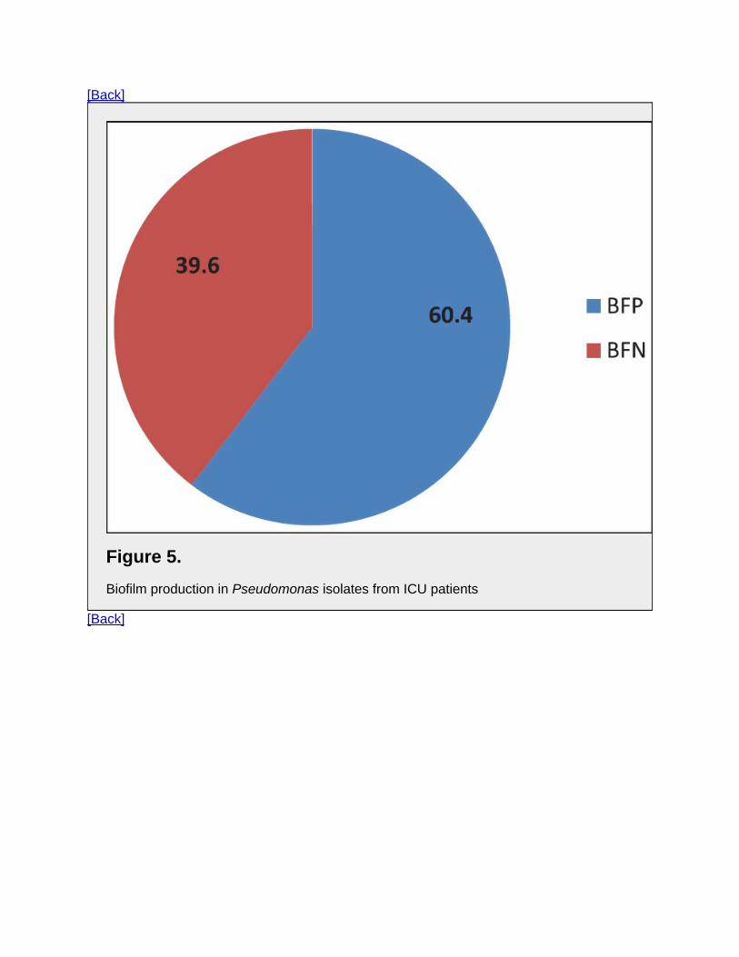
Figure 5 depicts the biofilm production in Pseudomonas isolates from ICUpatients. Among the 35 isolates, 21 (60.4 %) were biofilm producers. Figure 6shows antibiotic resistance profile of biofilm-positive (BFP) and biofilm-negative (BFN) Pseudomonas isolates.
View larger version
Figure 5. Biofilm production in Pseudomonas isolates from ICUpatients
View larger version
Figure 6. Antibiotic resistance profile of BFP and BFNPseudomonas isolates
DISCUSSIONPseudomonas spp. are one of the most common gram-negative pathogensassociated with infections in ICU patients including bacteremia, urinary tractinfections, and surgical site infections, but they predominate as agents oflower respiratory tract infections.[1]
In this study, Pseudomonas spp. represented 28% of isolates, similar resultswere reported by Hadadi et al.,[22] on other hand Chim et al.[23] andIzquierdo-Cubas et al.[24] found that Pseudomonas spp. represented 15.6% of
the total isolates.
In this study lower respiratory tract infections (LRTIs) were the mostcommon infection in ICU patients (34.3%). Our results agreed with Hadadi etal.,[22] who found that LRTI in ICU represented 29.5%. and also with that ofYetkin et al.[25] and Izquierdo-Cubas et al.[24] On the other hand, this resultwas not comparable with that of Al-Ghamdi et al.,[26] who reported thatLRTIs represented 8.9% of cases.
In our study Pseudomonas spp. were frequently isolated from LRTIs (40%).This agreed with Abd El-Fattah[27] and El Masry.[28]
In our study, age distribution revealed that patients >50 years had higher riskfor acquiring infections than younger patients. This result was in agreementwith Stéphan et al.,[29] who reported that elderly patients had a higherincidence of infection in ICU compared with younger patients because theaging process leads to variable decline of physiologic functions anddifferential changes in other organ systems.
Our study showed that the most important risk factors significantly associatedwith infections caused by Pseudomonas spp. in ICU were endotrachealintubation and mechanical ventilation (P = <0.0001). Comorbid conditionssignificantly associated with ICU infections were COPD (P = 0.001) andhypertension (P = 0.004). Duration of ICU stay of 7-15 days (P=0.03) and>15 days (P = 0.0006) were also statistically significant factors associatedwith ICU infections. These results agreed with Giamarellou et al.[30] andBaran et al.[31]
Management of infections due to Pseudomonas spp. represents a difficulttherapeutic challenge due to the increasing resistance levels of these
organisms to most classes of antimicrobial agents. Our study revealed that onan average 76.1% and 70.2% of the isolated Pseudomonas spp. were resistantto fluoroquinolones and aminoglycosides, respectively. The problem ofbacterial resistance to commonly used antibiotics is worldwide. Antibioticresistance is a greater problem in developing countries especially due to easyavailability of antibiotics over the counter.[32]
Resistances of Pseudomonas spp. to ceftazidime, cefotaxime andcefpodoxime were 68.5%, 67.1% and 65.7%, respectively. Comparable resultswere reported by McGowan[33] and Hadadia et al.[22]
ESβL-producing gram-negative bacilli have emerged as a serious problem inhospitalized patients. They pose a serious threat to the current β-lactamtherapy as well as other antimicrobial agents. Screening by the disk diffusionmethod in this study revealed that 14.3% of Pseudomonas isolates were ESβLproducers. These results were comparable to that of Aggarwal et al.,[34]however lower than results reported by Jiang et al.[35]
Confirmation with DDST and disk potentiation tests revealed that 8.5% and11.4% Pseudomonas isolates in this study were ESβL producers in agreementwith Jiang et al.[35] and Supriya et al.[36]
For AmpC production, both disk diffusion test and screening by cefoxitindisk revealed the same results that 51.4% of Pseudomonas spp. were AmpCproducers. Similar results were reported by Altun et al.[37]
Those isolates confirmed phenotypically as ESBL and AmpC producers weresubjected to PCR for the detection of gene responsible for the production ofESBL and AmpC. The most prevalent gene for ESBL production was blaCTX-
M which was detected in 11.4% of Pseudomonas isolates while blaTEM and
blaSHV genes were not detected in any of the isolates. These results are inagreement with Picao et al.[38]
PCR detected the blaAmpC gene in 42.8%. These results are comparable tothat of Khanal et al.[39]
According to the disk diffusion method, results of our study revealed that14.3% of Pseudomonas isolates were carbapenem resistant. The modifiedHodge test revealed that 8.5% of Pseudomonas isolates were MβL producers.When tested by the imipenem-EDTA DDST, 11.4% of Pseudomonas spp.were MBL producers. However, no resistance was observed for polymyxin Band colistin. This result agreed with Behera et al.[40]
Comparison between modified Hodge test and imipenem-EDTA DDST inour study revealed that the imipenem-EDTA DDST was more sensitive fordetecting MBL. The same observation was reported by Jesudason et al.[41]Mereuţă et al.[42] reported that double-disk and combined disk tests areuseful, simple and accessible to clinical laboratories but PCR is needed toconfirm the presence of the MβL gene in bacteria and to determine type ofthe enzymes.
Biofilms have an enormous impact on healthcare and are estimated to beassociated with 65% of infections in ICU patients. Biofilm growth isassociated with an increased level of mutations as well as with quorum-sensing-regulated mechanisms. Antimicrobial resistance is an innate featureof bacterial biofilms.[43] Many studies have shown that biofilm formation ishigher in MDR strains. In this study, among 35 isolates, 21 (60.4 %) werebiofilm producers. Carlos et al. reported biofilm formation in P. aeruginosa in83% of clinical strains and that biofilm formation was prevalent among
isolates with a MDR phenotype.[44] In our study we found higher antibioticresistance in biofilm producers as compared to the negative biofilmproducers.
CONCLUSIONIn this study amikacin, tobramycin, imipenem, polymyxin B and colistindemonstrated maximum sensitivity against Pseudomonas species. Therefore,use of these antibiotics should be restricted to severe infections especially incritically ill ICU patients, in order to avoid rapid emergence of resistantstrains.
Carbapenem resistance not only has enormous therapeutic implications, but isalso important from the point of view of infection control. Such stains areknown for rapid intra institutional spread and therefore, must be notified toinfection control teams. Higher antibiotic resistances were seen in strongbiofilm producers are due so testing for biofilm formation.
It is important to implement an easy, discriminatory, reproducible and cheapdetection of MβL, ESβL and AmpC producers in our hospitals. Double disksynergy method for detection of ESBL and MBLs and estimation of cefoxitinresistance by the disk diffusion method for AmpC detection are not only costeffective and highly sensitive but also easy to perform.
Regular antimicrobial susceptibility surveillance is essential. An effectivenational and state level area-wise monitoring of the resistance patternsantibiotic policy and draft guidelines should be introduced to preserve theeffectiveness of antibiotics and for better patient management.
FootnotesSource of Support: Nil.
Conflict of Interest: None declared.
Articles from Journal of Global Infectious Diseases are provided herecourtesy of Wolters Kluwer -- Medknow Publications
PMC Copyright Notice
The articles available from the PMC site are protected by copyright, even though access is free.Copyright is held by the respective authors or publishers who provide these articles to PMC. Usersof PMC are responsible for complying with the terms and conditions defined by the copyrightholder.
Users should assume that standard copyright protection applies to articles in PMC, unless an articlecontains an explicit license statement that gives a user additional reuse or redistribution rights. PMCdoes not allow automated/bulk downloading of articles that have standard copyright protection.
See the copyright notice on the PMC site, https://www.ncbi.nlm.nih.gov/pmc/about/copyright/, forfurther details and specific exceptions.
REFERENCES1. Nseir S, Di Pompeo C, Brisson H, Dewavrin F, Tissier S, Diarra M, et al., authors. Intensive care
unit-acquired Stenotrophomonas maltophilia: Incidence, risk factors, and outcome. Critical Care.2006;10:R143 [PubMed]
2. Aloush V, Navon-Venezia S, Seigman-Igra Y, Cabili S, Carmeli Y, authors. Multidrug-resistantPseudomonas aeruginosa: Risk factors and clinical impact. Antimicrob Agents Chemother.2006;50:43–8. [PubMed]
3. Livermore DM, author. Multiple mechanisms of antimicrobial resistance in Pseudomonasaeruginosa: Our worst nightmare? Clin Infect Dis. 2002;34:634–40. [PubMed]
4. Queenan AM, Bush K, authors. Carbapenemases: The versatile β-lactamases. Clin Microbiol
Rev. 2007;20:440–58. [PubMed]
5. Cornaglia G, Giamarellou H, Rossolini GM, authors. Metallo-β-lactamases: A last frontier for β-lactams? Lancet Infect Dis. 2011;11:381–93. [PubMed]
6. Walsh TR, Toleman MA, Poirel L, Nordmann P, authors. Metallo-beta-lactamases: The quietbefore the storm? Clin Microbiol Rev. 2005;18:306–25. [PubMed]
7. Poirel L, Naas T, Nicolas D, Collet L, Bellais S, Cavallo JD, et al., authors. Characterization ofVIM-2, a carbapenem-hydrolyzing metallo β-lactamase and its plasmid- and integron-borne genefrom a Pseudomonas aeruginosa clinical isolate in France. Antimicrob Agents Chemother.2000;44:891–7. [PubMed]
8. Dunne WM Jr, author. Bacterial adhesion: Seen any good biofilms lately? Clin Microbiol Rev.2002;15:155–66. [PubMed]
9. Obritsch MD, Fish DN, MacLaren R, Jung R, authors. National surveillance of antimicrobialresistance in Pseudomonas aeruginosa isolates obtained from intensive care unit patients from1993 to 2002. Antimicrob Agents Chemother. 2004;48:4606–10. [PubMed]
10. Collee JG, Duguid JP, Fraser AG, Marmion BP, Simmons A, authors. Laboratory strategy in thediagnosis of infective syndrome. Mackie and McCartney Practical Microbiology. 1996. 14th ed.London: Churchill Livingstone; p. 53–94
11. Collee JG, Marr W, authors. Culture of bacteria. Mackie and McCartney Practical Microbiology.1996. 14th ed. London: Churchill Livingstone; p. 113–30
12. Clinical and Laboratory Standards Institute: Performance standards for antimicrobialsusceptibility testing: Seventeenth informational supplement. M100-S17. 2007. Wayne, PA:Clinical and Laboratory Standards Institute;
13. Peterson DL, Bonomo RA, authors. Extended spectrum beta lactamases: A clinical update. ClinMicrobiol Rev. 2005;18:657–86. [PubMed]
14. Jarlier V, Nicolas MH, Fournier G, Philippon A, authors. Extended-spectrum beta lactamasesconferring transferable resistance to newer beta-lactam agents in Enterobacteriaceae: Hospitalprevelance and susceptibility patterns. Rev Infect Dis. 1988;10:867–78. [PubMed]
15. Shahid M, Malik A, Agarwal M, Singhal S, authors. Phenotypic detection of the extendedspectrum and AmpC β-lactamases by a new spot inoculation method and modified three-dimensional extract test: Comparison with conventional three- dimensional extract test. JAntimicrob Chemother. 2004;54:684–7. [PubMed]
16. Lee K, Lim JB, Yum JH, Yong D, Chong Y, Kim JM, et al., authors. blaVIM-2 cassette-containing novel integrons in metallo-β-lactamase-producing Pseudomonas aeruginosa andPseudomonas putida isolates disseminated in a Korean hospital. Antimicrob Agents Chemother.2002;46:1053–8. [PubMed]
17. Shahid M, Ensor VM, Hawkey PM, authors. Emergence and dissemination ofEnterobacteriaceae with plasmid-mediated CMY-6 and CTX-M-15 beta-lactamases in acommunity in North-India. World J Microbiol Biotech. 2009;25:1439–46
18. Shahid M, Malik A, Adil M, Jahan N, Malik R, authors. Comparison of beta-lactamase genes inclinical and food bacterial isolates in India. J Infect Dev Ctries. 2009;3:593–8. [PubMed]
19. Feria C, Ferreira E, Correia JD, Goncalves J, Canica M, authors. Patterns and mechanisms ofresistance to beta-lactams and beta-lactamase inhibitors in uropathogenic Escherichia coliisolated from dogs in Portugal. J Antimicrob Chemother. 2002;49:77–85. [PubMed]
20. Hassan A, Usman J, Kaleem F, Omair M, Khalid A, Iqbal M, authors. Evaluation of differentdetection methods of biofilm formation in the clinical isolates. Braz J Infect Dis. 2011;15:305–11. [PubMed]
21. Mathur T, Singhal S, Khan S, Upadhyay DJ, Fatma T, Rattan A, authors. Detection of biofilmformation among the clinical isolates of Staphylococci: Evaluation of three different screeningmethods. Indian J Med Microbiol. 2006;24:25–9. [PubMed]
22. Hadadi A, Rasoulinejad M, Maleki Z, Yonesian M, Shirani A, Kourorian Z, authors.Antimicrobial resistance pattern of Gram-negative bacilli of nosocomial origin at 2 universityhospitals in Iran. Diag Microbiol Infect Dis. 2008;60:301–5
23. Chim H, Hock Tan B, Song C, authors. Five-year review of infections in a burn intensive careunit: High incidence of Acinetobacter baumannii in a tropical climate. Burns. 2007;33:100–14.[PubMed]
24. Izquierdo-Cubas F, Zambrano A, Frómeta I, Gutiérrez A, Bastanzuri M, Guanche H, et al.,authors. National Prevalence of Nosocomial Infections. Cuba 2004. J Hosp Infect. 2008;68:234–40. [PubMed]
25. Yetkin G, Otlu B, Cicek A, Kuzucu C, Durmaz R, authors. Clinical, microbiologic, andepidemiologic characteristics of Pseudomonas aeruginosa infections in a University Hospital.Am J Infect Control. 2006;34:188–92. [PubMed]
26. Al-Ghamdi S, Gedebou M, Bilal NE, authors. Nosocomial infections and misuse of antibiotics ina provincial community hospital, Saudi Arabia. J Hosp Infec. 2002;50:115–21. [PubMed]
27. Abd El-Fattah SM, author. Evaluation of antibiotic resistance among Gram-ve bacilli isolatedfrom critically ill patients: Relation to risk factors and liberal use of antibiotics. M.Sc Thesis inMedical Microbiology an Immunology. Faculty of Medicine. 2008. Giza, Egypt: CairoUniversity; p. 66–7
28. El Masry E, author. Extended spectrum beta-lactamase-producing Enterobacteriaceae and P.aeruginosa causing nosocomial pneumonia in Menoufiya university hospitals. M.Sc Thesis inMedical Microbiology and Immunology. Faculty of Medicine. 2007. Menoufiya University; p.
97–106
29. Stéphan F, Cheffi A, Bonnet F, authors. Nosocomial Infections and Outcome of Critically IllElderly Patients after Surgery. Anesthesiology. 2001;94:407–14. [PubMed]
30. Giamarellou H, Antoniadou A, Kanellakopoulou K, authors. Acinetobacter baumannii: Auniversal threat to public health? J Hosp Infect. 2007;67:245–52. [PubMed]
31. Baran G, Erbay A, Bodur H, Ongürü P, Akinci E, Balaban N, et al., authors. Risk factors fornosocomial imipenem-resistant Acinetobacter baumannii infections. Int J Infect Dis.2007;12:16–21. [PubMed]
32. Martinez JL, Baquero F, authors. Interactions among strategies associated with bacterialinfection: Pathogenicity, epidemicity and antibiotic resistance. Clin Microbiol Rev.2002;15:647–79. [PubMed]
33. McGowan JE Jr, author. Resistance in nonfermenting gram-negative bacteria: multidrugresistance to the maximum”. Am J Med. 2006;119(Suppl 1):S29–36. [PubMed]
34. Aggarwal R, Chaudhary U, Bala K, authors. Detection of extended-spectrum beta-lactamase inPseudomonas aeruginosa. Indian J Pathol Microbiol. 2008;51:222–4. [PubMed]
35. Jiang X, Zhang Z, Li M, Zhou D, Ruan F, Lu Y, authors. Detection of extendedspectrum beta-lactamases in clinical isolates of Pseudomonas aeruginosa. Antimicrob Agents Chemother.2006;50:2990–5. [PubMed]
36. Supriya S, Jalgaonkar V, Ahamad S, Hassani U, authors. Evaluation of extended spectrum betalactamase in urinary isolates. Indian J Med Res. 2004;120:553–6. [PubMed]
37. Altun Ş, Tufan ZK, Yağcı S, Önde U, Bulut C, authors. Extended Spectrum Betalactamases,AmpC and Metallo Beta-lactamases in emerging multi-drug resistant Gram-negative bacteria inintensive care units. 2013;2:707 . doi:10.4172/scientificreports.
38. Picão RC, Gales AC, authors. Extended-spectrum beta-lactamase producing (ESBL)Pseudomonas aeruginosa: Nightmare or imagination? Prática Hospitalar. 2007;49:79–84
39. Khanal S, Joshi DR, Bhatta DR, Devkota U, Pokhrel BM, authors. β-Lactamase-ProducingMultidrug-Resistant Bacterial Pathogens from Tracheal Aspirates of Intensive Care Unit Patientsat National Institute of Neurological and Allied Sciences, Nepal. ISRN Microbiol.2013;2013:847569 [PubMed]
40. Behera B, Mathur P, Das A, Kapil A, Sharma V, authors. An evaluation of four differentphenotypic techniques for detection of metallo-beta-lactamase producing Pseudomonasaeruginosa. Indian J Med Microbiol. 2008;26:233–7. [PubMed]
41. Jesudason MV, Kandathi AJ, Balaji V, authors. Comparison of the methods for the detection ofthe carbapenamase and the metallo-beta- lactamases production in the clinical isolates. Indian JMed Res. 2005;121:780–3. [PubMed]

42. Mereuţă AI, Poiati A, Tuchiluş C, Dorneanu O, Nistor S, Copăcianu B, authors. Screeningmethods for detection of metallo-beta-lactamase producing gram-ve rods. Rev Med Chir SocMed Nat Iasi. 2008;109:387–91. [PubMed]
43. Høiby N, Bjarnsholt T, Givskov M, Molin S, Ciofu O, authors. Antibiotic resistance of bacterialbiofilms. Int J Antimicrob Agents. 2010;35:322–32. [PubMed]
44. Sanchez CJ Jr, Mende K, Beckius ML, Akers KS, Romano DR, Wenke JC, et al., authors.Biofilm formation by clinical isolates and the implications in chronic infections. BMC InfectDis. 2013;13:47 [PubMed]

[Back]
Figure 1.The various strains of micro-organisms isolated from the patients admitted in ICU
[Back]
[Back]
Table 1.Distribution of Pseudomonas spp. according to the type of specimen
[Back]
[Back]
Table 2.Number and percentage of total and Pseudomonas spp.-positive cases in relation to riskfactors
[Back]

[Back]
Figure 2.Antibiotic resistance pattern of Pseudomonas spp. detected by the disk diffusionmethod
[Back]
[Back]
Table 3.Number and percentage of ESâL producing Pseudomonas spp. detected by phenotypicmethods
[Back]
[Back]
Table 4.Number and percentage of AmpC producing Pseudomonas spp. detected by phenotypicmethods
[Back]

[Back]
Table 5.Number and percentage of MBL producing Pseudomonas spp. detected by phenotypicmethods
[Back]
[Back]
Figure 3.2% agarose gel electrophoresis showing results of pcr for the detection of BLActx-mgene. lanes m are showing ladders (o’ range rulertm 100 bp + 500 bp). Lanes 2-6 areshowing test strains positive for BLActx-m gene (593 bp). Lane 1- positive control
[Back]

[Back]
Figure 4.
2% agarose gel electrophoresis showing results of pcr for the detection of BLAampcgene. Lanes m are showing ladders (o’ range rulertm 100 bp + 500 bp). Lanes 2-6 areshowing test strains positive for BLAampc gene (634 bp). Lane 1 1- positive control
[Back]
[Back]
Table 6.bla (CTX-M, SHV, TEM) and blaAmpC gene distribution among Pseudomonas isolates fromICU patients
[Back]
[Back]
Figure 5.Biofilm production in Pseudomonas isolates from ICU patients
[Back]

[Back]
Figure 6.Antibiotic resistance profile of BFP and BFN Pseudomonas isolates
[Back]