journal of inorganic biochemistry - ipen · and the oxldl labeled with horseradish peroxidase (hrp)...
TRANSCRIPT

Journal of Inorganic Biochemistry 123 (2013) 11–17
Contents lists available at SciVerse ScienceDirect
Journal of Inorganic Biochemistry
j ourna l homepage: www.e lsev ie r .com/ locate / j inorgb io
Luminescent material based on the [Eu(TTA)3(H2O)2] complex incorporated intomodified silica particles for biological applications
Ana Valéria S. Lourenço a, Cláudia A. Kodaira b,1, Eduardo M. Ramos-Sanchez c,2, Maria Cláudia F.C. Felinto d,3,Hiro Goto e,4, Magnus Gidlund c,2, Oscar L. Malta f,5, Hermi F. Brito a,⁎a Instituto de Química, Universidade de São Paulo, CP 26077, 05508-000 São Paulo, Brazilb Luminetech Marcadores Ópticos Ltda — CIETEC, Av. Prof. Lineu Prestes, 2242 Sala 9 2º andar, 05508-000 São Paulo, SP, Brazilc Departamento de Imunologia, Instituto de Ciências Biomédicas-IV, Universidade de São Paulo, Av. Lineu Prestes, 1730, 05508-900 São Paulo, Brazild Instituto de Pesquisas Energéticas e Nucleares, Av. Lineu Prestes, 2242, 05508-000 São Paulo, Brazile Faculdade de Medicina e Instituto de Medicina Tropical de São Paulo, Universidade de São Paulo, Av. Dr. Enéas de Carvalho Aguiar, 470, 05403-000 São Paulo, Brazilf Departamento de Química Fundamental, CCEN, Universidade Federal de Pernambuco Cidade Universitária, 50670-901 Recife, Brazil
⁎ Corresponding author. Tel.: +55 11 3091 3708; faxE-mail address: [email protected] (H.F. Brito).
1 Tel.: +55 11 3039 8432.2 Tel.: +55 11 3091 7382; fax: +55 11 3091 7224.3 Tel.: +55 11 3133 9343; fax: +55 11 3133 9018.4 Tel.: +55 11 3061 7023; fax: +55 11 3061 8270.5 Tel.: +55 81 2126 7459; fax: +55 81 2126 8442.
0162-0134/$ – see front matter © 2013 Elsevier Inc. Allhttp://dx.doi.org/10.1016/j.jinorgbio.2013.02.006
a b s t r a c t
a r t i c l e i n f oArticle history:Received 17 October 2012Received in revised form 16 February 2013Accepted 18 February 2013Available online 26 February 2013
Keywords:Europium complexFunctionalized silicaLuminescence propertiesBioassayOxidized low density lipoprotein
Amino-functionalized luminescent silica particles were investigated for use in immunoassays. The particles wereprepared by the Stöber method where the [Eu(TTA)3(H2O)2] complex (TTA: 3-thenoyltrifluoroacetonate) wasincorporated into silica particles during the hydrolysis and condensation of TEOS: tetraethylorthosilicate. Then,the amino groups were introduced in the particle surface using APTS: 3-aminopropyltriethoxisilane. Theresulting particles were characterized by scanning electron microscopy (SEM), X ray diffraction (XRD) andphotoluminescence spectroscopy. In order to demonstrate the viability of the use of luminescent particles as op-tical markers, an enzyme–substrate reaction was performed using HRP: horseradish peroxidase. It was possibleto verify the binding of HRP-oxidized LDL (low density lipoprotein) and anti-oxLDL antibody-luminescent silicaparticles through the evaluation of the presence ofHRP. The bioassay data open a broadfield for the developmentof protein-tagged luminescent particles for use in biomedical sciences.
© 2013 Elsevier Inc. All rights reserved.
1. Introduction
Nowadays the development of luminescent complexes incorpo-rating silica, which combine the characteristics of inorganic matricesand optical properties of the rare earth complexes, has been the sub-ject of several studies [1–4]. There has been an increasing interest inthis field of research, where silica particles were shown to be veryuseful platforms for lighting and luminescent markers or probes[5–9]. The trivalent rare earths (RE3+) show unique properties [10]and have been widely applied in biological assays [11]. The criteriathat should accomplish a RE3+ luminescent probe to be helpful inthe design of bioassays in diagnostic or drug discovery (high through-put screening) are defined as brightness, absorption wavelength, lu-minescence decay, instrumentation crosstalk, stability, lipophilicity/hydrophilicity, photobleaching, quenching phenomenon, conjugationchemistry, and synthesis practicability.
: +55 11 3091 3146.
rights reserved.
The 4f orbitals are shielded from the chemical environment by the5s and 5p filled orbitals, producing very narrow bands in their emissionand absorption spectra from the compounds and long lifetimes [10,12].Furthermore, the surface of the silica particles incorporating RE3+-complexes can be easily modified, providing new opportunities fortheir applications in biological assays, enabling conjugation with bio-molecules such as proteins, peptides, sugars, antibodies, etc. [13–15].
Ischemic coronary artery disease consequent of atheroscleroticprocess may lead to an inadequate blood circulation in the myocardi-um due to partial or complete obstruction of the coronary arteries[16]. Atherosclerosis is responsible for more than 30% of the totalnumber of recorded deaths in the urban centers. The process is achronic inflammatory disease characterized by the accumulation oflipid and vascular smooth muscle cell proliferation on the wall ofthe artery [17,18].
Clinical evidences demonstrate that high plasma concentration oflow density lipoprotein (LDL) is a risk factor for atherosclerosis devel-opment. Indeed the modified forms of LDL as oxidized LDL (oxLDL)are the main components responsible for the formation of foam cells,contributing to the installation of the atherosclerotic process [19,20].
This study presents a preparation of a luminescent markerbased on silica particles incorporating [Eu(TTA)3(H2O)2], whereTTA: 3-thenoyltrifluoroacetonate. In addition, the recognition of theconjugation between the marker incubated with anti-oxLDL antibody

Fig. 1. Representative scheme of the immobilization of anti-oxLDL antibody particles inEu-TTA-Si using glutaraldehyde as a spacer.
12 A.V.S. Lourenço et al. / Journal of Inorganic Biochemistry 123 (2013) 11–17
and the oxLDL labeled with horseradish peroxidase (HRP) was inves-tigated, using enzyme–substrate reaction.
2. Experimental
2.1. Preparation of the [Eu(TTA)3(H2O)2] complex incorporated withamino-functionalized silica particles
The luminescent amino-functionalized silica particles were pre-pared using the modified Stöber method [21]. Complex diaquatris(thenoyltrifluoroacetonate) europium(III) (1.9 mmol), named asEu-TTA, prepared by the method reported elsewhere [22,23] wasdissolved in 100 mL of ethyl alcohol (Aldrich). Afterwards, 1.4 mmolof TEOS: tetraethylorthosilicate (Fluka) and 2 mL of hydrous ammonia28% (Synth) were added to the previous solution at room temperaturefor 12 h under stirring. Then, the precipitate was centrifuged andwashedwith water and ethyl alcohol. Subsequently, the resulting parti-cleswere redispersed in ethyl alcohol (100 mL) and functionalizedwith0.2 mmol of APTES: 3-aminopropyltriethoxisilane (Fluka) and stirredat room temperature for 5 h [24]. Finally, the luminescent amino-functionalized silica particles (named as Eu-TTA-Si) were centrifuged,washed with water and ethyl alcohol and dried at 100 °C overnight.
X ray powder diffraction patterns were recorded on a RigakuMiniflex using Cu Kα radiation (30 kV and 15 mA) in the rangefrom 5 to 70° (2θ). The scanning electron microscopy (SEM) micro-graphs were obtained in a Field Emission Scanning Electron Micro-scope model JEOL JSM 7401F. The solid samples were deposited ona double-sided copper tape, attached to the sample holder. The exci-tation and emission spectra of the complex and amino-functionalizedmaterial at liquid nitrogen temperature (77 K) were recorded at anangle of 22.5° (front face) in a spectrofluorimeter (SPEX-Fluorog 2)with double grating 0.22 monochromator (SPEX 1680) and a 450 WXenon lamp as excitation source. All spectra were recorded using adetector mode correction. The luminescence decay curves of theemitting levels were measured at room temperature using a phos-phorimeter SPEX 1934D accessory coupled to the spectrofluorometer.
2.2. Antigen-antibody (Ag–Ab) interaction on the surface of theluminescent amino-functionalized silica particles
2.2.1. Bioconjugation of Eu-TTA-Si particles with anti-oxLDL antibodyThe process for the bioconjugation of anti-oxLDL antibody [19,25]
onto the luminescent amino-functionalized silica particles is shownin Fig. 1 [11]. The Eu-TTA-Si particles (1.0 mg) were dispersed inphosphate-buffered saline (PBS) solution (pH 7.4) containing 5%glutaraldehyde — GA (Aldrich) and kept at 4 °C for 12 h. After that,these particles were centrifuged, washed four times with a PBS solu-tion and redispersed in 1.0 mL of PBS solution. At that time, 30 μL ofanti-oxLDL antibody solution (1.9 mg mL−1) was added and incubat-ed overnight at 4 °C. Then, 1.6 mg of sodium borohydride (NaBH4
from Merck), was added and left to react for 30 min at 4 °C. Toblock the unreacted aldehyde sites, 50 μL of glycine (Vetec) solution0.5 mol L−1 was added, and this mixture was maintained for 2 h at4 °C. The conjugate system Eu-TTA-Si particle-GA-anti-oxLDL wasobtained and washed four times with PBS solution to remove theexcess of glycine, and afterwards stored at 4 °C.
2.2.2. Conjugation of HRP-GA with oxLDLDifferent HRP amounts from Aldrich (2 to 370 units) were added
in 1.0 mL of PBS buffer (pH 7.4) and 18.75 μL of glutaraldehydesolution (50%) and kept for 12 h at 4 °C. The HRP concentration(in U mL−1) was found as the difference between initial and finalenzyme amount remaining in the solution, at all times the specific activ-ity was expressed in terms of pyrogallol units (one pyrogallol unit willform 1.0 mg purpurogallin from pyrogallol in 20 s with pH 6.0 at20 °C). The solutions were dialyzed in a molecular porous membrane
tubing (Spectrum Laboratories, 12–14,000 (molecular weight cut-off))against PBS solution for 24 h at 4 °C. After this step, 85 μL of oxLDLsolution (3.0 mg mL−1), obtained as described in references [26,27],was incubated to themixture for 12 h at 4 °C. Besides, 1.6 mgof sodiumborohydridewas added and after 30 min, 50 μL of glycine (0.5 mol L−1)was added and left to react for 2 h at 4 °C. Then this solution wasdialyzed against PBS solution for 24 h at 4 °C. The representation of theHRP-GA-oxLDL bioconjugated system is illustrated in Fig. 2.
2.2.3. HRP-GA-oxLDL with Eu-TTA-Si particle-GA-anti-oxLDL antibodyreaction
The HRP-GA-oxLDL solution was added to Eu-TTA-Si particle-GA-anti-oxLDL antibody and incubated overnight at 4 °C (Fig. 3). After re-moval of the supernatant, the solid precipitate was washed fifteentimes with PBS. Both the supernatant and washing water werereserved for further analysis to determine unbound HRP-GA-oxLDL.
2.2.4. Measurement of the HRP enzyme concentrationThe optical density measurements of the supernatant and the
washings were performed on flat bottomed 96-well polystyrene
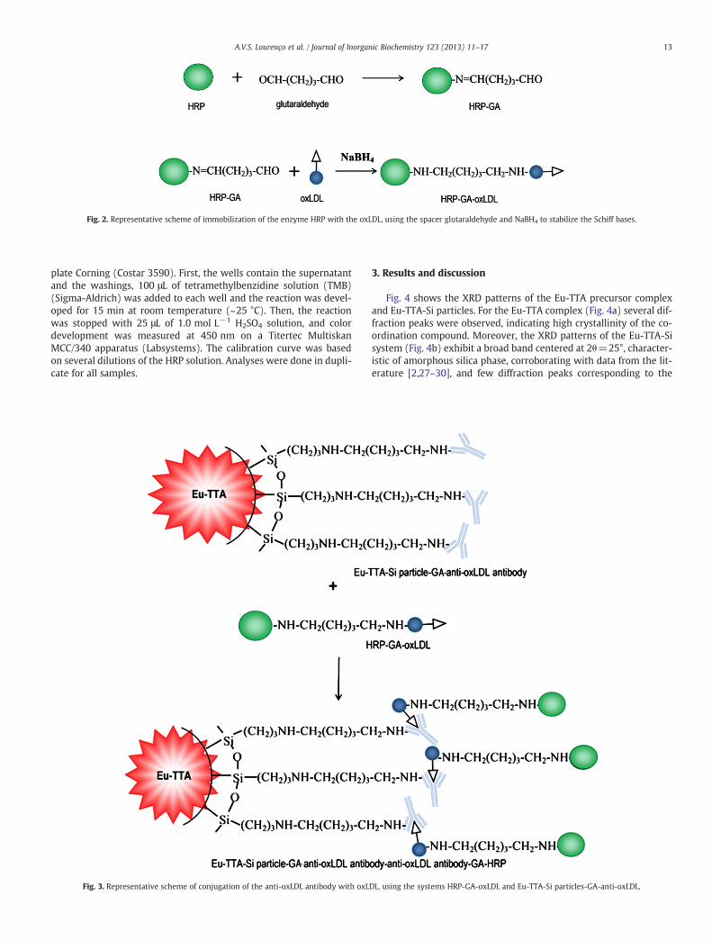
Fig. 2. Representative scheme of immobilization of the enzyme HRP with the oxLDL, using the spacer glutaraldehyde and NaBH4 to stabilize the Schiff bases.
13A.V.S. Lourenço et al. / Journal of Inorganic Biochemistry 123 (2013) 11–17
plate Corning (Costar 3590). First, the wells contain the supernatantand the washings, 100 μL of tetramethylbenzidine solution (TMB)(Sigma-Aldrich) was added to each well and the reaction was devel-oped for 15 min at room temperature (~25 °C). Then, the reactionwas stopped with 25 μL of 1.0 mol L−1 H2SO4 solution, and colordevelopment was measured at 450 nm on a Titertec MultiskanMCC/340 apparatus (Labsystems). The calibration curve was basedon several dilutions of the HRP solution. Analyses were done in dupli-cate for all samples.
Fig. 3. Representative scheme of conjugation of the anti-oxLDL antibody with oxL
3. Results and discussion
Fig. 4 shows the XRD patterns of the Eu-TTA precursor complexand Eu-TTA-Si particles. For the Eu-TTA complex (Fig. 4a) several dif-fraction peaks were observed, indicating high crystallinity of the co-ordination compound. Moreover, the XRD patterns of the Eu-TTA-Sisystem (Fig. 4b) exhibit a broad band centered at 2θ=25°, character-istic of amorphous silica phase, corroborating with data from the lit-erature [2,27–30], and few diffraction peaks corresponding to the
DL, using the systems HRP-GA-oxLDL and Eu-TTA-Si particles-GA-anti-oxLDL.

10 20 30 40 50 60 70
b) Eu-TTA-Si
I/I
0 / a
rb. u
nit
2θ / °
a) Eu-TTA
Fig. 4. X ray diffraction patterns of a) Eu-TTA complex and b) Eu-TTA-Si material.
250 300 350 400 450 500 550 600
λem.
= 613 nm
7 F 05 D
2
7 F 05 D
0
7 F 05 D
1
7 F 05 L 6
→
→
→
a) Eu-TTA
Inte
nsit
y / a
rb. u
nit
λ λ / nm
b) Eu-TTA-Si
λem.
= 613 nm
Fig. 6. Excitation spectra obtained at 77 K of a) Eu-TTA complex and b) Eu-TTA-Simaterial.
14 A.V.S. Lourenço et al. / Journal of Inorganic Biochemistry 123 (2013) 11–17
Eu-TTA complex precursor. This indicates different interactionsbetween the rare earth complex and the silica network in the processof silanization for the system prepared by the Stöber method [31–33].
SEM was used to observe the differences in the morphology of theEu-TTA and Eu-TTA-Si (Fig. 5). The SEM image of Eu-TTA-Si (Fig. 5b)reveals a homogeneous material with a rough and porous surface,suggesting a silica coating in the Eu-TTA precursor complex (Fig. 5a).
The concentration of 10,899 nmol mg−1 of amino groups on theEu-TTA-Si particle surface was quantified using the ninhydrin method[34,35]. Then, the potential for biological application of these particleswas demonstrated with the quantification of the amine groups afterthe conjugation with glutaraldehyde spacer [11]. The concentrationof amine groups decreases to 54.53%, suggesting that these groupsreact with the glutaraldehyde.
3.1. Luminescence marker study
Fig. 6 shows the excitation spectra of the Eu-TTA complex and theEu-TTA-Si material recorded at liquid nitrogen (77 K) in the range of250–600 nm, monitored at the 5D0→
7F2 hypersensitive transition(613 nm). The excitation spectrum of the Eu-TTA complex, Fig. 6a, dis-plays a broad absorption band covering the spectral range from 250 to475 nm overlapped with narrow bands assigned to the 7F0→ 5L6(394 nm), 5D2→
7F0 (464 nm) transitions of the trivalent europiumion. In Fig. 6b, the spectrum of Eu-TTA-Si shows a broad absorption
Fig. 5. The scanning electron micrographs of a)
band, in the range 300–425 nm, which is attributed to S0→S1allowed transition in the TTA ligands. The excitation spectrum alsodisplays three narrow bands attributed to intraconfigurational transi-tions of the Eu3+ ion (Fig. 6b), assigned to 7F0→ 5L6 (394 nm),7F0→ 5D2 (464 nm) and 7F0→ 5D1 (525 nm). The Eu-TTA-Si materialexhibits a different spectral profile compared with the Eu-TTA com-plex, suggesting that there was an effective interaction between theEu-TTA complex and the silica network.
The emission spectra (Fig. 7) were also recorded at 77 K in therange of 420–750 nm, under excitation in the 7F0→ 5L6 transition.The spectra exhibit narrow bands attributed to 5D0→
7FJ transitions(where J=0, 1, 2, 3 and 4), dominated by the 5D0→
7F2 hypersensi-tive transition. However, the intensity of the 5D0→
7F2 transition inrelation to the 5D0→
7F1 transition decreases in the emission spectrafor the Eu-TTA-Si material compared with the Eu-TTA complex,suggesting that the rare earth ion is in a site with higher symmetrywhen coated by the silica network.
It is important to notice the absence of a broad band between 450and 600 nm, referring to the triplet state (T) of the TTA ligand, demon-strating an efficient ligand-to-metal energy transfer. It is also observed
Eu-TTA complex and b) Eu-TTA-Si material.

450 500 550 600 650 700 750
5 D0
7 F 4
5 D0
7 F 35 D
07 F 2
5 D0
7 F 1→
→→
→
→5 D
07 F 0
λex.
= 396 nm
b) Eu-TTA-Si
Inte
nsit
y / a
rb. u
nit
λ λ / nm
a) Eu-TTA
λex.
= 393 nm
Fig. 7. Emission spectra of a) Eu-TTA complex and b) Eu-TTA-Si material, obtainedat 77 K.
15A.V.S. Lourenço et al. / Journal of Inorganic Biochemistry 123 (2013) 11–17
as a broadened peak of the 5D0→7FJ transitions in the Eu-TTA-Si
material (Fig. 7b) as compared with the complex, which can beexplained by a non-homogeneity of Eu3+ sites due to the silica porousmicrostructure.
The CIE (Comission Internacionale de l'Eclairage) x,y chromaticitydiagram of Eu-TTA and Eu-TTA-Si systems is displayed in Fig. 8 [36].
Eu-TTAEu-TTA-Si
Fig. 8. CIE chromaticity diagram showing the x,y emission color coordinates for Eu-TTAand Eu-TTA-Si systems.
When the complex was incorporated in silica matrix, a gradual shiftin the x,y color coordinates from the red to blue was observed.
The luminescence decay curves of the 5D0 emitting level of theEu-TTA complex and modified silica material were recorded underexcitation at 394 nm and emission at 614 nm. These data were ad-justed with a mono-exponential decay for the complex andbi-exponential for the modified silica material. The data in Table 1show that after the silanization of the Eu-TTA complex the lifetime in-creases in relation to the complex precursor due to the rigidity of thesilica network (Si–O) and substitution of water molecules in the firstcoordination sphere [37,38]. This behavior clearly indicates an inter-action of the silica network with the complex, changing the environ-ment of the Eu3+ ion.
The experimental intensity parameters Ωλ (λ=2 and 4) were de-termined for the Eu-TTA complex and Eu-TTA-Si material from theemission spectral data. The Ωλ, also known as Judd–Ofelt parameters[39], are determined by the intensities of the transitions 5D0→
7FJ(J=2 and 4) of Eu3+ ion [12,22,40], where the mechanisms of forcedelectric dipole and dynamic coupling are considered simultaneously.Therefore, the luminescence intensity (I) is expressed in terms ofthe area under the emission curves of the transitions from the 5D0
level to the levels 7FJ (J=2 and 4), and is defined as:
I0→J ¼ �hω0→JA0→JN0 ð1Þ
where �hω0 is the energy of the transition (in cm−1), N0 is the popu-lation of the emitting level 5D0 and A0→ J is the coefficient of sponta-neous emission. For the experimental determination of the emissioncoefficient A0→ J from the emission spectra the magnetic dipole usedallowed 5D0→
7F1 transition, which is formally insensitive to thechemical environment around the Eu3+ compound and, consequent-ly, can be used as a reference [41]. The values ofΩλ are obtained from:
A0→J ¼4e2ω3
3�hc31
2J þ 1χ ∑λ¼2;4
Ωλ5D0‖U
λð Þ‖7FJ
D E2 ð2Þ
where χ ¼ n0 n20þ2ð Þ29 is the Lorentz local field correction and n0 is the
refractive index of the medium (n0=1.5). The squared reduced ma-trix elements 5D0‖U
λð Þ‖7FJ i2D
are tabulated in the literature [42],and their values are 0.0032 and 0.0023 for J=2 and 4, respectively.The coefficients of spontaneous emission (A0→ J), are obtained from:
A0→J ¼σ0→1
S0→1
� �S0→J
σ0→J
!A0→1 ð3Þ
where S0→ J corresponds to the area under the curve related to thetransition 5D0→
7FJ and σ0→ J is the energy barycenter of thetransition.
The emission quantum efficiency (η) of the 5D0 excited state is de-termined according to the following expression [43]:
η ¼ Arad
Arad þ Anrad: ð4Þ
Table 1Experimental intensity parameters (Ωλ) (10−20 cm−2), lifetimes τ (ms), emission co-efficient rates Arad (s−1) Anrad (s−1), and emission quantum efficiencies η (%) forEu-TTA complex and Eu-TTA-Si material.
Samples Ω2
(10−20 cm−2)Ω4
(10−20 cm−2)Arad
(s−1)Anrad
(s−1)Atot
(s−1)τ(ms)
η(%)
Eu-TTA 32 5 1168 3180 4348 0.23 27Eu-TTA-Si 8 9 410 2090 2500 0.40⁎ 16
⁎ τaverage=(A1τ12)+(A2τ22)/(A1τ1+A2τ2), where τ1 and τ2 are short- andlong-lifetimes, with corresponding intensity coefficients A1 and A2.

0 50 100 150 200 250 300 350 400
0
80
160
240
320
400[H
RP]
qua
ntif
ied
/ U m
L-1
[HRP] added / U mL-1
added particle supernatant
Fig. 9. Quantification of the HRP units in the Eu-TTA-Si-GA-Ag-Ab-GA-HRP system as afunction of the enzyme content added.
16 A.V.S. Lourenço et al. / Journal of Inorganic Biochemistry 123 (2013) 11–17
The total decay rate corresponds to Atot ¼ 1τ ¼ Arad þ Anrad, where
Arad ¼ ∑JA0→J and Anrad are the total radiative and non-radiative
rates, respectively.A decrease is observed in the value of the parameter Ω2 of the
Eu-TTA-Si material when compared with the complex, due to the re-duced intensity of the 5D0→
7F2 transition. This fact together with thehigher value of the intensity parameter Ω4 for the system with silica,Eu-TTA-Si, reflects the highly uncommon intensity of the 5D0→
7F4transition observed in the emission spectra, corroborating with theproposal discussed in Refs. [44,45] in which these two facts indicatethat the site symmetry of the rare earth ion has changed towardshigher symmetry. The value of the quantum efficiency (η) of theEu-TTA-Si material (Table 1) is lower than the one determined forthe precursor complex. This result is a consequence of the consider-ably more accentuated decrease of the radiative contribution (Arad)to the total decay rate, in spite of the decrease in the nonradiativecomponent (Anrad).
3.2. Antigen-antibody reaction
In order to verify the occurrence of the Ag–Ab reaction on the sur-face of the Eu-TTA-Si particle, the assay was performed using the Agcoupled with the HRP enzyme. The system used was the Eu-TTA-Siparticle-GA-anti-oxLDL antibody that reacts with HRP-GA-oxLDL.Then, the concentration of the HRP was determined by colorimetricassay using an enzyme-substrate reaction. The formed Schiff basesfrom the reaction of the amino and aldehyde groups were reducedwith the addition of sodium borohydride. However, the remainingaldehyde groups of the spacer GA were blocked by adding glycine.
The absorbance data were recorded using an ELISA plate reader,whereas the degree of color development in each well is proportionalto the concentration of the enzyme. Fig. 9 illustrates the HRP concen-tration calculated in the Eu-TTA-Si particle-GA-anti-oxLDL antibody-oxLDL-GA-HRP system which varies in the oxLDL-GA-HRP conjugate.These data were obtained by the difference between the initialamount of added HRP and the one measured in the supernatant,resulting in a quantification of the HRP in the Eu-TTA-Si particle-GA-anti-oxLDL antibody-oxLDL-GA-HRP system.
The absorbance results showed that with increasing concentrationof the HRP, 20 to 370 U mL−1, an increase in the enzyme–substratereaction is observed. Moreover, with the maximum concentration ofthe enzyme used (370 U mL−1) they did not observe the occurrenceof saturation of the system. It was also noted the absence of enzymein the washings, indicating that the Ag–Ab linkage is stable.
4. Conclusion
The Eu-TTA-Si luminescent marker was effectively prepared bythe Stöber method. IR, SEM and XRD analyses suggest that the com-plex precursor interacts with the silica network. The ninhydrin meth-od confirmed the presence of the amino groups in the particle surfaceand showed the capacity of these groups to react with the glutaralde-hyde spacer. The assay using the enzyme HRP-tagged antigen demon-strated the ability of the aminated silica particles to interact withbiomolecules. These data open new area for the development ofprotein-tagged luminescent particles for use in biomedical sciences.The particles and the conjugate that were developed in this studyare potential candidates for development of the protocol for thequantification of the oxLDL or for the study of atherosclerotic process.
Acknowledgments
This work was supported by CAPES (Coordenação deAperfeiçoamento de Pessoal de Nível Superior), CNPq (ConselhoNacional de Desenvolvimento Científico e Tecnológico) and FAPESP(Fundação de Amparo à Pesquisa do Estado de São Paulo) Brazilianagencies, Nanobiotec-Brazil/CAPES (Coordenação de Aperfeiçoamentode Pessoal de Nível Superior) and INCT-INAMI (Instituto Brasileiro deNanotecnologia para Marcadores Integrados — CNPq).
References
[1] A.G. Macedo, M.A. Martins, S.E.M. Fernandes, A. Barros-Timmons, T. Trindade, L.D.Carlos, J. Rocha, Opt. Mater. 32 (2010) 1622–1628.
[2] A.V.S. Lourenço, C.A. Kodaira, E.R. Souza, M.C.F.C. Felinto, O.L. Malta, H.F. Brito,Opt. Mater. 33 (2011) 1548–1552.
[3] B. Cojocaru, C. Tiseanu, V.I. Parvulescu, J. Non-Cryst. Solids 356 (2010)1854–1858.
[4] K. Binnemans, Chem. Rev. 109 (2009) 4283–4374.[5] A.P. Duarte, M. Gressier, M.-J. Menu, J. Dexpert-Ghys, J.M.A. Caiut, S.J.L. Ribeiro,
J. Phys. Chem. C 116 (2012) 505–515.[6] L. Tian, Z. Dai, L. Zhang, R. Zhang, Z. Ye, J. Wu, D. Jin, J. Yuan, Nanoscale 4 (2012)
3551–3557.[7] Y. Wu, X. Xu, Q. Tang, Y. Li, Nanotechnology 23 (2012) 205103.[8] S.L.C. Pinho, H. Faneca, C.F.G.C. Geraldes, M.-H. Delville, L.D. Carlos, J. Rocha, Bio-
materials 33 (2012) 925–935.[9] Y. Zhou, X. Xia, Y. Xu, W. Ke, W. Yang, Q. Li, Anal. Chim. Acta 722 (2012) 95–99.
[10] A.A.S. Araujo, H.F. Brito, O.L. Malta, J.R. Matos, E.E.S. Teotonio, S. Storpirtis, C.M.S.Izumi, J. Inorg. Biochem. 88 (2002) 87–93.
[11] C.A. Kodaira, A.V.S. Lourenço, M.C.F.C. Felinto, E.M.R. Sanchez, F.J.O. Rios, L.A.O.Nunes, M. Gidlund, O.L. Malta, H.F. Brito, J. Lumin. 131 (2011) 727–731.
[12] H.F. Brito, O.L. Malta, H.F. Brito, O.L. Malta, M.C.F.C. Felinto, E.E.S. Teotonio, TheChemistry of Metal Enolates, in: J. Zabicky (Ed.), John Wiley & Sons Ltd., England,2009, pp. 131–184.
[13] J. Kim, J.E. Lee, J. Lee, J.H. Yu, B.C. Kim, K. An, Y. Hwang, C.H. Shin, J.G. Park, J. Kim,T.J. Hyeon, J. Am. Chem. Soc. 128 (2006) 688–689.
[14] D.K. Yi, S.T. Selvan, S.S. Lee, G.C. Papaefthymiou, D. Kundaliya, J.Y. Ying, J. Am.Chem. Soc. 127 (2005) 4990–4991.
[15] D. Kehrloesser, R.-P. Baumann, H.-C. Kim, N. Hampp, Langmuir 27 (2011)4149–4155.
[16] R. Stocker, J.F. Keaney Jr., Physiol. Rev. 84 (2004) 1381–1478.[17] R. Ross, Nature 362 (1993) 801–809.[18] J.L. Fernandes, J.L. Orford, C. Garcia, O.R. Coelho, M. Gidlund, M.H.S.L. Blotta,
J. Autoimmun. 23 (2004) 345–352.[19] M. Gidlund, N.R.T. Damasceno, D.S.P. Abdalla, H. Goto, Braz. J. Med. Biochem. Res.
29 (1996) 1625–1628.[20] L. Van Tits, J. De Graaf, H. Hak-Lemmers, S. Bredie, P. Demarker, P. Holvoet, A.
Stalenhoef, Lab. Invest. 83 (2003) 13–21.[21] W. Stöber, A. Fink, E. Bohn, J. Colloid Interface Sci. 26 (1968) 62–69.[22] O.L. Malta, H.F. Brito, J.F.S. Menezes, F.R. Gonçalves e Silva, S. Alves Jr., F.S. Farias
Jr., A.V.M. de Andrade, J. Lumin. 75 (1997) 255–268.[23] J. Kai, D.F. Parra, H.F. Brito, J. Mater. Chem. 18 (2008) 4549–4554.[24] J. Zhang, Y. Fu, J.R. Lakowicz, J. Phys. Chem. C 111 (2007) 1955–1961.[25] N.R.T. Damasceno, H. Goto, F.M.D. Rodrigues, C.T.S. Dias, F.S. Okawabata, D.S.P.
Abdalla, M. Gdilund, J. Nutr. 130 (2000) 2641–2647.[26] D.F. Ketelhuth, G.C. Tonini, M.D.T. Carvalho, R.F. Ramos, P. Boschcov, M. Gidlund,
Scand. J. Immunol. 68 (2008) 456–462.[27] E.C. Fernvik, D.F.J. Ketelhuth, M. Russo, M. Gidlund, J. Clin. Immunol. 24 (2004)
170–176.[28] J.L. Liu, B. Yan, J. Phys. Chem. B 112 (2008) 10898–10907.[29] C. Ren, J. Li, J. Sun, X. Chen, Z. Hua, D. Xue, J. Lumin. 130 (2010) 65–69.[30] H. Hoffmann, P.B. Staudt, T.M.H. Costa, C.C. Moroand, E.V. Benvenutti, Surf. Inter-
face Anal. 33 (2002) 631–634.

17A.V.S. Lourenço et al. / Journal of Inorganic Biochemistry 123 (2013) 11–17
[31] B. Feng, R.Y. Hong, L.S. Wang, L. Guo, H.Z. Li, J. Ding, Y. Zheng, D.G. Wei, ColloidsSurf. A 328 (2008) 52–59.
[32] P. Yang, Z. Quan, L. Lu, S. Huang, J. Lin, Biomaterials 29 (2008) 692–702.[33] M. Yu, J. Lin, J. Fang, Chem. Mater. 17 (2005) 1783–1791.[34] D. Shapilov, V.G. Kayumov, A.I. Krashenyuk, J. Anal. Chem. USSR 38 (1983)
436–438.[35] I. Taylor, A.G. Howard, Anal. Chim. Acta 271 (1993) 77–82.[36] P.A. Santa-Cruz, F.S. Teles, SpectraLux Software v.1.0, Ponto Quântico Nanodispositivos/
RENAMI, 2003.[37] V. Divya, S. Biju, R.L. Varma, M.L.P. Reddy, J. Mater. Chem. 20 (2010) 5220–5227.[38] X. Guo, L. Fu, H. Zhang, L.D. Carlos, C. Peng, J. Guo, J. Yu, R. Denga, L. Sun, New J. Chem.
29 (2005) 1351–1358.[39] G.S. Ofelt, J. Chem. Phys. 37 (1962) 511–520;
B.R. Judd, Phys. Rev. 127 (1962) 750–761.
[40] W.M. Faustino, S.A. Junior, L.C. Thompson, G.F. de Sá, O.L. Malta, A.M. Simas, Int. J.Quantum Chem. 103 (2005) 572–579.
[41] O.L. Malta, M.A.C. dos Santos, L.C. Thompson, N.K. Ito, J. Lumin. 69 (1996) 77–84.[42] W.T. Carnall, H. Crosswhite, H.M. Crosswhite, Energy structure and transition
probabilities of the trivalent lanthanides in LaF3, Argonne National LaboratoryReport, 1977, (unnumbered).
[43] G.F. Sá, O.L. Malta, C.D. Donega, A.M. Simas, R.L. Longo, P.A. Santa-Cruz, E.F. SilvaJr., Coord. Chem. Rev. 196 (2000) 165–195.
[44] R.A.S. Ferreira, S.S. Nobre, C.M. Granadeiro, H.I.S. Nogueira, L.D. Carlos, O.L. Malta,J. Lumin. 121 (2006) 561–567.
[45] M. Bettinelli, A. Speghini, F. Piccinelli, A.N.C. Neto, O.L. Malta, J. Lumin. 131 (2011)1026–1028.