journal of integrative oncology - omicsonline.org · figure 1: sono-mammography of left breast...
TRANSCRIPT

Primary Squamous Cell Carcinoma of Breast in a Young Female: AnInstitutional Experience with Review of LiteratureAbhishek Purkayastha1*, Sankalp Singh1, Niharika Bisht1, Divya Shelly2, Reena Bharadwaj2, Harinder Pal Singh3, Amul Kapoor3, Deepak Mulajkar3, SameerGupta4 and Richa Joshi4
1Department of Radiation Oncology, Command Hospital (Southern Command), Pune, India2Department of Onco-Pathology and Molecular Science, Armed Forces Medical College, Pune, India3Department of Medical Oncology, Command Hospital (Southern Command), Pune, India4Department of Surgical Oncology, Command Hospital (Southern Command), Pune, India*Corresponding author: Abhishek Purkayastha, Department of Radiation Oncology, Command Hospital (Southern Command), Pune, India, Tel: 9650901736; E-mail:[email protected]
Received date: January 17, 2018; Accepted date: January 25, 2018; Published date: February 16, 2018
Copyright: © 2018 Abhishek P, et al. This is an open-access article distributed under the terms of the Creative Commons Attribution License, which permits unrestricteduse, distribution, and reproduction in any medium, provided the original author and source are credited.
Abstract
Invasive or infiltrating ductal carcinoma is the most common type of breast cancer while primary squamous cellcarcinoma of breast is extremely rare occurring in 0.04%-0.1% of all breast cancer cases known for aggressivebehaviour and poor prognosis. We hereby report a case of primary squamous cell carcinoma of breast in a 31-year-old female who presented with a left breast lump of 2 months duration. Lumpectomy done at another non-oncologycentre showed invasive ductal carcinoma with margin positivity for which she underwent modified radicalmastectomy at our institute. Post-operative immune-histopathology revealed triple negative squamous cellcarcinoma. Her metastatic work-up with whole-body positron emission tomography scan was negative. She wastreated with adjuvant chemotherapy doxorubicin, cyclophosphamide and docetaxel followed by loco-regionalradiotherapy to left chest wall which she tolerated well. No hormonal therapy was given in view of hormone receptornegativity. She is presently on follow-up for more than 6 months without any evidence of recurrence or distantmetastasis. This case is being presented to highlight its extreme rarity, it’s occurrence in a young female, thediagnostic and therapeutic challenges it presented and the overall prognosis of this neoplasm.
Keywords Primary; Squamous cell carcinoma; Breast; Young; Female
Abbreviations SqCC: squamous cell carcinoma; IDC: invasive ductalcarcinomas; RT: radiotherapy; BIRADS: Breast Imaging Reporting andData System; HPR: histopathology report; IHC:immunohistochemistry; PET: positron emission tomography; MRM:modified radical mastectomy; ER: estrogen receptor; PR: progesteronereceptor; Her-2/Neu: human epidermal growth factor receptor-2;EGFR: epidermal growth factor receptor; BCS: breast conservationsurgery; CT: computed tomography; MRI: magnetic res¬onanceimaging; OS: overall survival; RFS: relapse free survival; DFS: diseasefree survival.
IntroductionPrimary SqCC of the breast is diagnosed when squamous
component comprises more than 90% of the malignant cells [1]. SqCCof the breast is an extremely rare neoplasm accounting for only0.04%-0.1% of all breast tumors [2]. This variety is highly aggressivewith more incidence of loco-regional recurrence, distant disseminationand treatment failures associated with a poor prognosis as compared toIDC [3]. With only solitary case reports and few case series withoutany randomized controlled trials in world literature, and managementprotocols are extrapolated from treatment algorithms of IDC. Upfrontmastectomy followed by chemotherapy and or RT with addition ofhormonal therapy in few selected cases of hormone receptor positivityis the current strategy being followed worldwide. As the aetiology andpathogenesis of this unusual entity being still unclear [4] there are still
no universally accepted guidelines for its diagnosis and treatmentapproach, thus posing a diagnostic and therapeutic dilemma fortreating oncologists.
Case ReportA 31-year-old female with no known co-morbidity presented with a
history of lump left breast of 2 months duration. The lump wasgradually progressive in size, not associated with pain, nipple dischargeand retraction or dimpling of the skin. There was no family history ofbreast malignancy. Physical examination revealed a firm, non-tenderand mobile mass of approximately 4 × 3 cm in the infero-lateralquadrant of left breast. The skin and the nipple-areola complex werenot involved. No enlarged axillary lymph nodes were noted onipsilateral side. Right breast was normal. Sono-Mammography (Figure1) showed a 40 × 33 mm well-defined hypoechoic solid irregular massin left infero-lateral quadrant with obscured margins without anymicrocalcification or other associated abnormalities. Margins invasiveat places and smoothly lobulated at other places suggestive of BIRADSIII. She was advised biopsy from the left breast lump for a definitediagnosis.
Jour
nal o
f Integrative Oncology
ISSN: 2329-6771
Journal of Integrative Oncology Purkayastha et al., J Integr Oncol 2018, 7:1DOI: 10.4172/2329-6771.1000204
Case Report Open Access
J Integr Oncol, an open access journalISSN:2329-6771
Volume 7 • Issue 1 • 1000204

Figure 1: Sono-mammography of left breast showing well-definedhypoechoic solid irregular mass in left infero-lateral quadrant withobscured margins without any microcalcification or otherassociated abnormalities.
However she presented to us after 12 weeks undergoinglumpectomy of left breast without any axillary clearance from a non-oncology centre. HPR of the lumpectomy specimen from the othercentre showed IDC grade-III with margin positivity for which sheunderwent left MRM along with left axillary lymphadenectomy at ourcentre. Post-operative HPR (Figure 2) reported by onco-pathologist atour centre however revealed a tumour composed of sheets and clustersof round to polyhedral cells with pleomorphic hyperchromatic nuclei,prominent nucleoli and a moderate amount of eosinophilic cytoplasm.Individual cell keratinization was also noted. Multinucleated tumourcells and brisk mitoses were seen. At places, cystic spaces with centralareas of necrosis and lining of the similar tumour cells were also noted.No areas of adenocarcinoma were seen. No chondroid, osseous ormesenchymal differentiation was seen. A total of 17 axillary lymph-nodes were dissected, all were free of any tumor deposits. On IHC, thetumour cells were positive for CK5/6 and p63 (Figure 3); while werenegative for estrogen and progesterone receptors and Her2neu (Figure4). Since the overlying skin and nipple areola complex were free oftumour and metastatic work-up with PET scan was negaative, hencethe diagnosis of primary SqCC of the breast was rendered. She receivedadjuvant chemotherapy 4 cycles of doxorubicin and cyclophosphamide(AC) followed by 4 cycles of docetaxel. The patient was further treatedwith loco-regional RT to left chest wall to a conventional dosageschedule of 50 Gy in 25 fractions 5 days a week which she toleratedwell. No hormonal therapy was given in view of triple negative disease.Presently she is on close follow-up for more than 6 months without anyevidence of local or distant faluire.
Figure 2: Post-MRM HPR showing a tumour composed of sheetsand clusters of round to polyhedral cells with pleomorphichyperchromatic nuclei, prominent nucleoli and a moderate amountof eosinophilic cytoplasm. Individual cell keratinization was alsonoted. No areas of adenocarcinoma were seen (H & E 100 X).
Figure 3: IHC of the tumour cells showing positivity for keratin 5/6(CK5/6) and p63 (100 X).
Citation: Abhishek Purkayastha, Sankalp Singh, Niharika Bisht, Divya Shelly, Reena Bharadwaj, et al. (2018) Primary Squamous Cell Carcinomaof Breast in a Young Female: An Institutional Experience with Review of Literature. J Integr Oncol 7: 204. doi:10.4172/2329-6771.1000204
Page 2 of 4
J Integr Oncol, an open access journalISSN:2329-6771
Volume 7 • Issue 1 • 1000204
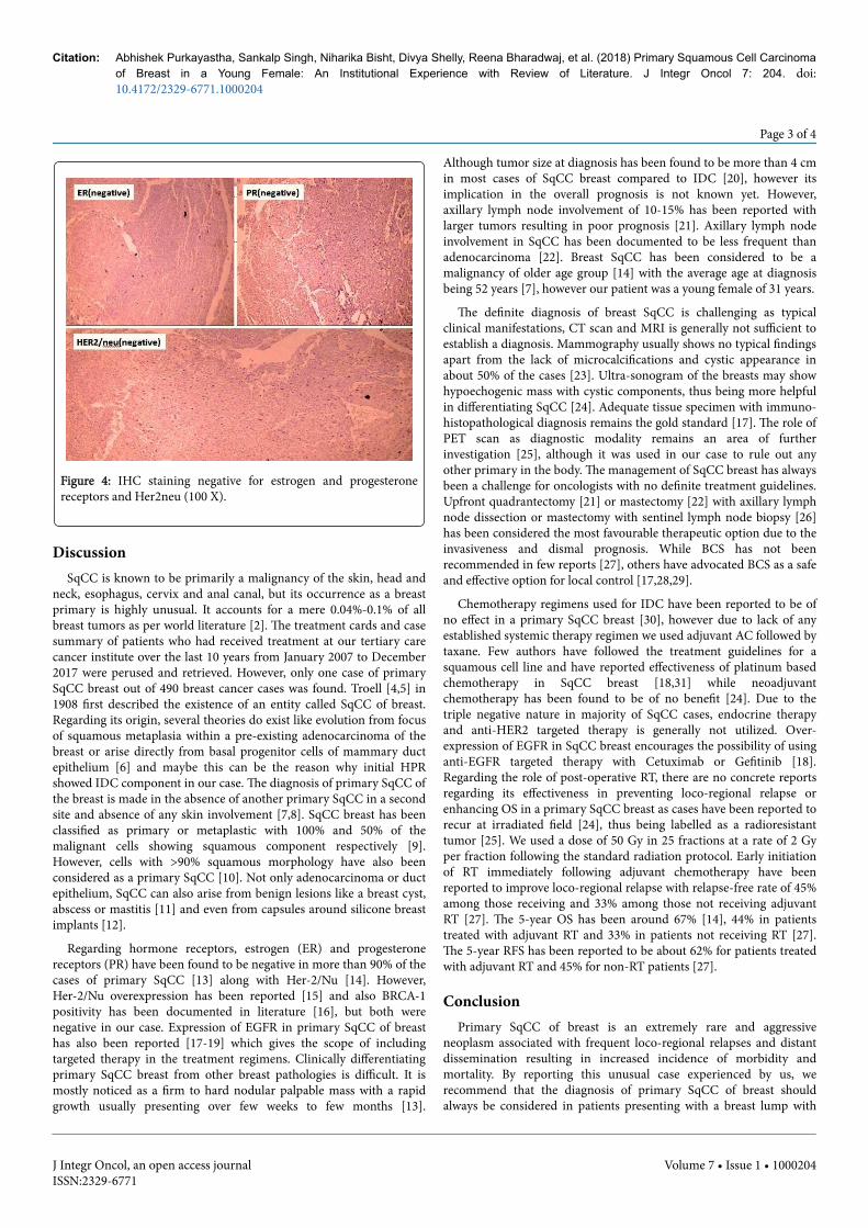
Figure 4: IHC staining negative for estrogen and progesteronereceptors and Her2neu (100 X).
DiscussionSqCC is known to be primarily a malignancy of the skin, head and
neck, esophagus, cervix and anal canal, but its occurrence as a breastprimary is highly unusual. It accounts for a mere 0.04%-0.1% of allbreast tumors as per world literature [2]. The treatment cards and casesummary of patients who had received treatment at our tertiary carecancer institute over the last 10 years from January 2007 to December2017 were perused and retrieved. However, only one case of primarySqCC breast out of 490 breast cancer cases was found. Troell [4,5] in1908 first described the existence of an entity called SqCC of breast.Regarding its origin, several theories do exist like evolution from focusof squamous metaplasia within a pre-existing adenocarcinoma of thebreast or arise directly from basal progenitor cells of mammary ductepithelium [6] and maybe this can be the reason why initial HPRshowed IDC component in our case. The diagnosis of primary SqCC ofthe breast is made in the absence of another primary SqCC in a secondsite and absence of any skin involvement [7,8]. SqCC breast has beenclassified as primary or metaplastic with 100% and 50% of themalignant cells showing squamous component respectively [9].However, cells with >90% squamous morphology have also beenconsidered as a primary SqCC [10]. Not only adenocarcinoma or ductepithelium, SqCC can also arise from benign lesions like a breast cyst,abscess or mastitis [11] and even from capsules around silicone breastimplants [12].
Regarding hormone receptors, estrogen (ER) and progesteronereceptors (PR) have been found to be negative in more than 90% of thecases of primary SqCC [13] along with Her-2/Nu [14]. However,Her-2/Nu overexpression has been reported [15] and also BRCA-1positivity has been documented in literature [16], but both werenegative in our case. Expression of EGFR in primary SqCC of breasthas also been reported [17-19] which gives the scope of includingtargeted therapy in the treatment regimens. Clinically differentiatingprimary SqCC breast from other breast pathologies is difficult. It ismostly noticed as a firm to hard nodular palpable mass with a rapidgrowth usually presenting over few weeks to few months [13].
Although tumor size at diagnosis has been found to be more than 4 cmin most cases of SqCC breast compared to IDC [20], however itsimplication in the overall prognosis is not known yet. However,axillary lymph node involvement of 10-15% has been reported withlarger tumors resulting in poor prognosis [21]. Axillary lymph nodeinvolvement in SqCC has been documented to be less frequent thanadenocarcinoma [22]. Breast SqCC has been considered to be amalignancy of older age group [14] with the average age at diagnosisbeing 52 years [7], however our patient was a young female of 31 years.
The definite diagnosis of breast SqCC is challenging as typicalclinical manifestations, CT scan and MRI is generally not sufficient toestablish a diagnosis. Mammography usually shows no typical findingsapart from the lack of microcalcifications and cystic appearance inabout 50% of the cases [23]. Ultra-sonogram of the breasts may showhypoechogenic mass with cystic components, thus being more helpfulin differentiating SqCC [24]. Adequate tissue specimen with immuno-histopathological diagnosis remains the gold standard [17]. The role ofPET scan as diagnostic modality remains an area of furtherinvestigation [25], although it was used in our case to rule out anyother primary in the body. The management of SqCC breast has alwaysbeen a challenge for oncologists with no definite treatment guidelines.Upfront quadrantectomy [21] or mastectomy [22] with axillary lymphnode dissection or mastectomy with sentinel lymph node biopsy [26]has been considered the most favourable therapeutic option due to theinvasiveness and dismal prognosis. While BCS has not beenrecommended in few reports [27], others have advocated BCS as a safeand effective option for local control [17,28,29].
Chemotherapy regimens used for IDC have been reported to be ofno effect in a primary SqCC breast [30], however due to lack of anyestablished systemic therapy regimen we used adjuvant AC followed bytaxane. Few authors have followed the treatment guidelines for asquamous cell line and have reported effectiveness of platinum basedchemotherapy in SqCC breast [18,31] while neoadjuvantchemotherapy has been found to be of no benefit [24]. Due to thetriple negative nature in majority of SqCC cases, endocrine therapyand anti-HER2 targeted therapy is generally not utilized. Over-expression of EGFR in SqCC breast encourages the possibility of usinganti-EGFR targeted therapy with Cetuximab or Gefitinib [18].Regarding the role of post-operative RT, there are no concrete reportsregarding its effectiveness in preventing loco-regional relapse orenhancing OS in a primary SqCC breast as cases have been reported torecur at irradiated field [24], thus being labelled as a radioresistanttumor [25]. We used a dose of 50 Gy in 25 fractions at a rate of 2 Gyper fraction following the standard radiation protocol. Early initiationof RT immediately following adjuvant chemotherapy have beenreported to improve loco-regional relapse with relapse-free rate of 45%among those receiving and 33% among those not receiving adjuvantRT [27]. The 5-year OS has been around 67% [14], 44% in patientstreated with adjuvant RT and 33% in patients not receiving RT [27].The 5-year RFS has been reported to be about 62% for patients treatedwith adjuvant RT and 45% for non-RT patients [27].
ConclusionPrimary SqCC of breast is an extremely rare and aggressive
neoplasm associated with frequent loco-regional relapses and distantdissemination resulting in increased incidence of morbidity andmortality. By reporting this unusual case experienced by us, werecommend that the diagnosis of primary SqCC of breast shouldalways be considered in patients presenting with a breast lump with
Citation: Abhishek Purkayastha, Sankalp Singh, Niharika Bisht, Divya Shelly, Reena Bharadwaj, et al. (2018) Primary Squamous Cell Carcinomaof Breast in a Young Female: An Institutional Experience with Review of Literature. J Integr Oncol 7: 204. doi:10.4172/2329-6771.1000204
Page 3 of 4
J Integr Oncol, an open access journalISSN:2329-6771
Volume 7 • Issue 1 • 1000204
supporting IHC differentiating it from other common breastpathologies so as to initiate an appropriate management strategy.Presently there are no definite treatment guidelines for this disease perse as most information is based on solitary case reports and small caseseries. Also a better understanding and interpretation of the molecularand biological mechanism of the disease process may help to devicetherapeutic strategies to manage this entity. The role of EGFR andother signalling pathways in this disease needs to be investigatedfurther to establish their role as a potential treatment target. Moresophisticated diagnostic techniques, gene profiling, novel targetedregimens, optimal RT dosages should be devised which can improvethe DFS, OS and RFS of the patients.
AcknowledgementWe thank the patient and her relatives for allowing us to publish her
case.
We also like to thank department of Radiology and NuclearMedicine, Command Hospital (Southern Command), Pune, India.
The manuscript has been read and approved by all the authors, therequirements for authorship have been met, and each author believesthat the manuscript represents honest work.
References1. Rosen PR (2001) Rosen’s Breast Pathology. Philadelphia, PA: Lippincott
Williams & Wilkins 455-461.2. Gupta C, Malani AK, Weigand RT, Rangineni G (2006) Pure primary
squamous cell carcinoma of the breast: A rare presentation andclinicopathologic comparison with usual ductal carcinoma of the breast.Pathol Res Pract 202: 465-469.
3. Mitra B, Pal M, Debnath S, Paul B, Saha TN, et al. (2011) Primarysquamous cell carcinoma of breast with ipsilateral axillary lymph nodemetastasis: An unusual case. Int J Surg Case Rep 2: 194-197.
4. Bhosale SJ, Kshirsagar AY, Deshmukh SJ, Jagtap SV, Langade YB (2013)Squamous cell carcinoma of the breast. Am J Case Rep 14: 188-190.
5. Troell A (1908) Zwei Falle von Palttenepithelcarcinom. Nord Med Ark 1:1-11.
6. Farrand R, Lavigne R, Lokich J, McAuley R, Sparling S, et al (1979)Epidermoid carcinoma of the Breast. J Surg Oncol 12: 207-212.
7. Weigel RJ, Ikeda DM, Nowels KW (1996) Primary squamous cellcarcinoma of the breast. South Med J 89: 511-515.
8. Sheen-Chen S, Chen Y, Chou FF, Eng HL (1992) Primary squamous cellcarcinoma of the breast. South Med J 85: 207-209.
9. Honda M, Saji S, Horiguchi S, Suzuki E, Aruga T, et al. (2011)Clinicopathological analysis of ten patients with metaplastic squamouscell carcinoma of the breast. Surg Today 41: 328-332.
10. Porzio R, Cordini C, Orsi N, Brigati F, Paties CT, et al. (2014) Primarysquamous cell carcinoma of the breast after cured bilateral breast cancer.In Vivo 28: 1155-1158.
11. Murialdo R, Boy D, Musizzano Y, Tixi L, Ballestrero A, et al. (2009)Squamous cell carcinoma of the breast: a case report. Cases J 2: 7336.
12. Kitchen SB, Paletta CE, Shehadi SI, Bauer WC (1994) Epithelialization ofthe lining of a breast implant capsule. Possible origins of squamous cell
carcinoma associated with a breast implant capsule. Cancer 73:1449-1452.
13. Siegelmann-Danieli N, Murphy TJ, Meschter SC, Stein M.E, Prichard J(2005) Primary squamous cell carcinoma of the breast. Clinical of breastcancer 6: 270–272.
14. Behranwala KA, Nasiri N, Abdullah N, Trott PA, Gui GPH (2003)Squamous cell carcinoma of the breast: clinico-pathologic implicationsand outcome. Eur J Surg Oncol 29: 386-389.
15. Karamouzis MV, Fida A, Apostolikas N, Rigatos G (2006) A case of Her-2positive squamous cell breast carcinoma: an unusual presentation of anunusual clinical entity. Eur J Surg Oncol 32: 1250–1251.
16. Breuer A, Kandel M, Fisseler EA, Sutter C, Schwaab E, et al. (2007)BRCA-1 germline mutation in a women with metaplastic squamous cellbreast cancer. Onkologie 30: 316–318.
17. Chen XL, Luo J, Xu FL (2016) Squamous cell carcinoma of the breast:particularity and clinical management. Int J Clin Exp Med 9:14167-14174.
18. Wang J, Zhang X, He J, Yang M, Tang J, et al. (2014) Co-expression ofEGFR and CK5/6 in primary squamous cell carcinoma of the breast. MedOncol 31: 172.
19. Kimura F, Iwaya K, Kawaguchi T, Kaise H, Yamada K, et al. (2010)Epidermal growth factor-dependent enhancement of invasiveness ofsquamous cell carcinoma of the breast. Cancer Sci 101: 1133-1140.
20. Gupta N, Vashisht R, Nimbran V, Gupta R, Dhingra N, et al. (2012)Primary squamous cell carcinoma of the breast: case report andmanagement decisions. J Cancer Res Ther 8: 323-325.
21. Punzo C, Fortarezza F, De Ruvo V, Minafra M, Laforgia R, et al. (2017)Primitive squamous cell carcinoma of the breast (SCCB): case report ofan uncommon variant of metaplastic carcinoma. G Chir 38: 139-142.
22. Menes T, Schachter J, Morgenstern S (2003) Primary squamous cellcarcinoma (SqCC) of the breast. AM J Clin Oncol 26: 571-573.
23. Zoltan B, Lawrence K, Coleman J (2001) Pure squamous cell carcinomaof the breast in a patient with previous adenocarcinoma of the breast: acase report and review of the literature. Am Surg 67: 671-673.
24. Flikweert ER, Hofstee M, Liem M (2008) Squamous cell carcinoma of thebreast: a case report. World J Surg Oncol 6: 135.
25. Healy CF, Feeley L, Leen E, Walsh TN (2006) Primary squamous cellcarcinoma of the breast: value of positron emission tomography scanningin confirming the diagnosis. Clin Breast Cancer 5: 413-415.
26. Lim GH, Acosta HA, Gudi MA (2017) Natural history of metaplasticsquamous cell breast cancer: a case report and literature review onsurgical management. Gland Surg 6: 738-741.
27. Hennessy BT, Krishnamurthy S, Giordano S, Buchholz TA, Kau SW, et al.(2005) Squamous cell carcinoma of the breast. J Clin Oncol 23:7827-7835.
28. Carbone S, Lobo AR, Lamacchia A, Almenar GA, Martin Hernandez R,et al. (2012) Primary squamous cell carcinoma of the breast: A rare casereport. Rep Pract Oncol Radiother 17: 363-366.
29. Zhang X, Zhang B, Zang F, Zhao L, Yuan Z, et al. (2016) Clinical featuresand treatment of squamous cell carcinoma of the breast. Onco targets andTherapy 9: 3181-3185.
30. Rostock RA, Bauer TW, Eggleston JC (1984) Primary squamouscarcinoma of the breast: A review Breast 10: 27-31.
31. Tsung SH (2012) Primary pure squamous cell carcinoma of the breastmight be sensitive to Cisplatin-based chemotherapy. Case Rep Oncol 5:561-565.
Citation: Abhishek Purkayastha, Sankalp Singh, Niharika Bisht, Divya Shelly, Reena Bharadwaj, et al. (2018) Primary Squamous Cell Carcinomaof Breast in a Young Female: An Institutional Experience with Review of Literature. J Integr Oncol 7: 204. doi:10.4172/2329-6771.1000204
Page 4 of 4
J Integr Oncol, an open access journalISSN:2329-6771
Volume 7 • Issue 1 • 1000204