journal of proteomics & bioinformatics open access of proteomics & bioinformatics - open...
TRANSCRIPT

Journal of Proteomics & Bioinformatics - Open Access
JPB/Vol.2/October 2009
J Proteomics Bioinform Volume 2(10) : 455-462 (2009) - 455
ISSN:0974-276X JPB, an open access journal
Research Article OPEN ACCESS Freely available online doi:10.4172/jpb.1000105
Abstract
Background: We have earlier shown that exposure of
human endothelial cell line EA.hy926 to 900 MHz GSM
mobile phone radiation causes changes in the expression
of numerous proteins. Here, we have examined the effects
of 1800 MHz GSM mobile phone signal on the proteome
of the same cell line.
Results: EA.hy926 cells were exposed for one hour to
1800 MHz GSM signal, simulating mobile phone talking
conditions, at an average specific absorption rate (SAR)
of 2.0 W/kg at 37±0.3°C. Sham samples were produced
simultaneously in the same conditions but without the
radiation exposure. Cells were harvested immediately after
1-hour exposure to the radiation, and proteins were
extracted and separated using 2-dimensional
electrophoresis (2DE). In total, 10 experimental replicates
were generated from both exposed and sham samples.
About 900 protein spots were detected in the 2DE-gels
using PDQuest software and eight of them were found to
be differentially expressed in exposed cells (p<0.05, t-test).
Three out of these eight proteins were identified using
Maldi-ToF mass spectrometry (MS). These proteins are:
spermidine synthase (SRM), 78 kDa glucose-regulated
protein (55 kDa fragment) (GRP78) and proteasome
subunit alpha type 1 (PSA1). Due to the lack of the
availability of commercial antibodies we were able to
further examine expression of only GRP78. Using SDS-
PAGE and western blot method we were not able to confirm
the result obtained for GRP78 using 2DE. Additionally,
we have not seen any effect of 1800GSM exposure on the
expression of vimentin and Hsp27 - proteins that were
affected by the 900 MHz GSM exposure in our earlier
studies.
Conclusions: Our results suggest that the 900GSM and
1800GSM exposures might affect the expression of some
proteins in the EA.hy926 cell line. The observed here
discrepancy between the expression changes of GRP78
detected with 1DE and 2DE confirms the importance of
validation of the results obtained with 2DE using other
methods, e.g. western blot.
*Corresponding author: Dariusz Leszczynski, PhD, STUK-Radiation
and Nuclear Safety Authority, Laippatie 4, 00880 Helsinki, Finland,
E-mail: [email protected]
Received September 22, 2009; Accepted October 26, 2009; Published
October 26, 2009
Citation: Nylund R, Tammio H, Kuster N, Leszczynski D (2009) Proteomic
Analysis of the Response of Human Endothelial Cell Line EA.hy926 to
1800 GSM Mobile Phone Radiation. J Proteomics Bioinform 2: 455-462.
doi:10.4172/jpb.1000105
Copyright: © 2009 Nylund R, et al. This is an open-access article
distributed under the terms of the Creative Commons Attribution License,
which permits unrestricted use, distribution, and reproduction in any
medium, provided the original author and source are credited.
Proteomic Analysis of the Response of Human EndothelialCell Line EA.hy926 to 1800 GSM Mobile Phone Radiation
Reetta Nylund1, Hanna Tammio1, Niels Kuster2, Dariusz Leszczynski1,*
1STUK - Radiation and Nuclear Safety Authority, Helsinki, Finland2IT’IS Foundation, Swiss Federal Institute of Technology, Zurich, Switzerland
Abbreviations: 2DE: Two-dimensional electrophoresis;
CHAPS: 3-[(3- Cholamidopropyl)dimethylammonio]-1-
propanesulfonate; Da: Dalton; ddH2O: Double distilled water;
DMEM: Dulbecco’s Modified Eagle’s Medium; DTT:
Dithioreitol; EA.hy926: Human endothelial cell line; ECL
Enhanced chemiluminescence; GSM: Global System for Mobile
Communications; HAT: (mixture of) sodium hypoxanthine,
aminopterin, and thymidine; HRP: Horseradish peroxidase; IAA:
Iodoacetamide; IEF: Isoelectric focusing; IPG: Immobilized pH
gradient; LR: Linear-reflectron; MALDI-TOF: Matrix-assisted
laser desorption/ionization time of flight; MS: Mass
spectrometry/ mass spectrometer; NH4HCO
3:
Ammoniumbicarbonate; PAGE: Polyacrylamide gel
electrophoresis; PBS: Phosphate buffered saline; pI: Isoelectric
point; PMF: Peptide mass fingerprint; PMSF:
Phenylmethylsulphonyl fluoride; PVDF: Polyvinylidene
Fluoride; RF-EMF: Radiofrequency modulated electromagnetic
field; SAR: Specific absorption rate; SDS: Sodium dodecyl
sulphate; Tris-HCl: Tris(hydroxymethyl)aminomethane
hydrochloride; Versene: Chelating agent containing EDTA
Background
The use of mobile phones has widely increased over the past
decade. However, the issue of potential health effects induced
by mobile phone radiation remains controversial and further
research is needed to fill-up the existing gaps in the knowledge
about the biological and physiological effects of this low-level
energy radiation.
We have proposed that the use of high-throughput screening
techniques of transcriptomics and proteomics, as tools to find
genes and proteins responding to mobile phone radiation, might
help the process of finding out whether mobile phone radiation
might cause any health risk (Leszczynski and Joenväärä, 2001;
Leszczynski, 2006; Leszczynski and Meltz, 2006). Proteomics
approach has been so far used only in a few in vitro studies
(Leszczynski et al., 2002; Leszczynski et al., 2004; Nylund and
Leszczynski, 2004; Nylund and Leszczynski, 2006; Zeng et al.,
2006; Li et al., 2007) and in a single in vivo human volunteer
study (Karinen et al., 2008). Such a small number of published
studies does not allow for making any generalized conclusions
about of the possible effects of mobile phone exposures on the
cell proteome and on the cell physiology. Only by performing
more of this kind of studies, the proteomic database can be
Journal of Proteomics & Bioinformatics - Open Access
JPB/Vol.2/October 2009
J Proteomics Bioinform Volume 2(10) : 455-462 (2009) - 456
ISSN:0974-276X JPB, an open access journal
expanded and, with the help of that, the impact of mobile phone
radiation on cell proteome will be possible to assess.
We have previously determined that the 900 MHz GSM mobile
phone radiation signal alters expression of several tens of proteins
in the human endothelial cell line EA.hy926 (Leszczynski, et
al., 2002; Nylund and Leszczynski, 2004; Nylund and
Leszczynski, 2006). In the present study we have examined
whether the 1800 MHz GSM mobile phone radiation signal
exposure will also affect protein expression in EA.hy926 cells.
Protein expression was determined using 2DE proteomics and
results were compared with the earlier study that used 900 MHz
GSM mobile phone radiation.
Materials and Methods
In Vitro Cell Model and Cell Culture Conditions
Brain capillary endothelial cells are one of the potential tar-
gets of the mobile phone radiation. In some animal studies it
has been shown that mobile phone radiation might affect func-
tion of the blood-brain barrier. That is why we have selected to
examine in vitro effects of mobile phone radiation on endothe-
lial cells. Human endothelial cell line EA.hy926 was selected
because of the uniformity of cell cultures from batch to batch
and because of easy and fast means to generate large quantities
of cells for experiments. Neither of the above is possible to
achieve with primary endothelial cells, known for slow growth
and for the variability between batches isolated from different
human donors.
Human endothelial cell line EA.hy926 (a gift from Dr. Cora-
Jean S. Edgell North Carolina University at Chapel Hill, NC,
USA) was grown in Dulbecco’s MEM (DMEM), supplemented
with antibiotics, 10% foetal bovine serum, L-glutamine and HAT-
supplement (Sigma, USA). For the mobile phone radiation
experiments, cells were removed from culture flasks by brief
trypsinization, washed in cell culture medium and seeded at a
density of 0.4x10⁶cells/dish in 35 mm-diameter Petri dishes
(NUNC, Denmark). After an overnight culturing the semi-
confluent monolayers of EA.hy926 were exposed to mobile
phone radiation or sham exposed.
Exposure to Mobile Phone Radiation Signal
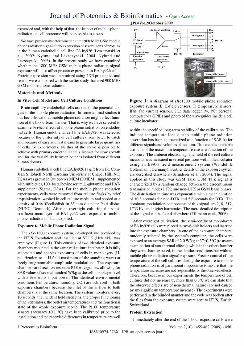
The sXc-1800 exposure system, developed and provided by
the IT’IS Foundation and installed at STUK (Helsinki), was
employed (Figure 1). This consists of two identical exposure
chambers mounted in the same cell culture incubator. It is fully
automated and enables exposures of cells in monolayers (H-
polarization or at H-field maximum of the standing wave) at
freely programmable amplitude modulations. The exposure
chambers are based on resonant R18 waveguides, allowing for
SAR values of several hundred W/kg at the cell monolayer level
with a few watts input power. The identical environmental
conditions (temperature, humidity, CO2) are achieved in both
exposure chambers because the inlet of the airflow to both
chambers is at the same location. The system monitors, every
10 seconds, the incident field strengths, the proper functioning
of the ventilators, the outlet air temperatures and the functional
state of the whole exposure set-up. The Pt100 temperature
sensors (accuracy ±0.1 °C) have been calibrated prior to the
installation and the recorded differences in temperature are well
within the specified long-term stability of the calibration. The
induced temperature load due to mobile phone radiation
absorption has been characterized as a function of SAR (t) for
different signals and volumes of medium. This enables a reliable
estimate of the maximum temperature rise as a function of the
exposure. The ambient electromagnetic field of the cell culture
incubator was measured in several positions within the incubator
using an EFA-3 field measurement system (Wandel &
Goltermann, Germany). Further details of the exposure system
are described elsewhere (Schuderer et al., 2004). The signal
applied in this study was GSM Talk. GSM Talk signal is
characterized by a random change between the discontinuous
transmission mode (DTX) and non-DTX or GSM Basic phases.
The distribution in time was exponential with a mean duration
of 10.8 seconds for non-DTX and 5.6 seconds for DTX. The
dominant modulation components of this signal are 2, 8, 217,
1733 Hz, and higher harmonics. The more detailed description
of the signal can be found elsewhere (Tillmann et al., 2006).
After overnight cultivation, the semi-confluent monolayers
of EA.hy926 cells were placed in two 6-dish holders and inserted
into the exposure chambers. In one of the exposure chambers,
randomly selected by the system’s computer, the cells were
exposed to an average SAR of 2.0 W/kg at 37±0.3°C (to assure
examination of non-thermal effects), while in the other chamber
they were sham-exposed, in the similar conditions but without
mobile phone radiation signal exposure. Precise control of the
temperature of the cell cultures during the exposure to mobile
phone radiation is of paramount importance to assure that the
temperature increases are not responsible for the observed effects.
Therefore, because in our experiments the temperature of cell
cultures did not increase by more than 0.3oC we can state that
the observed effects are of non-thermal nature (are not caused
by any significant temperature increase). The experiments were
performed in the blinded manner and the code was broken after
the files from the exposure system were sent to IT’IS, Zurich,
Switzerland.
Protein Extraction
Immediately after the end of the 1-hour exposure cells were
Figure 1: A diagram of sXc1800 mobile phone radiation
exposure system (E: E-field sensors, T: temperarure sensors,
Ifan: fan current sensors, DL: data logger i/o, PC: personal
computer via GPIB) and photo of the waveguides inside a cell
culture incubator.
RF signalgenerator
Functiongenerator
PC
GSM burst
PC
AM
DL, PC
5W linear amplifier
GSM framegenerator
Switch
blank
DL, PC
Waveguide
Waveguide
Incubator
DL, PC
Ifan
Ifan
T
T
E
E
Waveguide (open)
Waveguide (with cover)

Journal of Proteomics & Bioinformatics - Open Access
JPB/Vol.2/October 2009
J Proteomics Bioinform Volume 2(10) : 455-462 (2009) - 457
ISSN:0974-276X JPB, an open access journal
quickly washed with PBS and harvested with versene. Proteins
were extracted with a buffer consisting of 8 M Urea, 1 M
Thiourea, 4% Chaps, 10 mM DTT, 2% IPG buffer pH 4-7, 1
mM sodium orthovanadate and 1 mM PMSF. Protein
concentrations were measured using Bradford method. The
250µg of total protein was used for two-dimensional gel
electrophoresis (2DE).
2DE
The isoelectric focusing was performed using an IPGphor
apparatus (GE Healthcare, USA) and 24 cm long ready IEF strips
pH 4-7 (GE Healthcare). The samples were loaded using in-gel
rehydration in a buffer containing 9 M Urea, 2% Chaps, 0.2%
DTT, 0.5% IPG buffer pH 4-7 for 4 hours. IEF was run at 20°C
using step-and-hold methods as follows: 50 V 8 h; 100 V 1 h;
500 V 1 h; 1000 V 1 h; 2000 V 1 h; 8000 V until 95000 Vhrs
were achieved. Before SDS-PAGE the IEF strips were
equilibrated for 15 min with 6 M urea, 30% glycerol, 50 mM
Tris-HCl, 2% SDS, and 10 mg/mL DTT and then for another 15
min in the same buffer, in which DTT was replaced by 25 mg/
mL iodoacetamide (IAA). SDS-PAGE was run in 10% gel using
Ettan DALTsix Electrophoresis system (GE Healthcare) at the
constant power setting of 3.5W/gel for the first 0.5 hours and
then 13W/gel. After electrophoresis the gels were silver stained.
Gels were fixed (30% ethanol, 0.5% acetic acid), washed with
20% ethanol and ddH2O, sensitized with sodium thiosulfate (0.2
g/L), incubated in the silver nitrate solution (2 g/L) and developed
(potassium anhydride 30 g/L, 37% formaldehyde 0.7 mL/L,
sodium thiosulfate 0.01 g/L). The development was stopped with
Tris 50 g/L + 0.5% acetic acid, and then the gels were washed
twice with ddH2O and scanned.
Data Analysis
The silver stained gels were scanned using GS-710
densitometer (Bio-Rad, USA) and analyzed using PDQuest 7.2
software (Bio-Rad). In total, ten gels from both sham and
exposed samples were analysed. The normalized spot volumes
of the proteins from sham and exposed sample gels were
statistically analyzed using student t-test at the confidence level
of 95%. Protein spots, that visually appeared as technical
artefacts (e.g. background areas of silver staining, irregular-
shaped dust particles, air bubbles) but were erroneously detected
by the software, were manually removed from the analysis.
In-gel Digestions for Mass Spectrometry Protein
Identification
Proteins of interest were extracted from several gels and in-
gel digested. Before digestion the proteins were reduced with
20 mM DTT in 0.1M ammonium-bi-carbonate (NH4HCO
3) and
alkylated with 55 mM IAA in NH4HCO
3. Proteins were digested
overnight at +37°C with modified trypsin (sequencing grade
modified trypsin, porcine, Promega, USA) in 50 mM NH4HCO
3.
After overnight digestion, resulting peptides were extracted from
gels with 25 mM NH4HCO
3 and twice with 5% formic acid.
Peptides were concentrated and de-salted using C-18 ZipTips
(Millipore, USA) according to the manufacturer’s instructions
with the exception of elution solution (60% acetonitrile).
Mass Spectrometry Identification of Proteins
Tryptic digestions were mixed 1:1 with α-cyano-4-
hydroxycinnamic acid matrix and analyzed with MALDI-TOF-
LR-MS (Waters, USA) operating in a positive ion reflectron
mode. The mass spectra were externally calibrated with ACTH
clip 18-39 (MW 2465.199 Da, Sigma, USA) and internally
calibrated with trypsin autolysis peaks (1045.564/2211.108 Da).
The peptide mass fingerprints for protein identification were
searched automatically at the accuracy of 20-50ppm from
UniProt database with ProteinLynx-software (Waters) operating
along the instrument. Statistically significantly affected proteins
were also searched manually using Matrix Science Mascot
Peptide Mass Fingerprint search tool (www.matrixscience.com).
Western Blotting
Immediately after the end of the RF-EMF exposure the cells
were washed with PBS and harvested with versene. Proteins
were extracted with 2% SDS, 1% protease inhibitor cocktail
(Sigma, USA). Protein concentrations were measured using
Lowry method (Bio-Rad). In total, five replicates were produced.
Proteins were separated on 7.5% (GRP78) or 10% (Hsp27,
Vimentin) 1D SDS-PAGE and blotted on a PVDF-membrane,
blocked with 2% non-fat dry milk, and exposed to primary
antibody. The polyclonal Bip (GRP78, Cell Signalling
Technology, USA), monoclonal Hsp27 (StressGene, Canada),
and vimentin (Zymed, USA) antibodies were used. The
respective secondary antibody containing a horseradish
peroxidase (HRP)-conjugate (Dako, Denmark) was used. The
signal was detected using enhanced chemiluminescence (ECL)
(Millipore, USA). Autoradiography films were scanned with GS-
710 densitometer (Bio-Rad) and analysed with Phoretix software
(Molecular Probes, USA).
Results and Discussion
In this study we have examined protein expression levels in
EA.hy926 cells after the exposure to 1800 MHz GSM mobile
phone radiation. Protein expression pattern of EA.hy926 cells
was analysed using 2DE with the pH range of 4 - 7 and the gel
percentage of 10%, allowing a good separation at the molecular
weight (MW) range of approximately 15-150 kDa. In total, 10
replicates were generated from both exposed and sham samples.
Such high number of replicates is necessary in order to diminish
technical and biological variability, when using silver staining
technique to visualize proteins in 2DE gels.
Using PDQuest 7.2 software, about 900 protein spots were
detected in the gels. Protein spots, that visually appeared as
technical artefacts but were detected by the software, were
manually removed from the analysis. Statistical significance of
the observed differences in proteins expression levels was
determined using student t-test, at the confidence level of 95%,
with the assumption of the independent samples. The analysis
has revealed eight protein spots which were found to be
differentially expressed (p<0.05) (Figure 2). Expression of the
four of the proteins was found to be down-regulated and four
up-regulated by the mobile phone radiation exposure. Down-
regulation ratios varied between 0.33-0.47 and up-regulation
ratios varied from 1.47 to 2.46.
Comparison of the changes in protein expression pattern
observed here and in the earlier study (Nylund and Leszczynski,
2004), shows that exposure to 900 MHz GSM signal has caused
expression changes in a larger number of proteins spots and the

Journal of Proteomics & Bioinformatics - Open Access
JPB/Vol.2/October 2009
J Proteomics Bioinform Volume 2(10) : 455-462 (2009) - 458
ISSN:0974-276X JPB, an open access journal
Figure 2: A representative gel image showing protein spots with altering expression levels and histograms showing average expression
levels and standard deviations of the sham and exposed samples as well as ratio between RF and sham exposed sample (ratio >1
describes up-regulation and ratio <1 down-regulation of the protein). Also t-test p-values are shown.
Figure 3: A gel image showing the identified protein spots in the EA.hy926 2DE map.
pH 4 7MW250
150
100
75
50
37
25
20
M6PBPC1
AHSA1
STOML2SAKS1
MEP50TMOD3
CCT5
PDIA3
ACTBRPSA
GSTP1
GRP75
VCL
PDC6I
TBB2CVIME
PSMC6
EIF3I
HSP7C frag
TPM3 CLIC1
HSP27
ST1A3
TBB2C
PHB
STRAP
VIME fragGRP78 frag
GDIR1
PNPH
PRDX6
RBBP4
RAD23
HSP60
TBA1C
GDIR2
PSA6
TPIS
GSTO1
LDHBKPYM frag.
CALR
CALU
PSA1
GRP78 frag
SRM
PSMC2RUVB2
KRT8KRT7
NPM
EF1D
ENPL
CCT1
HSP27
VIME fragACTG1
HSP60

Journal of Proteomics & Bioinformatics - Open Access
JPB/Vol.2/October 2009
J Proteomics Bioinform Volume 2(10) : 455-462 (2009) - 459
ISSN:0974-276X JPB, an open access journal
Gene name Protein name Access
code
Sequence
coverage (%)
MW(kDa)/pI
theoretical
MW(kDa)/pI
measured
ACTB Actin, cytoplasmic 1 P60709 31.5 41.7/5.4 43/5.4
ACTG1 Actin, cytoplasmic 2 P63261 40 41.8/5.4 43/5.5
AHSA1 Activator of 90 kDa heat shock
protein ATPase homolog 1, p38
O95433 72.8 38.3/5.5 40/5.8
CALR Calreticulin precursor P27797 49 48.1/4.3 58/4.3
CALU Calumenin O43852 45 37.1/4.5 44/4.5
CLIC1 Chloride intracellular channel
protein 1
O00299 46.5 26.9/5.1 29/5.3
CCT1/
TCPA
T-complex protein 1 subunit alpha P17987 60.3 60.3/6.0 60/6.3
CCT5/
TCPE
T-complex protein 1 subunit
epsilon
P48643 48.2 59.6/5.6 60/5.9
EF1D Elongation factor 1-delta P29692 44.8 31.1/4.9 36/5.2
EIF3I Eukaryotic translation initiation
factor 3 subunit I
Q13347 23 36.5/5.4 37/5.8
ENPL Endoplasmin precursor P14625 27.5 92.4/4.8 120/4.9
GDIR1 Rho GDP-dissociation inhibitor 1 P52565 52.5 23.2/5.0 26/5.2
GDIR2/
ARHGDIB
Rho GDP-dissociation inhibitor 2 P52566 35.3 23.0/5.1 25/5.3
GRP75 Stress-70 protein, mitochondrial
(Precursor)
P38646 56 73.6/6.1 74/5.8
GRP78
(frag.)
78kDa glucose-regulated protein
(Precursor) (frag)
P11021 26 72.4/5.1 54/5.9
GRP78
(frag.)
78kDa glucose-regulated protein
(Precursor) (frag)
P11021 33.6 72.4/5.1 48/4.8
GSTO1 Glutathione transferase omega-1 P78417 45.6 27.5/6.6 27/6.5
GSTP1 Glutathione S-transferase P P09211 53.8 23.3/5.5 23/5.9
HSP27 Heat shock protein beta-1 P04792 48.3 22.8/6.3 26/5.9
HSP27 Heat shock protein beta-1 P04792 37.1 22.8/6.3 26/6.4
HSP60 60 kDa heat shock protein P10809 51 61.0/5.8 61/5.6
HSP60 60 kDa heat shock protein P10809 52.7 61.0/5.8 61/5.4
HSP7C
frag.
Heat shock cognate 71 kDa protein
(frag)
P11142 25.7 71.2/5.4 40/5.1
KPYM frag. Pyruvate kinase isozymes M1/M2
(frag)
P14618 46.5 58.0/8.2 36/6.3
KRT7 Keratin, type II cytoskeletal 7 P08729 64.8 51.4/5.6 54/5.8
KRT8 Keratin, type II cytoskeletal 8 P05787 57.1 53.7/5.6 54/5.9
LDHB L-lactate dehydrogenase B chain P07195 46.7 36.5/6.0 36/6.1
M6PBP1C mannose-6-phosphate receptor
binding protein 1C
O60664 64.3 47.0/5.4 48/5.4
MEP50 Methylosome protein 50 Q9BQA1 31.6 36.7/5.1 41/5.3
NPM Nucleophosmin P06748 44.6 32.5/4.7 37/4.9
PDCD6IP Programmed cell death 6-
interacting protein
Q8WUM4 58.8 96.0/6.4 105/6.8
PDIA3 Protein disulfide-isomerase A3
(Precursor)
P30101 49.5 56.7/6.3 56/6.2
PHB Prohibitin P35232 42.6 29.8/5.7 28/5.8
PNPH Purine nucleoside phosphorylase P00491 59.5 32.1/6.9 30/6.9
PRDX6 Peroxiredoxin-6 P30041 48.7 25.0/6.3 25/6.8
PSA1 Proteasome subunit α type 1 P25786 27 29.5/6.6 28/6.8
PSA6 Proteasome subunit α type 6 P60900 52.8 27.4/6.7 26/6.8
PSMC3/
PRS6A
26S protease regulatory subunit 6A P17980 85 49.2/5.2 49/5.3
PSMC2/
PRS7
26S protease regulatory subunit 7 P35998 53.6 48.6/5.9 48/6.2
RPSA 40S ribosomal prot SA P08865 31.2 32.9/4.8 40/4.8
RAD23 UV excision repair protein RAD23
homolog B
P54727 27.1 43.1/4.8 57/4.9

Journal of Proteomics & Bioinformatics - Open Access
JPB/Vol.2/October 2009
J Proteomics Bioinform Volume 2(10) : 455-462 (2009) - 460
ISSN:0974-276X JPB, an open access journal
RBBP4 Histone-binding protein BBP4 Q09028 31.1 47.7/4.8 53/4.8
RUVB2 RuvB-like 2 Q9Y230 69.1 51.1/5.6 52/5.9
SAKS1 SAPK substrate protein 1 Q04323 50.5 33.3/5.3 39/5.4
SRM Spermidine synthase P19623 19.9 33.8/5.4 33/5.4
ST1A3 Sulfotransferase 1A3/1A4 P50224 52.6 36.4/5.8 35/6.0
STOML2 Stomatin-like protein 2 Q9UJZ1 46.6 38.5/6.9 40/5.7
STRAP Serine-threonine kinase receptor-associated protein Q9Y3F4 56.3 38.4/5.0 38/5.2
TBA1C Tubulin alpha-1C chain Q9BQE3 44.5 49.9/5.0 58/5.3
TBB2C Tubulin beta-2C chain P68371 50.1 49.8/4.8 51/5.1
TBB2C (frag) Tubulin beta-2C chain (frag) P68371 35.3 49.8/4.8 36/5.8
TMOD3 Tropomodulin3 Q9NYL9 36.9 39.6/5.1 40/5.4
TPIS Triosephosphate isomerase P60174 80 26.7/6.9 24/6.6
TPM3 Tropomyosin 3 Q5VU58 69 29.2/4.8 30/4.8
VIME Vimentin P08670 78 53.6/5.1 54/5.3
VIME Vimentin (fragment) P08670 51.1 53.6/5.1 49/4.9
VIME Vimentin (fragment) P08670 66.3 53.6/5.1 47/4.8
VCL Vinculin P18206 32.8 123.7/5.6 130/6.5
Table 1: All proteins that were identified by MS in EA.hy926 2DE gels.
spot # Expression
(exposed/sham)
Protein name Access code Sequence
coverage (%)
Mascot score
4 down SRM P19623 19.9 74
5 up GRP78 fragment P11021 26 101
7 down PSA1 P25786 27 111
Table 2: Identified proteins that altered their expression after exposure to 1800 MHz GSM radiation.
changes induced by both exposures were detected in different
proteins spots. Previously, using 900 MHz GSM signal, total of
38 protein spots were found to be affected after the mobile phone
exposure (Nylund and Leszczynski, 2004), out of which 28 was
in the pH range of 4 - 7, as compared with 8 proteins spots that
were found here to be statistically significantly affected by 1800
MHz GSM exposure in the same pH range. The number of
statistically significantly affected proteins is small (below the
number of expected false positives). However, it is possible that
some of these proteins might indeed be responding to mobile
phone radiation. As shown in our earlier study (Nylund and
Leszczynski, 2004), the number of statistically significantly
affected proteins might be lower than the expected number of
false positives but further analysis using western blot might show
that some of the affected proteins (in that particular study -
vimentin), might indeed respond to the mobile phone radiation.
Using peptide mass fingerprint (PMF) technique and Maldi-
ToF MS, total of 50 protein spots were identified in 2DE gels of
EA.hy926 exposed to 1800 MHz GSM mobile phone radiation
(Figure 3; Table 1). Among the identified proteins were proteins
that we have shown earlier to be affected by 900 MHz GSM
radiation: vimentin and Hsp27 (Leszczynski et al., 2002; Nylund
and Leszczynski, 2004). Expression of neither of them was
altered in a statistically significant manner in 2DE by 1800 MHz
GSM radiation (not shown).
Among the 50 identified protein spots were 8 proteins that
expression was statistically significantly affected by 1800 MHz
GSM radiation. Three of these eight protein spots were
successfully identified (Table 2):
• spot #4 - spermidine synthase (P19623 SRM) (Wahlfors et
al., 1990), regulates amine and bioamine biosynthesis,
• spot #5 - 78 kDa glucose regulated protein (fragment)
(P11021 GRP78) (Ting and Lee, 1988), member of the heat
shock protein 70 family, facilitates the assembly of
multimeric protein complexes inside the endoplasmic
reticulum. The molecular weight of this protein 72.4 kDa,
while the affected protein spot observed here was only a
fragment of ca. 55 kDa.
• spot #7 - proteasome subunit alpha type 1 (P25786 PSA1)
(Silva-Pereira et al., 1992), is a part of large proteasome
complex.
Identification of the other five proteins spots with Maldi-ToF
was not successful due to low amount of protein in the spots.
Using western blot technique we have attempted to confirm
the 2DE results for some of the proteins. Expression changes of
GRP78 were examined using polyclonal antibody (Bip/GRP78,
Cell Signalling Technology). Two protein bands were detected
with MW of 75 kDa (represents the whole protein) and 55 kDa
(represents GRP78 fragment identified from our 2DE gels).
However, neither of the protein bands appeared to be affected
by radiation exposure (Figure 4A). Thus, the western blot
technique did not confirm the results obtained with 2DE. Two
other identified proteins, SRM and PSA1, were not analyzed
using western blot because the corresponding antibodies were
not commercially available. Also the western blot experiments
for vimentin and Hsp27 have shown a lack of effect of 1800
MHz GSM radiation. For vimentin, using the same antibody as
previously (Nylund and Leszczynski, 2004), only a single band
was observed in western blot, while in the earlier study the 900
MHz GSM radiation has caused appearance of an additional
low-molecular weight vimentin band (Nylund and Leszczynski,
2004). For the single vimentin band observed here there was no

Journal of Proteomics & Bioinformatics - Open Access
JPB/Vol.2/October 2009
J Proteomics Bioinform Volume 2(10) : 455-462 (2009) - 461
ISSN:0974-276X JPB, an open access journal
change in the expression following the radiation exposure
(Figure 4B). For Hsp27, the 2DE gel analyses have shown a
statistically non-significant slight increase in the expression but
western blot did not show any difference between Hsp27
expression in sham and exposed cells (Figure 4C).
Future Perspectives
In our previous and in the present study we have used two
common mobile phone frequencies, 900 MHz and 1800 MHz,
to determine if these radiation frequencies could have any impact
on cell proteome. The observed here discrepancy between the
responses of EA.hy926 cells to 1800 MHz GSM radiation and
the previously published responses of EA.hy926 cells to 900
MHz GSM might be caused either by the different exposure
frequencies or by technical differences between the exposure
set-ups or by both of the above. The major difference, besides
the frequency, between the 900 GSM and 1800 MHz GSM
exposure chambers, appears to be the distribution of radiation
field within the cell culture dish. In 900 MHz GSM set-up there
was non-uniform SAR distribution (Leszczynski et al., 2002).
It means that the cells growing in the certain areas of the culture
dish were exposed to much higher SAR (over 5.0 W/kg) as
compared to the average SAR for the whole cell culture dish
(2.4 W/kg) (Leszczynski et al., 2002). In the contrast, the 1800
MHz GSM set-up had very uniform SAR distribution and the
cells throughout the cell culture dish were exposed to the same
level (2.0 W/kg) of radiation. The possibility of the field-
distribution-related effect is supported by our new results
showing that stress kinases are activated by the 1800 MHz
radiation at 5.0 W/kg but not at 2.0 W/kg (manuscript in
preparation). Therefore, there is a need to compare side-by-side
the effects of 900 MHz and 1800 MHz frequencies on protein
expression and on stress response in EA.hy926 cells using
different SAR values.
Summary Conclusions
Our results suggest that the 900 MHz GSM and 1800 MHz
GSM exposures might affect the expression of some proteins in
the EA.hy926 cell line. The observed here discrepancy between
the expression changes of GRP78 detected with 1DE and 2DE
confirms the importance of validation of the results obtained
with 2DE using non-high-throughput methods, as e.g. western
blot. However, one serious limitation of this approach is the
availability of specific antibodies or possession of an animal
facility permitting to produce specific antibodies.
Authors’ Contributions
RN developed the proteomics system used here, performed
all the analyses presented here, and wrote the draft manuscript.
HT performed the 2DE experiments. NK provided the exposure
set-up used here. DL obtained the funding of the study and
coordinated execution of this project and wrote the final version
of the manuscript. All authors have read and approved the final
version of the manuscript.
Acknowledgements
We thank Ms. Pia Kontturi for very skilful assistance in
performing peptide digests for MS as well as for western blots.
We would also like to thank Ms. Marja Huuskonen for the help
in the cell cultivation. The IT’IS personnel (Denis Spät and
Manuel Murbach) we would like to thank for decoding the files
from the exposures. This study was funded by internal funding
from STUK and IT’IS.
References
1. Karinen A, Heinävaara S, Nylund R, Leszczynski D (2008)
Mobile phone radiation might alter protein expression in
human skin. BMC Genomics 11: 77.
2. Leszczynski D (2006) The need for a new approach in studies
of the biological effects of electromagnetic fields. Proteomics
6: 4671-4673.
3. Leszczynski D, Joenväärä S (2001) Proteomics: new way to
determine possible biological effects of mobile phone
radiation. Nature Genetics 27: 67.
Figure 4: Western blots and densitogram bar-graph analyses (mean ± SD) for GRP78 protein (A), vimentin (B) and Hsp27 protein
(C). For all western blots EA.hy926 cells were exposed for one hour to 2.0 W/kg 1800 MHz GSM signal using talk-conditions. The
experiments were repeated five times. S = sham sample; RF = exposed sample.
A
C RF C RF C RF C RF C RF
Grp78
0
100
200
300
400
Sham RF Sham RFMW ca 75 kDa MW ca 55 kDa
Ratios=0.99 & 1.01p=0.93 & 0.94
B
C RF C RF C RF C RF C RF
Vimentin
0
50
100
150
200
Sham RF
Ratio=0.98p=0.85
C
C RF C RF C RF C RF C RF
Hsp27
0
50
100
150
200
250
Sham RF
Ratio=1.1p=0.65
A
C RF C RF C RF C RF C RFC RF C RF C RF C RF C RF
Grp78
0
100
200
300
400
Sham RF Sham RFMW ca 75 kDa MW ca 55 kDa
Ratios=0.99 & 1.01p=0.93 & 0.94
B
C RF C RF C RF C RF C RFC RF C RF C RF C RF C RF
Vimentin
0
50
100
150
200
Sham RF
Ratio=0.98p=0.85
Vimentin
0
50
100
150
200
Sham RF
Ratio=0.98p=0.85
C
C RF C RF C RF C RF C RFC RF C RF C RF C RF C RF
Hsp27
0
50
100
150
200
250
Sham RF
Ratio=1.1p=0.65
» CrossRef » Pubmed» Google Scholar
» CrossRef » Pubmed » Google Scholar

Journal of Proteomics & Bioinformatics - Open Access
JPB/Vol.2/October 2009
J Proteomics Bioinform Volume 2(10) : 455-462 (2009) - 462
ISSN:0974-276X JPB, an open access journal
4. Leszczynski D, Joenväärä S, Reivinen J, Kuokka R (2002)
Non-thermal activation of the hsp27/p38MAPK stress
pathway by mobile phone radiation in human endothelial
cells: Molecular mechanism for cancer- and blood-brain
barrier-related effects. Differentiation 70: 120-129.
5. Leszczynski D, Meltz ML (2006) Questions and answers
concerning applicability of proteomics and transcriptomics
in EMF research. Proteomics 6: 4674-4677.
6. Leszczynski D, Nylund R, Joenväärä S, Reivinen J (2004)
Applicability of discovery science approach to determine
biological effects of mobile phone radiation. Proteomics 4:
426-431.
7. Li HW, Yao K, Jin HY, Sun LX, Lu DQ, et al. (2007)
Proteomic analysis of human lens epithelial cells exposed to
microwaves. Jpn J Ophthalmol 51: 412-416.
8. Nylund R, Leszczynski D (2004) Proteomics analysis of
human endothelial cell line EA.hy926 after exposure to GSM
900 radiation. Proteomics 4: 1359-1365.
9. Nylund R, Leszczynski D (2006) Mobile phone radiation
causes changes in gene and protein expression in human
endothelial cell lines and the response seems to be genome-
and proteome-dependent. Proteomics 6: 4769-4780.
10. Schuderer J, Samaras T, Oesch W, Spät D, Kuster N (2004)
High peak SAR exposure unit with tight exposure and
environmental control for invitro experiments at 1800 MHz.
IEEE Transact Microwave Theor Tech 52: 2057-2066.
11. Silva-Pereira I, Bey F, Coux O, Scherrer K (1992) Two
mRNAs exist for the Hs PROS-30 gene encoding a
component of human prosomes. Gene 120: 235-242.
12. Tillmann T, Ernst H, Ebert S, Kuster N, BehnkeW, et al.
(2006) Carcinogenicity study of GSM and DCS wireless
communication signals in B6C3F1 mice. Bioelectromagnetics
28: 173-187.
13. Ting J, Lee AS (1988) Human gene encoding the 78,000-
dalton glucose-regulated protein and its pseudogene:
structure, conservation, and regulation. DNA 7: 275-286.
14. Wahlfors J, Alhonen L, Kauppinen L, Hyvönen T, Jänne J,
et al. (1990) Human spermidine synthase: cloning and
primary structure. DNA Cell Biol 9: 103-110.
15. Zeng Q, Chen G, Weng Y, Wang L, Chiang H, et al. (2006)
Effects of global system for mobile communications 1800
MHz radiofrequency electromagnetic fields on gene and
protein expression in MCF-7 cells. Proteomics 6: 4732-4738.
» CrossRef » Pubmed » Google Scholar
» CrossRef » Pubmed» Google Scholar
» CrossRef » Pubmed » Google Scholar
» CrossRef » Pubmed» Google Scholar
» CrossRef » Pubmed» Google Scholar
» CrossRef » Pubmed » Google Scholar
» CrossRef » Google Scholar
» Pubmed » Google Scholar
» CrossRef » Pubmed » Google Scholar
» CrossRef » Pubmed » Google Scholar
» CrossRef » Pubmed» Google Scholar
» CrossRef » Pubmed » Google Scholar