journal of usc student dental research
TRANSCRIPT

Journal of USC Student Dental ResearchPremier Publication
2009

page � Explorer JournalSC February �009 page �
February �009 page �
AAs human beings we make observations and express opinions on a daily basis. Often the inter-pretations of our observations and our formed opin-ions are biased by our past experiences. As health care providers our opinions about the management of different oral conditions, as well as utilization of health care products, are sought regularly by patients, friends, family members and colleagues. It is best to give recommend to you all that are sup-ported by solid scientific research.
I have had the good fortune to be surrounded by scholars throughout my professional life. Through collaborative research work with these scholars I have changed my opinions about many things in general and about saliva - spit - in par-ticular. What used to be considered one of the least respected body fluids, today is considered one of the most precious body fluids. Over the past 25 years we have developed and examined the most reproducible methods for saliva collection, defined low saliva flow rates and investigated its impact on the increased prevalence of soft and hard tissue diseases (fungal infections and dental caries). We have also evaluated the impact of different medi-cations and medical conditions, such as Sjogren’s syndrome, HIV infection, and Highly Active Anti Retroviral Therapy (HAART), on salivary gland function and developed a test that identifies indi-viduals at risk for dental caries with significant accuracy.
Our contributions to the literature have brought us national and international visibility, and
the outcomes of our research work have helped to enhance the quality of life for others.The op-portunity to develop ideas and search for answers
to unknowns has been one of the highlights of my academic career and is among the most reward-ing experiences of my life. Developing hypoth-eses, collecting facts, generating evidence and documenting outcomes enable us to challenge our capabilities to the maximum and keep our state of mind alert and engaged. Despite my demanding ad-ministrative activities in recent years, I continue to do research, because it energizes me and keeps my sense of curiosity alive. It is a fascinating experi-ence and I recommended it to you all.
Mahvash Navazesh, D.M.D.Professor and Chair Division of Diagnostic SciencesAssociate Dean Academic Affairs and Student Life.
Our contributions to the literature have brought us national and in-ternational visibility, and the out-comes of our research work have helped to enhance the quality of life for others.
RESEARCH & INNOVATION
Paving the Way for the Future of Student Research at the USC School of Dentistry

page � Explorer JournalSC February �009 page �
RESEARCH & INNOVATION
The Center for Craniofacial Molecular Biology (CCMB) was estab-lished in October of 1989, under the leadership of Dr. Harold C. Slavkin, who later became the sixth director of the National Institute of Dental and Craniofacial Research in 1995. Dr. Slavkin is a world renowned scientist in craniofacial developmental biology and oral health research.
CCMB’s dedication to leadership in craniofacial biology research and steadfast commitment to open access for all continued and expanded dur-ing the tenure of our second director, Dr. Charles Shuler who served from 1995 until 2007. Over the years, CCMB has produced ground-breaking research and trained many scientists who are now leaders in oral health research around the world.
Today, as we honor the past and carry forward a rich legacy, we are well positioned to take advantage of the recent extraordinary advances in biomedical sciences and the inspiring academic environment at USC. Our research scientists are exploring the molecular mechanisms of craniofacial birth defects, such as cleft lip and cleft palate, transcriptional regulation and biomineralization, wound healing and oral cancer biology, and stem cell biology and tissue regeneration. In the coming years, we will continue to contribute to the scientific advancements in human craniofacial health while expanding the research horizon with innovative technologies. We encourage interdisciplinary approaches to further the knowledge that will improve human health. Ultimately, we will create something that is greater than ourselves and seize the opportunity of today to invest in the CCMB of tomorrow.
Yang Chai, DDS, PhD
Message from Dr. Yang Chai Director of the Center for Craniofacial Molecular Biology

February �009 page �
Welcome to the Explorer Journalfrom Dean Harold Slavkin
RESEARCH & INNOVATION
Dear Students, Faculty and Alumni,
We all can be proud of the premiere issue of the USC Journal of Stu-dent Dental Research. Founded by our second year periodontics resident Dr. Shervin Molayem, this journal affirms the work of those students who choose to engage in research during their predoctoral studies as it creates a forum specifically dedicated to publishing their findings.
As you all know, the USC School of Dentistry has established an interna-tional reputation for its research enterprise. It is essential to our mission that we develop future leaders in the field of research who will continue the work of generating new knowledge, promoting oral health, and investigating the cause, prevention, diagnosis and treatment of disease. For many of our students, this effort represents their first foray into the world of research and publication. My hope, and the hope of the many faculty members who mentor them, is that publication in The USC Journal of Student Dental Research marks the beginning of many promising research careers.
The School of Dentistry looks forward with great anticipation to what lies ahead. I hope that everyone takes the time to read and appreciate the articles and abstracts in this first issue of this journal.
Sincerely,
Dean Harold C. Slavkin, DDSG. Donald and Marian James MontgomeryProfessor of Dentistry

page � Explorer JournalSC February �009 page �
While it is an impossible task to capture the vivid and lively research activities of the USC dental students in a still life photo, we have made our best attempt for the cover of the Explorer. Our students are an integral part of the research efforts of an institution that is in the spotlight as one of the world’s leading institutions in dental research. At USC SOD, we are given the chance to “explore” the many opportunities in dentistry as we strive to reach our greatest potential as practitioners and professionals. The explorer is the signature instrument in the hands of the dental students in training as research-based practicing clinicians. The Explorer’s logo is symbolic in highlighting the real life texture of this symbol of dentistry as felt by on our fingertips everyday.
The Explorer Logo and the premier cover were designed by S. Jossein Shahangian DDS.
PublisherStudents of the University of Southern
California School of Dentistry925 West 34th Street
Los Angeles, CA 90089
Founder
Rita Y. Chuang
Editorial Board
Co Editor-in-Chief
Dr. Shervin Molayem
Tin LuongCo Editor-in-Chief
Goldwyn JequintoContributing Editors & Writers
Bina JoshiChad Tomazin
Michael C. MeruProduction Designer & Layout
About the Cover
All views are the authors and do not necessarily represent those of the student body at the University of Southern California School of Dentistry nor of the editors of the Explorer Journal, unless such statements have been officially adopted by the University. The Explorer Journal editorial board reserves the right to reduce, revise or reject any material submitted for publication. Articles and photos published in the Explorer Journal are the property of the Explorer Journal and may be reproduced or reprinted only after written permission has been granted. The editors and founder reserve the right to ac-cept, reject, discontinue or edit any article, letter, or abstract submitted for publication.
The Explorer Journal is published annually by members of the student body of the University of Southern California School of Dentistry. Any questions may be sent to either Editor-in-Chief at: [email protected] (Tin Luong) or [email protected] (Rita Chuang)
Dr. Mahvash Navazesh
Faculty Contributors
Introduction
Dr. Harold SlavkinDean’s Welcome
Dr. Yang ChaiCCMB MessageDr. Dennis ManganParting Words

February �009 page �
In This Issue
RESEARCH & INNOVATION
Feature Articles
Departments
9 Cutting Edge Research in Advanced Periodontics
12 Innovations in Fixed Prosthodontics
13 Exploring Dental Anesthesiology
15 A Look in to Orthodontics
2 Paving the Way for the Future of Research at USCSD
17 Submitting an Abstract
18 Eleven Student Abstracts
30 Message from the Editors
31 Parting Words

page � Explorer JournalSC February �009 page 9

February �009 page 9
CUTTING EDGE RESEARCH
Looking Into the Future of Research WithOur Graduate Studies Programs
Dr. Nowzari et al, found that renal transplant patients affected by periodontitis are at risk of harbor-ing active HCMV regardless of prior prophylactic antiviral therapy. Although patients in the present study received systemic antiviral therapy for the first 3 months’ post transplantation, overgrown gingival tissue and periodontal pockets were sites of active HCMV replication. This pilot study linking HCMV with Periodontitis/Gingivitis in Renal Transplant Out-comes began in 2003 with the purpose “to compare 1) presence or absence of human cytomegalovirus (HCMV) 2) presence or absence of known patho-gens, 2) presence or absence of gingival overgrowth, and transplant complications among renal transplant patients, who are (prior to and after transplantation): 1) treated for periodontitis/gingivitis, 2) periodontally healthy subjects (no periodontal treatment needed, 3) untreated for periodontitis/gingivitis (historical data and attrition subjects). The significance of the pilot study investigated the “overgrowth of gingival and other deep or shallow pockets of the periodontium that may serve as a reservoir for viruses such as HCMV and be detectable there even when the virus is not
detected in serum.” Dr. Nowzari was the first investigator to sug-
gest as well as provide support for the possible link between HCMV and periodontitis in renal transplant rejection, and worked in collaboration with investiga-tors at the National Institute of Transplantation. This clinical trial is currently on-going, where the data col-lected will be used to form a basis for a larger study in the near future.
The second area emphasized cytokine-microbiol-ogy-virology monitoring after implant placement that may help to develop profiles of variables that can help to explain interaction between the immune system and alveolar bone. Dr. Nowzari et al, found cytokines (TNF-a and IL-1B) studied are potent stimulators of bone resorption, they are likely to be involved in an inflammatory response that can lead to early bone loss following implant placement, implying that implant design, independent of a bacterial challenge, may contribute to a destructive, repetitive, acute inflamma-tory response.
The data from his study provides the basis for
The Advanced Periodontics Program has focused the bulk of their efforts towards three main areas: HCMV-associated Peri-odontitis/Gingivitis in relation to Renal Transplant Outcomes, cytokine microbiology virology variables in implant placement and protocol, and Impact of systemic disease-associated gingi-val enlargement on pediatric patients.
(continued on page 8)
further developing and testing hypotheses related to cytokine-microbiology-virology profiles, implant designs, and peri-implant bone and papillae loss. Descriptive information at the molecular and cellu-lar levels after implant placement is important in the emerging field of osteoimmunology and may help to formulate hypotheses and intervention strategies in periodontology and implantology.
A simple and logical analysis of current pediat-ric health trends reveals that gingival overgrowth is
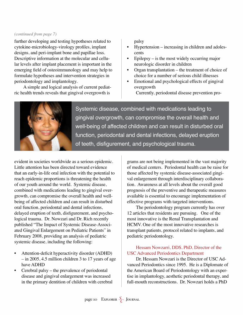
evident in societies worldwide as a serious epidemic. Little attention has been directed toward evidence that an early-in-life oral infection with the potential to reach epidemic proportions is threatening the health of our youth around the world. Systemic disease, combined with medications leading to gingival over-growth, can compromise the overall health and well-being of affected children and can result in disturbed oral function, periodontal and dental infections, delayed eruption of teeth, disfigurement, and psycho-logical trauma. Dr. Nowzari and Dr. Rich recently published “The Impact of Systemic Disease-Associ-ated Gingival Enlargement on Pediatric Patients” in February 2008, providing an analysis of pediatric systemic disease, including the following:
Attention-deficit hyperactivity disorder (ADHD) – in 2005, 4.5 million children 3 to 17 years of age have ADHD Cerebral palsy – the prevalence of periodontal disease and gingival enlargement was increased in the primary dentition of children with cerebral
•
•
palsyHypertension – increasing in children and adoles-cents Epilepsy – is the most widely occurring major neurologic disorder in childrenOrgan transplantation – the treatment of choice of choice for a number of serious child illnessesEmotional and psychological effects of gingival overgrowthCurrently, periodontal disease prevention pro-
grams are not being implemented in the vast majority of medical centers. Periodontal health can be raise for those affected by systemic disease-associated gingi-val enlargement through interdisciplinary collabora-tion. Awareness at all levels about the overall good prognosis of the preventive and therapeutic measures available is essential to encourage implementation of effective programs with targeted interventions.
The periodontology program currently has over 12 articles that residents are pursuing. One of the most innovative is the Renal Transplantation and HCMV. One of the most innovative researches is transplant patients, protocol related to implants, and pediatric periodontology.
Hessam Nowzarri, DDS, PhD, Director of the USC Advanced Periodontics Department
Dr. Hessam Nowzari is the Director of USC Ad-vanced Periodontics since 1995. He is a Diplomate of the American Board of Periodontology with an exper-tise in implantology, aesthetic periodontal therapy, and full-mouth reconstructions. Dr. Nowzari holds a PhD
•
•
•
•
(continued from page 7)
Systemic disease, combined with medications leading to gingival overgrowth, can compromise the overall health and well-being of affected children and can result in disturbed oral function, periodontal and dental infections, delayed eruption of teeth, disfigurement, and psychological trauma.
page 10 Explorer JournalSC

in Biology and Health Sciences. He is the founder of the Taipei Academy of Reconstructive Dentistry in Taiwan and one of only two American members of the Dniepropetrovsk State Academy of Medical Sciences in Ukraine. Since 2002, Dr. Nowzari has served as one of the selected scientific experts on an international panel charged with assessing and rank-ing research proposals for the Ministry of Education and Research, section of medical engineering, in Italy. He was elected to the Italian Board of Experts for evaluating medical products for patient safety and health integrity in 2004. He is the Editor of one of the leading reference publications, Aesthetic Periodontal Therapy: PERIODONTOLOGY 2000.
Sandra Rich, GDH, BA, MPH, PhD, Associate Professor
Dr. Sandra Rich is a dental hygienist (University of Minnesota) with a master’s degree in Public Health (UCLA), and a doctorate in educational psychology and higher education (USC). She is an Associate Professor
in the Department of Periodontology at the USC School of Dentistry. Her current position involves working with both undergraduate D.D.S. students and graduate resi-dents in periodontology. Prior to her position with the USC Advanced Periodontics Program, she was with the Dental Hygiene Program at USC and served as Direc-tor of that program for five years. She has served since 1998 as a committee member on the Health Sciences Institutional Review Board (HSIRB) at USC, which is charged with protecting the rights and welfare of hu-man research subjects in research studies. Dr. Rich’s own research interests include aspects of the behavioral sciences as applied to patient and student education, and clinical research topics in periodontology. She has published articles in a wide range of dental, peer-reviewed journals and authored a textbook chapter in Dental Hygiene Theory and Practice. She has contrib-uted to literature on problem-based learning (PBL) with an article in the Journal of Dental Education comparing performance of pre-clinical, periodontics, DDS PBL students with traditional DDS students.
February �009 page 11
CUTTING EDGE RESEARCH
Innovations in Fixed Prosthodontics
This software will be used to analyze the best site for implant placement. The limitation of the software currently resides in the fact that implant companies have their own implant-specific software that is not universal, and that the software has not yet become affordable in the mainstream market.
Currently, the DDS program boasts of loading implants in 2 months as opposed to 6-9 months years ago, thanks to new implant designs whose surfaces are treated with aluminum oxide blasing, acid etch-ing, or titanium blasting.
The Prosthodontics program explored fabricat-ing custom abutements for implants using Cadcam technology. Two materials used include zirconia and titanium. The drawbacks of zirconia include ce-ramic fatigue, susceptibility to fracture, and lack of a proven track record for use. Therefore, it is not fully embraced by the school right now. Titanium, on the other hand, is an esthetic concern to patients due to its metallic color and its weaker strength.
With regard to the predoctoral program, the extent of current DDS education is limited to plac-ing single unit anterior implants. Dr. Cho envisions multiple-unit anterior implant placements by DDS students in the near future.
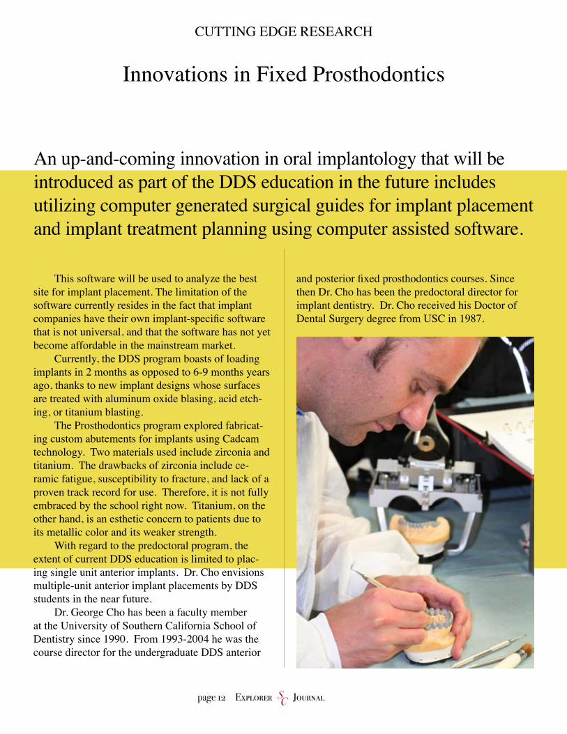
Dr. George Cho has been a faculty member at the University of Southern California School of Dentistry since 1990. From 1993-2004 he was the course director for the undergraduate DDS anterior
and posterior fixed prosthodontics courses. Since then Dr. Cho has been the predoctoral director for implant dentistry. Dr. Cho received his Doctor of Dental Surgery degree from USC in 1987.
An up-and-coming innovation in oral implantology that will beintroduced as part of the DDS education in the future includesutilizing computer generated surgical guides for implant placement and implant treatment planning using computer assisted software.
page 1� Explorer JournalSC
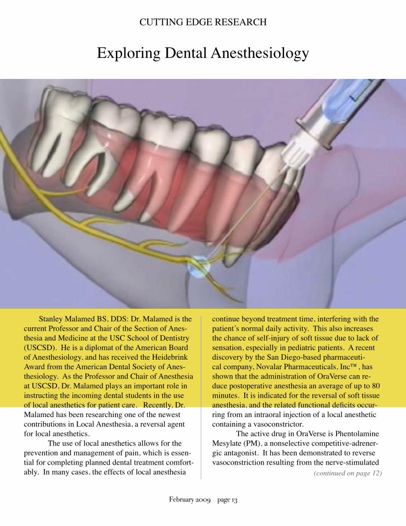
Stanley Malamed BS, DDS: Dr. Malamed is the current Professor and Chair of the Section of Anes-thesia and Medicine at the USC School of Dentistry (USCSD). He is a diplomat of the American Board of Anesthesiology, and has received the Heidebrink Award from the American Dental Society of Anes-thesiology. As the Professor and Chair of Anesthesia at USCSD, Dr. Malamed plays an important role in instructing the incoming dental students in the use of local anesthetics for patient care. Recently, Dr. Malamed has been researching one of the newest contributions in Local Anesthesia, a reversal agent for local anesthetics.
The use of local anesthetics allows for the prevention and management of pain, which is essen-tial for completing planned dental treatment comfort-ably. In many cases, the effects of local anesthesia
continue beyond treatment time, interfering with the patient’s normal daily activity. This also increases the chance of self-injury of soft tissue due to lack of sensation, especially in pediatric patients. A recent discovery by the San Diego-based pharmaceuti-cal company, Novalar Pharmaceuticals, Inc™, has shown that the administration of OraVerse can re-duce postoperative anesthesia an average of up to 80 minutes. It is indicated for the reversal of soft tissue anesthesia, and the related functional deficits occur-ring from an intraoral injection of a local anesthetic containing a vasoconstrictor.
The active drug in OraVerse is Phentolamine Mesylate (PM), a nonselective competitive-adrener-gic antagonist. It has been demonstrated to reverse vasoconstriction resulting from the nerve-stimulated
Exploring Dental Anesthesiology
(continued on page 12)
CUTTING EDGE RESEARCH
February �009 page 1�

release of norepinephrine or locally applied norepi-nephrine or epinephrine. When administered after dental treatment it will increase the rate of redistribu-tion of local anesthetic from the submucosa into the cardiovascular system.
The current dosage of OraVerse based on clini-cal trials will be 0.4mg/1.7mL solution per cartridge. It should be administered following the dental procedure using the same location and technique used for the administration of the local anesthetic. The recommended dose is based on the number of cartridges of local anesthetic with vasoconstrictor administered. The adult-dosage is 1 to 2 cartridges of PM (0.4 to 0.8 mg), and the child-dosage is 0.5 to 1 cartridge (0.2 to 0.4 mg). OraVerse is to be packaged in cartridges identical, with exception to labeling, to traditional local anesthetic carpules, and may be administered with any dental local anesthetic syringe and dental injection needle. OraVerse is cur-
rently not recommended for use in children less than six years in age or weighing less than 33 pounds.
Dr. Malamed is also exploring new methods of administering local anesthetics in order to increase patient comfort and safety. Single Tooth Anesthesia (STA), from Milestone Scientific, is a computer as-sisted local anesthesia device. It utilizes a Dynamic Pressure Sensing system in order to provide intra-ligamentary injections into the periodontal ligament. This will allow the practitioner to solely anesthetize the tooth/teeth planned for treatment, minimizing the use of regional blocks, which can leave the patient with prolonged facial and lingual anesthesia. The STA’s ability to slowly administer local anesthet-ics at a steady rate is also very helpful in reducing discomfort experienced during palatal injections. Dr. Malamed has allowed members of the IV Team to utilize an STA for dental procedures resulting in pain free injections and patient satisfaction.
(continued from page 11)
page 1� Explorer JournalSC

Researchers analyzed periapical radiographs be-fore and after fixed edgewise orthodontic treatment. As expected, the maxillary incisors demonstrated the greatest degree of resorption.
Dr. Sameshima’s interest in two- and three-di-mensional orthodontic imaging using cone beam computed tomography and clinical problems related to root resorption, have earned him several grants from the American Association of Orthodontists Foundation.
James Mah BSc, DDS, MS, DMSc: Dr. Mah is an orthodontic expert specializing in digital cra-niofacial imaging. He has been developing a new three-dimensional imaging technology that enables a clinician to virtually reconstruct the patient’s mouth inside and out. The technology, combined with a customized computer program, produces highly accurate 3-D images of the patient’s head and neck. Now being developed at the school’s Craniofacial Virtual Reality Laboratory, it employs virtual reality to advance dentistry, and not just in the laboratory. Such futuristic tools are being used to assist craniofa-cial dental specialists, orthodontists and surgeons in diagnosing and treating patients. Mah used the imag-ing technology to isolate the position of impacted tooth and pinpoint its proximity to surrounding struc-
tures: other teeth, nerves and bones. Armed with this information, Mah could make a definitive diagnosis and plan the boy’s treatment in fine detail, including surgery to expose his hidden tooth, then orthodontia to move it to the best possible position. In the hands of Jonathan’s dental surgeon, the same information became a blueprint for mapping the operation.
Stephen Yen, DMD, Ph.D Dr. Yen is involved in cutting edge research mainly involving surgi-cal orthodontic treatments for cleft lip and palate patients and molecular determinants of facial over-growth. He is the head orthodontist of the Cranio-facial Team at Children’s Hospital Los Angeles. His other research interests include: developing an animal model for studying mandibular distraction side effects and segmental movement. The objec-tive of this project was to develop an animal model that would allow spring forces to close an anterior open bite side effect created by bilateral mandibular distraction. Also, he is interested in developing clini-cal strategies for closing large palatal fistulae in cleft palate children. The long-term goal of this research is to develop spring-guided distraction of bony seg-ments to close large palatal openings in children with large palatal clefts or acquired palatal deformities secondary to tumor resection
A Look into Orthodontics
CUTTING EDGE RESEARCH
Glenn Sameshima DDS, PhD: Dr. Sameshima is the current Program Director for the Advanced Education in Orthodon-tics. Drs. Sameshima and Sinclair undertook a large-scale root resorption study at the USC Department of Orthodontics. The study included 1,000 patients from six private practices in Southern California.
February �009 page 1�

page 1� Explorer JournalSC
The SRG is committed to cultivating interests in scientific inquiry and promoting cutting-edge research among dental students. With USCSD’s learner-centered philosophy, SRG strives to help students gain an appre-ciation of evidence-based dentistry established by sys-tematic methods. We strongly believe that exposing and immersing students to basic science and clinical research in dental school will enhance their clinical judgment as future practitioners who make clinical decisions based on current research evidence rather than commercial trends.
This organization is comprised of distinctive stu-dents who have taken initiatives in projects leading to discoveries, inventions, and effective skills in promoting oral and systemic health. Members have presented their research in the form of posters, abstracts, and oral presen-
tations in conferences across the country. Students have also exhibited in Taiwan, Hong Kong, China, Australia, and Mexico, where they have established lasting interna-tional networks and gained valuable experiences that will help them develop into future leaders in dentistry.
We welcome you to expand your horizons in the research and practice of evidence-based dentistry by joining the Student Research Group. The SRG is a tight-knit community that provides wonderful opportunities for budding researchers to collaborate with dedicated research mentors and fellow student researchers while building lasting friendships. We look forward to meeting you and celebrating the fruits of research!
Warmest Regards, Rita Y. Chuang
A Message from the SRG President
STUDENT RESEARCH GROUP (SRG)
Dear Colleagues, Research is celebrated with passion at the USC School of Dentistry!
From Left to Right: Joe Field, Matt MacGinnis, Rita Chuang, and George Abichaker

February �009 page 1�
USC School of Dentistry Continuing Education
Rate: Views: 9,914
2:32 / 2:32
You can now view USCSD CE videos onYouTube!
•Esthetics•Esthetics
•Periodontics •Periodontics
•Implants•Implants
•Endodontics•Endodontics
•Oral Surgery•Oral Surgery
•Oral Medicine•Oral Medicine
A Message from the SRG President
STUDENT RESEARCH GROUP (SRG)

page 1� Explorer JournalSC
RRules for Preparation of Abstracts1. An abstract must have a short, specific title (no abbreviations) that clearly indicates the nature of the investigation.2. Briefly describe the objectives of the study unless they are contained in the title. If applicable, include a brief statement of methods. State findings in sufficient detail to support conclusions. Abstracts should not de-scribe research in which the chemical identity or source of the reagent is proprietary or cannot be revealed.3. Use generic names wherever possible. Product-based names are to be used only when necessary.4. Avoid beginning sentences with numerals.5. Standard abbreviations may be used without defi-nition. Nonstandard abbreviations (minimum num-ber) should be placed in parentheses after the first use of the word or phrase abbreviated.6. No references, credits or grant support should be included in the abstract.7. No identifying information about patients partici-pating in a study or trial should be used (name or personal information).8. Check category for submission carefully to assure proper scientific consideration.9. Proofread abstracts carefully to avoid errors before submission. No corrections will be allowed once abstract has been submitted.
Writing an Abstract1. Title: make the title dynamic and conclusive, rather than descriptive.2. Structure: A well written abstract should have the following identified sections: Introduction, Hypoth-esis, Methods, Results and Conclusions.3. Category: Carefully choose the correct category
for submission of your work. The submitting author will be required to check a box verifying that all co-authors have read and approved the submission of the abstract. This will replace downloading and faxing a signed copy of the submitted abstract.
Abstracts must contain the following information, in order:1. Abstract Title, Capitalize appropriate words (e.g. In situ Hybridization Studies of Osteocalcin mRNA in Developing Rat Bone)2. Your Name (e.g. Ima Student)3. School, Class Year (e.g. USC, 07’ ) note: you must be a current USC dental student/resident in order to submit an abstract4. Research Sponsor, Title, Department, School or Organization(e.g. Sponsored by Ima Faculty, DDS, Dept. of Oral Medicine and Diagnostic Sciences, USCSOD, Brigham & Women’s Hospital.) 5. Abstract Text (500 word maximum)_Include: Hy-pothesis tested, Methods used, Data, Statistical analy-sis (where appro priate), Conclusions (underline)
All submissions will be reviewed for content by se-lect USC faculty members. Authors will be contacted should revisions be necessary.
GETTING STARTED
Submitting Your Abstract
Sharing your knowledge via ab-stracts will not only further research at USCSD, but it will allow you to recieve feedback from your peers.
February �009 page 19
USCSD ABSTRACTS
BMP2 signaling is initiated by immobilized BMP2 ligand
Purpose: To determine if immobilized BMP2 induces SMAD phosphorylation in cultured cells.
Methods: C2C12 mouse skeletal muscle cells were cultured to 75%-80% confluence in 12-well culture dishes and starved overnight in serum-free Minimal Essential Media (MEM)-a prior to BMP2 stimu-lation. To assess stimulation by soluble BMP2, cells were stimulated for 0, 5, 10, 20, 40, and 80 minutes with 0.1 and 1.0 mL/mL BMP2. To immobilize BMP2, two commercial anti-BMP2 antibodies were covalently coupled to ferrite epoxy beads and loaded with 10 mg/ml human BMP2 for 15 min. Cells were stimulated for 0, 20, 40, and 80 min with 2 ml packed bead volume/well, and lysed in RIPA buffer with protease and phosphatase inhibitors. The lysates resolved by
10% SDS-PAGE, transferred to nitrocellulose, and immunoblotted for phosphorylated SMAD1/5/8. BMP2-induced SMAD 1/5/8 phosphorylation was visualized by chemiluminescence.
Results: BMP2-induced SMAD 1/5/8 phosphorylation was not substantially affected by immobilization to anti-BMP2 beads. SMAD 1/5/8 phosphorylation induced by the bead-antibody-ligand complex peaked at 40 and 80 minutes of stimulation; this was similar to that induced by soluble BMP2. Conclusions: These results suggest that immo-bilized BMP2 initiates SMAD phosphorylation comparable to that induced by soluble BMP2. Thus, BMP2 recruited to or immobilized on den-tal implants may be expected confer biological activity.
Laura Harshbarger, Susan M. Smith, Matt K. Lee, and Homayoun H. Zadeh
Background: Bone morphogenic proteins (BMPs) are growth factors that induce bone and cartilage formation. Soluble BMP2 interacts with specific receptors to induce the phosphorylation of SMAD 1/5/8 signaling proteins that regulate gene expression and, ultimately, osteoblast func-tion. Since BMP2 regulates bone regeneration, recruitment of BMP2 to dental appliances may accelerate implant osseo-integration. However, it is unclear if immobilized BMP2 re-tains the ability to initiate SMAD 1/5/8 phosphorylation.
page �0 Explorer JournalSC
USCSD ABSTRACTS
Leucine-rich amelogenin peptide induces osteogenesis in mouse embryonic stem cells via activation of Wnt and
BMP signaling pathways. Rungnapa Warotayanont, Malcom Snead, Baruch Frenkel, and Yan Zhou
Background: The majority of the organic enamel matrix protein is amelogenin. Leucine-rich amelogenin peptide (LRAP), an amelogenin splice product, has been shown to pro-mote osteogenesis. However, the pathway(s) involved in LRAP-mediated osteogenic effect is still unclear, and the function(s) of LRAP in stem cells remains unknown.
Purpose: (1) To determine the effect of LRAP on osteogenesis in mouse embryonic stem (ES) cell. (2) To determine the responsible signaling pathway(s) for the osteogenic effect of LRAP.
Methods: Mouse ES cells were induced to osteogenic induc-tion in the presence of LRAP. The resulting embryoid bodies (EB) were analyzed for osteogenic markers by RT-PCR and mineral formation by Alizarin red stain-ing. The expression of b-catenin and pSmad1/5/8 for
LRAP-treated EB was determined by Western immu-noblotting. The BMP-Smad activity in LRAP-treated EB was detected by using a stably transfected lucifer-ase reporter driven by BMP-Smad binding elements. The Wnt promoter activity for LRAP-treated EB was detected by transient transfection of luciferase reporter plasmid containing TCF/LEF binding elements.
Results: ES cells in response to LRAP showed significant increase in BSP and Osx expression, and significant increase in calcium and mineral deposition. The expression of b-catenin and Wnt-reporter activity was upregulated within 24h after LRAP treatment. The expression of pSmad1/5/8 and BMP-Smad-reporter ac-tivity was upregulated in 4 days after LRAP treatment. Conclusions: LRAP induces osteogenesis in ES cells via the activation of Wnt and BMP signaling pathways at the early and late stages of osteogenic differentiation, respectively.
February �009 page �1
USCSD ABSTRACTS
Early Eruption Timing and Sexual Dimorphism of the Permanent Dentition in Mexican Adolescents:A Comparison with the Caucasian Standards
Jennifer Rojas and Glenn Sameshima
Purpose: The purpose of this study was to determine the av-erage eruption age of the posterior permanent den-tition in Mexican adolescents in reference to the current standards based on a Caucasian sample, and to determine if sexual dimorphism is present in the eruption timing of the posterior permanent dentition of the Mexican sample.
Methods: 257 consecutive re-cords were obtained from a university orthodontic program in Mexico City using established inclu-sion-ary and exclusionary criteria. The eruption of the canines and poste-rior secondary dentition was recorded for girls and boys from 8 to 12 years of age. The data was compared statistically to the Caucasian data from a previous study. The study was approved by the DSC Institutional Review Board.
Results: Eruption timing differences start showing in 8 y.o. females with significant differences evident by age 10 for males (p=.001, upper bicuspids) and females (p=.006, upper canines). For all age
groups, Mexican children presented more erupted teeth that Caucasian chil-dren. For the Mexi-can sample, evidence of sexual dimorphism was present in erup-tion timing by age 10, however differ-ences were not statis-tically significant.
Conclusions: The posterior perma-
nent dentition erupts earlier in Mexican patients. Effective diagnosis and treatment planning for patients of Mexican heritage may require assess-ment at an earlier age.
The posterior permanent denti-tion erupts earlier in Mexican patients. Effective diagnosis and treatment planning for patients of Mexican heritage may require assessment at an earlier age.
Background: Although a standard of normal dental eruption has long been established, variance to these norms is common.
page �� Explorer JournalSC

USCSD ABSTRACTS
Functional Restoration of Implants on the Day of Sur-gical Placement in the Fully Edentulous Mandible: A
Case SeriesAlina Krivitsky Aalam, Alexandre Aalam, and Hessam Nowzari
Background: The reconstruction of a functional occlusion in the presence of severe residual ridge resorption remains a significant challenge for dental practitioners. Removable appliances are unsuccessful in severely resorbed cases and the advanced degree of bone loss precludes conventional, implant-retained prosthe-sis, immediate loading of dental implants shortens the treatment time and makes it possible to give the patient a fixed, esthetic appearance during the provi-sional and final treatment period.
Purpose:The purpose of this article is to report the clinical experience and outcome of a study of the functional rehabilitation of 16 completely edentulous mandibles with immediately loaded, cross-arch, screw-retained, hybrid prostheses at the University of Southern Cali-fornia.
Methods: After signing informed consent forms, 16 patients (9 male, 7 femaie) aged 47 to 84 years (mean age, 62.6+/-11.6 years) received 90 BrSnemark System Mk III dental implants (Nobel Biocare USA, Yorba Linda, CA, USA). Stability and radiographs of the dental implants were evaluated at the time of surgery, 3 months, 1 year, and 3 years post-loading.
Results: Three implants failed to meet the criteria of suc-cess, bringing the cumulative success rate to 96.6%, resulting in a 100% prosthetic success rate at 3 years. Thirty-nine (43.3%) of the dental implants placed
were 15 mm in length. Seventy-seven (85.5%) of the dental implants were placed in high-density bone. At 3 years post-loading, the average bone loss was -1.2+/-0.1 mm.
Conclusions: Within the limitations of this study, restoration of implants by unreinforced hybrid prostheses at the time of place-ment provided satisfactory results. The outcome was stable at 3 years post-restoration. Mandibular rehabilitation by functional loading of the implants on the day of the insertion requires the comprehension and proper application of surgical and restorative principles.
February �009 page ��
In contrast, a Th2 environment stabilizes the plaques and may be protective in atherosclerosis. One goal would be to pro-duce a Th2 environment within the arterial wall that will lead to a stable lesion that is at lower risk of rupturing. Autopsy analy-ses of hundreds of leprosy patients demonstrated that ath-erosclerosis, myocardial infarction and stroke were significantly less common than expected. Further examination revealed curious resemblances between lepromatous leprosy lesions and athero-sclerotic plaques. In addition, structural and histological simi-larities between oxiDL and rnycobactena have been confirmed in several literature reviews.
Purpose: We hypothesize that the molecular mimicry between the oxLDL and mycobacterial ligands will allow us to extrapolate the mechanism by which the mycobac-teria utilize PPAR-gamma to create an irnmunosup-pressive Th2 environment to aid in plaque stabiliza-tion in atherosclerosis. In addition, we hypothesize that the Th2 environment created by lepromatous mycobacteria may explain the paucity of atheroscle-rosis ir leprosy patients reported. Further, we hy-pothesize that atherosclerotic lesions in leproma-tous
leprosy patients will be more stabte than found in matched controls.
Methods:1. Elucidate the mechanism by which nycobacteria induce PPAR-gamma, activation. We have obtained various purified mycobac-terial lipids from Colorado State University, which we will test for their ability to (a) activate a PPAR-gamma,-luciferase reporter assay (b) activate downstream target genes of PPAR-gamma, by real-time quant tative PCR (c) alter den-dritic cell functions, such as T cell activation.2. Characterize the phospholiplds in the lepromatous lesions that cross-react with anti-oxLDL antibodies, (a) Determine whether these lipids are mycobacte-rial or host in origin by performing Western blots with EO6 antibody (anti-oxLDL) on mycobacterial
USCSD ABSTRACTS
Applying Mycobacterial Suppression of Host Immunity to AtherosclerosisRita Chuang and Dan Cruz
Background: Recent cardiovascular research has established the central role of inflammation in atherosclerosis develop-ment. This inflammatory lesion is characterized by elevated Thl cytokines which cause lesion progression, plaque rupture, and thrombosis.
It is our goal to determine whether blocking this activation can lead to a more effective immune response against the organism.
page �� Explorer JournalSC

extracts and also on extracts of infected monocyte/macrophages and (b) confocal microscopy utilizing specific antibodies against oxLDLand mycobacteria, and assessing for co-localization.3. Explore whether lepromatous leprosy patients, characterized by a weak, Th2 cell mediated immu-nity against mycobacteria, are protected from other Th 1 diseases such as atherosclerosis.Results: Mycobacterial infection of human monocyte-derived DCs results in upregulation of FA8P-4, a PPAR-gamma downstream target gene, and this activation can be blocked with the PPAR specific antagonist, GR9662. Activation of PPAR-gamma in mono-cyte-derived APCs has been demonstrated to result in (a) a reduction of CD1 molecules, (b) decreased IL-12 production, and in some reports, (c) inefficient activation of lymphocytes in a mixed lymphocyte reaction (MLR). In general, PPAR-gamma activa-tion can lead to APCs that promote a Th2 response
in vitro. Furthermore, it is already established that in-fection witn mycobacteria results in decreased IL-12 and CD1 expression. We aim to determine if PPAR-gamma signaling accounts for these effects,
Conclusions: We demonstrate that mycobacteria activate a nuclear receptor, PPAR-gamma, which results in decreased levels of CD1. Interestingly, we also show that the levels of PPAR-gamma are elevated in leprosy le-sions that are characterized by a weak cell-mediated immune response (Th2-type response). It is our goal to further characterize the components in mycobac-tera that activate PPAR-gamma, and to determine whether blocking this activation can lead to a more effective immune response against the organism. In addition, we propose that understanding mycobacte-rial chronic infections will shed light on other chron-ic inflammatory conditions, such as atherosclerosis.
February �009 page ��

Background: Cytomegaiovirus (CMV) is one of the most common causes of major birth defects in humans. Of the ap-proximately 8400 children born each year in the U.S. with CMV-induced birth defects, more than 1/3 of these children exhibit hypoplasia and hypocalcification of tooth enamel.
Purpose: Our objective was to initiate the investigation of the pathogenesis of CMV-induced tooth defects.
Methods: Mouse Cap stage (E15) mandibular first molars were infected with mouse CMV(mCMV) in vitro in a chemi-cally-defined organ culture system and analyzed utilizing histological and immunolocalization methodologies. The antiviral, acyclovir, was used to inhibit mCMV replica-tion and comparisons made between mCMV-infected and acyclovir-treated, mCMV-infected teeth-Results: Active infection of Cap stage molars for up to 15 days in vitro results in smaller, developmentally-delayed and dysmor-phic molars characterized by shallow, broad and misshapen cusps, infected and affected dental papilla mesenchyrne, poorly differ-entiated odontoblasts and ameloblasts, and no dentin matrix. Initial protein local-ization studies suggest that the pathogenesis is mediated through NF-_B signaling and that there appears to be an unusual interaction between abnormal mesenchymal cells and surrounding matrix. Rescue with acyclovir indicates that mCMV replication is necessary to initiate and sustain progressive tooth dysmorphogenesis.
Conclusions: Our results indicate that mCMV-induced changes in signaling pathways severely delays, but does not com-pletely interrupt, tooth morphogenesis. Importantly, our results demonstrate that this well-defined embryonic mouse organ culture sys-tem can be utilized to delineate the molecular mechanism underlying the CMV-induced tooth defects that characterize the Amelogenesis Imper-fecta phenocopy seen in many CMV-infected children.
USCSD ABSTRACTS
Cytomegalovirus Inhibition of Embryonic Mouse Tooth Development: A Model of the Human
Amelogenesis Imperfecta PhenocopyGeorge Abichaker, Pablo Bringas Jr, Nolan Jangaard, Tina Jaskoll, and Michael Melnick
page �� Explorer JournalSC

Background: In the tumor microenvironment immunosuppres-sion is generally associated with tumor growth. Non steroidal anti-inflammatory drugs (NSAIDs) are clinically shown to have significant anti-carcinogenic effects. Particularly, sulindac has received con-siderable attention as an antican-cer drug since it is capable of inhibiting tumor growth in different types of tumors such as oral squa-mous cell carcinoma. Although an extensive number of studies have previously established
the preventive role of sulindac in tumor genera-tion and progression, none has clearly delineated either the mechanisms or the involvement of im-mune cells in this process.
Purpose: The objective of our study was to investigate the effect of sul/ndac treatment on squamous eel! car-cinoma growth and in the survival and function of immune cells.
Methods: Squamous eel. carcinoma cell line and Peripheral Blood Mononuclear Cells (PBMCs), containing lymphocytes and monocytes, were use for the ex-
periments. Tumor cell viability was determined by MTT Assay. Immune cell proliferation was meas-ured through incorporation of tritiated thymidine into DMA. The cytokine secretion levels were measured using cytokine multiplex kits. Different concentra-tions of sulindac were used to evaluate the growth inhibitory effect on tumor cells.
Results: The results showed a decreased in the viability of tumor cells treated with different concentrations of
sulindac. Treatment of oral tumors with sulindac resulted in a significant decrease in the secretion of IL-6 and VEGF. Moreover, the immune cells showed significantly increased secretion of IFN- _ and GM-CSF, in addition to increased proliferation when in the presence of sulindac treated tumor cells.
Conclusions: We show that sulindac inhibits tumor growth and promotes survival and increased function of immune cells. The present data suggest the potential benefi-cial effects of sulindac administration as an adjunc-tive therapy for chemotherapeutic inhibition of tumor growth.
USCSD ABSTRACTS
Role of Non Steroidal Anti-Inflammatory Drugs in Decreasing Squamous Cell Carcinoma Growth and
Increasing Immune ActivationAntonia Teruel, Anahid Jewett, and Glenn Clark
The present data suggest the potential beneficial effects of sulin-dac administration as an adjunctive therapy for chemotherapeutic inhibition of tumor growth.
February �009 page ��

USCSD ABSTRACTS
Comparison of Normalized Referenced Facial Form Classification and Implant Denture Function
Purpose: The purpose of this study was to determine the effect of alternate definitions for facial form classification and the rela-tionship to the func-tional outcomes in dentitions restored with com-plete dentures supported with and without dental implants.
Methods: A randomized controlled clinical trial was un-dertaken to compare treatment success rates, and functional and percep-tual outcomes with mandibular complete dentures (CD) and implant overdentures (IOD) in average denture wear-ers. Pre- and post-insert ion cephalometric ra-diographs were taken and evaluated for 25 CD subjects and 44 IOD subjects. The radiographs were evaluated by two clinicians using Dolphin software. (Dolphin 10.0 - Dolphin Imaging, Chatsworth CA.) Masticatory perform-ances were evaluated with peanuts using standard-ized (PS) and swallowing threshold performance
(SWT) tests. Comparison of PS and SWT were evaluated for CD and IOD comparison, and for treatment-type with normalized (age, gender, ethnicity) and non-normalized facial form cat-egories.
Results: Significant differences were seen in distributions of facial forms (p<0.001), with greater repre-sentation of dolico-fada! form patterns with the normalized method (12%) compared to the non-normalized method (3%). Masticatory perfor-mance data for dolico-facial form was lower than for other forms for both normalized and non-nor-malized classifications, with the non-nor-malized classification being the lowest.
Conclusions: Facial form evaluation is a valuable assessment for prosthodontic evaluation of implant treatment and functional profiling.
Camille Nakamura, Hideki Ikeda, Neal Garret, and Kent Ochiai
Background: The use of cephalometric measurements was previously proposed to classify facial form for prosth-odontic diagnostic purposes; however, the effect of age, gender and ethnic profile on cephalometric facial form categorization for implant prosthodontic patients has not been demonstrated.
page �� Explorer JournalSC

USCSD ABSTRACTS
Integrin Linked Kinase is Necessary for Medial Edge Epithelia Transformation During Palatal Fusion
Daniela Schmid, Xiao-Mei, Matthew Lee, and Chuck Shuler
Background; The fusion of the secondary palate is a complex event that requires medial edge epithelial cells (MEE) to under-go epithelial-mesenchymal transformation (EMT). EMT is regulated by both integrins and growth factors, and disruption of their intra-cellular signaling pathways may prevent palatal fusion. Integrin-linked kinase (ILK) is a cytoplasmic serine/threonine kinase that mediates signal transmission from integrins and growth factor receptors and has been implicated in the regulation of EMT.
Purpose: To determine that ILK activity is necessary for palatal fusion.
Methods: Mouse fetuses were examined by immunohistochernistry in vivo and in vitro. Palate cultures were transfected with ILK siRNA and results analyzed by Western blotting, H&E and immunostaining. siRNA transfected cultures were treated with EGF and ILK downstream signaling was assessed us-
ing phospho specific antibodies to GSK-3 Akt.
Results: To assess this, we first assessed ILK expression in vivo and in cultured mouse palatal shelves. Immunohisto-chemistry demonstrated that ILK expression is restricted to the MEE and oral epithelia. Next, ILK expression was silenced by siRNA transfec-tion into cultured palatal tis-sues. This resulted in significantly decreased ILK expres-sion and the persistence of midline MEE cells. Finally, EGF-induced ILK signaling was assessed in palate cultures treated with either ILK siRNA or non-silenc-ing control siRNA. The resulting phosphorylation of the downstream ILK mediators GSK-3_ and Akt was then assessed. ILK silencing significantly reduced the phos-phorylation of both proteins.
Conclusions: These results demonstrate that ILK signaling is necessary for palatal fusion and MEE involution.
February �009 page �9

Background: Tissue Engineering is defined as the science devel-oping techniques for fabrication of new tissues for replacement and regeneration of lost tissues based on principles of cell and developmental biology. Mineral trioxide aggregate (MTA) has been clinically used in dentistry for repair of root defects, pulp capping, partial pulpotomy, apexification, perforation repair, resorption repair, and as a root-end filling material. In vitro and in vivo MTA studies suggest that MTA (Portland cement) promote hard tissue formation and it is now widely used in endodontics as a root-filling material.
Purpose: To determine if MTA has the ability to regenerate periodontal tissues using a rat animal model.
Methods: A periodontal fenestrat on defect was surgically cre-ated bilaterally on the maxilla of 45-days old rats. A periodontal flap over the mesial root of maxillary first molar was created and a low speed bur was used to penetrate the marginal bone to create the defect on
root. The mesial root of the first maxillary molar was denuded of its PDL, overlying cementum, and super-ficial dentin. The defects were approximately 1.5x2 mm. The surgical defects was either left untreated or covered with MTA. The tissues were re-positioned and sealed using Dermabond. The animals were given 10% dextrose solution supplemented with Amoxicillin and Ibuprofen daily for 7 days and kept on a soft d-et until they were euthanized 8 weeks after the surgery. Maxillas were collected, decalci-fied and embeded in paraffin. Serial sections were prepared along the full length of the defect. Sections were stained with Goldner’s Trichrome for histologi-cal evaluation jsing light microscopy. All animal protocols were approved by the DSC IACUC.
Results: Although still under analysis, the data obtained up till now indicates that all roots treated produced a thick layer of terti-ary dentin in response to the sur-gical defect. The data also suggests that treatment of the surgical defect with MTA offers several benefits to the healing and regeneration of the defect. De-pending of the seventy of the defect, MTA appears to preserve the pulp from necrosis, seerns to protect the root from resorption and appears to induce regenera-tion of PDL ard bone. However, it does not appear to induce cementurn formation and root re-attachment.
Conclusions: Our results suggest that MTA indeed has an effect on protecting the pulp from necrosis and resorption and although it has the ability to induce formation of bone and Periodontal ligament, it does not provide complete regeneration of root to bone attachment.
USCSD ABSTRACTS
Effect of Mineral Trioxide Aggregate (MTA) on Regeneration of Periodontal Tissues
Eric Nordberg, Drew Brueckner, Agustine Kim, Nathan Ostler, Keith Tarn, Mahbod Rashidi Torghi, Yu-Jen Wu, and Maggie Zeichner-David
page �0 Explorer JournalSC
USCSD ABSTRACTS
Comparative Growth, Cognitive, and Neuroanatomical Outcomes Following Maternal Deprivation or
Perinatal Brain Injury in RatsWei-Cheng Taif, L Gundamraj, and Jack Turman
Methods: Neonatal brain injury or MD was initiated at postnatal day (p) 3 or 4, respectively, using a rat model- On P3 we induced hypoxic ischemic (HI) brain injury using
the Levine procedure (n = 18). MD was performed by isolating pups from dam for 5 con-secutive days (P4-8, n = 16). Control (CL) animals with no HI or MD were used for comparisons (n = 8). Growth measures includ-ed daily weight, length and head crown-snout length (HCSL) through P23. Cognitive outcomes were as-sessed using a neonatal odor habituation paradigm, and a postweaning open field paradigm. Rats were perfused between P25-27, brains removed, and cryostat sections were collected.
Results: HI animals had reduced growth when compared to
MD and CL groups. From P3-13 MD animals showed increased growth. The cognitive phenotype of the MD group included: increased arousal following odor presentation. In the open field the MD group was char-
acterized by decreased grooming and rearing, whereas HI animals were characterized by decreased wall time. Histological data showed that MD animals had a significant reduction of frontal pole hemispheric and corpus callosum areas com-pared to Hi/Sham animals.
Conclusions: Our findings reveal distinct growth, cognitive and neuroanatomical patterns associated with these pertur-bations, and serve as a foundation for our future studies that will investigate the cumulative effects of MD and HI brain injury,
Background: Neonatal brain injury and maternal deprivation (MD) are common perturbations encountered by infants in the NICU. Purpose: We are interested in differentiating the outcomes that re-sult from either of these perturbations.
Our findings reveal distinct growth, cognitive and neuroanatomical patterns associated with these perturbations.
February �009 page �1

We are very thrilled to introduce the inaugural publication of The Explorer, Journal of USC Student Dental Research. From assembling a group of driven dental students, interviewing dental faculty, planning and editing layout, to seeing the final product has proven to be an immensely satisfying experience. In the process, we have gained a deeper appreciation of the rich traditions and research culture at the school of dentistry, as well as the tenacity and dedication that is required to create a new journal.
The Explorer highlights the cutting-edge research in which the USC School of Dentistry takes great pride. From winning research abstracts from the annual USC Research Day to innovations in each of the Advanced Education Programs at the school, it is our sincere hope that the journal will initiate curiosity and discussions among pre and post-doctoral students here at USC and other institutions to encourage more D.D.S. students to pursue innovative research in their career and contribute to the knowledge pool for the advancement of our profession.
It has been an immensely gratifying experience putting together this journal with our team. The Student Research Group (SRG) will carry on this wonderful tradition by publishing future issues to update the USCSD community as well as the student body on the research innovations at USC. We are proud to present to you The Explorer: Journal of USC Student Dental Research. Enjoy the premiere issue!
Sincerely, Rita Chuang & Tin Luong
From Left to Right: Goldwyn Jequinto, Chad Tomazin, Rita Chuang, Dr. Shervin Molayan, Michael Meru, Bina Joshi, & Tin Luong.
FINAL WORDS
Message from the Co-Editor’s in Chief
page �� Explorer JournalSC

To get to that answer, a definition of research is needed. Research is defined as a diligent, studious, and systematic inquiry aimed at discovering facts, confirming or revising existing theories or laws of nature, or using new information for practical application. In an academic environment such as the USC School of Dentistry, re-search is an important component of our purpose/mission, goals and objectives along with education, service, and leadership. Research is the fuel for the engine of tech-nology and the numerous applications to improve the human condition. Research ranges from basic or fundamental inquiry and discovery to bedside and chairside outcomes research and policies that improve standards of care for all people.
For students wanting to get started “doing research”, here are my recommendations: Begin by talking with several our research faculty. Their names and contact information are listed on USCSD website (http://www.usc.edu/hsc/dental/research/index.htm).Find an area of research that interests you: infectious diseases, bioimaging, nanotechnology, tissue regeneration, stem cells, education, biobehavior and so on. Match your interest with a faculty member – contact them to see if there are projects ongoing that meet your interests. Carve out time and commitment from your busy schedule to work on the project- quality research is time con-suming! (USCSD faculty only want quality research efforts in their labs!) Dig into the literature and learn all that you can about the specific topic you’re working on – talk with other col-leagues in the labs – share ideas and experiences with others involved in research. Join the Student Research Group to network and meet others with like interests in research.Talk with me – explore your options and consider your goals.
I believe these simple steps will give you the resources to get you started in research and the rewards will be many.
There is a thrill in being the first to learn something that nobody else in the world knows or do something that nobody else has done. That is what we call innovation and discovery! The adrenaline rush is unbelievable … the first to dis-cover: a new virulence gene in a bacterium; a technique for identifying the early stages of cancer; a way to turn stem cells into teeth; an image of a unique biofilm on infected bone tissue; an inexpensive test to identify kids or seniors who are especially susceptible to dental caries; or, a computer-based approach to improve dental education. In addition, research teaches us to think critically and apply scientific methodology to move dentistry forward. The recognition and respect you receive as a student of science elevates your stature as a dental practitioner and is an experience you will cherish for a lifetime.
Dennis Mangan, Ph.D.Associate Dean for Innovation and Discovery (Research) [email protected] - (213) 740-5735
•
•
••
•
••
Message from Dr. Dennis Mangan,Associate Dean for Innovation and Discovery
FINAL WORDS
I’ve often been asked by USC students, “How do I get started in research?”
February �009 page ��

JANUARY - MAY 2009
U S C S C H O O L O F D E N T I S T RYCO N T I N U I N G E D U C AT I O NY o u r T r o j a n H o m e f o r L i f e - L o n g L e a r n i n g
USC 34TH ANNUAL PERIODONTAL ANDIMPLANT SYMPOSIUM AND WORKSHOPSTHUR - SUN, JAN 22 - 25
MASTERING MOLAR ENDODONTICSFRI - SAT, JAN 30 - 31
ESTHETIC PERIODONTAL SURGERY FOR THEGENERAL PRACTITIONER - MODULE ISACRAMENTO: SAT, JAN 31
PORCELAIN VENEERS: OPTIMIZING LONG-TERMESTHETIC OUTCOME USING DFED ANDMANAGING OCCLUSAL DISEASEFRI, FEB 6
EMERGING DISEASES, INFECTION CONTROL ANDCALIFORNIA DENTAL PRACTICE ACTSAT, FEB 7
ESTHETIC PERIODONTAL SURGERY FOR THEGENERAL PRACTITIONERMODULE I: FRI, FEB 20MODULE II: SAT, FEB 21MODULE III: SUN, FEB 22
CHRONIC OROFACIAL, ORODENTAL ANDHEADACHE PAINS FOR THE DENTISTFRI - SAT, FEB 20 - 21
NEW APPROACHES FOR ANTIMICROBIALTREATMENT OF PERIODONTAL DISEASESACRAMENTO: FRI, FEB 27
UPDATES IN PEDIATRIC DENTISTRY: PERIODONTAL HEALTH AND DISEASES INCHILDREN AND ADOLESCENTSFRI, FEB 27
USC NEW ODONTIC SEMINARFRI, MAR 6FRI, APR 24FRI, JUNE 12
ORAL SURGERY FOR THE GENERALPRACTITIONERSAT, MAR 7
USC RUTH RAGLAND 23RD DENTALHYGIENE SYMPOSIUMPART I: SAT, MAR 7PART II: SUN, MAR 8
FUNDAMENTALS OF RESTORATIVE IMPLANTDENTISTRY FOR THE GENERAL DENTISTPART I: FRI, MAR 13PART II: SAT, MAR 14
ESTHETIC FULL-MOUTH IMPLANTRECONSTRUCTIONMODULE I: SAT, MAR 14MODULE II: FRI, MAR 20MODULE III: SAT, MAR 21
APPLIED HYPNOSIS: TREAT PAIN, TMD &OTHER DENTAL CONDITIONSSAT - SUN, MAR 14 - 15
A NEW APPROACH TO REDUCING STRESS FORDENTISTS AND THEIR STAFF, AND FORDENTAL PATIENTSFRI, MAR 27
MASTERING BONE GRAFTING FOR ESTHETICIMPLANT SITE DEVELOPMENT - LECTURE & CADAVER WORKSHOPMODULE I: FRI, MAR 27MODULE II: SAT, MAR 28
IMPLANT THERAPY IN THE ESTHETIC ZONEFRI - SUN, APR 3 - 5
COMMON ORAL LESIONS (1): UPDATE ONEARLY ORAL CANCER AND PRECANCERDETECTIONSAT, APR 4
DIGITAL PHOTOGRAPHY IN DENTISTRYPART I: FRI, APR 17PART II: SAT, APR 18OBSTRUCTIVE SLEEP APNEA, SNORING ANDDENTAL ADVANCEMENTFRI - SAT, APR 17 - 18
USC ESTHETIC DENTISTRY CONTINUUM2009MODULE I: FRI - SUN, APR 24 - 26MODULE II: FRI - SUN, MAY 29 - 31MODULE III: FRI - SUN, JUN 26 - 28
MAKING SENSE OUT OF REMOVABLEPARTIAL DENTURE DESIGNSAT, APR 25
IMPLANT THERAPY IN COMPROMISED SITESFRI - SUN, MAY 1 - 3
ADVANCED BONE AND SOFT TISSUEAUGMENTATION - CADAVER WORKSHOPSUN, MAY 3
NEW APPROACHES FOR ANTIMICROBIALTREATMENT OF PERIODONTAL DISEASEFRI, MAY 8
THE USES OF PHOTOSHOP & POWERPOINTIN MODERN DAY DENTISTRY: A HANDS-ON COURSESAT, MAY 9
PHYSICAL EVALUATIONMON, MAY 18
EMERGENCY MEDICINETUES, MAY 19
MONITORING AND CLINICAL EMERGENCYMEDICINEWED, MAY 20
SCHEDULE2009COURSE
University of Southern CaliforniaSchool of DentistryOffice of Continuing Education
925 W 34th St Rm 201JLos Angeles, Ca 90089-0641t. 213.821.2127f. [email protected]
View more CE courses and register online atUSCDENTALCE.ORG
photo credit: christopher creighton

Courage To Explore
USC School of DentistryRESEARCH - EDUCATION - LEADERSHIP