july 15–17, 2014 presentation and poster proceedings
TRANSCRIPT

Montana State University Center for Biofilm Engineering Bozeman
JULY 15–17, 2014 presentation and poster
PROCEEDINGS
K Gorham, MSU News

CBE Montana Biofilm Science & Technology Meeting: July 2014
abstracts
Presentation ToC | Presentation Abstracts Poster ToC | Poster Abstracts
1
Montana Biofilm Science & Technology Meeting: July 15-17, 2014
Table of Contents: Presentation Abstracts
SESSION 1: Biofilm Infection
7 Biofilm model of delayed healing in the rabbit ear: Clinical implications of virulence, host response, and treatment Thomas A. Mustoe, MD, Professor, Plastic Surgery, Northwestern University
8 Whack-a-mole, chess and the fight against chronic infections Pradeep Singh, MD, Professor, Medicine and Microbiology, University of Washington
8 Prosthetic joint infection update Robin Patel, MD, Professor, Medicine and Microbiology, Mayo Clinic Medical College
8 Transport limitations in heterogeneous systems Isaac Klapper, Professor, Mathematics, Temple University
9 Atmospheric plasma for annihilation of wound biofilms Garth James, Associate Research Professor, Chemical and Biological Engineering, MSU-CBE
9 Gel-entrapped Staphylococcus aureus as a model of biofilm infection Breana Pabst, Research Assistant, MSU-CBE
SESSION 2: Microscopy
10 Time-lapse confocal microscopy of gel-entrapped bacteria as models of infection Betsey Pitts, Research Scientist and Microscope Facilities Manager, MSU-CBE
10 Visualization of cell surface interactions of environmental samples using confocal microscopy
Heidi Smith, PhD student, Land Resources and Environmental Sciences, MSU-CBE
11 Experience with the microscopy Treatment Flow Cell Lindsey Lorenz, Research Assistant, MSU-CBE; and Kelli Buckingham-Meyer, Research Assistant, MSU-CBE
11 FISH on! Optimization and utility of Fluorescence In Situ Hybridization (FISH) in detecting industry-relevant environmental microbes
Dana Skorupa, Postdoctoral Research Associate, MSU-CBE

CBE Montana Biofilm Science & Technology Meeting: July 2014
abstracts
Presentation ToC | Presentation Abstracts Poster ToC | Poster Abstracts
2
SESSION 3: DNA Sequencing in Practice Sequencing: Trials and tribulations
Matthew Fields, Associate Professor, Microbiology, MSU-CBE
12 Molecular diagnostics in wound care Randy Wolcott, MD, Southwest Regional Wound Care Center, Lubbock, TX, USA
12 Bacterial community changes with depth and metal geochemistry from mined material
Chiachi Hwang, Research Professional, MSU-CBE 13 I did not know because I could not grow…the impact of molecular methods on
microbial control in industrial systems Vic Keasler, Senior RD&E Group Leader, Nalco, Inc.
Young Investigators
14 Genetic requirements in spatially organized polymicrobial wound infection Keith Turner, Postdoctoral Fellow, Center for Infectious Disease, The University of Texas at Austin
15 Analyzing secondary metabolite production by 3D-printed bacterial populations using scanning electrochemical microscopy
Jodi Connell, Postdoctoral Fellow, Center for Infectious Disease, The University of Texas at Austin
Special Presentations
State of the CBE, 2014 and Presentation of CBE Awards Phil Stewart, CBE Director
SESSION 4: U.S. Regulatory Review 16 Biofilm claims for antimicrobial products: U.S. EPA regulatory perspective Stephen Tomasino, Senior Science Advisor, U.S. EPA Office of Pesticide Programs –
Microbiology Laboratory Branch 16 FDA/CBE joint workshop recap Phil Stewart, CBE Director
17 Biofilm claims—EPA rules and implications John Wood, Senior Director Agency Relations (Law and Regulatory Affairs), Ecolab, Inc.
17 Biofilm test methods and impact on regulatory guidelines LaShanda Glenn, Scientist, Procter & Gamble

CBE Montana Biofilm Science & Technology Meeting: July 2014
abstracts
Presentation ToC | Presentation Abstracts Poster ToC | Poster Abstracts
3
SESSION 5: New CBE Capabilities: Micromechanics & Microfluidics 18 Tools for measuring biofilm mechanical properties
James Wilking, Assistant Professor, Chemical and Biological Engineering, MSU-CBE
18 Drop-based microfluidics for biological applications: From colloidal dispersions to high-throughput assaying Connie Chang, Assistant Research Professor, Chemical and Biological Engineering, MSU-CBE
SESSION 6: Bacterial Survival in Industry and the Environment
18 Hot water disinfection of planktonic and biofilm bacteria Mark Pasmore, Research Manager, Baxter Healthcare Corporation Diane Walker, Research Engineer, MSU-CBE
19 Systems analysis of iron-limited growth: Insights into pathogen metabolic acclimation to host Ross Carlson, Associate Professor, Chemical and Biological Engineering, MSU-CBE
19 Field-scale plugging of hydraulic fractures using ureolytic bacteria Al Cunningham, Robin Gerlach, and Adie Phillips, MSU-CBE
20 Presence, pervasiveness, and persistence of wastewater pathogen Escherichia coli O157:H7 in model treatment wetlands Rachel VanKempen-Fryling, PhD student, Microbiology, MSU-CBE
21 Monitoring Chlorella survival during algal biofuel production using a community ecology approach Tisza Bell, PhD student, Microbiology, MSU-CBE
22 Biofouling on household reverse osmosis water treatment membranes Stephen Markwardt, master’s student, Environmental Engineering, MSU-CBE

CBE Montana Biofilm Science & Technology Meeting: July 2014
abstracts
Presentation ToC | Presentation Abstracts Poster ToC | Poster Abstracts
4
Table of Contents: Poster Abstracts
Industry and Agency Posters 23 The Sharklet micropattern limits bacterial adherence and biofilm:
A potential technological improvement for endotracheal tube design Ethan Mann, Sharklet Technologies, Inc.
24 The use of CDC biofilm reactor to test cleaning and disinfection capabilities on rouged stainless steel Amanda Deal, STERIS
Academic Posters (non-CBE) 25 Using Surface Plasmon Resonance imaging (SPRi) to evaluate bacterial activity
on surfaces Edgar Goluch, Northeastern University
25 Effect of removal of biofilm on titanium surface applied by ultrasonic water flow
technology Takashi Takiguchi, Showa University School of Dentistry, Japan
26 Toward a broader appreciation for backyard biofilms: Soils
Tony Hartshorn, Montana State University
Center for Biofilm Engineering posters
27 #619: Applicability of MICP in subsurface and fractured environments Adie Phillips, CBE staff researcher
28 #620: Using biomineralization sealing for leakage mitigation in shale Robin Gerlach, CBE faculty, chemical and biological engineering
29 #621: A ruggedness analysis of sonication in the Single Tube Method (ASTM E2871-12) Blaine Fritz, CBE post-baccalaureate researcher
30 #622: Biofilm parameter estimation using inverse methods Ben Jackson, CBE PhD student, mathematics
30 #623: Toward multiscale metabolic network analysis of an anaerobic microbial community Kris Hunt, PhD student, chemical and biological engineering
31 #624: Optimization of media for the hydrolysis of urea and precipitation of calcium carbonate with S. pasteurii Eric Troyer, undergraduate student, chemical and biological engineering
31 #625: The ESENCYA project: Environmental SENsory perception in CYAnobacterial biofilms: Understanding biodeterioration of outdoor stone materials in a changing environment Federica Villa, CBE visiting researcher, University of Milan, Italy

CBE Montana Biofilm Science & Technology Meeting: July 2014
abstracts
Presentation ToC | Presentation Abstracts Poster ToC | Poster Abstracts
5
33 #626: Development of a clinically relevant model flow system for observing struvite formation by Proteus mirabilis biofilms Trace Hobbs, undergraduate student, chemical and biological engineering
33 #627: Low field Magnetic Resonance for in situ bioremediation monitoring Catherine Kirkland, PhD student, civil engineering
34 #628: The effects of UV light on biofilm formation and pigment production of Antarctic Janthinobacterium sp. strain CG23_2 Christine Foreman, for Emily Bermel, undergraduate student, chemical and biological engineering
35 #629: Chemotaxis of Antarctic and Arctic microbial life towards various carbon sources using a capillary motility method Shu Ying Wee, undergraduate student, chemical and biological engineering
36 #630: Microbial diversity and ecophysiology of cryoconite granules from the Dry Valleys, Antarctica Heidi Smith, PhD student, land resources and environmental sciences
36 #632: Analyses of accumulated lipids and secreted proteins from an extremophilic lignocellulose-degrading fungus Logan Boucher, undergraduate student, chemical and biological engineering
37 #633: Amphiphilic siderophores produced by haloalkaliphiles: A story of iron and vesicle self-assembly Luis O. Serrano Figueroa, PhD student, microbiology
38 #634: Denitrification at the microscale in a treatment wetland system Justin Spengler, PhD student, molecular biosciences
38 #635: Time-lapse confocal microscopy of gel-entrapped bacteria as models of infection Betsey Pitts, CBE staff researcher
39 #636: Biofilm growth and particle size relationships Sara Altenburg, CBE staff researcher
39 #637: Investigating culture effects of light irradiance and initial biomass concentration in Nannochloropsis Oceanica sp. using response surface methodology Todd Pedersen, CBE master’s student, chemical and biological engineering
40 #638: Biogenic coal bed methane enhancement: Methods for field-relevant experiments Katherine Davis, PhD student, chemical and biological engineering
40 #639: Growth of two alkaliphilic microalgal isolates in recycled harvest water supplemented with anaerobic digestate Ashley Berninghaus, undergraduate student, chemical and biological engineering
41 #640: Growth and lipid productivity of two alkaliphilic microalgal isolates in municipal wastewater Luke Halverson, master’s student, chemical and biological engineering
42 #641: Imaging the extracellular matrix of Pseudomonas aeruginosa biofilms Mike Franklin, for Amanda Richards, undergraduate student, microbiology
CBE Montana Biofilm Science & Technology Meeting: July 2014
abstracts
Presentation ToC | Presentation Abstracts Poster ToC | Poster Abstracts
6
42 #642: Treatment-associated bias assessment of the Single Tube Method ASTM E2871-12 Diane K. Walker, CBE staff researcher
43 #643: Comparison of bacterial transfer and biofilm formation on intraluminal catheter surfaces among fourteen connectors in a clinically simulated in vitro model Elinor deLancey Pulcini, CBE faculty researcher
44 #644: MALDI-IMS imaging of chlorhexidine and bacteria on an in vitro human skin model Maggie Butler, Bioscience Laboratories, Inc.
44 #645: Novel anti-biofilm materials for medical devices based on biofunctionalized surfaces Cristina Cattό, CBE visiting researcher, University of Milan, Italy
45 #646: How does the biofilm structure influence the local flow regime? An investigation of biofilm carriers originating from waste water treatment Pia Herrling, CBE visiting researcher, Karlsruhe Institute of Technology, Germany
46 #647: Developing copious biofilm growth in porous media with low cost nutrient Andrew Bender, undergraduate student, chemical and biological engineering

CBE Montana Biofilm Science & Technology Meeting: July 2014
abstracts
Presentation ToC | Presentation Abstracts Poster ToC | Poster Abstracts
7
Presentation Abstracts
SESSION 1: Biofilm Infection Biofilm model of delayed healing in the rabbit ear: Clinical implications of virulence, host response, and treatment Presenter: Thomas A Mustoe, MD, Professor, Plastic Surgery Co-authors: Seok Hong PhD, Robert Galiano MD, Kai Leung, PhD Affiliation: Northwestern University, Chicago, IL, USA. Although it is now well accepted that biofilm in wounds is an important reason for their chronicity, much is still not known. We have developed a standardized model in our validated rabbit ear model which we have utilized for many years to study many aspects of normal and delayed healing. The model has been highly reproducible, and has been used to study the impact of ischemia, ischemia reperfusion, and scarring, as well as the effects of various therapeutic strategies including the impact of growth factors. With the inoculation of bacteria (Staphylococcus aureus, Pseudomonas aeruginosa, Klebsiella, and Staphylococcus epidermidis), we have found biofilm formation within 12 hours—and a steady state of bacterial number after several days—regardless of the size of the original inoculation. In contradistinction to planktonic bacterial acute infections, the host response is limited to the immediate area surrounding the wound and the wounds go on to heal, although in delayed fashion. There are significant variations in virulence (as defined by the amount of delay in wound healing) between bacterial species, and inoculation with both P. aeruginosa and S. aureus are much more virulent than either alone. The effect of the host on limiting bacterial counts is significant. In ischemic wounds the steady state bacterial count is a full log higher than in non-ischemic wounds. Virulence is not strictly related to bacterial counts, in that steady state P. aeruginosa counts are substantially lower than other bacteria—yet P. aeruginosa has the greatest impact on wound healing. Detailed transcriptome analysis has added to the insights on the importance of the host inflammatory response. Utilizing bacterial mutants, we have found that P. aeruginosa mutants deficient in biofilm production have a much greater effect on virulence than quorum sensing mutants, emphasizing the importance of biofilm production. P. aeruginosa is the most virulent bacteria, with the greatest amount of biofilm formed clinically, while Klebsiella actually has less effect on wound healing than S. epidermidis. We have used the model to investigate various therapeutic strategies. As expected, topical antibiotics are ineffective unless combined with a strategy to reduce biofilm, such as high pressure water irrigation done daily. Other strategies to reduce biofilm, such as ultrasound mist, or dressings designed to disperse biofilm and allow silver penetration, have also been effective. Bacterial phage active against inoculated bacteria have only been effective with a mutant bacteria deficient in biofilm formation (in S. aureus). Recently we have made progress in identifying the signaling pathway involved in the biofilm mediated inhibition of keratinocyte migration that contributes to the delay in wound healing.
1. Gurjala AN, Geringer MR, Seth AK, Hong SJ, Smeltzer MS, Galiano RD, Leung KP, Mustoe TA. “Development of a novel, highly quantitative in vivo model for the study of biofilm-impaired cutaneous wound healing,” Wound Repair and Regeneration, 2011; 19:400–410.
2. Seth, et al. Wound Repair and Regeneration, 2012; 20:214–225. 3. Seth, et al. Plastic and Reconstructive Surgery, 2012; 129:354–361.

CBE Montana Biofilm Science & Technology Meeting: July 2014
abstracts
Presentation ToC | Presentation Abstracts Poster ToC | Poster Abstracts
8
4. Seth, et al. J Am Coll Surg, 2012; 215(3):388–399. 5. Seth, et al. PLOS ONE, 2012; 7(8):e42897. 6. Seth, et al. Journal of Surgical Research, Nov 2012; 178(1):330–338. 7. Seth, et al. Wound Repair and Regeneration, 2013; 21(2):266–74. 8. Seth, et al. Plast Reconstr Surg, 2013; 131(2):225–34. 9. Cheng, et al. Antimicrob Agents Chemother, 2014; 58(2):1208.
10. Leung KP. BMC Clinical Pathology, 2014; 14:20. Whack-a-mole, chess and the fight against chronic infections Presenter: Pradeep Singh, MD, Professor, Medicine and Microbiology Affiliation: University of Washington, Seattle, WA, USA Recent observations suggest that the bacterial lineages that cause chronic cystic fibrosis (CF) infections can generate clonally related genetic variants during infection; however, the origins and clinical consequences of this diversity are poorly understood. A similar observation has been made in cancer, where neoplastic cells diversify during tumor growth. In tumors, regional differences in environmental conditions are thought to be a key driver of diversifying evolution, as different tumor regions have been found to harbor distinct clonal variants. We studied lungs from 10 CF patients and found that the clonally related Pseudomonas aeruginosa in different lung regions exhibited extensive genetic and phenotypic diversity. An in-depth study of 1,200 regional isolates identified P. aeruginosa in severely diseased areas that had evolved increased nutritional versatility, resistance to host defenses, and hyperactivity of the Type 3 secretion system, a key determinant of invasive virulence. Furthermore, bacteria from lobar regions clustered together phylogenetically, suggesting that sustained exposure to local conditions contributed to their divergent evolution. These data suggest that heterogeneous tissue environments within infected organs contribute to the diversification of bacteria during chronic infection and may promote pathogenic adaptations that accelerate disease. Prosthetic joint infection update Presenter: Robin Patel, MD, Professor, Medicine and Microbiology Affiliation: Mayo Clinic College of Medicine, Rochester, MN, USA. Increasing numbers of prosthetic joint infections (PJI) are encountered in clinical practice due to the increasing number of prosthetic joints being implanted. PJI may be challenging to diagnose and treat, and misdiagnosis or mistreatment can have adverse outcomes on the patient and the healthcare industry. In this presentation, Dr. Patel will review the pathogenesis, microbiology, clinical manifestations, diagnosis and management of PJI. Transport limitations in heterogeneous systems Presenter: Isaac Klapper, Professor, Mathematics Affiliation: Temple University, Philadelphia, PA, and
Center for Biofilm Engineering, Montana State University, Bozeman, MT, USA. Biofilm form and function is often dominated by diffusive transport limitation. In fact, some of the most fundamental questions about biofilms concern productivity in the face of such constraints: for example, given available supplies of certain substrates, at what rate can these substrates be turned into product? Models of transport in homogeneous biofilms will be discussed and extensions to heterogeneous environments such as host infections will be considered.
CBE Montana Biofilm Science & Technology Meeting: July 2014
abstracts
Presentation ToC | Presentation Abstracts Poster ToC | Poster Abstracts
9
Atmospheric plasma for annihilation of wound biofilms Presenter: Garth James1, Associate Research Professor, Chemical and Biological Engineering Co-Authors: Kelly Kirker1, Steve Fisher1, Kimberly Kelly-Wintenberg2, Alan Wintenberg2 Affiliation: 1 Center for Biofilm Engineering, Montana State University, Bozeman, MT, USA. 2 Advanced Plasma Products Inc., Knoxville, TN, USA. Chronic non-healing wounds are an increasing burden on healthcare systems and cause considerable morbidity and mortality. The control of bioburden is a well-recognized aspect of wound care; however, the recalcitrance of microbial biofilms to commonly used antimicrobial agents makes treatment difficult. Plasma is one of the four fundamental states of matter and consists of an ionized gas. With conventional technologies plasma is created under vacuum or at high temperatures. In contrast, Advanced Plasma Products (APP) Inc.'s One Atmosphere Uniform Glow Discharge Plasma (OAUGDP®) electrically breaks down air at standard pressure and ambient temperatures, creating highly reactive chemical species. APP is collaborating with the Center for Biofilm Engineering to develop an Atmospheric Plasma Wound Applicator (APWA) capable of destroying wound biofilms and stimulating healing, while causing minimal damage to surrounding tissues. Using a colony drip flow reactor to simulate biofilms in chronic wounds, we have achieved 8–9 log reductions of viable bacteria of various wound pathogens including methicillin-resistant Staphylococcus aureus, Pseudomonas aeruginosa, and multi-drug-resistant Acinetobacter baumannii with a single treatment of 20 minutes or less. In contrast, a 20-minute treatment of cultured human keratinocytes did not affect their viability, as determined using an XTT (2,3-bis[2-Methoxy- 4-nitro-5-sulfophenyl]- 2H-tetrazolium-5- carboxanilide) assay. Overall, the APWA appears to be a promising technology for eliminating biofilms in chronic wounds without significant damage to the host. Gel-entrapped Staphylococcus aureus as a model of biofilm infection Presenter: Breana Pabst, Research Assistant Co-Authors: Betsey Pitts, Ellen Lauchnor, Phil Stewart Affiliation: Center for Biofilm Engineering, Montana State University, Bozeman, MT, USA. The goal of this research was to develop an experimental model that more realistically captures the structure and characteristics of in vivo biofilm infections—specifically in lung or wound tissues. In these types of infections, biofilm forms as pockets of cell aggregates interspersed in a layer of mucus or host matrix material. This was modeled by seeding Staphylococcus aureus tagged with green fluorescent protein in agarose gel and pipetting the gel into a glass capillary tube so that only the ends were directly exposed to nutrients. This model demonstrates key features of biofilm infection: growth in dense aggregates, antibiotic tolerance, oxygen concentration gradients, and localized expression of the lactate dehydrogenase gene in regions of diminished oxygen. Confocal microscopy was used to visualize the biofilm structure in the modeled system. Images showed that the bacteria formed in discrete pockets distributed throughout the gel matrix. These aggregates grew larger with time and also developed a size gradient, with the clusters being bigger at the nutrient interface. Antibiotic resistance was tested by exposing both the gel biofilms and planktonic cultures of the same strain to three antibiotics (oxacillin, minocycline, and ciprofloxacin) at 20X their minimum inhibitory concentrations. The log reduction in viable cell numbers was much less in the gel model compared to planktonic bacteria. This showed that the gel biofilm model demonstrated antibiotic tolerance, which is a characteristic feature of biofilm infection. Another universal feature of biofilms is an oxygen concentration gradient. Oxygen concentrations were measured in the gel system using a microelectrode. Oxygen profiles showed that the oxygen concentration decreased with depth into the gel and reached zero at a depth of approximately 500 µm. To further study the oxygen availability, a

CBE Montana Biofilm Science & Technology Meeting: July 2014
abstracts
Presentation ToC | Presentation Abstracts Poster ToC | Poster Abstracts
10
reporter strain with the lactate dehydrogenase gene tagged with green fluorescent protein was used in the gel model. Since this strain fluoresces under conditions of low oxygen, confocal microscopy was utilized to visualize areas of diminished oxygen. These findings support that this experimental gel model accurately captures important features of biofilm-infected lung or wound tissues. SESSION 2: Microscopy Time-lapse confocal microscopy of gel-entrapped bacteria as models of infection Presenter: Betsey Pitts, Research Scientist/Facilities Manager, Microscopy Co-Authors: Phil Stewart, Fernanda Godoy Affiliation: Center for Biofilm Engineering, Montana State University, Bozeman, MT, USA. Some biofilm infections, such as those in the cystic fibrosis lung and chronic dermal wounds, do not involve a foreign metal or polymer surface to which the biofilm attaches. Instead, microorganisms are distributed as small aggregates in a layer of mucus or necrotic tissue. To simulate these structures in vitro, green fluorescent protein-tagged Staphylococcus aureus was seeded into low-melting-temperature agarose gels, which were then cast into films or hemispherical shapes with a characteristic dimension on the order of one millimeter. Growth, antimicrobial treatment, and regrowth were observed by time-lapse confocal microscopy using an environmental chamber to expose the gel biofilm to medium and to maintain a constant relative humidity and temperature of 37°C. Bacteria grew within the gel during 24 hours, creating small aggregates approximately 10–30 microns in diameter. Quantitative image analysis was used to measure the integrated biomass during the growth phase. The specific growth rate during the first 12 hours of incubation was 0.4 h-1. In some experiments, bacterial growth ruptured the gel, causing the dramatic release of a cloud of planktonic cells. Young gel biofilms were more susceptible to antimicrobial treatments than were older biofilms. For example, a 5h-old gel biofilm exposed to nisin for 1 hour subsequently lost all green fluorescence, indicating a complete loss of membrane integrity. In contrast, 24 h-old gel biofilms lost some green fluorescence but then sometimes exhibited robust regrowth from the interior of the gel structure. The gel biofilm system was also used to ascertain the penetration of nisin into the structure by first loading bacterial cells with a fluorescent dye (via staining with calcein-AM). The loss of red calcein fluorescence at the center of the gel biofilm confirmed the penetration and action of the peptide. Agarose gel biofilms are transparent and ideally suited to investigation by confocal microscopy. The experiments described above are each illustrated by compelling time-lapse video.
Visualization of cell surface interactions of environmental samples using confocal microscopy Presenter: Heidi Smith, PhD student, Land Resources and Environmental Sciences Co-Authors: Betsey Pitts, Amber Schmit, and Christine Foreman Affiliation: Center for Biofilm Engineering, Montana State University, Bozeman, MT USA. Icy ecosystems represent a distinct biome, but little is known about the role of microbes in biogeochemical cycling from these environments and the effect of transferred nutrients on downstream aquatic ecosystems. This study focused on cryoconites, which are formed by windblown sediments that settle on a glacial surface to an equilibrium depth within the ice. Cryoconite holes provide an aqueous environment in the ice and contain biologically active aggregations of microbes associated with sediment granules. We were interested in the visualization and quantification of bacterial cell surface interactions with environmental cryoconite sediment particles. Traditionally, scanning electron microscopy (SEM) has been used to visualize cell-particle interactions. This method typically requires cell dehydration, and vacuum steps, which compromise cell integrity and attachment. Additionally,

CBE Montana Biofilm Science & Technology Meeting: July 2014
abstracts
Presentation ToC | Presentation Abstracts Poster ToC | Poster Abstracts
11
fluorescent nucleotide and biofilm stains cannot be used in combination with SEM, making the confirmation of cellular material impossible. As a result of the limitations associated with SEM, we turned to using a combination of reflection and fluorescence confocal imaging. These methodologies allowed us to collect 3-dimensional pictures of both the sediment surfaces and the attached microbial communities. In addition to high-resolution sediment imaging, we were able to image and quantify extracellular polymeric substances, live/dead and autotrophic organisms from environmental Antarctic cryoconite sediments. Experience with the microscopy Treatment Flow Cell Presenter: Lindsey Lorenz, Research Assistant and Kelli Buckingham-Meyer, Research Assistant Affiliation: Center for Biofilm Engineering, Montana State University, Bozeman, MT, USA. The microscopy Treatment Flow Cell (TFC) was developed in collaboration with BioSurface Technologies Corp. and researchers at the Center for Biofilm Engineering to produce a flow cell designed to hold coupons with biofilm grown in the CDC biofilm reactor. During the last three years, methods for operation of the TFC have been modified to create movie files that show treatment efficacy and removal in real time. In response to testing requests, the Standardized Biofilm Methods laboratory has tested multiple disinfectants from various manufacturers in attempts to visualize biofilm removal. This presentation describes the history, design, operation, challenges, and future steps regarding the microscopy Treatment Flow Cell. Movies demonstrating real-time effects of various treatments in the TFC will be featured. FISH on! Optimization and utility of fluorescence in situ hybridization (FISH) in detecting industry-relevant environmental microbes. Presenter: Dana Skorupa1, Postdoctoral Research Associate Co-Authors: Brent Peyton1, Abbie Richards1, Ross Carlson1, and Chuck Pettigrew2 Affiliation: 1Center for Biofilm Engineering, Montana State University, Bozeman, MT, USA. 2Procter and Gamble, Cincinnati, OH, USA. Fluorescence in situ hybridization (FISH) is a rapid, non-invasive, and culture-independent means of detecting microbial communities, and has numerous environmental, industrial, and clinical applications. However, successful detection of targeted microorganisms is sometimes difficult, requiring stringent optimization and use of hybridization controls. In this study, two industry-relevant bacterial species were targeted for FISH identification, with the hope of utilizing this tool for routine monitoring at several sub-system points within an industrial plant. Successful method development involved the improvement of probe design and sensitivity, hybridization and wash conditions, and minimization of binding to non-target organisms. Following optimization, proper targeting of probe-labeled organisms was confirmed using both epifluorescence and confocal scanning laser microscopy. The incorporated modifications resulted in significant improvement to the overall signal intensity and probe specificity, and highlight the importance of tailoring FISH design and analysis to the specific microorganisms of interest.

CBE Montana Biofilm Science & Technology Meeting: July 2014
abstracts
Presentation ToC | Presentation Abstracts Poster ToC | Poster Abstracts
12
SESSION 3: DNA Sequencing in Practice Sequencing: Trials and tribulations Presenter: Matthew Fields, Associate Professor, Microbiology Affiliation: Center for Biofilm Engineering, Montana State University, Bozeman, MT, USA. Abstract not available. Molecular diagnosis of medical biofilm Presenter: Randy Wolcott, MD Affiliation: Southwest Regional Wound Care Center, Lubbock, TX, USA.
www.woundcarecenter.net Medical microbiology treats the issue of biofilm as the “elephant in the room.” Although an incredible amount of work has been done to define the nature of biofilm, medicine is just now connecting biofilm’s role in chronic infections. If all chronic infections mainly produced by biofilm phenotype microorganisms were combined, we would find approximately 17 million people developing a chronic infection each year, and over 500,000 dying with or from their chronic infection. In 1999, Costerton and Stewart proposed a viable model of biofilm infection. Since that time, molecular methods have illuminated many of the intricate strategies that biofilm uses for attachment, maintaining senescence and procuring nutrition for its decidedly paracytic mode of infection. The small effector proteins and other molecules that individual bacterial species utilize for themselves, or in synergy with other microbial species in highly diverse biofilms, define how the biofilm commandeers a host niche. Understanding these subcellular strategies to produce chronic infection requires sophisticated technologies. The first step in the use of DNA based technologies (molecular methods) is to simply define which species of microorganisms (gene pool) are present in a biofilm. PCR and sequencing technologies can fully quantitate (bacterial load) and identify microorganisms with DNA certainty. Identifying the major constituents of a polymicrobial biofilm allows chronic infections such as diabetic foot ulcers, venous leg ulcers and decubitus ulcers to be specifically treated with the appropriate antibiotics and antiseptics that collapse the biofilm. By removing this impediment to wound healing, the number of wounds healed at 90 days jumps from 38% to 81%. The bottom line is: by utilizing molecular methods to identify and quantitate a biofilm producing a chronic infection, clinical outcomes can be significantly improved. Bacterial community changes with depth and metal geochemistry from mined material Presenter: Chiachi Hwang, Research Professional Co-author: Matthew W. Fields Affiliation: Center for Biofilm Engineering, Montana State University, Bozeman, MT, USA. Next generation sequencing technologies have generated massive amounts of data on microbial community diversity and dynamics. Principles in microbial ecology can be applied to various industrial settings in order to better appreciate the microbial heterogeneity at different scales, in time and space, which could potentially lead to improvements in engineering designs and product stability. Novel predictive models are also being developed to aid in comparative analysis; they hold great potential in providing a better understanding of microbial responses to environmental influences. Here, a bacterial community from a spent ore gold heap leach facility was characterized using SSUrRNA gene pyrosequencing to assess the potential for in situ biomining at the site. Multiple populations identified from the spent ore could be classified into ‘functional groups’, e.g., organic acid production, metal-oxidation, sulfur-oxidation, and cyanide production/degradation, which are all biogeochemical

CBE Montana Biofilm Science & Technology Meeting: July 2014
abstracts
Presentation ToC | Presentation Abstracts Poster ToC | Poster Abstracts
13
processes relevant to biomining. Microbial community structure was also impacted by both sample depth and metal levels. A better understanding of the interactions between microbes and the local environment will allow improved design of in situ engineering configurations that stimulate microbial activities for more efficient metal recovery and subsequent site closure. The concepts demonstrated here can also be applied to other industrial settings to aid with improvements in process control, product quality, and cost or risk assessment strategies. I did not know because I could not grow…the impact of molecular methods on microbial control in industrial systems Presenter: Vic Keasler, Senior RD&E Group Leader Microbiology & Global Biotechnology Anchor Co-Authors: Laura Rice, Corporate Scientist Affiliation: Ecolab, Inc. Microorganisms have been linked to operational issues in industrial settings for decades. Their presence, and particularly their existence as biofilms, can lead to a number of issues such as poor product quality, reduced process efficiency, and asset integrity risks. Traditional culture-based methods have historically been used to identify and quantify microbial risk, but it is now well documented that the organisms present often go undetected by conventional culture-based methods. That can be a result of incorrect media, incubation temperatures, or even the presence of viable, but not culturable organisms. Regardless, sole reliance on culture-based methods for microbial detection in industrial settings increases the risk for production issues when these problematic organisms are not accounted for. Over the past few years, there has been a large movement away from these culture-based methods towards DNA-based detection across a variety of industrial settings. The work that has been done to date has revealed a very diverse microbial population and in many cases, a much larger microbial risk than previously known. In addition to providing insight into the actual population, DNA-based methods provide unique insight into how to solve the problems—simply by having a better understanding of the organisms present. This talk will highlight the value of DNA-based methods in two industrial settings: Pulp and Paper as well as Oil and Gas. Several field case studies will be discussed and the impact of DNA-based methods for diagnosing and solving problems related to microbial growth will be reviewed. The challenges associated with DNA-based methods will also be discussed including what has to be done to move these technologies into a field setting and how to manage the very large data sets that can be generated.
CBE Montana Biofilm Science & Technology Meeting: July 2014
abstracts
Presentation ToC | Presentation Abstracts Poster ToC | Poster Abstracts
14
Young Investigators Genetic requirements in spatially organized polymicrobial wound infection Presenter: Keith H. Turner, Postdoctoral Fellow Co-Authors: Jake Everett, Rebecca Gabrilska, Kendra P. Rumbaugh, and Marvin Whiteley Affiliation: Department of Molecular Biosciences, Institute of Cellular and Molecular Biology,
Center for Infectious Disease, The University of Texas at Austin, Austin, TX, USA.
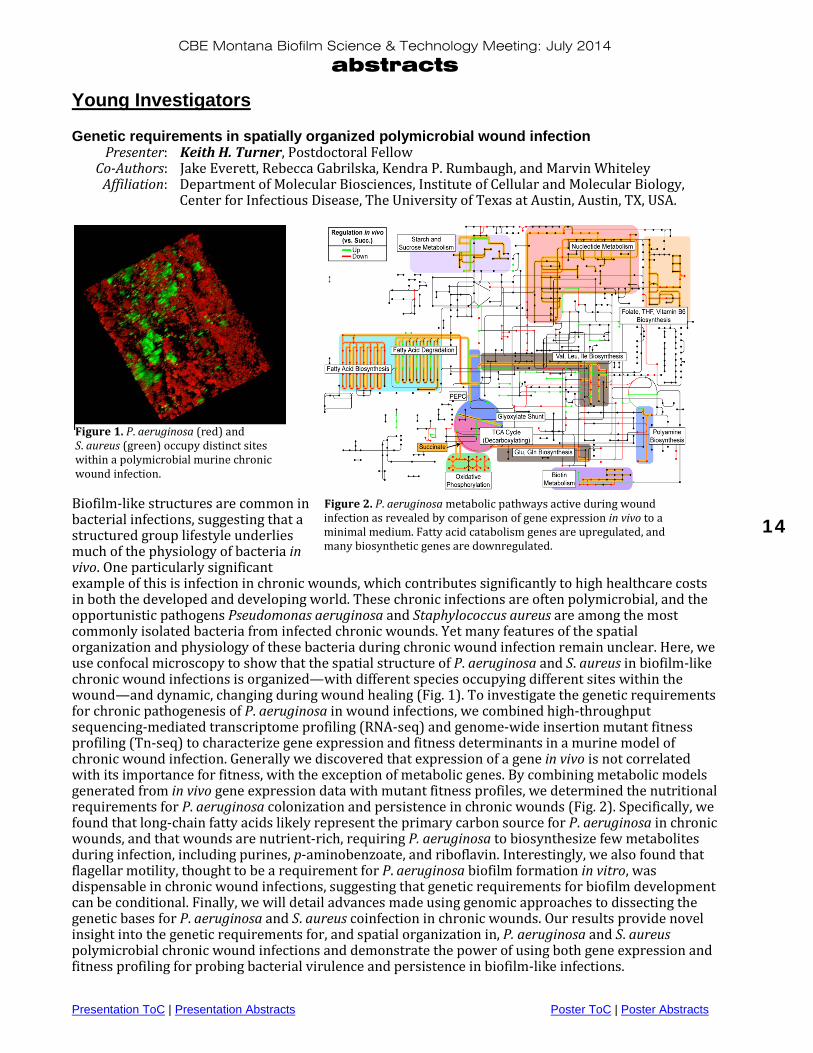
Biofilm-like structures are common in bacterial infections, suggesting that a structured group lifestyle underlies much of the physiology of bacteria in vivo. One particularly significant example of this is infection in chronic wounds, which contributes significantly to high healthcare costs in both the developed and developing world. These chronic infections are often polymicrobial, and the opportunistic pathogens Pseudomonas aeruginosa and Staphylococcus aureus are among the most commonly isolated bacteria from infected chronic wounds. Yet many features of the spatial organization and physiology of these bacteria during chronic wound infection remain unclear. Here, we use confocal microscopy to show that the spatial structure of P. aeruginosa and S. aureus in biofilm-like chronic wound infections is organized—with different species occupying different sites within the wound—and dynamic, changing during wound healing (Fig. 1). To investigate the genetic requirements for chronic pathogenesis of P. aeruginosa in wound infections, we combined high-throughput sequencing-mediated transcriptome profiling (RNA-seq) and genome-wide insertion mutant fitness profiling (Tn-seq) to characterize gene expression and fitness determinants in a murine model of chronic wound infection. Generally we discovered that expression of a gene in vivo is not correlated with its importance for fitness, with the exception of metabolic genes. By combining metabolic models generated from in vivo gene expression data with mutant fitness profiles, we determined the nutritional requirements for P. aeruginosa colonization and persistence in chronic wounds (Fig. 2). Specifically, we found that long-chain fatty acids likely represent the primary carbon source for P. aeruginosa in chronic wounds, and that wounds are nutrient-rich, requiring P. aeruginosa to biosynthesize few metabolites during infection, including purines, p-aminobenzoate, and riboflavin. Interestingly, we also found that flagellar motility, thought to be a requirement for P. aeruginosa biofilm formation in vitro, was dispensable in chronic wound infections, suggesting that genetic requirements for biofilm development can be conditional. Finally, we will detail advances made using genomic approaches to dissecting the genetic bases for P. aeruginosa and S. aureus coinfection in chronic wounds. Our results provide novel insight into the genetic requirements for, and spatial organization in, P. aeruginosa and S. aureus polymicrobial chronic wound infections and demonstrate the power of using both gene expression and fitness profiling for probing bacterial virulence and persistence in biofilm-like infections.
Figure 2. P. aeruginosa metabolic pathways active during wound infection as revealed by comparison of gene expression in vivo to a minimal medium. Fatty acid catabolism genes are upregulated, and many biosynthetic genes are downregulated.
Figure 1. P. aeruginosa (red) and S. aureus (green) occupy distinct sites within a polymicrobial murine chronic wound infection.
CBE Montana Biofilm Science & Technology Meeting: July 2014
abstracts
Presentation ToC | Presentation Abstracts Poster ToC | Poster Abstracts
15
Analyzing secondary metabolite production by 3D-printed bacterial populations using scanning electrochemical microscopy Presenter: Jodi L. Connell, Postdoctoral Fellow Co-Authors: Jiyeon Kim, Allen J. Bard, and Marvin Whiteley Affiliation: Department of Molecular Biosciences, Institute of Cellular and Molecular Biology,
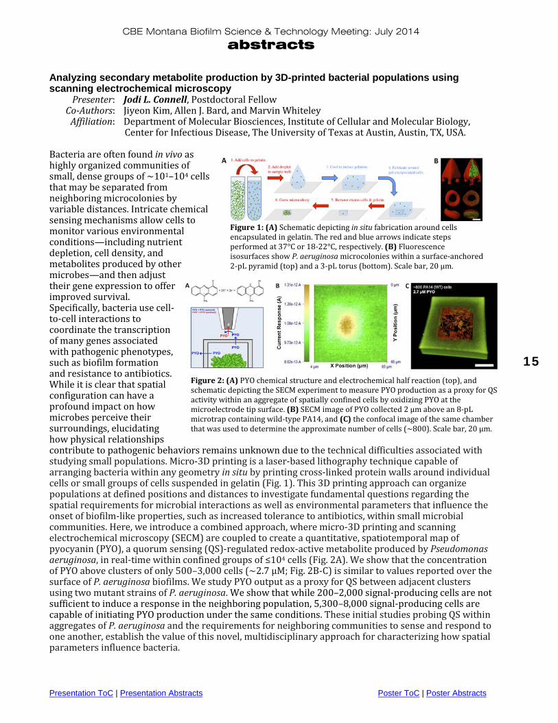
Center for Infectious Disease, The University of Texas at Austin, Austin, TX, USA. Bacteria are often found in vivo as highly organized communities of small, dense groups of ~101–104 cells that may be separated from neighboring microcolonies by variable distances. Intricate chemical sensing mechanisms allow cells to monitor various environmental conditions—including nutrient depletion, cell density, and metabolites produced by other microbes—and then adjust their gene expression to offer improved survival. Specifically, bacteria use cell-to-cell interactions to coordinate the transcription of many genes associated with pathogenic phenotypes, such as biofilm formation and resistance to antibiotics. While it is clear that spatial configuration can have a profound impact on how microbes perceive their surroundings, elucidating how physical relationships contribute to pathogenic behaviors remains unknown due to the technical difficulties associated with studying small populations. Micro-3D printing is a laser-based lithography technique capable of arranging bacteria within any geometry in situ by printing cross-linked protein walls around individual cells or small groups of cells suspended in gelatin (Fig. 1). This 3D printing approach can organize populations at defined positions and distances to investigate fundamental questions regarding the spatial requirements for microbial interactions as well as environmental parameters that influence the onset of biofilm-like properties, such as increased tolerance to antibiotics, within small microbial communities. Here, we introduce a combined approach, where micro-3D printing and scanning electrochemical microscopy (SECM) are coupled to create a quantitative, spatiotemporal map of pyocyanin (PYO), a quorum sensing (QS)-regulated redox-active metabolite produced by Pseudomonas aeruginosa, in real-time within confined groups of ≤104 cells (Fig. 2A). We show that the concentration of PYO above clusters of only 500–3,000 cells (~2.7 µM; Fig. 2B-C) is similar to values reported over the surface of P. aeruginosa biofilms. We study PYO output as a proxy for QS between adjacent clusters using two mutant strains of P. aeruginosa. We show that while 200–2,000 signal-producing cells are not sufficient to induce a response in the neighboring population, 5,300–8,000 signal-producing cells are capable of initiating PYO production under the same conditions. These initial studies probing QS within aggregates of P. aeruginosa and the requirements for neighboring communities to sense and respond to one another, establish the value of this novel, multidisciplinary approach for characterizing how spatial parameters influence bacteria.
Figure 1: (A) Schematic depicting in situ fabrication around cells encapsulated in gelatin. The red and blue arrows indicate steps performed at 37°C or 18-22°C, respectively. (B) Fluorescence isosurfaces show P. aeruginosa microcolonies within a surface-anchored 2-pL pyramid (top) and a 3-pL torus (bottom). Scale bar, 20 µm.
Figure 2: (A) PYO chemical structure and electrochemical half reaction (top), and schematic depicting the SECM experiment to measure PYO production as a proxy for QS activity within an aggregate of spatially confined cells by oxidizing PYO at the microelectrode tip surface. (B) SECM image of PYO collected 2 µm above an 8-pL microtrap containing wild-type PA14, and (C) the confocal image of the same chamber that was used to determine the approximate number of cells (~800). Scale bar, 20 µm.

CBE Montana Biofilm Science & Technology Meeting: July 2014
abstracts
Presentation ToC | Presentation Abstracts Poster ToC | Poster Abstracts
16
SESSION 4: U.S. Regulatory Review Biofilm claims for antimicrobial products: U.S. EPA regulatory perspective Presenter: Stephen Tomasino, Senior Science Advisor Affiliation: U.S. EPA Office of Pesticide Programs—Microbiology Laboratory Branch The U.S. Environmental Protection Agency’s (EPA), Office of Pesticide Programs (OPP), Office of Chemical Safety and Pollution Prevention (OCSPP), under the statutory authority of the Federal Insecticide, Fungicide, and Rodenticide Act (FIFRA), is responsible for the registration of antimicrobial products, including products intended to treat biofilm on environmental surfaces in household and health care settings. Under FIFRA, antimicrobial products are considered as pesticides. Thus, public health products with claims to prevent, destroy, repel or mitigate biofilm on an inanimate environmental surface would require registration under FIFRA—including the submission of product efficacy data. EPA’s perspective on biofilm methodology, potential label claims, and the technical aspects under consideration for regulatory guidance will be provided during the session. Biofilms express unique characteristics, and therefore require specific and relevant test methods for measuring product efficacy. The choice of method will dictate the type of label claim. Formal efficacy test guidelines have not been established for biofilm claims; however, a draft guidance document is under preparation by the EPA to inform registrants of which test methodology and microorganisms are appropriate to support a specific biofilm claim. Currently, the EPA is considering the use of ASTM method E2871-12 (Evaluating Disinfectant Efficacy against Pseudomonas aeruginosa Biofilm using the Single Tube Method) as a regulatory method; this quantitative method was collaboratively developed by the OPP Microbiology Laboratory Branch (MLB) and the Montana State University Center for Biofilm Engineering. To gain further experience with the Single Tube Method, MLB conducted a series of in-house efficacy tests on several EPA-registered disinfectants (without biofilm claims) against P. aeruginosa (ATCC No. 15442), and also conducted testing against Staphylococcus aureus (ATCC No. 6538) biofilm. Bacterial biofilms were produced using the CDC biofilm reactor procedure (ASTM method E2562-12). The test chemicals included a wide range of active ingredients, thus neutralizer confirmation was required prior to testing. The initiative was used to improve and verify an in-house standard operating procedure for the Single Tube Method and to provide the Agency with technical input on the development of regulatory guidance. Also, data from a recently completed ASTM Interlaboratory Study (Research Report E35-1008) will be used to inform the Agency on best practices for use of the methodology. Additional collaborative studies have been initiated to verify the test conditions for the generation and testing of S. aureus biofilm. MLB is also interested in revising the ASTM standards, and plans to work with Dr. Darla Goeres of Montana State University to seek technical consensus and approval of the proposed revisions. FDA/CBE joint workshop recap Presenter: Phil Stewart, CBE Director Affiliation: Center for Biofilm Engineering, Montana State University, Bozeman, MT, USA. A public workshop entitled “Biofilms, Medical Devices, and Anti-Biofilm Technology—Challenges and Opportunities” was held on the White Oak Campus of the Food and Drug Administration (FDA) in Silver Spring, Maryland, on February 20, 2014. The meeting was co-sponsored by the U.S. Food and Drug Administration and the Center for Biofilm Engineering (CBE) at Montana State University. This event brought together academic researchers and clinicians, scientists from FDA and the U.S. Environmental Protection Agency (EPA), and many representatives from industry. Topics addressed included the public health impact of biofilms, the challenges and opportunities of translating biofilm science into

CBE Montana Biofilm Science & Technology Meeting: July 2014
abstracts
Presentation ToC | Presentation Abstracts Poster ToC | Poster Abstracts
17
new anti-biofilm technologies, methods for measuring biofilms and for evaluating anti-biofilm chemistries and materials, and discussion of critical research needs to advance the development of these technologies. This presentation will summarize highlights from the workshop. Biofilm claims—EPA rules and implications Presenter: John Wood, Senior Director Agency Relations (Law and Regulatory Affairs) Affiliation: Ecolab, Inc. This presentation provides a historical background of EPA’s regulation of biofilm claims from 1982 to the present, EPA’s 2008 clarification on the use of biofilm and slime claims on EPA registered antimicrobial product labels, information published by EPA’s Antimicrobials Division regarding biofilm removal claims, and what’s next for EPA and industry on methods and claims. Biofilm test methods and impact on regulatory guidelines Presenter: LaShanda Glenn, Scientist Co-Authors: Jeff VanKomen, Senior Scientist; and Chuck Pettigrew, Principal Scientist Affiliation: Procter & Gamble, Cincinnati, OH, USA. Biofilms are important to public health in a variety of situations, from homes to commercial and institutional settings, as evidenced by over 5,000 publications related to biofilms in public health settings. Products intended to treat biofilms on inanimate surfaces are regulated by the EPA as pesticides. Consumers and professionals have a need to understand how products perform in their specific use situations and how they should be used. To meet this need there should be a clearly defined process for testing products for efficacy against biofilms, support claims for registration, and communicate to the user. A number of methods have been developed for assessing product performance for specific applications. Of these, attention has recently focused on two ASTM methods as appropriate for supporting public health claims. These are ASTM E2562-12 “Standard Test Method for Quantification of Pseudomonas aeruginosa Biofilm Grown with High Shear and Continuous Flow using CDC Biofilm Reactor” and ASTM E2871-13 “Standard Test Method for Evaluating Disinfectant Efficacy Against Pseudomonas aeruginosa Biofilm Grown in a CDC Bioreactor Using Single Tube Method.” Important characteristics for the inclusion of these methods in developing regulatory guidelines include intended use of the product, specific organisms to be evaluated, growth conditions of the organisms in the environment and in standardized tests, exposure conditions, efficacy determination, success criteria and others. Appropriate testing with these methods could support a variety of claims that help the user determine the suitability for their specific application. Additional research is essential to optimize these test methods not only for the required organisms for biofilm claims, but also additional organisms that may be of interest in specific environments. This presentation outlines an industry perspective on proposed biofilm methods and observations that could potentially impact the ability of industry to meet consumer needs.

CBE Montana Biofilm Science & Technology Meeting: July 2014
abstracts
Presentation ToC | Presentation Abstracts Poster ToC | Poster Abstracts
18
SESSION 5: New CBE Capabilities: Micromechanics & Microfluidics Tools for measuring biofilm mechanical properties Presenter: James N. Wilking, Assistant Professor, Chemical and Biological Engineering Affiliation: Center for Biofilm Engineering, Montana State University, Bozeman, MT, USA. Knowledge of biofilm mechanical properties is essential for a variety of industrial applications; beneficial biofilms must be repaired and maintained and detrimental biofilms must be removed. Despite this critical need, biofilm mechanical properties remain poorly understood. This is due to at least two factors. First, biofilms are typically too thin and heterogeneous for traditional mechanical measurements. Second, lack of knowledge regarding the composition of the extracellular matrix and the genes responsible for biofilm formation has prevented the development of a material-based understanding of biofilm mechanics. In this talk, I will present a variety of micromechanical measurement capabilities, including optical force spectroscopy, thermally driven microrheology and actively driven microrheology that we are currently developing for use in our lab. I will also briefly discuss the application of these techniques to the study of model microbial biofilms. Drop-based microfluidics for biological applications: From colloidal dispersions to high-throughput assaying Presenter: Connie Chang, Assistant Research Professor, Chemical and Biological Engineering Affiliation: Center for Biofilm Engineering, Montana State University, Bozeman, MT, USA. Using drop-based microfluidics, emulsion drops can be created one at a time in microscale channels within microfluidic devices. These drops have volumes that range from picoliters to nanoliters and are created at high-throughput rates, up to thousands per second. Monodisperse emulsions can be used for applications in pharmaceuticals, oil recovery, catalysis, and encapsulation technology in food and cosmetics. Here, drop-based microfluidics is presented as a method for engineering emulsion-templated materials, including liquid-, polymer-, and hydrogel-based particles with controlled functionality and tunable mechanical properties. Drop-based microfluidics will also be presented as a method for high-throughput assaying and sensing for biological applications. SESSION 6: Bacterial Survival in Industry and the Environment Hot water disinfection of planktonic and biofilm bacteria Presenters: Mark Pasmore1, Research Manager Diane K. Walker2, Research Engineer Co-Authors: Laura Wahlen1, Al Parker2, Paul Sturman2 Affiliation: 1Baxter International, Inc. 2Center for Biofilm Engineering, Montana State University, Bozeman, MT, USA. Baxter was interested in understanding the efficacy of hot water in controlling microorganism contamination in the manufacturing environment. A study was developed to assess the hot water inactivation of a Sphingomonas parapaucimobilis biofilm, an organism isolated from a production facility, to simulate a representative challenge. Experiments were conducted at Baxter (planktonic) and CBE (biofilm) to test the effect of exposure to 65, 70, 75 and 80 °C water at various contact times. For both planktonic and biofilm tests, the results clearly demonstrate a temperature/time dependence on kill rate, with biofilm being substantially more tolerant to treatment—an observation that is also typically seen in efficacy testing with antimicrobial agents.

CBE Montana Biofilm Science & Technology Meeting: July 2014
abstracts
Presentation ToC | Presentation Abstracts Poster ToC | Poster Abstracts
19
Systems analysis of iron-limited growth: Insights into pathogen metabolic acclimation to host Presenter: Ross Carlson1,2, Associate Professor, Chemical and Biological Engineering Co-Authors: James Folsom1, Postdoctoral Researcher; Albert Parker1, Biostatistician Affiliation: 1Center for Biofilm Engineering, and 2Department of Chemical and Biological Engineering, Montana State University,
Bozeman, MT, USA. Iron bioavailability is a major limiter of bacterial growth in mammalian host tissue and thus represents an important area of study. Escherichia coli K-12 metabolism was studied at four levels of iron limitation in chemostats using physiological and proteomic analyses. The data documented an E. coli acclimation gradient, where progressively more severe iron scarcity resulted in a larger percentage of substrate carbon being directed into an overflow metabolism accompanied by a decrease in biomass yield on glucose. Acetate was the primary secreted organic byproduct for moderate levels of iron limitation, but as stress increased, the metabolism shifted to secrete primarily lactate (~ 70% of catabolized glucose carbon). Proteomic analysis reinforced the physiological data and quantified relative increases in glycolysis enzyme abundance and decreases in tricarboxylic acid (TCA) cycle enzyme abundance with increasing iron-limitation stress. The combined data indicated that E. coli responds to limiting iron by investing the scarce resource into essential enzymes at the cost of catabolic efficiency (i.e., down regulating high ATP-yielding pathways containing enzymes with large iron requirements like the TCA cycle). Acclimation to iron-limited growth was contrasted experimentally with acclimation to glucose-limited growth to identify both general and nutrient-specific acclimation strategies. While the iron-limited cultures maximized biomass yields on iron and increased expression of iron acquisition strategies, the glucose-limited cultures maximized biomass yields on glucose and increased expression of carbon acquisition strategies. This study quantifies ecologically competitive acclimations to nutrient limitations, yielding knowledge essential for understanding medically relevant bacterial responses to host and to developing intervention strategies. Field-scale plugging of hydraulic fractures using ureolytic bacteria Presenter: Al Cunningham1,2, Professor, Civil Engineering; Robin Gerlach1,3, Associate Professor,
Chemical and Biological Engineering; and Adie Phillips1,3, Research Engineer Co-Authors: Randy Hiebert, Jim Kirksey (Schlumberger), Ellen Lauchnor, Lee Spangler Affiliation: 1Center for Biofilm Engineering, 2Department of Civil Engineering, 3Department of Chemical and Biological Engineering, Montana State University,
Bozeman, MT, USA.
Ureolytic microorganisms (biofilms) contribute urease, which catalyzes the hydrolysis of urea. In the presence of calcium, this reaction can create saturation conditions favorable for precipitation of calcium carbonate (“biomineralization”). Applications such as sealing hydraulic fractures with biomineralization have been successfully demonstrated in the laboratory. A field study was initiated in
a Figure 1. a) Filling the bailer with ureolytic microorganisms for delivery downhole. b) 9 5/8” Gorgas #1 well head.
b

CBE Montana Biofilm Science & Technology Meeting: July 2014
abstracts
Presentation ToC | Presentation Abstracts Poster ToC | Poster Abstracts
20
April 2014 at a 4915’ deep Gorgas #1 well at the Southern Company Gorgas Steam Generation Plant in Walker County, Alabama. First, tubing and packer were set to isolate the Fayetteville Sandstone at approximately 1118’ below ground surface. The Sandstone was perforated with 6 shots in 60 degree phasing before 24 g/L NaCl amended water (brine) was pumped to increase the pressure until formation breakdown. The formation fractured with a surface pressure of 960 psi with a 2.5 gallons per minute (gpm) brine flow rate. After a 6-hour injection test at 0.5 gpm, where the pressure averaged 510 psi, the pump was shut down and the well was shut in to initiate an 84-hour pressure falloff test. Following the pressure fall off test, ureolytic microorganisms were cultured and placed via bailer delivery into the zone near the perforations. Brine was pumped at 0.5 gpm through the tubing string to push the cells into the fracture and formation (inoculate). Concentrated calcium and growth component containing media were also dropped into the perforated zone by bailer delivery and brine was pumped at 0.5 gpm to dilute and push the reagents into the fracture and formation. After 3 days of injection of 21 calcium pulses and 5 inoculations with ureolytic microorganisms, injection into the formation was reduced. During the fourth day, the brine injection flow rate was reduced to 0.14 gpm to avoid increased pressure and re-fracture as the final inoculation and 3 more calcium pulses were delivered. Pressure falloff was monitored for the first five minutes after terminating pumping. The falloff pressure continued to improve (less pressure falloff over time) over the course of the experiment and ended with a promising 7% pressure falloff, down from 35% over 5 minutes prior to biomineralization treatment. At the termination of the experiment, the formation was again fractured. The post- experiment fracturing pressure occurred at 1198 psi while pumping brine at 0.5 gpm. Prior to biomineralization, the downhole fracture extension pressure was approximately 1420 psi, but after treatment was approximately 1640 psi. After re-fracturing, a surface pressure of 1116 psi at 3.3 gpm was necessary to inject working fluid into the formation, indicating reduced ability to inject after biomineralization treatment. Viable ureolytic microorganisms were recovered from a sample collected with a Kuster Sampler from the mixing zone below the packer and near the facture during the experiment. These promising results suggest the potential for sealing hydraulic fractures at the field scale with ureolytic microorganism biomineralization treatments. Presence, pervasiveness, and persistence of wastewater pathogen Escherichia coli O157:H7 in model treatment wetlands Presenter: Rachel VanKempen-Fryling, PhD student, Microbiology Co-Authors: Anne Camper, Professor, Civil Engineering Affiliation: Center for Biofilm Engineering, Montana State University, Bozeman, MT, USA. Previous research has shown that treatment wetlands (TWs) efficiently remove a variety of pollutants including a several log-order reduction of pathogens from influent to effluent. However, pathogen removal mechanisms are not well documented and there is evidence to suggest that pathogen cells sequestered in a sub-surface wetland may remain viable months after inoculation. Esherichia coli is a common pathogen in domestic and agricultural wastewater and the O157:H7 strain is the main cause of outbreaks and infection in the United States. To assess apparent persistence of E. coli within the TW matrix allowing for later release, direct measurements of E. coli levels within a planted gravel matrix and root rhizosphere were taken. The intent is to better understand initial attachment and persistence of E. coli within existing biofilms surrounding roots and abiotic attachment sites. Initial experiments were performed in hydroponic reactors (300mL volume) containing either glass wool as an abiotic control or roots of Carex utriculata or Schoenoplectus acutus at an average of 49.3mL ± 26.4mL per 300mL (three replicates per experimental treatment). The reactors were fed a constant flow of simulated wastewater at a 2-hour residence time. The influent was inoculated with E. coli O157:H7- containing a gene for the DsRed fluorescent protein, and the total effluent was collected over three residence times to determine initial wash-out. Root samples were excised and analyzed via epifluorescent microscopy cell counts and DNA extraction for RT-PCR. E. coli O157:H7 was detected on

CBE Montana Biofilm Science & Technology Meeting: July 2014
abstracts
Presentation ToC | Presentation Abstracts Poster ToC | Poster Abstracts
21
the root surface at 2 hours post-inoculation, and was visible under microscopy as single cells. Microcolonies began forming at 24 hours post-inoculation and were detected for up to 1 week post-inoculation. Image analysis determined that the number of microcolonies with >100 cells increased 1 week post-inoculation, supporting the view that E. coli O157:H7 is capable of growth within biofilms surrounding wetland plant roots. An abiotic surface of nylon ranging from 0.77mm to 1.022mm in diameter was used in the hydroponic reactor systems to compare attachment to the root versus an inert surface. Collection of results is ongoing. Current experiments repeated the general procedure using 50 cm tall by 20 cm diameter microcosms planted with Carex utriculata and Schoenoplectus acutus compared to gravel-only controls. Experimental microcosms are run in duplicate. Initial results show consistent evidence of E. coli survival on roots comparable to that of the hydroponic systems. Keywords: biofilm, epiflorescence, PCR Carex, Schoenoplectus Monitoring Chlorella survival during algal biofuel production using a community ecology approach Presenter: Tisza Bell, PhD student, Microbiology Co-Authors: Peyton BM1, Prithiviraj B2, Wahlen BD3, Fields MW1 Affiliation: 1Center for Biofilm Engineering, Montana State University, Bozeman, MT, USA. 2University of Colorado, Boulder, CO, USA. 3Utah State University, Logan, UT, USA.
Figure 1. A) The blue line depicts the growth curve for Chlorella vulgaris during the 16-day pond run measured in total suspended solids (TSS). The green curve shows the traditional growth curve for algae. Starred points indicate samples that were pyrosequenced. B) Chao diversity for each domain plotted over time. Archaea started with a high Chao diversity, which quickly declined. Eukarya maintained a steady diversity level composed almost entirely of C. vulgaris. Bacterial diversity steadily increased with time. The majority of research on algal biofuel production has been conducted on single species isolates in closed systems that are costly to maintain. In contrast, open ponds are estimated to be an order of magnitude less expensive to run than closed systems, but overall productivity is typically lower than in closed systems. Use of extremophile alkaliphilic algae may help overcome some of the constraints associated with large-scale biofuel production. It is hypothesized that open alkaline systems are not easily colonized by other species because the restrictive environment limits diversity that allows for better selective control and management of a desired biofuel-producing algal species. In this study, we monitored the microbial community in an outdoor 2,000 liter open raceway pond. A previously isolated high lipid-producing alkaliphilic alga, Chlorella vulgaris, was cultivated for

CBE Montana Biofilm Science & Technology Meeting: July 2014
abstracts
Presentation ToC | Presentation Abstracts Poster ToC | Poster Abstracts
22
approximately two weeks before being harvested. Community DNA samples were collected over the two-week period in conjunction with water chemistry. Universal primers for the SSU rRNA gene sequences for Eukarya, Bacteria, and Archaea were used for barcoded pyrosequence determination. The most influential parameters on C. vulgaris abundance were pH and phosphate. Results also indicated that the pond system did not remain mono-algal, but was colonized by other microbial organisms, further contributing to fluctuations in the abundance of C. vulgaris. However, likely due to the high pH of the system, C. vulgaris remained the dominant organism and never represented less than 49% of the community. The characterization of the microbial community dynamics of an alkaliphilic open pond system will provide significant insight into control and optimization for biomass production. Biofouling on household reverse osmosis water treatment membranes Presenter: Stephen Markwardt, master’s student, Environmental Engineering Co-Authors: Anne K. Camper, Professor, Civil Engineering Affiliation: Center for Biofilm Engineering, Montana State University, Bozeman, MT, USA. Reverse osmosis (RO) is a principal method for creating potable water and is being used widely for the desalination of ocean and brackish waters. RO technology has also been scaled down for household and small community applications. In these applications, RO is being used to treat marginal-quality fresh water from natural or municipal sources. Increasing future demands for potable water and changing climate patterns may create a greater need for RO water treatment, as lower quality sources of water must be exploited. While RO water treatment shows great promise, it suffers from one very large problem: membrane fouling. There are four types of membrane fouling: inorganic fouling, colloidal fouling, organic fouling, and biofouling. Of the fouling types, biofouling is the most troublesome. Traditional means for assessing fouling, such as pressure and flux monitoring and membrane autopsies, have severe drawbacks. Monitoring pressure and flux cannot determine the type of fouling. While membrane autopsies can assess the type of fouling, this is an endpoint because it permanently destroys the membrane. Therefore, new non-destructive, real-time methods are required for assessing fouling type. This presentation will cover the basic aspects of RO water treatment, describe some of the problems associated with it, and delve into some research work concerning fouling in household RO water treatment membranes.

CBE Montana Biofilm Science & Technology Meeting: July 2014
abstracts
Presentation ToC | Presentation Abstracts Poster ToC | Poster Abstracts
23
Poster Abstracts
Industry & Agency Posters Title: The Sharklet micropattern limits bacterial adherence and biofilm:
A potential technological improvement for endotracheal tube design Date: July 2014 Authors: Ethan E. Mann1, May RM1, Mettetal MR1, Hoffman MG1, Sogo MJ1, Parker AE2,
O’Toole GA3, Brennan AB4, Reddy ST1 Affiliation: 1Sharklet Technologies, Inc., Aurora, CO, USA. 2Center for Biofilm Engineering and the Department of Mathematical Sciences,
Montana State University, Bozeman, MT, USA. 3Geisel School of Medicine at Dartmouth, Hanover, NH, USA.
4Department of Materials Science and Engineering, University of Florida, Gainesville, Florida, USA.
Background: Airway management of patients on mechanical ventilation (MV) has garnered greater attention lately with updated CDC definitions of ventilator-associated events (VAEs), which include ventilator-associated conditions (VACs), VACs with infection present (iVACs), and ventilator-associated pneumonia (VAP). The lumen of endotracheal tubes (ET) and any host secretions are easy targets for bacterial colonization due to the inability for mucociliary transport to clear microorganisms, which can ultimately result in VAP. To combat this issue, implementation of the Sharklet micropattern, a novel microscopic ordered surface topography, on ET surfaces may provide an innovative strategy for iVAC or VAP prevention. Bacterial attachment and biofilm growth were evaluated in culture medium as well as mucin-modified medium to simulate the interactions with host secretions in the tracheal environment in vitro. Methods: The top five pathogens associated with ET-related pneumonia—Methicillin Resistant Staphylococcus aureus (MRSA), Pseudomonas aeruginosa, Klebsiella pneumoniae, Acinetobacter baumannii, and Escherichia coli—were evaluated for attachment to micropatterned and unpatterned silicone surfaces in a short-term colonization assay. Two key pathogens, MRSA and P. aeruginosa, were cultured in growth media on the test and control surfaces using a static biofilm assay and a drip-flow
S. aureus TSBP. aeruginosa TSB+mucinP. aeruginosa TSB
1.6
1.4
1.2
1.0
0.8
0.6
0.4
0.2
Biofilm Cond.
CFU
Log
Red
ucti
on
Boxplot of CFU Log Reduction
Figure 1. Sharklet reduces biofilm accumulation. Sharklet and smooth samples were compared in the drip-flow biofilm assay for 48-h (P. aeruginosa) and 96-h (S. aureus).

CBE Montana Biofilm Science & Technology Meeting: July 2014
abstracts
Presentation ToC | Presentation Abstracts Poster ToC | Poster Abstracts
24
biofilm assay. P. aeruginosa was further evaluated for biofilm formation on test and control surfaces in a mucin-modified medium using both the static biofilm and drip-flow assays. Results are reported as mean log reductions and derived median percent reductions based on t-tests, which also provide p values. All experiments were replicated at least three times. Results: Sharklet micropatterned surfaces demonstrated reductions in microbial colonization for a broad range of species, with up to 99.9% (p<0.05) reduction compared to unpatterned controls. In static biofilm growth conditions, Sharklet reduced MRSA and P. aeruginosa biofilm 67% (p=0.12) and 52% (p=0.05), respectively. Using static biofilm conditions with mucin-modified media, Sharklet reduced P. aeruginosa biofilm 58% (p<0.01) compared to unpatterned controls. In the drip-flow assay, MRSA and P. aeruginosa biofilm was reduced 83% (p<0.05) and 90% (p<0.01), respectively, on Sharklet compared to unpatterned surfaces. In the drip-flow assay with mucin-conditioned media, P. aeruginosa biofilm was reduced 85% (p=0.058) on Sharklet compared to unpatterned surfaces. Conclusions: The Sharklet micropattern reduces colonization and biofilm formation of predominant VAP-associated pathogens in vitro. Implementation of the Sharklet micropattern on endotracheal tubes may prevent or prolong the onset of VAP without the need for antimicrobial agents. Title: The use of CDC biofilm reactor to test cleaning and disinfection capabilities
on rouged stainless steel Date: 07/2014 Authors: Amanda Deal, Klein D, Lopolito P, Spencer Schwarz Affiliation: STERIS Manufacturers of cGMP products in a highly regulated and technically challenging environment encounter many cleaning and disinfection issues. Among these is biofilm, commonly found in manufacturing vessels, utility lines, and other processing equipment. Where stainless steel is used in production, one may also find rouge, an iron oxide deposit caused by the oxidation of steel by aqueous solutions. Surfaces contaminated with biofilm are difficult to clean and difficult to disinfect. It is reasonable to suppose that the presence of rouge hinders the ability of detergents and biocides to remove biofilm by increasing the surface area of contaminated substrates. Further, a rouged surface may exacerbate a microbial excursion and promote the development of biofilm. Here we demonstrate the challenge to cleaning and disinfection posed by P. aeruginosa biofilm formed on rouged stainless steel coupons using the CDC biofilm reactor system. Cleaning and disinfection were evaluated using total organic carbon surface analysis, visual cleanliness and microbial efficacy testing. The data collected indicate that rouged surfaces are more difficult to clean and more difficult to disinfect. The demonstrated increase in resistance to remediation highlights the need to employ effective cleaning, preventative maintenance and disinfection strategies in a contamination control program.
CBE Montana Biofilm Science & Technology Meeting: July 2014
abstracts
Presentation ToC | Presentation Abstracts Poster ToC | Poster Abstracts
25
Academic Posters (non-CBE) Title: Using Surface Plasmon Resonance imaging (SPRi) to evaluate bacterial
activity on surfaces Date: 07/2014 Authors: Edgar Goluch, Abadian PN Affiliation: Department of Chemical Engineering, Northeastern University, Boston, MA, USA. Sponsored by: Partial support from the U.S. National Science Foundation under Grant No. 1125535,
and a Northeastern University Tier 1 Interdisciplinary Research Seed Grant. Surface plasmon resonance imaging (SPRi) provides continuous, label-free, high-spatial-resolution monitoring of physical changes that occur on surfaces that are up to one square centimeter in area. The Goluch Group utilizes SPRi technology to address the challenges of quantitatively evaluating the efficacy of materials in preventing bacterial adhesion when exposed to flowing fluids. A multiplexed analysis format is particularly important in bacterial attachment studies, as adhesion events are very sensitive to their local micro-environment, which is difficult to reproduce and control between experiments. In this study, the effectiveness of bovine serum albumin (BSA), casein, and penicillin/streptomycin surface coatings to prevent Pseudomonas aeruginosa and Staphylococcus aureus adhesion is investigated with SPRi. The coatings were deposited on different sections of a single gold SPRi sensing surface and monitored for 24 hours while being exposed to a continuous flow of growth medium containing bacterial cells. We found that casein most effectively inhibits attachment of cells to the gold surface over a 24-hour period, with an 80% decrease in adhesion versus a bare gold surface for P. aeruginosa and a 60% decrease for S. aureus. Title: Effect of removal of biofilm on titanium surface applied by ultrasonic water
flow technology Date: 07/2014 Authors: Matsuo Yamamoto1, Masanori Sato2, Shunjirou Kume3, Kei Saito3, Takashi Takiguchi1 Affiliation: 1Department of Periodontology, Showa University School of Dentistry, Japan. 2Honda Electronics Co., LTD, Aichi, Japan. 3GC Corporation R&D Center, Tokyo, Japan. Objective: Microbial biofilm stimulates inflammatory processes of peri-implantitis, and possible dental implant loss. Although the most important step in management of peri-implantitis is removal of biofilm from the titanium surface, an effective method is still not established. On the other hand, the ultrasonic water flow cleaner has been used for precise cleaning of silicon wafers; this technology involves action of vibrational acceleration and rectilinear flow. The aim of this study was to demonstrate the removal of biofilm from titanium surfaces by using the ultrasonic water flow medical device development. Methods: Optimal conditions were identified by the correlation between sound pressure and removal of biofilm. Plaque biofilm was formed on titanium specimens kept intra-orally for 72h with eight volunteers. For each titanium specimen, residual plaque biofilm (RPB) areas were evaluated as a percentage of the scanned surface selected at random by digital microscope. The decontaminated titanium surfaces were analyzed by energy dispersive X-ray spectroscopy (EDX) and scanning electron microscope (SEM). Results: The optimal ultrasonic condition was 320 kHz at an intensity of 12W. Our data shows the ultrasonic water flow was effective for considerable reduction of biofilm from microstructure titanium. After exposure to the ultrasonic water flow for 3 min, microorganisms or water-insoluble glucans were not observed on the titanium surfaces. EDX revealed that the chemical composition of the titanium surface had not changed in relation to the ultrasonic water flow.

CBE Montana Biofilm Science & Technology Meeting: July 2014
abstracts
Presentation ToC | Presentation Abstracts Poster ToC | Poster Abstracts
26
Conclusion: These results suggested that the ultrasonic water flow exposure in a non-contact mode effectively removed adherent biofilm on microstructure titanium. Title: Toward a broader appreciation for backyard biofilms: Soils Date: 07/2014 Authors: Tony Hartshorn, Sugden J, Sanchez D, Atkins S, McDermott T, Zabinski C Affiliation: Department of Land Resources and Environmental Sciences, Montana State
University, Bozeman, MT, USA. Sponsored by: MSU VP for Research and Economic Development, MSU College of Agriculture Bacteria are most often associated with biofilms, which can be defined as “gated microbial communities,” in contrast to their traditional depiction as planktonic (isolated) individuals. Our laboratory is encouraging those working in the field of biofilm engineering to consider the relatively poorly characterized biofilms in the broader (but ungated!) “Yellowstone Club” represented by northern Rocky Mountains and northern Great Plains soils (Figure 1). We seek to explore whether the patterns and processes that dominate biofilms are fractal. That is, are the structures and functions we can define for biofilms at the micron scale the same at the meter scale? Here we frame a cross-scale biofilm (CSB) approach (Figure 2), relying on research sites arrayed across a continuum of disturbances, with four primary endpoints: (i) unusually high levels of mechanical disturbance; (ii) unusually high levels of acidity; (iii) unusually high levels of hydrocarbons (±halogens); and (iv) unusually high levels of salinity and/or sodicity. At the core of our CSB framework lie relatively undisturbed ecosystems such as unharvested fields and forests, as well as undrained wetlands.
Figure 1. (above) Attempt to set a world record for soybean harvest in Monkton, Canada (120 combines on a 160-acre field). This image illustrates biofilm engineering at the meter- or kilometer-scale, versus the more traditional micron scale associated with biofilms. Source.
Figure 2. (right) Disturbance gradients provide an approach to characterizing biofilm patterns and processes across scales.

CBE Montana Biofilm Science & Technology Meeting: July 2014
abstracts
Presentation ToC | Presentation Abstracts Poster ToC | Poster Abstracts
27
At the outer perimeter lie sites representing intensively cultivated fields (Bozeman’s Post and Ft. Ellis farms), intensively harvested forests (Mt. Ellis), acid mine drainage (Clark Fork), hydrocarbons (Bozeman’s Idaho Pole Plant Superfund site; soils affected by the 2011 Yellowstone River oil spill), and, finally, the saline-sodic soils of the Hailstone National Wildlife Refuge. We look forward to collaborating on approaches for quantifying the degree to which biofilms are fractal. To the extent that biofilms are fractal, we look forward to developing applications where microbial communities are managed for ecosystem-level (kilometer-scale) outcomes; to the extent that biofilms are not fractal, we look forward to developing scale-specific applications where manipulations of the biofilms at one scale might drive processes at alternate scales. Center for Biofilm Engineering Posters CBE Poster #619 Title: Applicability of MICP in subsurface and fractured environments Date: 12/2013 Authors: Adrienne J. Phillips 1,2* Eldring J1, Hiebert R5, Lauchnor E1, Mitchell AC6, Esposito R7,
Gerlach R1,2, Cunningham A1,3, Spangler L4 Affiliation: 1Center for Biofilm Engineering, 2Department of Chemical and Biological Engineering, 3Department of Civil Engineering, and 4Energy Research Institute, Montana State University, Bozeman, MT, USA. 5Center for Advanced Mineral and Metallurgical Processing at Montana Tech, Butte, Montana, USA. 6Institute of Geography and Earth Sciences, Aberystwyth University, Aberystwyth, Ceredigion UK SY23 3DB. 7Southern Company, Birmingham, Alabama, USA. Sponsored by: U.S. Department of Energy Subsurface leakage mitigation strategies using ureolytic biofilm- or microbially induced calcite (CaCO3) precipitation (MICP) have been investigated for sealing high permeability regions, such as fractures under subsurface relevant conditions. This technology may help in the deep subsurface to improve security of geologically stored carbon dioxide, seal subsurface hydraulic fractures, or enhance oil recovery. Sealing technologies using low-viscosity fluids, such as those used to promote MICP are advantageous since they may penetrate small aperture fractures not reachable by cement-based sealing technologies. MICP has also been researched by others for applications including: consolidating porous materials, improving or repairing construction materials, and remediating environmental concerns. First, injection strategies to control saturation conditions and region-specific precipitation were developed in two-foot long sand-filled columns. Sporosarcina pasteurii biofilms were promoted and calcium and urea solutions were injected to stimulate mineralization. These injection strategies resulted in: 1) promoting homogeneous CaCO3 distribution along the flow path, 2) minimizing near-injection point plugging, and 3) enhancing precipitation efficiency by periodically reviving ureolytic activity. Second, the developed injection strategies were used to reduce permeability and ultimately twice seal a hydraulically fractured, 74 cm diameter (meso-scale) Boyles Sandstone core under ambient pressures. Third, a novel high pressure test vessel was developed to study MICP at subsurface relevant pressures (up to 96 bar). The fractured core’s permeability was reduced by more than two orders of magnitude after promoting MICP under 44 bar of confining pressure. In a recent high pressure meso- scale MICP experiment, non-homogeneous, preferential flow paths were observed as cemented regions

CBE Montana Biofilm Science & Technology Meeting: July 2014
abstracts
Presentation ToC | Presentation Abstracts Poster ToC | Poster Abstracts
28
in a porous media sand pack. The preferential cementation was hypothesized to be formed due to density differences and gravity-driven flow between urea/calcium medium and the confining fluids.
These studies suggest biofilm-induced CaCO3 precipitation technologies may potentially strengthen or seal high permeability regions or fractures, but point to the need to further study and model MICP under relevant subsurface pressure conditions. CBE Poster #620 Title: Using biomineralization sealing for leakage mitigation in shale Date: 08/2013 Authors: Robin Gerlach1,2, Rothman A1,2, Hiebert R4, Cunningham A1,3, Busch A5 Affiliation: 1Center for Biofilm Engineering; 2Department of Chemical and Biological Engineering; and 3Department of Civil Engineering, Montana State University, Bozeman, MT, USA. 4Montana Emergent Technologies, Inc., Butte, MT, USA. 5Shell Global Solutions. Sponsored by: US Department of Energy and the Carbon Capture Project 3 (CCP3) Estimates of the number of abandoned wells in the U.S. and abroad range in the millions; all of them have a high probability of eventual leakage. We are currently focusing on developing technologies for sealing unwanted leakage pathways in fractured shales. We are investigating the feasibility of a plugging technology based on the microbially induced precipitation of carbonate minerals. Microbes can hydrolyze urea to ultimately change the saturation state of various minerals, including carbonates, such as calcium carbonate. We have demonstrated that the resulting bio-cement (calcite) can cement together heavily fractured shale and drastically reduce the permeability of fractures in shale cores. We propose this technology for mitigating leakage from abandoned wells and as an alternative to more traditional cement-based plugging technologies. We have demonstrated the principal feasibility of this technology for ensuring geologic CO2 storage in deep saline aquifers through the plugging of small aperture leaks such as fractures or delamination interfaces in the vicinity of injection wells. Fractured shale might reduce production efficiency, as well as pose a risk to the environment due to leakage of hydrocarbons in the form of gas and liquid. The biomineralization technology can be delivered via low viscosity fluids and could potentially have significant advantages including a time- and space-dependent placement of bio-cement plugs in the immediate vicinity of wells, as well as further away from the wellbore in the rock formation.

CBE Montana Biofilm Science & Technology Meeting: July 2014
abstracts
Presentation ToC | Presentation Abstracts Poster ToC | Poster Abstracts
29
CBE Poster #621 Title: A ruggedness analysis of sonication in the Single Tube Method
(ASTM E2871-12) Date: 07/2014 Authors: Blaine Fritz1, Walker DK1, Parker AE1,2 Affiliation: 1Center for Biofilm Engineering, Montana State University, Bozeman, MT, USA. 2Department of Mathematical Sciences, Montana State University, Bozeman, MT, USA.
The effect of varying sonication parameters (time, power, degas) on the biofilm responses in the Single Tube Method (STM) (ASTM E2871) became an important area of investigation after a 9-lab inter-laboratory study (ILS) examining repeatability and reproducibility of ASTM Method E2871, in which there was considerable heterogeneity among sonicators. This ruggedness analysis examined the effect that these small deviations from the method have on the log density (LD = log10(CFU/cm2)) of biofilm recovered, an important response in the STM. A Pseudomonas aeruginosa (ATCC 15442) biofilm was grown following ASTM E2562 and sampled with STM, which calls for a series of vortex (30s, high) and sonicate (45kHz, 30s, 20W) steps to remove and disaggregate the biofilm. Thirty-three coupons were sampled according to the STM and subjected to different combinations of power (20W, 80W, 100W), time (25s, 30s, 35s), and degas (Y/N). The data was analyzed by a centered regression analysis, which centered time and power on their median values (30s, 80W) to allow for a more meaningful interpretation of the resulting regression equations. There was a statistically significant difference in the effect of time and power depending on whether the sonicator was degassed (i.e., there was a significant 3-way interaction, p=0.045) prompting individual analyses of degassed and non-degassed LD values. For the non-degassed samples, neither power nor time significantly affected the mean LD of biofilm bacteria. For the degassed samples, power and time did have a significant effect (p=0.035). In other words, changing power and time seems to have more of an effect when the sonicator has been degassed. Figure 1 displays the regression equations from these analyses. The low values for the coefficients suggest that, although significant effects of power, time, and degas were reported for the degassed samples, the method is rugged over the range of values tested for the sonication parameters.
Figure 1. Regression equations from a centered regression analysis of degassed and non-degassed, log10 (CFU/cm2), biofilm data. Centered power and time are represented by (P-80) and (T-30), respectively. There is a significant difference between the two-way interacting effect of power and time depending on if the sonicator has been degassed. The low coefficient values suggest that the method is, however, rugged for practical purposes regardless of whether the sonicator has been degassed.

CBE Montana Biofilm Science & Technology Meeting: July 2014
abstracts
Presentation ToC | Presentation Abstracts Poster ToC | Poster Abstracts
30
CBE Poster #622 Title: Biofilm parameter estimation using inverse methods Date: 07/2014 Authors: Ben Jackson1,2, Connolly, J1,3, Parker, A1, Klapper, I1,4, Gerlach, R1,3 Affiliation: 1Center for Biofilm Engineering, 2Department of Mathematical Sciences, and 3Department of Chemical and Biological Engineering, Montana State University,
Bozeman, MT, USA. 4Department of Mathematics, Temple University, Philadelphia, PA, USA. Sponsored by: National Science Foundation grant 0934696 Microbially induced calcite precipitation (MICP) has potential applications in subsurface engineering. MICP takes place in complex systems that contain biofilms in which reaction rates that induce precipitation are not well known. We seek to characterize these rates in a biofilm system via two different approaches. In the first approach, we formulate a forward ODE (ordinary differential equation) model and then solve the inverse problem using basic Bayesian methods. In the second, a detailed COMSOL model is created to solve the Navier-Stokes equations and least squares minimization is used to fit experimental data. Both approaches make use of data from tube reactor experiments conducted at Montana State University’s Center for Biofilm Engineering. CBE Poster #623 Title: Toward multiscale metabolic network analysis of an anaerobic microbial
community Date: 07/2014 Authors: Kristopher Hunt1, Adam Z2, Bell T1, Camilleri L1, Connolly J1, Lohman E1, Michaud A2,
Smith H1, Taffs R1, Tigges M1, Folsom J1, Carlson R1, Fields M1, Foreman C1, Gerlach R1, Inskeep W2
Affiliation: 1Center for Biofilm Engineering, and 2Department of Land Resources and Environmental Sciences, Montana State
University, Bozeman, MT, USA. Sponsored by: National Science Foundation IGERT Program; NSF EFRI Program Microbial communities represent a growing interest to many disciplines including ecology, microbiology and engineering. Understanding microbial interactions within these communities provides fundamental insight into properties such as nutrient and energy cycling and provides a rational basis for the optimization of technologies such as anaerobic biogas synthesis. One method for probing community behaviors, including microbial composition and product yields, is through the construction of stoichiometric metabolic models and their investigation using elementary flux mode analysis. Unfortunately, this metabolic modeling method has limits regarding the complexity of systems it can examine; the method is generally limited to simplified, sub-genome scale analyses. Here, an automated, demand-based dissection of the metabolic networks into smaller more manageable sub-networks is shown to make elementary flux mode analysis feasible at increased levels of system complexity. This research expands the application of elementary flux mode analysis allowing for examination of more complex models such as genome-scale eukaryotic systems and anaerobic microbial communities.
CBE Montana Biofilm Science & Technology Meeting: July 2014
abstracts
Presentation ToC | Presentation Abstracts Poster ToC | Poster Abstracts
31
CBE Poster #624 Title: Optimization of media for the hydrolysis of urea and precipitation of calcium
carbonate with Sporosarcina pasteurii Date: 04/2014 Authors: Eric Troyer, Lauchnor E, Phillips A, Gerlach R Affiliation: Center for Biofilm Engineering, and Department of Chemical and Biological
Engineering, Montana State University, Bozeman, MT, USA. Sponsored by: U.S. Department of Energy (DOE) Calcium carbonate (CaCO3) precipitation is induced by the hydrolysis of urea, which many different soil bacteria catalyze with the enzyme urease. As part of an upcoming field scale test, Sporosarcina pasteurii will be used to induce the formation of CaCO3 in order to seal a well. This technology could ultimately be used to prevent leakage of geologically stored CO2 from the subsurface. In this study various sources and concentrations of urea, ammonium chloride, calcium, and nutrients were tested in order to find an optimized media for microbially induced calcite precipitation (MICP) that would allow for large scale MICP to be a cost effective endeavor. Results from batch tests showed that S. pasteurii was able to grow, hydrolyze urea, and precipitate CaCO3 using fertilizer as a source of urea, ice-melt as a source of calcium, yeast extract and molasses as a nutrient source, and less ammonium chloride than originally used. Additionally, a highly saline solution (2.4% NaCl), similar to groundwater composition in some CO2 injection sites, was used to grow the cultures, confirming that MICP using S. pasteurii is a viable approach in high salt or brine environments. Furthermore, up to 15 liters of liquid culture was grown using yeast extract and molasses broth to demonstrate feasibility of a large-scale injection of cells in the well-sealing field test. CBE Poster #625 Title: The ESENCYA project: Environmental SENsory perception in CYAnobacterial
biofilms: Understanding biodeterioration of outdoor stone materials in a changing environment
Date: 06/2014 Authors: Federica Villa1,2, Cappitelli F2, Klapper I3, El Moustaid F3, Jacob JM4, Pitts B1,
Carlson R1, Stewart PS1 Affiliation: 1Center for Biofilm Engineering, Montana State University, Bozeman, MT, USA. 2Dipartimento di Scienze per gli Alimenti, la Nutrizione e l’Ambiente, Università degli
Studi di Milano, 20133 Milano, Italy. 3Department of Mathematics, Temple University, Philadelphia, PA 19122, USA. 4National Park Service, Northeast Region, Historic Architecture, Conservation, and
Engineering Program; New York, NY, USA. Sponsored by: European Union under the FP7 Marie Curie People program IOF (FP7-PEOPLE-2012-
IOF, grant agreement no. 328215) Many of the world’s most precious artworks are made of stone. Their irreversible deterioration due to biological attack is a worldwide concern. Cyanobacteria colonize outdoor lithic surfaces and develop into biofilms at the solid/air interface (subaerial biofilms, SABs), which, in turn might cause aesthetic, chemical and physical decay. The fragile character of the stone heritage material is further threatened by the unpredictable nature of impacts from environmental changes, posing challenges for conservation management. Although it has been estimated that at least 99% of the world's microbial biomass exists in biofilms, the role and behavior of cyanobacteria within the biofilm matrix and their complex interactions with the external environment is still unknown.

CBE Montana Biofilm Science & Technology Meeting: July 2014
abstracts
Presentation ToC | Presentation Abstracts Poster ToC | Poster Abstracts
32
ESENCYA project provides pioneering, interdisciplinary and multidisciplinary research to investigate perception of environmental changing in cyanobacteria within the biofilm matrix for sorting out temporal-spatial relationships and to elucidate microorganism-EPS, inter-organism, biofilm-atmosphere and biofilm-stone interactions. As never before there is the urgent need to advance our knowledge on this complex phenomenon in order to comprehend the molecular program embraced by SABs to sense changes in the environment and respond accordingly, laying the basis for novel preventive conservation strategies. In addition, with the advent of global warming, there is growing interest in processes that couple CO2 capture to chemical synthesis through the use of photosynthetic microorganisms. In this regard, the knowledge acquired from the ESENCYA project on cyanobacterial biofilms will promote future work involving cyanobacteria in biotechnological applications. The project spans sophisticated molecular, chemical, physical and data modeling techniques and it is approached from two complementary angles: i) lab-scale study to examine the sensory machinery of cyanobacterial biofilms by analyzing the cell’s capacity to sense different chemical and physical properties of the biofilm matrix, to integrate the incoming signals and to respond to them, triggering specific biodecay activities; ii) real heritage case studies to investigate the ecological landscape of cyanobacteria within subaerial biofilms. A light apparatus system has been created to host an elegant bioreactor and control the growth of the phototrophic microrganism. Cyanobacterial SABs have been grown in a modified drip flow reactor that provides a very low shear and high gas transfer environment for growing biofilms on microscope-slide shaped stone coupons. The configuration of the modified drip flow reactor allows simulation of different environmental conditions like acid rain, drought, increase in salinity, rainfall events etc. Several media and system configurations have been developed and tested to ensure and improve the growth of SABs. In addition, a protocol to reproduce a multi-species SAB has been successfully obtained. The artificial consortium system consisted of two of the major functional guilds found on stonework, including a photoautotrophic cyanobacterium and a chemoheterotroph. Investigations of the SAB dynamic, 3-D architecture and behavior have been carried out. The findings demonstrated the capability of the developed systems and methodologies to successfully reproduce complex sub-aerial biofilms at the laboratory scale, opening a new exciting research opportunity and new exploitation of the developed methodologies. Explorative investigations of outdoor artifacts located in the northeast regions of the USA have been performed in order to understand the nature of the alterations present on the stonework, and to create a map of the most interesting area to analyze in depth. Finally, modeling work is in progress to incorporate data on microbial community ecology and function into computational biofilm models that will predict biofilm-induced chemistry and its effect on stone (or other) substrata. The findings obtained so far will contribute to better understand the complexity of all the interactions encountered within SAB communities, and how these interactions may influence the biofilm outcome and behavior and the biodeterioration of the stone materials under different environmental conditions.

CBE Montana Biofilm Science & Technology Meeting: July 2014
abstracts
Presentation ToC | Presentation Abstracts Poster ToC | Poster Abstracts
33
CBE Poster #626 Title: Development of a clinically relevant model flow system for observing struvite
formation by Proteus mirabilis biofilms Date: 04/2014 Authors: Trace Hobbs1,2, Lauchnor E1,3, Lange D4, Gerlach R1,3 Affiliation: 1Center for Biofilm Engineering, 2Department of Chemistry and Biochemistry, and 3Department of Chemical and Biological Engineering, Montana State University,
Bozeman, MT, USA. 4The Stone Centre at Vancouver General Hospital, Vancouver, B.C., Canada. Sponsored by: NIH NIGMS Kidney stones form when ions in urine become supersaturated, resulting in mineral precipitation and aggregation. Struvite (MgNH4PO4 ∙6H2O) precipitation can be induced by bacteria associated with urinary tract infections, which often consist of Proteus mirabilis biofilms. P. mirabilis is a ureolytic bacterium; it produces urease, an enzyme that catalyzes the hydrolysis of urea (CO(NH2)2), generating ammonium (NH4+) and increasing the pH of the urine. As the pH rises and ammonium concentrations increase, struvite precipitation can occur in the presence of magnesium and phosphate (PO43-). Planktonic culture studies confirmed precipitation of struvite in artificial urine that correlated with growth of P. mirabilis or Escherichia coli MJK2 (a genetically engineered ureolytic and green fluorescent protein producing model organism). A model flow system has been developed to simulate biofilm formation in the kidney and ureters. The system is being used to investigate the process of microbially induced struvite formation. The flow system is filled with artificial urine and inoculated with ureolytic bacteria to simulate an infected kidney. Liquid and mineral samples have been analyzed to demonstrate that biofilm growth resulted in struvite formation based on mineral analyses and stoichiometric changes of the dissolved ions in the bulk fluid. The goal of developing the model flow system is to observe initial biofilm growth and struvite mineral formation, and ultimately to develop mechanistic relationships between the two in a clinically relevant system. CBE Poster #627 Title: Low field Magnetic Resonance for in situ bioremediation monitoring Date: 02/2014 Authors: Catherine M. Kirklanda,b, Hiebert Ra, Grunewald Ed, Walsh Dd, Seymour JDa,b,
Codd SLa,c Affiliation: aCenter for Biofilm Engineering, bDepartment of Chemical and Biological Engineering, and cDepartment of Mechanical and Industrial Engineering, Montana State University,
Bozeman, MT, USA. dVista-Clara, Mukilteo, WA, USA. Sponsored by: NIH NIGMS This research addresses the challenges associated with monitoring of the biogeochemical activity central to bioremediation of subsurface contaminants. Remediation efforts often include growth of a biofilm mat to contain or degrade chemical contaminants1. Previous research has indicated that nuclear magnetic resonance (NMR) is sensitive to the biogeochemical processes of biofilm growth2 and biofilm-induced iron redox reactions, which change the contaminants to an insoluble form in the subsurface3. Previous research conducted at MSU has shown that the Vista Clara low-field NMR spectrometer can

CBE Montana Biofilm Science & Technology Meeting: July 2014
abstracts
Presentation ToC | Presentation Abstracts Poster ToC | Poster Abstracts
34
detect biopolymers in laboratory samples of high and low susceptibility geological materials by measuring T2 relaxation times4. The current research focuses on the development of low-cost NMR technology that will support a) in situ monitoring over space and time, and b) NMR measurement and interpretation methods to allow better monitoring of biofilm growth and geochemical remediation processes in the subsurface. We investigate laboratory experiments by placing an in situ probe within a large-scale sand-filled bioreactor and use radial flow to stimulate biofilm growth. Successful development of this NMR technology has the potential to reduce costs and improve efficacy of monitoring subsurface remediation efforts. References: 1. Cunningham AB, Sharp R, Hiebert R, James G, “Subsurface biofilm barriers for the containment and remediation
of contaminated groundwater,” Bioremediation, 2003; 7:151–164. 2. Codd SL, Vogt S, Hornemann J, Phillips A, Maneval J, Romanenko K, Hansen L, Cunningham A, Seymour J, “NMR
relaxation measurements of biofouling in model and geological porous media,” Organic Geochemistry, 2011; 42:965–971.
3. Keating K, Knight R, Tufano K, “Nuclear magnetic resonance relaxation measurements as a means of monitoring iron mineralization processes,” Geophysical Research Letters, 2008; 35:L19405, doi10.1029/2008Gl035225.
4. Sanderlin A, Vogt S, Grunewald E, Bergin B, Codd SL, “Biofilm detection in natural unconsolidated porous media using a low-field magnetic resonance system,” Environ Sci Technol, 2013; 47:987–992.
CBE Poster #628 Title: The effects of UV light on biofilm formation and pigment production of
Antarctic Janthinobacterium sp. strain CG23_2 Date: 04/2014 Authors: Emily Bermel1,2/Christine Foreman1,2, Smith H2,3, Tigges M2,4 Affiliation: 1Department of Chemical and Biological Engineering, 2Center for Biofilm Engineering, 3Department of Land Resources and Environmental Sciences, and 4Department of Chemistry and Biochemistry, Montana State University, Bozeman,
MT, USA. Sponsored by: National Science Foundation, MSU Undergraduate Scholars Program, McNair Scholars
Program Organisms found in Antarctica have been shown to possess a variety of mechanisms to persist under low temperatures, freeze-thaw events, and UV radiation. A well-studied adaptation that bacteria have developed to cope with a variety of environmental stressors is biofilm formation. Although stress responses are receiving increased attention, relatively little is known about the responses of Antarctic bacterial isolates to UV stress. Organisms from supraglacial streams may offer insights into the requirements for the growth of microbes adapted to high levels of solar radiation, since they are continuously exposed during the Austral summers. The microorganism selected for this study was isolated from the Cotton Glacier supraglacial stream in Antarctica. This organism is Janthinobacterium sp. strain CG23_2, a pigmented, gram-negative bacterium. This organism was grown in a CDC bioreactor and the sunlight simulation was accomplished using a broad spectrum white light, UVA (Phillips Actinic BL PL-L 36W/10/4P 1CT/25), UVB (Philips PL-L 36W/01/4P 1CT/25). Samples were exposed to continuous UV radiation during the entire duration of the reactor run. Samples were collected every 24 hours during the run of the reactor for both biofilm and planktonic analysis. Samples were analyzed for cell abundances, protein and pigment content. Additionally, confocal scanning laser microscopy (CSLM) was used to image biofilm formation and cell localization throughout the course of the reactor runs. Oxidative stress response was analyzed by a combination of maleimide probes and proteomic analysis.

CBE Montana Biofilm Science & Technology Meeting: July 2014
abstracts
Presentation ToC | Presentation Abstracts Poster ToC | Poster Abstracts
35
Results suggest that increased biofilm formation is a reaction to UV stress. Moreover, the increased pigment abundance per cell is an additional indication of a UV stress response. CSLM showed that there are differences in biofilm structure and cell localization and viability throughout the course of the experiment. Proteomics data suggests that proteins undergo oxidative stress due to the UV radiation. In conclusion, the pigmented Janthinobacterium sp. strain CG23_2 has developed many different responses to UV stress. CBE Poster #629 Title: Chemotaxis of Antarctic and Arctic microbial life towards various carbon
sources using a capillary motility method Date: 04/2014 Authors: Shu Ying Wee1,2, Smith H1,3, Foreman C1,2 Affiliation: 1Center for Biofilm Engineering, 2Department of Chemical and Biological Engineering, and 3Department of Land Resources and Environmental Sciences, Montana State
University, Bozeman, MT, USA. Sponsored by: National Science Foundation This project aims to investigate the chemotaxis of motile heterotrophic bacteria from the Arctic and Antarctic towards various carbon sources. By identifying carbon sources that the bacteria are most attracted to, we can learn more about the organization of psychrotolerant microbial communities in their environment and how they process organic matter such as carbon. This knowledge can be applied to strengthen our understanding of the metabolism of carbon and the prediction of carbon fate for a changing environment. It is hypothesized that positive chemotaxis is expected from all the motile heterotrophic bacteria towards organic carbon sources. Additionally, it is hypothesized that different carbon sources will be stronger attractants for different motile bacteria based upon their metabolic capabilities. To investigate the chemotactic activity in the bacteria, a glass slide capillary motility chamber was designed and optimized. One microcapillary containing chemotaxis media and three microcapillaries containing the carbon sources were inserted into the chamber containing the bacterial suspension. The assay was then incubated for 30 minutes, after which the contents of the capillaries were flushed out to prepare slides for cell counts. The number of cells that migrated to the capillary with chemoattractants was compared to that in the chemotaxis media in order to obtain the quantity of cells attracted to a certain attractant as well as to compare the strength of the attraction. The selected carbon sources represent the different types of carbon sources that the isolates will typically be exposed to in their environment.

CBE Montana Biofilm Science & Technology Meeting: July 2014
abstracts
Presentation ToC | Presentation Abstracts Poster ToC | Poster Abstracts
36
CBE Poster #630 Title: Microbial diversity and ecophysiology of cryoconite granules from the
Dry Valleys, Antarctica Date: 04/2014 Authors: Heidi J. Smith1,2, Schmit AM1,3, Foster RA4, Foreman CM1,3 Affiliation: 1Center for Biofilm Engineering, 2Department of Land Resources and Environmental Sciences, and 3Department of Chemical and Biological Engineering, Montana State University,
Bozeman, MT, USA. 4Biogeochemistry Group, Max Planck Institute for Marine Microbiology, Bremen,
Germany. Sponsored by: National Science Foundation Icy ecosystems represent a distinct biome, but little is known about the role of microbes in biogeochemical cycling from these environments and the effect of transferred nutrients to downstream aquatic ecosystems. The transfer of nutrients is important, as glacial surface-melt volumes will increase as a response to climate warming. A detailed investigation of these glacial aquatic features examined the microbial diversity, activity, granule structure, and nutrient cycling of a cryoconite from the Canada Glacier in the McMurdo Dry Valleys, Antarctica. Cryoconite granules were analyzed by scanning electron microscopy, powder x-ray diffraction, step-wise thermogravimetric analysis, and reflection confocal scanning laser microscopy (CSLM) of fully hydrated cells. CSLM confirmed the association of microbial populations with sediment surfaces and the presence of biofilm. Carbon fixation and ammonia assimilation rates were determined at the single-cell level for Oscillatoriales and Bacteroidetes populations using Halogen In Situ Hybridization-Secondary Ion Mass Spectroscopy (HISH-nanoSIMS). 13C fixation and 15N uptake rates were calculated, and the successive uptake of fixed carbon compounds by heterotrophic Bacteroidetes was quantified. CBE Poster #632 Title: Analyses of accumulated lipids and secreted proteins from an extremophilic
lignocellulose-degrading fungus Date: 07/2014 Authors: Logan Boucher1,2, Macur R1,2 Affiliation: 1Center for Biofilm Engineering, and 2Department of Chemical and Biological Engineering, Montana State University, Bozeman, MT, USA. Sponsored by: MSU Undergraduate Scholars Program and Sustainable Bioproducts, LLC An extremophilic fungus isolated from Yellowstone National Park has been shown to be highly efficient for converting lignocellulosic feedstocks and industrial waste products to valuable oils and wax. Montana State University and Sustainable Bioproducts, LLC are working together to further optimize the use of this fungus for a variety of commercial purposes. Understanding the lipids accumulated by the fungus, especially the high-value oils and waxes, is an important step toward evaluating economic feasibility for commercialization. Furthermore, understanding the enzymes and enzyme systems responsible for the degradation of lignocellulose will provide important information that can be used to optimize conversion rates. Lipids were extracted using various techniques, transesterified, and analyzed using gas chromatography-mass spectroscopy (GC-MS) and gas chromatography-flame ionization detection (GC-FID). The combination of these GC methods enabled characterization of lipid profiles, which identified high-value lipids such as vaccenic and palmitoleic acids. Protein profiles were identified by precipitation of the secretome followed by step-wise purification and shotgun sequencing with a MS-MAXIS instrument at the MSU Mass Spectrometry, Proteomics, and Metabolomics Facility.
CBE Montana Biofilm Science & Technology Meeting: July 2014
abstracts
Presentation ToC | Presentation Abstracts Poster ToC | Poster Abstracts
37
Proteomic data were analyzed using an OPEN-MS pipeline that we have been developing. The pipeline enabled label-free quantification and identification of proteins and enabled us to determine that the fungus relies on a unique suite of enzymes to efficiently degrade lignocellulose. The lipid and protein data obtained using these techniques provided significant insight into the potential uses of this unique fungus for commercial applications. CBE Poster #633 Title: Amphiphilic siderophores produced by haloalkaliphiles: A story of iron and
vesicle self-assembly Date: 07/2014 Authors: Luis O’mar Serrano Figueroa1,2, 4, Pitts B1, Richards AM,1, 3 Affiliation: 1Center for Biofilm Engineering, 2Department of Microbiology and Immunology, 3Department of Chemical and Biological Engineering, and 4Molecular Biosciences Program Fellow, Montana State University, Bozeman, MT, USA. Sponsored by: National Science Foundation, and the MSU Molecular Biosciences Program Background: Soap Lake, located in Washington State, was the subject of an NSF funded Microbial Observatory and is a naturally occurring saline and alkaline lake. Several organisms inhabiting this lake have been identified as producers of siderophores that are unique in structure. Two isolates, SL01 and SL28, were the focus of this study of siderophore production, structure elucidation, and vesicle self-assembly. Methods: Bacterial isolates enriched from Soap Lake sediment and water samples were screened for siderophore production. Siderophore production was confirmed through the chrome azurol S agar plate method. Isolates SL01 and SL28 were found to produce relatively high concentrations of siderophores in liquid medium. Siderophore structure was determined using LC/MS/MS. Vesicle self-assembly studies were performed by dynamic light scattering (DLS) and epifluorescence microscopy. Results: Three new families (two from SL01 and one from SL28) of amphiphilic siderophores were produced by the bacterial isolates, microbes found to be most closely related to Halomonas variablis and Halomonas pantelleriensis, respectively. These siderophores resemble the amphiphilic aquachelin siderophores produced by Halomonas aquamarina strain DS40M3, a marine bacterium. Addition of ferric iron (Fe+3) at different equivalents demonstrated vesicle formation and this was confirmed by both DLS and epifluorescence microscopy. Conclusion: Bacteria thriving under saline and alkaline conditions are capable of producing unique siderophores resembling those produced by microbes inhabiting marine environments. Vesicle self-assembly was confirmed quantitatively and qualitatively.
CBE Montana Biofilm Science & Technology Meeting: July 2014
abstracts
Presentation ToC | Presentation Abstracts Poster ToC | Poster Abstracts
38
CBE Poster #634 Title: Denitrification at the microscale in a treatment wetland system Date: 06/2014 Authors: Justin W. Spengler1,2, Allen CR1,3, Burr MD1,4, Camper AK1,3, Stein OR3 Affiliation: 1Center for Biofilm Engineering, 2Department of Chemical and Biological Engineering, 3Department of Civil Engineering, and 4Department of Land Resources and Environmental Sciences, Montana State
University, Bozeman, MT, USA. Sponsored by: National Science Foundation, and MSU Molecular Biosciences Fellowship Program Treatment wetlands (TW) are an emerging technology for treating water of various qualities, including contaminated water and wastewater. The scope of this project aims to investigate the effects of exogenous carbon load and plant species on nitrogen removal from nitrate rich wastewaters, optimizing for minimal nitrous oxide (N2O) emissions. Because N2O is a potent greenhouse gas, understanding the causes and mechanisms of N2O emission compared to environmentally benign nitrogen (N2) gas emissions could play a large role in lessening the effects of global warming. Microcosm experiments were conducted under climate control to simulate a seasonally cold climate. Plants were grown for three years with synthetic wastewater at two carbon loading rates. During core sampling, roots, gravel, and a biomass fraction were isolated as separate habitats. Habitat samples from three plant species and one unplanted control were incubated for 3 hours in 10 mL incubation vials with excess carbon and nitrogen added consistently as either N2O or NO3-. One treatment had acetylene added to inhibit the N2O to N2 reaction. The treatments represented net uninhibited N2O production, total potential N2O consumption, and a total potential N2O production. Gas chromatography analysis revealed at least twice as much N2O reduction as N2O production in almost all cases. The zero-carbon load roots were more active than the two-carbon load roots. The zero-carbon load microcosms also showed more specificity in activity based on habitat than the two-carbon load microcosms. DNA extractions were performed of the isolated habitats from core samples, and qPCR of the nirS, nirK, and nosZ denitrification gene copy numbers will be correlated with the gas chromatography data. Preliminary qPCR results have suggested the root fraction also contains the most gene copy numbers. These results, when complete, will provide a more fundamental understanding of treatment wetland emission optimization. CBE Poster #635 Title: Time-lapse confocal microscopy of gel-entrapped bacteria as models of
infection Date: 05/2014 Authors: Betsey Pitts, Godoy F, Stewart PS Affiliation: Center for Biofilm Engineering, Montana State University, Bozeman, MT, USA. Some biofilm infections, such as those in the cystic fibrosis lung and chronic dermal wounds, do not involve a foreign metal or polymer surface to which the biofilm attaches. Instead, microorganisms are distributed as small aggregates in a layer of mucus or necrotic tissue. To simulate these structures in vitro, green fluorescent protein-tagged Staphylococcus aureus was seeded into low-melting temperature agarose gels, which were then cast into films or hemispherical shapes with a characteristic dimension on the order of one millimeter. Our experimental aim was to adjust parameters including gel thickness, cell density and medium concentration such that growth and antimicrobial tolerance characteristic of biofilm cells could be observed in the gels. Gfp-labeled S. aureus allowed for visualization of growth, response to antimicrobials, and potential regrowth after treatment.

CBE Montana Biofilm Science & Technology Meeting: July 2014
abstracts
Presentation ToC | Presentation Abstracts Poster ToC | Poster Abstracts
39
CBE Poster #636 Title: Biofilm growth and particle size relationships Date: 05/2014 Authors: Sara Altenburg1, Zelaya A1, Arkin AP2, Fields MW1 Affiliation: 1Center for Biofilm Engineering, Montana State University, Bozeman, MT, USA. 2Lawrence Berkeley National Laboratory, Berkeley, CA Sponsored by: US DOE, Office of Science; ENIGMA-http://enigma.lbl.gov Sulfate-reducing bacteria (SRB) occur naturally in a variety of anaerobic environments where sediments are present. In order to investigate the impact of physical surface scale on microbial interactions occurring in anaerobic habitats, attempts were made to standardize the growth of Desulfovibrio biofilm on various particle sizes using modified biofilm reactors. The standard coupon holders were modified to contain a mass of particles with continuous access to nutrients and Desulfovibrio culture, thus providing a surface for biofilm formation that could be easily removed at the end of the study period. The reactor systems have used environmental isolates, Desulfovibrio RCH1 (Hanford) and Desulfovibrio FW1012B (Oak Ridge) to characterize growth on glass beads (30 μm, 425 μm and 3,000 μm). The surface area to volume ratio decreased with increasing bead size, and ranged from 1500 to 58 to 20 cm-1, respectively. The biofilm protein per surface area (μg/cm2) was 25-fold and 50-fold greater for the intermediate and largest sized particles, respectively, compared to the smallest. A similar trend was observed for biofilm carbohydrate (17- and 40-fold increased) compared to the smallest bead size. However, the overall biofilm carbohydrate to protein ratio was similar for the tested particle sizes (0.11, 0.07, 0.09, respectively). Because the tested particle sizes are significantly larger than the dimension of cells, we propose that initial colonization of beads is not mass transfer limited. For the particle sizes tested, the amount of biofilm per unit area decreased with the particle size even as surface area/volume increased. CBE Poster #637 Title: Investigating culture effects of light irradiance and initial biomass
concentration in Nannochloropsis Oceanica sp. using response surface methodology
Date: 04/2014 Authors: Todd Pedersen1,2, Gardner RD1,2, Peyton BM1,2, Parker A1,2,3 Affiliation: 1Center for Biofilm Engineering, 2Department of Chemical and Biological Engineering, and 3Department of Mathematical Sciences, Montana State University, Bozeman, MT, USA. Sponsored by: Church & Dwight Co., Inc.; MSU Undergraduate Scholars Program Biomass produced from microalgae has the capacity to produce precursors for biofuels and bio-products; research in this field has been stimulated by a paradigm shift to renewable energy and sustainability. Biomass changes and content—such as chlorophyll, accessory pigments, and lipid concentrations—play a major role in optimizing culturing strategies for industrial realization. With traditional scientific research, it is common to control one variable and evaluate the response. Here, a statistical approach is demonstrated in which two control variables were investigated and results were used to generate three-dimensional surface response plots. The two control variables investigated were the initial concentration of biomass and the available photosynthetically active radiation (PAR). A two-phase experiment was conducted with Nannochloropsis Oceanica sp., which involved a growth phase for bulk biomass generation, and a re-suspension phase to investigate the two aforementioned parameters. Bulk culture was grown in a pH-controlled, fed batch reactor with daily NO3-
concentrations monitored to indicate the needs for nutrient addition. This culture was grown until peak

CBE Montana Biofilm Science & Technology Meeting: July 2014
abstracts
Presentation ToC | Presentation Abstracts Poster ToC | Poster Abstracts
40
chlorophyll concentrations were accumulated and then concentrated and re-suspended for the second phase. During the re-suspension phase, eight different experimental conditions were investigated, each representing a different combination of culture concentration and available PAR. Initial biomass concentrations were on the order of 1-3x10⁸ cells·mL-1, and available PAR ranged from 300–900 µmol Photons·m-2·s-1. Additionally, bicarbonate (HCO3-), an inorganic carbon source which has previously been shown to function as a lipid accumulation trigger, was added to promote neutral lipid accumulation. During the re-suspension phase medium pH, culture concentration, Nile Red fluorescence, medium dissolved inorganic carbon (DIC), chlorophyll concentration, and lipid content were monitored daily. Preliminary results have shown lipid accumulation on the basis of Nile Red fluorescence in all conditions with varying intensities, while rapid chlorophyll degradation has been seen in cultures with high light intensities. CBE Poster #638 Title: Biogenic coal bed methane enhancement: Methods for field-relevant
experiments Date: 04/2014 Authors: Katherine Davis1,3, Hodgskiss L1,4, Fields MW1,2, Cunningham AB1,4, Gerlach R1,3 Affiliation: 1Center for Biofilm Engineering, 2Department of Microbiology, 3Department of Chemical and Biological Engineering; and 4Department of Civil Engineering, Montana State University, Bozeman, MT, USA. Sponsored by: MSU Molecular Biosciences Program The largest fossil fuel resource in the United States is coal; most of this coal is deep in the subsurface, making it costly and potentially dangerous to extract. However, in many deep coal seams, methane, the main component of natural gas, has been discovered and successfully harvested. Coal bed methane (CBM) accounts for approximately 7.5% of the natural gas produced in the U.S. each year. Combustion of natural gas produces substantially less CO2 and toxic emissions (e.g., heavy metals) than combustion of coal or oil, making it a cleaner energy source. CBM can have both abiotic and biotic origins. The biotic production of CBM by methanogenic microbes is of particular interest for present and future natural gas sources, as it has the potential to enable the harvesting of energy from coal seams without the environmental impacts of mining and burning coal. Previous MSU research has shown that there is potential for enhancing the microbial processes that produce CBM. This project investigated the design of laboratory experiments to investigate methanogenesis enhancement while maintaining “field-relevant” conditions. CBE Poster #639 Title: Growth of two alkaliphilic microalgal isolates in recycled harvest water
supplemented with anaerobic digestate Date: 04/2014 Authors: Ashley Berninghaus, Halverson L, Gerlach R Affiliation: Center for Biofilm Engineering and the Department of Chemical and Biological
Engineering, Montana State University, Bozeman, MT, USA. Sponsored by: MSU Undergraduate Scholars Program In order for large-scale algae production facilities to be feasible, adequate supplies of water and nutrients must be available and able to sustain algal growth. Municipal wastewater, agricultural wastewater, and water produced as a byproduct of oil and gas extraction have been proposed as possible sources of low-quality and/or nutrient-rich water that could be used in the production of algal

CBE Montana Biofilm Science & Technology Meeting: July 2014
abstracts
Presentation ToC | Presentation Abstracts Poster ToC | Poster Abstracts
41
biomass. In this study, two alkaliphilic microalgal isolates were separately grown in non-sterile municipal wastewater until medium nitrogen depletion. The algae were then removed through centrifugation, and the water, supplemented with nutrients, was reused for subsequent growth. Nutrient additions included: anaerobic digestate, anaerobic digestate plus iron, and laboratory-grade NaNO3. Growth characteristics and nutrient removal efficiencies were determined for three generations of growth in recycled harvest water. The two strains were able to effectively utilize supplemented nutrients in recycled harvest water for each generation of growth. No inhibitory effects of recycled harvest water were shown for either strain. The addition of iron resulted in a significant increase in chlorophyll content of Scenedesmus sp. WC-1, but the addition did not affect the chlorophyll content of Chlorella sp. SLA-04. This study suggests harvest water remaining after algal growth can be reused and nutrients can be recycled through anaerobic digestion to support subsequent growth. CBE Poster #640 Title: Growth and lipid productivity of two alkaliphilic microalgal isolates in
municipal wastewater Date: 09/2013 Authors: Luke Halverson1,2, Lohman E1, Gardner R1, Peyton B1,2, Gerlach R1,2 Affiliation: 1Center for Biofilm Engineering; 2Department of Chemical and Biological Engineering, Montana State University,
Bozeman, MT, USA. Sponsored by: National Science Foundation and US Department of Energy In order for large-scale algae production facilities to be feasible, adequate supplies of water and nutrients must be available and able to sustain algal growth. Municipal wastewater, agricultural wastewater, and production water have been proposed as possible sources of low-quality, nutrient-rich water that could be used in the production of algal biomass. However, several challenges must be addressed prior to implementing any of these streams in a sustainable algal biomass production facility. The effect of “contaminating” microorganisms, bioavailability of nutrients, and nutrient recycling possibilities are just a few of the issues needing to be evaluated. In this study, growth characteristics and lipid productivity were determined for two alkaliphilic microalgal isolates grown in non-sterile, unamended municipal wastewater supplied with various forms of inorganic carbon (CO2 and HCO3-). The two strains out-competed other microorganisms over the duration of the experiments, utilized various forms of nitrogen and phosphorus, and reached similar cell concentrations as when grown in sterile basal medium. TAG content of Scenedesmus sp. WC-1 grown in wastewater increased upon the addition of bicarbonate while Chlorella sp. SLA-04 displayed a significant decrease in lipid content when bicarbonate was added at nitrogen depletion. This study suggests that municipal wastewater can be used as a suitable source of low-cost, nutrient-rich water for algal growth and that NaHCO3 may be used as a source of inorganic carbon.

CBE Montana Biofilm Science & Technology Meeting: July 2014
abstracts
Presentation ToC | Presentation Abstracts Poster ToC | Poster Abstracts
42
CBE Poster #641 Title: Imaging the extracellular matrix of Pseudomonas aeruginosa biofilms Date: 04/2014 Authors: Amanda Richards1,2, Pitts B1, Stewart PS1,3, Michael Franklin1,2 Affiliation: 1Center for Biofilm Engineering, 2Department of Microbiology, and 3Department of Chemical and Biological Engineering, Montana State University,
Bozeman, MT, USA. Sponsored by: NIH-NIGMS Biofilms are surface-associated microbial communities attached by self-produced extracellular matrix materials. The extracellular matrix is not well defined, but is thought to include secreted extracellular polysaccharides. The primary goal of this research was to characterize extracellular matrix of biofilms by using fluorescent probes and microscopic imaging. Pseudomonas aeruginosa, an opportunistic pathogenic bacterium, was used as the test organism, since it has the ability to produce three different extracellular polysaccharides, termed Psl, Pel, and alginate. P. aeruginosa PAO1, which produces Psl, was the main strain used. Three commercial fluorescent stains—Cell Mask Orange (CMO), Bodipy 630/650 X-SE (BOD), and Griffonia (Bandeiraea) simplicifolia lectin I (GSL-I)—were found to stain the matrix of this strain. One stain from the MSU fluorescent probe core facility was also found to stain PAO1. Interestingly, the matrix material of PAO1 was found to form a fibrous structure, with the cells attached to the matrix fibers. Two stains, CMO and BOD, appear to bind different components of the matrix, suggesting that the matrix may contain material other than polysaccharide. Recently, we analyzed P. aeruginosa PA14, which produces the Pel polysaccharide. Thus far, no stains have been found that effectively bind Pel, but CMO had some small sections of sporadic staining of the bottom layer of the matrix. These results demonstrate that the matrix material of P. aeruginosa biofilms is structured and forms adhesive material for the bacterial cells. Future work will examine the developmental process associated with biofilm extracellular matrix formation. CBE Poster #642 Title: Treatment-associated bias assessment of the Single Tube Method
ASTM E2871-12 Date: 07/2014 Authors: Danielle Goveia/Diane K. Walker, Fritz B, Lorenz L, Parker A, Goeres D Affiliation: Center for Biofilm Engineering, Montana State University, Bozeman, MT, USA. Sponsored by: Testing sponsored by CBE Industrial Associates Test chemicals provided by Sealed Air, a CBE Member Company Bias is defined as the systematic deviation from a true value. When it occurs, it can potentially lead to inaccurate conclusions about experimental results. For example, the log reduction value can be biased if there are different removal or disaggregation efficiencies for treated samples as compared to controls. If a disinfectant has a fixative effect, conventional harvesting techniques may inadequately remove the biofilm from a surface, resulting in artificially low viable cell counts for treated coupons. If the treatment has a clumping effect, disaggregation may not adequately create an even suspension of cells, again yielding low cell counts. In both instances, the result is an exaggerated (biased) log reduction. One simple strategy to confirm removal or disaggregation is to look at it visually. Microscopy would provide a qualitative evaluation of a coupon surface, as would a crystal violet assay (CRV), which could also

CBE Montana Biofilm Science & Technology Meeting: July 2014
abstracts
Presentation ToC | Presentation Abstracts Poster ToC | Poster Abstracts
43
provide a quantitative assessment of biofilm removal by absorbance values. The Single Tube Method is the latest ASTM Standard for use in biofilm disinfectant efficacy testing. The goal of this research was to test several treatments at various concentrations following the Single Tube Method and to look for removal or disaggregation bias via microscopy and CRV. The results demonstrate the value of visualizing biofilm after treatment exposure as a means to identify bias. CBE Poster #643 Title: Comparison of bacterial transfer and biofilm formation on intraluminal
catheter surfaces among fourteen connectors in a clinically simulated in vitro model
Date: 07/2014 Authors: deLancey Pulcini E,1, Ryder M2, Parker A1, James G1 Affiliation: 1Center for Biofilm Engineering, Montana State University, Bozeman, MT, USA. 2Ryder Science, Escondido, CA, USA. Sponsored by: ICU Medical, Inc. Background: Intraluminal biofilm is a source of catheter-related bloodstream infection (CRBSI) during the maintenance phase of catheterization. The design features of a needleless connector influence the passage of bacteria from the connector surface through the connector to the catheter hub and lumen. The purpose of this study was to compare the bacterial transfer rate of fourteen needleless connectors and biofilm formation throughout the connector-catheter system. Methods: An in vitro model was designed to simulate intermittent infusion over 4 days. Fourteen connectors were compared: MicroClave®, SmartSite®, MaxPlus®, ClearLink®, OneLink®, Invision Plus®, Bionector®, Q-Syte®, Biosite®, Neutron®, Caresite®, Kendall®, TKO®-6 and UltraSite® with the MicroClave® serving as the matched control for every run in a total of 21 runs. Each day, the surface of each connector was inoculated with Staphylococcus aureus (106 CFU/connector) and then attached to a sterile 5 Fr, 60 cm catheter. Each connector-catheter set was flushed with saline that was collected and plated. Each set was again inoculated and the flushing/locking procedure was repeated for a series of 18 accesses/day. On days 3 and 4, two connector-catheter sets were destructively sampled for biofilm analysis. Results: The MicroClave® and Neutron® connectors had statistically significantly smaller mean log densities (LD) of bacteria in the flush, when pooled over all flushes, inoculations, days, and runs, compared to any of the other connector types (p <=0.0023). The MicroClave® and Neutron® were not statistically significantly different (p = 1.000). The Q-Syte® and UltraSite® had the significantly largest mean LDs of bacteria in the flush compared to any of the other connector types (p <0.0024). The Q-Syte® and UltraSite® were not statistically significantly different (p = 0.9101). Regression analysis indicates that biofilm formation within the connector was the best predictor of the number of bacteria flushed into the bloodstream (R-squared=95%). Conclusions: The risk of bacterial transfer from a contaminated connector surface and from biofilm within the connector-catheter system into the bloodstream is dependent on the type of connector used.
CBE Montana Biofilm Science & Technology Meeting: July 2014
abstracts
Presentation ToC | Presentation Abstracts Poster ToC | Poster Abstracts
44
CBE Poster #644 Title: MALDI-IMS imaging of chlorhexidine and bacteria on an in vitro
human skin model Date: 07/2014 Authors: Maggie Butler1, Hamerly T2, Fisher S3, Hilmer J2, James G3, and Bothner B2 Affiliation: 1Bioscience Laboratories Inc., Bozeman, MT, USA. 2Department of Chemistry and Biochemistry, and 3Center for Biofilm Engineering, Montana State University, Bozeman, MT, USA. Sponsored by: Bioscience Laboratories, Inc. The presence of biofilm communities is increasingly associated with wound infections and their consequential resistance to antimicrobial agents. These biofilms are found on the skin surface and in the deeper layers of the skin, such as hair follicles and sebaceous glands. Topical application of antiseptic compounds (with and without permeation enhancers) is one of the most effective strategies to prevent and/or treat skin infections. Mapping the location and distribution of the biofilm and the antimicrobial agent within the skin may provide meaningful insight about the accessibility of the antiseptic reagent to the biofilm and its mechanism of action. Recent advances in imaging mass spectrometry (IMS) are providing unique approaches to directly study the spatial mapping of molecules in biological samples. In this study we used Matrix-assisted laser desorption/ionization-imaging mass spectrometry (MALDI-IMS) to generate two-dimensional molecular images of the antiseptic chlorhexidine digluconate (CHG) and Staphylococcus aureus bound to a full thickness, wounded, in vitro human skin model (MatTek Corporation). This model consists of stratified epidermal components and a fully developed basement membrane. CBE Poster #645 Title: Novel anti-biofilm materials for medical devices based on biofunctionalized surfaces Date: 07/2014 Authors: Cristina Cattò1, Villa F1,3, Dell’Orto S2, Villa S2, Gelain A2, Forlani F1, Cappitelli F1 Affiliation: 1Department of Food Environmental and Nutritional Sciences, Università degli Studi
di Milano, IT. 2Department of Pharmaceutical Sciences, Università degli Studi di Milano, IT. 3Center for Biofilm Engineering, Montana State University, Bozeman, MT, USA. Sponsored by: CARIPLO Foundation, IT The tendency of microorganisms to develop detrimental biofilms has been well documented for a number of implanted medical devices. Unfortunately, the traditional approaches including systemic antibiotic prophylaxis and local antimicrobial administrations are not consistently and universally effective against biofilm-associated infections, with devastating medical consequences in term of patient morbidity, mortality, prolonged hospitalization, and increased healthcare costs1. In this contest the development of new, improved, effective therapeutic solutions able to replace the currently dominant combination of drug/device products is becoming imperative. The aim of this work was to develop new, effective antibiotic-free therapeutic strategies able to resist biofilm over a working timescale. The ideal approach would create permanently non-leaching, long-lasting bio-hybrid materials by covalent functionalization of already used medical device polymers with bio-inspired non-toxic and antibiotic-free anti-biofilm compounds (http://www.anfomat.unimi.it/). The new technology would be able to interfere with the key steps that orchestrate device-pathogen interactions in order to hamper infection cascade. Depriving microorganisms of their virulence

CBE Montana Biofilm Science & Technology Meeting: July 2014
abstracts
Presentation ToC | Presentation Abstracts Poster ToC | Poster Abstracts
45
properties without affecting their existence may also decrease selection pressure for drug-resistant mutations, restoring the efficacy of traditional antimicrobial agents2. A new series of chemically modified molecules, related to zosteric acid and salicylic acid scaffold, were proved to be powerful anti-biofilm compounds and the structural characteristics responsible for their anti-biofilm activity were also investigated in order to identify the functional group necessary for their immobilization on the abiotic surface3. This biological study on E. coli growth led us to the identification of molecules as suitable anti-biofilm compounds for grafting the already used medical polymers. A physical and chemical treatment was employed to activate the polyethylene surface without changing bulk properties, and a linker was used to bind the selected bioactive compounds—providing the new materials. The anti-biofilm performance of these innovative materials was investigated against E. coli biofilm using specific biofilm reactors (CDC reactor) able to simulate flow conditions normally encountered in vivo.
1. Donlan RM, “Biofilm elimination on intravascular catheters: important considerations for the infectious disease
practitioner,” Clinical infectious diseases: An official publication of the Infectious Diseases Society of America, 2011; 52:1038–1045.
2. Villa F and Cappitelli F, “Plant-derived bioactive compounds at sub-lethal concentrations: Towards smart biocide-free antibiofilm strategies,” Phytochem Rev, 2013;12:245.
3. Villa S et al, “Structure activity relationship of zosteric acid analogues as inhibitors of biofilm formation by Escherichia coli,” New perspective in pharmaceutical chemistry (Savigliano (Italy), May 29–31, 2013).
CBE Poster #646 Title: How does the biofilm structure influence the local flow regime?
An investigation of biofilm carriers originating from waste water treatment Date: 06/2014 Authors: Maria P. Herrlinga, Guthausen Gb, Horn Ha, Lackner Sa Affiliation: aDepartment of Chemical Engineering and Process Engineering, Chair of Water
Chemistry and Water Technology, Karlsruhe Institute of Technology, Karlsruhe, Germany.
bPro2NMR, Institute for Biological Interfaces 2 and Institute for Mechanical Process Engineering and Mechanics, Karlsruhe Institute of Technology, Karlsruhe, Germany.
Sponsored by: German Carl-Zeiss Foundation, Karlsruhe House of Young Scientist (KHYS) Biofilms are omnipresent in natural aquatic systems1 as well as in technical systems such as in the field of water technology. On the one hand, biofilms can cause negative effects, for example biofouling in membrane devices. On the other hand, biofilms are of high benefit in water purification, for instance in waste water treatment (WWT) processes2. Besides traditional activated sludge systems, where the biomass is organized in the form of sludge flocks, in WWT biofilm systems are applied as well3. Biofilms attached to carrier materials are continuously circulated in moving bed biofilm reactors (MBBR) to provide good mixing2. It is well known that the presence and structure of biofilms influence the local flow regime and, consequently, the transport properties of substrates into the biofilm4. The interaction between the biofilm matrix and the bulk phase is mainly driven by the interplay between diffusion and advection in the boundary layer5. Therefore, we investigated the fluid-structure interactions of real biofilms from technical processes for a better understanding of the flow conditions and the substrate transport in MBBRs by means of Magnetic Resonance Imaging (MRI). In comparison, other studies mainly refer to pure cultures in idealized flow systems6,7. MRI proves to be the ideal tool for non-destructive accurate imaging of living biological samples and has great potential in biofilm research. MRI 2D and 3D images of biofilms on different carrier materials were taken to understand the physical structure more in detail. The biofilm matrix consists of several layers with varying densities including channels, solids and gas bubbles. Since the biofilm structure in biofilm carriers is highly heterogeneous, the influence on the local flow field must be considered. In a flow- through experiment, where a clean

CBE Montana Biofilm Science & Technology Meeting: July 2014
abstracts
Presentation ToC | Presentation Abstracts Poster ToC | Poster Abstracts
46
(A) and cultivated biofilm carrier (B) were exposed to the flow velocities of 0.21, 0.42 and 0.64 mm/s (experimental setup, see (C)), spatially resolved flow velocities were obtained. The velocity maps show a significant increase of the local flow velocities in the carrier due to the presence of the biofilm; see (D) and (E). Furthermore, increasing local flow velocities result in higher shear stress and a compression of the boundary layer on the liquid-biofilm interface, which favors mass transport into the biofilm. This is expected to lead to a better performance of the biofilm carriers. Furthermore, MRI provides input data to verify mathematical simulations: �̅� and 𝑣𝑚𝑎𝑥 for the clean carriers and the cultivated carriers and biofilm structure parameters including: biofilm thickness Lmax and biomass occupation. Unique information is presented to optimize carrier material geometry and ultimately the technical processes. References: 1Costerton JW, Annual Review of Microbiology, 1995; 49:711–745. 2Odegaard H, Water Science and Technology, 2006; 53:17. 3Lazarova V, Water Research, 1995; 29:2227–2245. 4Flemming H-C, Nature Reviews Microbiology, 2010; 8:623–633. 5Waesche S, Water Science and Technology, 2002; 41:357–360. 6Manz B et al, Biotechnol Bioeng, 2003; 84:424. 7Seymour JD et al, J Magn Reson, 2004; 167:322. CBE Poster #647 Title: Developing copious biofilm growth in porous media with low cost nutrient Date: 07/2014 Authors: Andrew T. Bender1,2, Kirkland C2,3, Phillips A2, Hiebert R1, Codd SL1,2
Affiliation: 1Center for Biofilm Engineering, 2Department of Mechanical and Industrial Engineering, and 3Department of Chemical and Biological Engineering, Montana State University,
Bozeman, MT, USA. Sponsored by: Montana State University Undergraduate Scholars Program In situ bioremediation is a commonly used subsurface process to remove contaminants from groundwater; however, monitoring growth of the thick biofilm characteristic of this process is incredibly difficult. Previous work at MSU has shown that magnetic resonance (MR) techniques can detect copious amounts of the extracellular polymeric substances (EPS) that characterize biofilm. The MSU College of Engineering MR research group is investigating using a low-cost, low-field MR spectrometer that can be inserted in a borehole to monitor the extent of the subsurface bioremediation process over time. An upcoming field project aims to monitor the EPS formation in a subsurface remediation technique, called a biobarrier, at a test facility near Butte, Montana. This research focused on developing copious biofilm growth parameters in porous media that are realistic for the large-scale
9.2 mm
A) B)
Flow
dire
ctio
n
C)
D) E)
Figure 1. Carrier material type K1 (AnoxKaldnes, Sweden): A) clean carrier, B) cultivated carrier, C) setup for flow experiment. Flow velocity maps (axial plane indicated as green layer in C): D) clean carrier, E) cultivated carrier, where the black lines indicate the biofilm surface.

CBE Montana Biofilm Science & Technology Meeting: July 2014
abstracts
Presentation ToC | Presentation Abstracts Poster ToC | Poster Abstracts
47
field study. Benchtop column experiments were used to compare and contrast biofilm growth characteristics of Bacillus mojavensis and Pseudomonas fluorescens strain CPC211A with a low-cost molasses growth media. The extent of the EPS formation was examined using a series of measurements that are indicators of biofilm growth, such as NMR T2 relaxation times, hydraulic conductivity reduction, cell population analysis of sand samples, nitrate reduction of influent substrate, and fluorescence microscopy. P. fluorescens strain CPC211A produced a visible biofilm, resulting in a 77.46% reduction in hydraulic conductivity and a population of fast T2 relaxation times that indicate biofilm growth. This work suggests P. fluorescens can be used to form a relatively impermeable biofilm in porous media that can be detected using NMR techniques and grown in a large-scale field study.