jurnal neuro
TRANSCRIPT
Körner et al. BMC Neurology 2013, 13:84http://www.biomedcentral.com/1471-2377/13/84
RESEARCH ARTICLE Open Access
Weight loss, dysphagia and supplement intake inpatients with amyotrophic lateral sclerosis (ALS):impact on quality of life and therapeutic optionsSonja Körner1*, Melanie Hendricks1, Katja Kollewe1, Antonia Zapf3, Reinhard Dengler1,2, Vincenzo Silani4,5
and Susanne Petri1,2
Abstract
Background: Weight loss is a frequent feature in the motor neuron disease Amyotrophic lateral sclerosis (ALS). Inthis study we investigated possible causes of weight loss in ALS, its impact on mood/quality of life (QOL) and thebenefit of high calorie nutritional/other dietary supplements and percutaneous endoscopic gastrostomy (PEG).
Methods: 121 ALS patients were interviewed and answered standardized questionnaires (Beck depressioninventory - II, SF36 Health Survey questionnaire, revised ALS functional rating scale). Two years after the initialsurvey we performed a follow-up interview.
Results: In our ALS-cohort, 56.3% of the patients suffered from weight loss. Weight loss had a negative impact onQOL and was associated with a shorter survival. Patients who took high calorie nutritional supplements respectivelyhad a PEG stated a great benefit regarding weight stabilization and/or QOL.38.2% of our patients had significant weight loss without suffering from dysphagia. To clarify the reasons for weightloss in these patients, we compared them with patients without weight loss. The two groups did not differregarding severity of disease, depression, frontotemporal dementia or fasciculations, but patients with weight lossdeclared more often increased respiratory work.
Conclusions: Weight loss is a serious issue in ALS and cannot always be attributed to dysphagia. Symptomatictreatment of weight loss (high calorie nutritional supplements and/ or PEG) should be offered more frequently.
Keywords: Amyotrophic lateral sclerosis, Nutrition, Dietary supplements, Weight loss, Dysphagia, High caloriesupplements
BackgroundAmyotrophic lateral sclerosis (ALS) is the most commonadult-onset neurodegenerative disorder of the motor sys-tem. It is characterized by loss of upper and lower motorneurons in the primary motor cortex, brainstem andspinal cord. The resulting paralyses are rapidly progressiveand lead to death due to respiratory failure within 2–5 years [1]. Weight loss is a frequent phenomenon in ALS.It occurs not only in association with dysphagia but alsodue to not yet fully understood disease-specific reasons.Hypotheses to explain weight loss in ALS include higher
* Correspondence: [email protected] of Neurology, Hannover Medical School, Carl-Neuberg-Str. 1,Hannover 30625, GermanyFull list of author information is available at the end of the article
© 2013 Körner et al.; licensee BioMed CentralCommons Attribution License (http://creativecreproduction in any medium, provided the or
waste of energy because of muscle fasciculations, increas-ing respiratory efforts, hypermetabolism and decreasedfood intake due to depression [2-4]. In any case it is wellknown that weight loss and a lower body-mass-index(BMI) are negative prognostic factors for survival in ALS[5-8]. High-energy diet prolonged survival in transgenicALS mice [9]. However, administration of high calorie nu-tritional supplements or percutaneous endoscopic gastros-tomy (PEG) in case of weight loss is not considered earlyand frequently enough.In contrast, self-medication with other dietary supple-
ments, also called “nutriceuticals “ or “functional food”has become increasingly popular among ALS patientsand according to the literature is used by up to 80% ofthem [7,10]. Dietary supplements are supposed to affect
Ltd. This is an Open Access article distributed under the terms of the Creativeommons.org/licenses/by/2.0), which permits unrestricted use, distribution, andiginal work is properly cited.
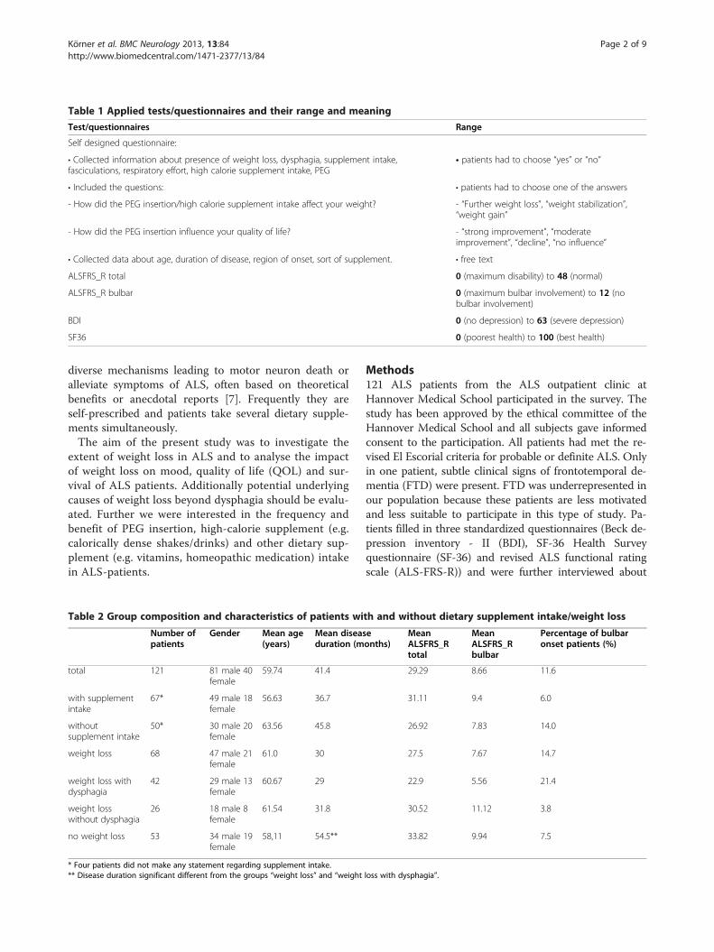
Table 1 Applied tests/questionnaires and their range and meaning
Test/questionnaires Range
Self designed questionnaire:
• Collected information about presence of weight loss, dysphagia, supplement intake,fasciculations, respiratory effort, high calorie supplement intake, PEG
• patients had to choose “yes” or “no”
• Included the questions: • patients had to choose one of the answers
- How did the PEG insertion/high calorie supplement intake affect your weight? - “Further weight loss”, “weight stabilization”,“weight gain”
- How did the PEG insertion influence your quality of life? - “strong improvement”, “moderateimprovement”, “decline”, “no influence”
• Collected data about age, duration of disease, region of onset, sort of supplement. • free text
ALSFRS_R total 0 (maximum disability) to 48 (normal)
ALSFRS_R bulbar 0 (maximum bulbar involvement) to 12 (nobulbar involvement)
BDI 0 (no depression) to 63 (severe depression)
SF36 0 (poorest health) to 100 (best health)
Körner et al. BMC Neurology 2013, 13:84 Page 2 of 9http://www.biomedcentral.com/1471-2377/13/84
diverse mechanisms leading to motor neuron death oralleviate symptoms of ALS, often based on theoreticalbenefits or anecdotal reports [7]. Frequently they areself-prescribed and patients take several dietary supple-ments simultaneously.The aim of the present study was to investigate the
extent of weight loss in ALS and to analyse the impactof weight loss on mood, quality of life (QOL) and sur-vival of ALS patients. Additionally potential underlyingcauses of weight loss beyond dysphagia should be evalu-ated. Further we were interested in the frequency andbenefit of PEG insertion, high-calorie supplement (e.g.calorically dense shakes/drinks) and other dietary sup-plement (e.g. vitamins, homeopathic medication) intakein ALS-patients.
Table 2 Group composition and characteristics of patients wi
Number ofpatients
Gender Mean age(years)
Mean diseasduration (m
total 121 81 male 40female
59.74 41.4
with supplementintake
67* 49 male 18female
56.63 36.7
withoutsupplement intake
50* 30 male 20female
63.56 45.8
weight loss 68 47 male 21female
61.0 30
weight loss withdysphagia
42 29 male 13female
60.67 29
weight losswithout dysphagia
26 18 male 8female
61.54 31.8
no weight loss 53 34 male 19female
58,11 54.5**
* Four patients did not make any statement regarding supplement intake.** Disease duration significant different from the groups “weight loss” and “weight
Methods121 ALS patients from the ALS outpatient clinic atHannover Medical School participated in the survey. Thestudy has been approved by the ethical committee of theHannover Medical School and all subjects gave informedconsent to the participation. All patients had met the re-vised El Escorial criteria for probable or definite ALS. Onlyin one patient, subtle clinical signs of frontotemporal de-mentia (FTD) were present. FTD was underrepresented inour population because these patients are less motivatedand less suitable to participate in this type of study. Pa-tients filled in three standardized questionnaires (Beck de-pression inventory - II (BDI), SF-36 Health Surveyquestionnaire (SF-36) and revised ALS functional ratingscale (ALS-FRS-R)) and were further interviewed about
th and without dietary supplement intake/weight loss
eonths)
MeanALSFRS_Rtotal
MeanALSFRS_Rbulbar
Percentage of bulbaronset patients (%)
29.29 8.66 11.6
31.11 9.4 6.0
26.92 7.83 14.0
27.5 7.67 14.7
22.9 5.56 21.4
30.52 11.12 3.8
33.82 9.94 7.5
loss with dysphagia”.

Körner et al. BMC Neurology 2013, 13:84 Page 3 of 9http://www.biomedcentral.com/1471-2377/13/84
weight loss, dysphagia, food habits and their intake of diet-ary supplements. It was documented if patients sufferedfasciculations or respiratory distress (yes or no).Two years after the initial survey, 61.2% of the patients
or their relatives were available for a short follow-uptelephone interview to find out whether the patient wasstill alive and whether the patient still used dietarysupplements.The ALSFRS_R is a well-established and widely used
score for the functional status of patients with ALS [11].It is based on 12 items, each rated on a 0–4 point scale.The rate of functional disability ranges from 0 (max-imum disability) to 48 (normal) points. Three items ofthe ALSFRS_R assess bulbar involvement (speech, saliva-tion, swallowing), which therefore can be rated from 0
Figure 1 Mean scores of ALSFRS_R and SF36 questionnaire of patientsupplements/PEG. Mean scores of ALSFRS_R (A) and SF36 questionnaire (significantly lower scores at the ALSFRS_R and the SF36 subscales “physicasupplements and 76.9% of patients who had undergone PEG reported sub** p < 0.01.
(maximum bulbar involvement) to 12 (no bulbarinvolvement).The BDI is a 21-question multiple-choice self-report
inventory and a commonly used instrument for quantify-ing levels of depression. Each of the 21 items is scoredon a scale value of 0 (symptom not present) to 3 (symp-tom very intense) leading to an overall-range of 63. Thecutoffs used are 0–8: no depression, 9–13: minimal de-pression, 14–19: mild depression, 20–28: moderate de-pression, 29–63 severe depression [12].The SF36 questionnaire is a multi-purpose, short-form
health survey with 36 questions. It is a self-administeredQOL scoring system that includes eight independentscales: 1. physical functioning (limitations in physical ac-tivities), 2. Physical role (limitations in usual role activities
s without and with weight loss and impact of high calorieB) of patients without and with weight loss. The latter group showedl functioning” and “vitality”. 58.3% of patients consuming high caloriesequent weight stabilization or even weight gain (C and D). * p < 0.05,

Table 3 Multiple regression analysis of the SF36 subscale“vitality” and weight loss (A and B) respectively the SF36subscale “social functioning” and supplement intake (C)
A
Vitality Regression coefficient p-value
(SF-36 scale)
ALSFRS_R 0,416 0,013
Weight loss −9,281 0,011
B
Vitality Regression coefficient p-value
(SF-36 scale)
Respiratory distress −10.093 0,005
Weight loss −9,457 0,008
C
Social Functioning Regression coefficient p-value
(SF-36 scale)
ALSFRS_R 0,628 0,010
Supplement intake 14,342 0,008
Multiple regression analysis of the SF36 subscale “vitality” and weight loss (Aand B) respectively the SF36 subscale “social functioning” and supplementintake (C) taking into account the influence of the ALSFRS_R score/respiratorydistress. The calculations showed an independent significant influence of bothweight loss/supplement intake and ALSFRS_R score/respiratory distress on“vitality” respectively “social functioning”: Patients with weight loss and lowerALSFRS_R showed worse results regarding “vitality” (A and B) and patientswith supplement intake and higher ALSFRS_R showed better results regarding“social functioning” (C).
Körner et al. BMC Neurology 2013, 13:84 Page 4 of 9http://www.biomedcentral.com/1471-2377/13/84
because of physical health problems), 3. Bodily pain, 4.General health perception, 5. Vitality (energy and fa-tigue), 6. social functioning (limitations in social activitiesbecause of physical or emotional problems), 7. Emotionalrole (limitations in usual role activities because of emo-tional problems), 8. Mental health (psychological distressand well-being) [13]. The SF-36 questionnaire is widelyused and appropriate in ALS patients [14].The tests and questionnaires used in this study are
briefly summarized in Table 1.Patients were divided in the following groups: “weight
loss” (defined as >3 kg since disease onset), againsubdivided in “weight loss without/with dysphagia” and“no weight loss” and, for the second aspect of the study,“with supplement intake” and “without supplement in-take” (supplement defined here as dietary supplements(e.g. vitamins)). The groups did not differ regarding genderor site of onset. Disease severity (ALSFRS_R), extent of de-pression (BDI) and quality of life (SF36) were comparedbetween these patient groups (group composition seeTable 2) using t-tests for independent samples. To identifycorrelations between weight loss or supplement intakeand QOL independent from disease severity (ALSFRS-R)we performed multiple regression analysis (dependentvariable: SF-36 subscale, predictor variables: ALSFRS-Rand weight loss or supplement intake). Statistical analyseswere performed using SPSS V. 19 (SPSS, Chicago, IL) soft-ware, a p-value of <0.05 was considered significant.
Results56.2% (n = 68) of the patients in our cohort reportedabout weight loss. Weight loss was associated with a sig-nificantly worse ALSFRS_R score and also with higherdepression (BDI, not significant) and significantly lowerQOL scores (SF36) regarding the subscales “physicalfunctioning” and “vitality” (Figure 1A and B). Multipleregression analysis identified the ALSFRS_R score asconfounding factor, showing that the differences in BDIand “physical functioning” were probably caused by thediscrepancy in the ALSFRS_R. But the difference in theSF36 subscale “vitality” between patients with and with-out weight loss remained highly significant (Table 3A),which means that patients with weight loss feel moreoften exhausted, tired and spiritless, regardless of thedisease stage. Multiple regression analysis showed thatthis influence of weight loss on vitality was independentof respiratory distress which had a significant effect onvitality as well (Table 3B).33.8% (n = 23) of patients with weight loss consumed
high calorie supplements and 60.8% (n = 14) of thesereported subsequent weight stabilization or even weightgain (Figure 1C). 25.5% (n = 13) of patients with dysphagiahad undergone PEG; 76.9% (n = 10) of these patients de-clared weight stabilization or weight gain (Figure 1D) and
84.6% (n = 11) stated an improvement of QOL after PEGinsertion. Remarkably, no patient indicated deterioration ofQOL after PEG insertion (although this is often suspectedby patients and relatives prior to the procedure).38.2% (n = 26) of patients with weight loss did not suf-
fer from dysphagia (according to self-reported statementand ALSFRS_R). This patient group did not differ frompatients without weight loss regarding ALSFRS_R (totaland bulbar) and depression (BDI) nor did they reportchanges in their eating habits. Patients with dysphagiaon the other hand showed significantly lower ALSFRS_Rscores, mainly due to the bulbar subscore (Figure 2A).The prevalence of fasciculations in patients with weightloss without dysphagia was equal to patients withoutweight loss. They did, however, more often declare in-creased respiratory efforts compared to patients withoutweight loss (Figure 2B).The telephone survey after two years showed that
weight loss was a strong negative prognostic factor:Kaplan- Meier survival curves of patients with and with-out weight loss showed significantly shorter survival of pa-tients with weight loss (log rank p = 0.001) (Figure 2C).54.5% (n = 67) of the patients stated regular intake of
other (not high-calorie) dietary supplements (e.g. vitamins).44.8% (n = 30) of them consumed more than one supple-ment, some up to five simultaneously or preparations

Figure 2 Comparison of patients with weight loss with/withoutdysphagia and patients without weight loss. Patients withweight loss and dysphagia differed significantly from patientswithout weight loss in BDI and ALSFRS_R (total and bulbar). Patientswith weight loss without dysphagia, on the other hand, did nothave higher BDI/ lower ALSFRS_R scores than patients withoutweight loss (A). Weight loss in patients without dysphagia thereforedoes not seem to be directly related to a more advanced diseasestage or increased depression. Patients with weight loss anddysphagia significantly more often declared increased respiratorywork than patients without weight loss. Patients with weight losswithout dysphagia showed a tendency towards increased respiratorywork compared to patients without weight loss (p = 0.12). Therewere no differences regarding frequency of fasciculations betweenthe groups (B). Follow-up by telephone two years after the initialsurvey highlighted the prognostic value of weight loss: Kaplan-Meiersurvival analysis for ALS patients with and without weight lossrevealed significantly shorter survival of ALS patients with weightloss (log rank p = 0.001) (C).
Körner et al. BMC Neurology 2013, 13:84 Page 5 of 9http://www.biomedcentral.com/1471-2377/13/84
containing up to seven ingredients. Overall 23 differentsupplements were mentioned (Table 4). Analysis of theALSFRS_R showed an inverse correlation between diseaseseverity and supplement intake (i.e. patients taking diet-ary supplements were significantly less impaired thanthose who did not) (Figure 3A). There also was a signifi-cant difference in the BDI scores as well as the SF-36subscales “physical functioning”, “vitality” and “socialfunctioning” between the two groups: patients with sup-plement intake were significantly better regarding moodand QOL (Figure 3B and C). However, multiple regres-sion analysis again showed that these differences aremainly attributable to the discrepancies in disease severityas assessed by the ALSFRS_R (=confounding factor). Onlythe difference in “social functioning” between patientswith and without supplement intake remained highly sig-nificant, showing an independent influence of both sup-plement intake and ALSFRS_R (Table 3C). Hence patientsusing dietary supplements are feeling less affected in their“social functioning” which means contact or visit familymembers, friends and neighbours. However it could alsobe the other way around, meaning that patients with amore active social life are more likely to start taking diet-ary supplements. At the follow-up telephone interviewtwo years after the first survey, 42.9% of the patients whohad initially reported use of dietary supplements now de-clared that they had stopped any supplement intake.
DiscussionAccording to the literature, weight loss is associated withshorter survival. In our ALS population it was accordinglyconnected with a significantly worse mean ALSFRS_Rscore and a significantly higher death rate after two years.Weight loss had a significant impact on QOL regardingthe SF36 subitem “vitality”, meaning that it made patientsfeel more exhausted, tired and spiritless, regardless of the

Table 4 Nutritional supplements and functional foodstaken in our population [15]
Nutritionalsupplement
Mechanism of action(hypothesis)
Percentage (%) inour population
Magnesium Against cramps 63,6
Vitamin E Antioxidant 40,3
Vitamin B (6 + 12) Other agent 13,6
Vitamin C Antioxidant 10,6
Vitamins, notspecified
Antioxidant 7,6
Homeopathicmedication
4,5
Folate Other 4,5
Calcium Bone regeneration 3,0
Carnitin Antioxidant/Mitochondrial stabilizer
3,0
CoQ10 Antioxidant/Mitochondrial stabilizer
3,0
Schussler salts 3,0
L-Arginin Muscle regeneration 3,0
Zinc Other 3,0
Vitamin A Antioxidant 1,5
Lipoic acid (Omega3) Antioxidant/Anti-glutamate
1,5
Grape seed extract Antioxidant/Anti-glutamate
1,5
Vitamin D Other 1,5
Lycopin (tomato) Antioxidant, Radicalscavenger
1,5
Selen Radical scavenger 1,5
Enzymes (Papain,(Chymo)Trypsin)
Antithrombotic,eupeptic
1,5
Protein preparation Muscle regeneration 1,5
Willow capsules Against pain 1,5
Chlorella Against heavy metals 1,5
Himalaya salt 1,5
Not specified 12,1
Körner et al. BMC Neurology 2013, 13:84 Page 6 of 9http://www.biomedcentral.com/1471-2377/13/84
respective disease stage. Nevertheless, high calorie nutri-tional supplements were only consumed by one third ofpatients with weight loss, mainly due to the fact that thecosts are often not covered by the respective health insur-ances. According to our survey, substantial benefit can beobtained from high calorie supplements and they shouldtherefore be given more frequently. Only one quarter ofpatients with dysphagia had undergone PEG placement.Most of these patients reported weight stabilization andimprovement of QOL. The decision about PEG insertionis mostly based on nutritional status and presence ofdysphagia [16,17]. From clinical practise it is known,that patients are often cautious accepting a PEG, evenwhen food intake becomes time consuming and laborious.
Neurologists also tend to postpone the discussion of PEG[18]. As a result, PEG placement is often initiated toolate and therefore associated with higher complicationrates as it is known that FVC >50% at the time of PEGinsertion is an important prognostic factor [16,18-20].Our data confirm that PEG placement can stabilizebody weight [10,21,22] and suggest additionally an im-pact on the QOL. The impact of PEG on QOL has beenstudied very little so far. Beside anecdotal impressions,only one study collected quantitative data: here, the ma-jority of the patients mentioned stabilized nutritional andhydrational status as most positive effect of PEG. Fewerpatients listed less fatigue or less time spent on meals andimproved psychological wellbeing as positive effects ofPEG [21,23]. Interestingly, in our cohort, even patientswho had undergone further weight loss after PEG in-sertion, unanimously stated improvement of QOL. Thissuggests that other factors such as reduction of time con-suming and tiring food and fluid intake with frequentchoking are equally important for patients.A further important result of our study is that weight loss
without dysphagia is frequent in ALS. We aimed to definewhat distinguishes these patients from patients withoutweight loss. As the groups did not differ regarding site ofonset and gender, these factors could not be relevant. Whilethorough neuropsychological testing had not been routinelyperformed, clinical signs of dementia were only detectablein one of the patients without weight loss, so that weightloss in the patients without dysphagia in our cohort couldnot be attributed to major cognitive or behavioural abnor-malities. Patients with weight loss without dysphagia didnot show differences in disease severity, grade of depres-sion or presence of muscle fasciculations, but they did re-port higher respiratory efforts. It remains unclear how farthis contributes to the weight loss. Two studies in ALS pa-tients did not detect a relation between forced vital cap-acity (FVC) and energy expenditure (REE) [2,24] whiletwo others suggest that increased respiratory efforts causeelevated REE [25,26]. Based on the existing literature, thisincreased energy consumption in ALS can probably notonly be explained by respiratory insufficiency but ratherby general hypermetabolism which occurs in about 50% ofALS patients and whose origin has not yet been fully elu-cidated [4,24].The proportion of patients taking other, not high cal-
orie dietary supplements such as vitamins was lower inour cohort (with 54.5%) than in the literature, were thepercentage of supplement intake among ALS-patients isestimated as approximately 80% [7,10].The observed differences in disease severity, depres-
sion and QOL are most probably not a direct effect ofthese diverse dietary supplements. While one must takeinto account a certain placebo-effect, the most likely ex-planation is that patients self-medicating with dietary

Figure 3 Mean scores of ALSFRS-R, BDI and SF36 of patients with and without intake of dietary supplements. Mean scores of ALSFRS-R(A), BDI (B) and SF36 (C) of patients with and without intake of other (not high calorie) dietary supplements. Supplement intake was associatedwith significantly higher scores at the ALSFRS_R and the SF36 subscales “physical functioning”, “vitality” and “social functioning” and significantlylower scores in BDI. * p < 0.05.
Körner et al. BMC Neurology 2013, 13:84 Page 7 of 9http://www.biomedcentral.com/1471-2377/13/84

Körner et al. BMC Neurology 2013, 13:84 Page 8 of 9http://www.biomedcentral.com/1471-2377/13/84
supplements presumably represent a more hopeful andoptimistic subgroup. This assumption is supported bythe fact that they had higher SF36 “social functioning”scores, i.e. felt less affected in their interactions withfamily members, friends and neighbours. A more activesocial life may also have provided increased stimulationsto try alternative treatment approaches. Discontinuationof dietary supplements over time (as revealed by ourtwo-year follow-up interview) presumably is the result ofloss of hope generally associated with further diseaseprogression.Although there is a lack of evidence for any rele-
vant benefit of dietary supplements, as long as thereis no clear contraindication and as long as efficientneuroprotective drugs beyond riluzole have not beendiscovered, self-medication with dietary supplementsmay represent hope and confidence for some pa-tients and thereby have a positive impact on the dis-ease course and quality of life.
ConclusionThe significance of the present study is limited becauseit is retrospective and based on subjective data from thepatients themselves. Nevertheless it provides valuable in-formation that can be used as a starting point for furtherprospective investigations.Even though malnutrition is a significant and inde-
pendent prognostic factor in survival, it is often inad-equately addressed in clinical practice. According to ourresults the effect of high calorie nutritional supplementsand PEG is often higher than expected. Patients, caregiversand physicians should therefore be encouraged to considerthese measures. However, their benefit still requires fur-ther confirmation by prospective studies.To evaluate the reasons of weight loss beside dysphagia,
further prospective analysis of patients without weight lossin comparison to patients suffering from weight loss notattributable to dysphagia, comparing clinical parameterssuch as fasciculations, spasticity and cognitive or behav-ioural abnormalities together with REE and FVC will cer-tainly provide more thorough understanding of thisphenomenon. In any case the existence of these differentphenotypes highlights once more the heterogeneity in theclinical presentation of ALS.Regarding dietary supplements, further studies are needed
to evaluate the safety and efficacy of numerous dietary sup-plements and to enable appropriate recommendations.
AbbreviationsALS: Amyotrophic lateral sclerosis; ALSFRS_R ALS: Functional ratingscale – revised version; BDI: Beck depression inventory; BMI: Body massindex; FVC: Flow vital capacity; PEG: Percutaneous endoscopic gastrostomy;QOL: Quality of life; REE: Resting energy expenditure; SF36: Short Form 36health survey.
Competing interestThe study was not externally funded. The authors declare no conflict ofinterest in regarding this manuscript.
Authors’ contributionsSK and SP designed the study and drafted the article. MH and KKcontributed to acquisition of data and revised the article critically forimportant intellectual content, SK and AZ analyzed and interpreted the data,AZ also revised the article critically for important intellectual content. RD andVS contributed to conception of the study and revised the article criticallyfor important intellectual content. All authors give final approval of theversion to be published.
AcknowledgmentsThe authors thank Andreas Niesel and Dagmar Conradt for technicalassistance.
Author details1Department of Neurology, Hannover Medical School, Carl-Neuberg-Str. 1,Hannover 30625, Germany. 2Center for Systems Neuroscience (ZSN),Hannover, Germany. 3Department of Medical Statistics, University Göttingen,Göttingen, Germany. 4Department of Neurology and Laboratory ofNeuroscience, IRCCS Istituto Auxologico Italiano, Milan 20149, Italy.5Department of Pathophysiology and Transplantation, “Dino Ferrari” Center,Università degli Studi di Milano, Milan 20149, Italy.
Received: 17 January 2013 Accepted: 11 July 2013Published: 12 July 2013
Reference1. Wijesekera LC, Leigh PN: Amyotrophic lateral sclerosis. Orphanet J Rare Dis
2009, 4:3.2. Bouteloup C, Desport JC, Clavelou P, Guy N, Derumeaux-Burel H, Ferrier A,
Couratier P: Hypermetabolism in ALS patients: an early and persistentphenomenon. J Neurol 2009, 256:1236–1242.
3. Vaisman N, Lusaus M, Nefussy B, Niv E, Comaneshter D, Hallack R, Drory VE:Do patients with amyotrophic lateral sclerosis (ALS) have increasedenergy needs? J Neurol Sci 2009, 279:26–29.
4. Desport JC, Torny F, Lacoste M, Preux PM, Couratier P: Hypermetabolism inALS: correlations with clinical and paraclinical parameters.Neurodegener Dis 2005, 2:202–207.
5. Dupuis L, Pradat PF, Ludolph AC, Loeffler JP: Energy metabolism inamyotrophic lateral sclerosis. Lancet Neurol 2011, 10:75–82.
6. Desport JC, Preux PM, Truong TC, Vallat JM, Sautereau D, Couratier P:Nutritional status is a prognostic factor for survival in ALS patients.Neurology 1999, 53:1059–1063.
7. Rosenfeld J, Ellis A: Nutrition and dietary supplements in motor neurondisease. Phys Med Rehabil Clin N Am 2008, 19:573–589.
8. Paganoni S, Deng J, Jaffa M, Cudkowicz ME, Wills AM: Body mass index,not dyslipidemia, is an independent predictor of survival in amyotrophiclateral sclerosis. Muscle Nerve 2011, 44:20–24.
9. Dupuis L, Oudart H, Rene F, de Aguilar JL G, Loeffler JP: Evidence fordefective energy homeostasis in amyotrophic lateral sclerosis: benefit ofa high-energy diet in a transgenic mouse model. model. Proc Natl AcadSci U S A 2004, 101:11159–11164.
10. Miller RG, Jackson CE, Kasarskis EJ, England JD, Forshew D, Johnston W,Kalra S, Katz JS, Mitsumoto H, Rosenfeld J, Shoesmith C, Strong MJ,Woolley SC: Practice parameter update: The care of the patient withamyotrophic lateral sclerosis: drug, nutritional, and respiratorytherapies (an evidence-based review): report of the Quality StandardsSubcommittee of the American Academy of Neurology. Neurology 2009,73:1218–1226.
11. Cedarbaum JM, Stambler N, Malta E, Fuller C, Hilt D, Thurmond B, NakanishiA: The ALSFRS-R: a revised ALS functional rating scale that incorporatesassessments of respiratory function. BDNF ALS Study Group (Phase III).J Neurol Sci 1999, 169:13–21.
12. Beck AT, Steer RA, Ball R, Ranieri W: Comparison of Beck DepressionInventories -IA and -II in psychiatric outpatients. J Pers Assess 1996,67:588–597.
Körner et al. BMC Neurology 2013, 13:84 Page 9 of 9http://www.biomedcentral.com/1471-2377/13/84
13. Ware JE Jr, Sherbourne CD: The MOS 36-item short-form health survey(SF-36). I. Conceptual framework and item selection. Med Care 1992,30:473–483.
14. Jenkinson C, Hobart J, Chandola T, Fitzpatrick R, Peto V, Swash M: Use ofthe short form health survey (SF-36) in patients with amyotrophic lateralsclerosis: tests of data quality, score reliability, response rate and scalingassumptions. J Neurol 2002, 249:178–183.
15. Cameron A, Rosenfeld J: Nutritional issues and supplements inamyotrophic lateral sclerosis and other neurodegenerative disorders.Curr Opin Clin Nutr Metab Care 2002, 5:631–643.
16. Andersen PM, Abrahams S, Borasio GD, de Carvalho M, Chio A, Van DammeP, Hardiman O, Kollewe K, Morrison KE, Petri S, Pradat PF, Silani V, Tomik B,Wasner M, Weber M: EFNS guidelines on the clinical management ofamyotrophic lateral sclerosis (MALS)–revised report of an EFNS taskforce. Eur J Neurol 2012, 19:360–375.
17. Heffernan C, Jenkinson C, Holmes T, Feder G, Kupfer R, Leigh PN, McGowanS, Rio A, Sidhu P: Nutritional management in MND/ALS patients: anevidence based review. Amyotroph Lateral Scler Other Motor Neuron Disord2004, 5:72–83.
18. Silani V, Kasarskis EJ, Yanagisawa N: Nutritional management inamyotrophic lateral sclerosis: a worldwide perspective. J Neurol 1998,245(Suppl 2):S13–S19.
19. Shaw AS, Ampong MA, Rio A, Al Chalabi A, Sellars ME, Ellis C, Shaw CE,Leigh NP, Sidhu PS: Survival of patients with ALS following institution ofenteral feeding is related to pre-procedure oximetry: a retrospectivereview of 98 patients in a single centre. Amyotroph Lateral Scler 2006,7:16–21.
20. Kasarskis EJ, Scarlata D, Hill R, Fuller C, Stambler N, Cedarbaum JM: Aretrospective study of percutaneous endoscopic gastrostomy in ALSpatients during the BDNF and CNTF trials. J Neurol Sci 1999, 169:118–125.
21. Katzberg HD, Benatar M: Enteral tube feeding for amyotrophic lateralsclerosis/motor neuron disease. Cochrane Database Syst Rev 2011. Issue 1.Art. No.: CD004030.
22. Mazzini L, Corra T, Zaccala M, Mora G, Del Piano M, Galante M:Percutaneous endoscopic gastrostomy and enteral nutrition inamyotrophic lateral sclerosis. J Neurol 1995, 242:695–698.
23. Mitsumoto H, Davidson M, Moore D, Gad N, Brandis M, Ringel S, RosenfeldJ, Shefner JM, Strong MJ, Sufit R, Anderson FA: Percutaneous endoscopicgastrostomy (PEG) in patients with ALS and bulbar dysfunction.Amyotroph Lateral Scler Other Motor Neuron Disord 2003, 4:177–185.
24. Desport JC, Preux PM, Magy L, Boirie Y, Vallat JM, Beaufrere B, Couratier P:Factors correlated with hypermetabolism in patients with amyotrophiclateral sclerosis. Am J Clin Nutr 2001, 74:328–334.
25. Kasarskis EJ, Berryman S, Vanderleest JG, Schneider AR, McClain CJ:Nutritional status of patients with amyotrophic lateral sclerosis: relationto the proximity of death. Am J Clin Nutr 1996, 63:130–137.
26. Shimizu T, Hayashi H, Tanabe H: Energy metabolism of ALS patients undermechanical ventilation and tube feeding. Rinsho Shinkeigaku 1991,31:255–259.
doi:10.1186/1471-2377-13-84Cite this article as: Körner et al.: Weight loss, dysphagia and supplementintake in patients with amyotrophic lateral sclerosis (ALS): impact onquality of life and therapeutic options. BMC Neurology 2013 13:84.
Submit your next manuscript to BioMed Centraland take full advantage of:
• Convenient online submission
• Thorough peer review
• No space constraints or color figure charges
• Immediate publication on acceptance
• Inclusion in PubMed, CAS, Scopus and Google Scholar
• Research which is freely available for redistribution
Submit your manuscript at www.biomedcentral.com/submit