jvi accepts, published online ahead of print on 23...
TRANSCRIPT

Enhanced Autointegration in Hyperstable Simian Immunodeficiency Virus Capsid 1
Mutants Blocked After Reverse Transcription 2
3
Christopher Tipper1 and Joseph Sodroski1,2,3,* 4
5
6
1Department of Cancer Immunology & AIDS, Dana-Farber Cancer Institute, 7
Harvard Medical School, Boston, MA 02215 8
9
2Department of Immunology & Infectious Disease, Harvard School of Public Health, 10
Boston, MA 02115 11
12
3Ragon Institute of Massachusetts General Hospital, Massachusetts Institute of 13
Technology and Harvard 14
15
*Corresponding author: 16 17 Joseph G. Sodroski, M.D. 18 Dana-Farber Cancer Institute 19 450 Brookline Avenue, CLSB 1010 20 Boston, MA 02215 21 Phone: 617-632-3371 22 Fax: 617-632-4338 23 Email: [email protected] 24 25 Running Title: Increased capsid stability and autointegration 26 Word Count (Abstract): 177 27 Word Count (Text): 6,072 28
29
30
Copyright © 2013, American Society for Microbiology. All Rights Reserved.J. Virol. doi:10.1128/JVI.03239-12 JVI Accepts, published online ahead of print on 23 January 2013
on June 24, 2018 by guesthttp://jvi.asm
.org/D
ownloaded from
2
Abstract 31
32
After entering a host cell, retroviruses such as simian immunodeficiency virus 33
(SIV) uncoat, disassembling the viral capsid. Too rapid and too slow rates of uncoating 34
can be detrimental to the efficiency of infection. Rapid uncoating typically leads to 35
blocks in reverse transcription, but the basis for replication defects associated with slow 36
uncoating is less clear. Here we characterize the phenotypes of two SIVmac239 37
mutants with changes, A87E and A87D, in the helix 4/5 loop of the capsid protein. 38
These mutant viruses exhibited normal capsid morphology but were significantly 39
attenuated for infectivity. The infectivity of wild-type and mutant SIVmac239 was not 40
decreased by aphidocolin-induced growth arrest of the target cells. In the cytosol of 41
infected cells, the A87E and A87D capsids remained in particulate form longer than the 42
wild-type SIVmac239 capsid, suggesting that the mutants uncoat more slowly than the 43
wild-type capsid. Both mutants exhibited much higher levels of autointegrated DNA 44
forms than wild-type SIVmac239. Thus, some changes in the helix 4/5 loop of the 45
SIVmac239 capsid protein result in capsid hyperstability and an increase in 46
autointegration. 47
on June 24, 2018 by guesthttp://jvi.asm
.org/D
ownloaded from

3
Introduction 48
49
Lentiviruses differ from most other retroviruses in that they have evolved to infect 50
mature individuals with competent immune systems (9). Indeed, lentiviruses rely upon 51
the cells of the host immune system to provide functions necessary to their replication 52
and dissemination. In so doing, they have evolved the ability to integrate their genome 53
into cells that are terminally differentiated, accessing the nucleus through the nuclear 54
pore (6). Only a fraction of viruses that gain entry to a cell are successful in navigating 55
to the nuclear compartment and creating a provirus capable of supporting the late 56
phase of retrovirus replication. This excess of defective events has complicated the 57
characterization of early-stage infection. 58
59
Following entry into the host cell, lentiviruses must proceed through several steps 60
on the way to generating a provirus. These early-phase processes include uncoating of 61
the core, reverse transcription of the RNA genome, nuclear entry of the pre-integration 62
complex, and integration. Changes in lentivirus capsid proteins have been shown to 63
accelerate or slow the uncoating process (13, 27, 28, 52, 54). Less expectedly, 64
alterations in the capsid protein can affect subsequent steps in the early phase of 65
lentivirus infection. An appreciation of the contribution of the capsid to multiple steps in 66
the lentiviral life cycle has made capsid an attractive target of therapeutic intervention 67
(3, 4, 10, 22, 23, 25, 31, 35, 41, 42, 51, 52). 68
69
on June 24, 2018 by guesthttp://jvi.asm
.org/D
ownloaded from

4
Two host proteins that bind the HIV-1 capsid and influence the uncoating process 70
have been identified. TRIM5α is a protein that mediates the premature, deleterious 71
disassembly of the capsid and therefore acts as a restriction factor (45). The prolyl cis-72
trans isomerase cyclophilin A (CypA) binds the HIV-1 capsid and modulates capsid 73
stability in either a positive or negative manner, depending on the type of infected cell 74
and the sequence of the helix 4/5 loop (CypA-binding loop) of the capsid protein (CA) 75
(17, 28, 38, 41, 44, 48, 55). In T lymphocytes, CypA stabilizes the HIV-1 capsid and 76
promotes infection (28). The CypA domain of the nuclear pore protein Nup358 has 77
been reported to be important for proper nuclear targeting of HIV-1 pre-integration 78
complexes (17, 28, 38, 41, 44, 48, 55). 79
80
The simian immunodeficiency viruses (SIVs) infect feral African monkeys and 81
apes and are the lentivirus ancestors of human immunodeficiency viruses (14, 15, 30, 82
37). The capsids of monkey SIVs do not interact with CypA (5, 47). In permissive cells 83
without a restricting TRIM5α protein, SIV infection can be studied in the absence of 84
either of these host capsid-binding proteins. We took advantage of this characteristic to 85
assess the function of the flexible helix 4/5 loop that extends above the capsid surface. 86
In HIV-1, this loop is the site of CypA binding, near CA residues Gly89 and Pro90 (49). 87
In contrast, the helix 4/5 loop of SIVmac239 does not contain a Gly-Pro motif, and does 88
not bind CypA. However, a structurally similar Ala-Pro motif occurs at capsid residues 89
Ala87 and Pro88 in the SIVmac239 helix 4/5 loop. Interestingly, the helix 4/5 loop of the 90
HIV-1 CA has an Ala-Pro pair (Ala92 and Pro93) C-terminal to the Gly-Pro motif. 91
Alteration of alanine 92 to glutamic acid, or the nearby glycine at position 94 to aspartic 92
on June 24, 2018 by guesthttp://jvi.asm
.org/D
ownloaded from
5
acid, renders HIV-1 CypA-independent in certain cell types (1, 55). In other cell types, 93
the infectivity of the A92E and G94D capsid mutants is inhibited by CypA binding. The 94
replication phenotype of the A92E and G94D capsid mutants is predicted by the level of 95
CypA in the target cell (28, 44, 55). 96
97
Because changes in the helix 4/5 loop of the HIV-1 CA can affect sensitivity to 98
CypA and TRIM5α (35), the interpretation of the mechanisms that underly the 99
phenotypes of HIV-1 CA helix 4/5 mutants is complicated. Here, we study the effect of 100
acidic substitutions in the Ala-Pro motif in the helix 4/5 loop of the SIVmac239 CA. We 101
characterize the phenotypes of the SIVmac239 A87E and A87D mutants, observing 102
decreases in the early phase of infection and slower capsid uncoating. Although 103
reverse transcription of these mutants was efficient, striking increases in the amounts of 104
autointegrated viral DNA were seen for both mutants. Models that fit this heretofore 105
unknown capsid function into current knowledge of early events in lentiviral infection are 106
discussed. 107
108
109
on June 24, 2018 by guesthttp://jvi.asm
.org/D
ownloaded from

6
Materials and Methods 110
111
Cell lines and culture 112
113
Adherent cell lines used in this study were grown in Dulbecco’s Modified Eagles 114
Medium (high glucose) (Invitrogen cat# 11965) supplemented with 10% FBS and 1% 115
penicillin/streptomycin. HEK 293T/17 (ATCC cat.# CRL-11268) and HeLa (ATCC cat.# 116
CCL-2) are of human origin. Cf2Th cells (ATCC Cat. #CRL-1430) are of canine origin. 117
118
Site-directed mutagenesis 119
120
All mutants were created by site-directed mutagenesis of either the full-length 121
pSIVmac239ΔnefΔenvEGFP plasmid (see below) or the shuttle vector SGag3 (36), 122
utilizing pfu Ultra Hotstart 2X Master Mix (Stratagene cat# 600630-51), 125 ng of each 123
mutagenic primer, and the following cycling protocol: 95°C for 1 minute; 18 cycles of 124
95°C for 30 seconds, 55°C for 1 minute, and 72°C for 12 minutes (full-length) or 5.5 125
minutes (SGag3), with a final 1-minute 72°C extension. SGag3 sequences were then 126
inserted into the full-length vector by DraIII/SbfI digestion and T4 ligation. All mutations 127
were verified by sequencing. Mutagenic primers (IDT) were based on the forward wild-128
type sequence: GCAGCACCCACAACCAGCTCCACAACAAGGACAACTTAGG. 129
130
on June 24, 2018 by guesthttp://jvi.asm
.org/D
ownloaded from

7
Production of Recombinant SIV variants expressing GFP 131
132
The simian immunodeficiency virus (SIV)-based pSIVmac239ΔnefΔenvEGFP 133
plasmid was derived from pSIVΔnefEGFP (2) (a kind gift from Dr. Ronald Desrosiers, 134
Harvard Medical School). Env sequences were deleted by first changing the ATG start 135
codon to TCC to create a BspE1 site. The 1.1 kb fragment between this and the native 136
BspE1 site at position 8013 (Genbank accession number M33262.1) was excised, 137
leaving the tat and rev genes and the Rev-responsive element intact. This construct, 138
and derivative mutants, were co-transfected into the desired cell type with a Rev-139
expressing construct and the vesicular stomatitis virus G glycoprotein-expressing 140
construct pHCMV-G, as described previously (35). HEK293T/17 cells were transfected 141
using Lipofectamine PLUS or Lipofectamine 2000 following the manufacturer’s protocol. 142
Recombinant viruses in the transfected cell supernatants were filtered (0.45-μ filter) and 143
titers were determined by a reverse transcriptase (RT) assay, as previously described 144
(40). If necessary, viruses were concentrated by PEG precipitation using PEG-it (SBI 145
Cat. #LV810A) and resuspended in an appropriate volume of 1X PBS. 146
147
Infection assays 148
149
One day before infection, HeLa and Cf2Th cells at a density of 1-3 x 10⁵ 150
cells/well were seeded into 24-well plates. On the day of infection, virus was added at 151
the desired titer as determined by RT activity, expressed in counts per minute (cpm). 152
on June 24, 2018 by guesthttp://jvi.asm
.org/D
ownloaded from
8
The medium was changed 2-4 hours afterwards, and 48 hours following infection, the 153
percentage of GFP-positive cells and median fluorescence were determined by FACS. 154
155
Gag processing assay 156
157
293T/17 cells in 24-well plates were transfected with a Rev-expressing plasmid 158
and the pSIVmac239ΔnefΔenvEGFP plasmids expressing the wild-type and mutant Gag 159
proteins. Sixteen hours post-transfection, the medium was replaced by DMEM plus 160
10% dialyzed FBS, 1% penicillin/streptomycin, 1X L-glutamine, 1X nonessential amino 161
acids, and 100-fold-diluted 35S-Met/Cys labeling mix (NEG072). Forty-eight hours post-162
transfection, viruses were purified by filtering (0.45-μ filter) followed by pelleting through 163
20% sucrose for 1 hour at 100,000 x g. Viral pellets were resuspended directly into 164
loading buffer. Approximately 25,000 cpm was loaded into each well of a 12% Bis-Tris 165
gel, which was run and exposed to film. 166
167
Electron Microscopy and Core Morphology Quantitation 168
169
Envelope-free viruses were produced and purified by anion-exchange 170
chromatography as described in reference (24). Purified virions were prepared for thin-171
section microscopy by the Harvard Medical School EM Facility. Fields containing 172
multiple identifiable virions were selected for analysis. Virions were binned into one of 173
three categories based upon the shape of their electron-dense core in the sectioning 174
plane: conical cores, well-defined circular cores, and unidentifiable “other.” 175
on June 24, 2018 by guesthttp://jvi.asm
.org/D
ownloaded from
9
176
Fate-of-capsid assays 177
178
Cf2Th cells were seeded into 75 cm2 flasks to achieve 80-90% confluence at the 179
time of infection. Flasks were pre-chilled on ice 10 minutes prior to adding 5 x 10⁶ cpm 180
of virus in a 4-ml volume. Flasks and virus were kept at 4°C for a half hour, then time 181
zero was marked by shifting them to a 37-degree incubator. Virus was washed off the 182
cells and fresh medium added 4 hours post-infection. At the appropriate time, flasks 183
were chilled on ice and washed three times with ice-cold 1X PBS. Cells were released 184
from the flask by pronase digestion (1 ml 7 μg/ml pronase in 1X PBS for 5 minutes), 185
transferred to 15-ml conical tubes, and washed three times with 10 ml cold PBS. The 186
pellet was resuspended in 250 μl cold hypo-lysis solution (10 mM Tris, pH 8, 10 mM 187
KCl, 1 mM EDTA), transferred to Eppendorf tubes, and kept on ice for 15 minutes. 188
Cells were then mechanically disrupted for 1 minute using a pellet pestle (Kontes 189
K49540) and non-cytosolic material removed by centrifugation at 1500 x g for 3 190
minutes. The supernatant was mixed with 250 μl fresh hypo-lysis buffer and 400 μl of 191
this mixture was layered on top of a 7-ml column of 50% sucrose/1X PBS in an ultra-192
clear SW41 tube (Beckman Coulter 344059); the remaining lysate was preserved in 1X 193
Laemmli buffer. The column was centrifuged 2 hours at 125,000 x g. The top 100 μl 194
was collected, and the pellet resuspended in 100 μl 1X PBS; concentrated Laemmli 195
buffer was added to bring both samples to a 1X final concentration. Samples were run 196
on 4-12% Bis-Tris gels and transferred to nitrocellulose filters. The filters were probed 197
with 1:1000 anti-SIVmac251 polyclonal serum (AIDS reagent program #2773) and 198
on June 24, 2018 by guesthttp://jvi.asm
.org/D
ownloaded from

10
1:40,000 HRP-conjugated goat anti-human IgG (Sigma A0293), and developed with the 199
femto-sensitivity chemiluminescence reagent (Thermo #34095). Densitometry was 200
performed using ImageQuantTL, using a consistent 50-pixel rolling-ball background 201
reduction. 202
203
Quantitative Real-Time PCR (qPCR) 204
205
HeLa cells were seeded into 6-well plates to achieve 80% confluence at the time of 206
infection. Viral suspensions were treated with DNAse Turbo (Ambion #AM2238) 207
following the manufacturer’s protocol for 1 hour at 37 degrees and then titered by RT 208
assay. Approximately 100,000 cpm of virus, expected to result in a multiplicity of 209
infection of ~0.5, was added to each well at time zero. For negative controls, viruses 210
were heat-inactivated at 56°C for 1 hour, and the infected cells were maintained in 100 211
μM AZT prior to incubation with the virus and for the duration of the experiment. In 212
some experiments, raltegravir was added to cells at a concentration of 1 μM for 1 hour 213
prior to addition of the virus, and maintained at that concentration throughout the 214
experiment (unless otherwise noted). Unbound virus was washed off the cells 2 hours 215
after virus-cell incubation was initiated. Cells were collected by trypsin digestion 2, 6, 216
12, 24 and 48 hours after virus-cell incubation. Total DNA was extracted by the Wizard 217
SV 96 Genomic System (Promega #A2371) and quantitated on a nanodrop 218
spectrophotometer. DNA was diluted to a final concentration of 10 ng/μl using double-219
distilled autoclaved water. Each qPCR reaction contained 100 ng of total DNA in a 50 220
μl reaction volume. The accumulation of product was assayed by TaqMan probe on an 221
on June 24, 2018 by guesthttp://jvi.asm
.org/D
ownloaded from

11
Applied Biosystems 7300. The primers and probe for the analysis of the early stage of 222
reverse transcription were: 223
224
Early Forward: GTC AAC TCG GTA CTC AAT AAT AAG 225
Early Reverse: GCG CCA ATC TGC TAG GGA T 226
Early Probe FAM-CTG TTA GGA CCC TTT CTG CTT TGG GAA ACC GAA G- 227
TAMRA 228
229
The primers and probe for the analysis of the late stage of reverse transcription were: 230
Late Forward: TTG GGA AAC CGA AGC AGG 231
Late Reverse: TCT CTC ACT CTC CTT CAA GTC CCT 232
Late Probe: FAM-AAA TCC CTA GAC GAT TGG CGC CTG AA-TAMRA 233
234
The primers and probe for the analysis of 2-LTR circles were: 235
2-LTR Forward: GGA AAC CGA AGC AGG AAA AT 236
2-LTR Reverse: CTG TGC CTC ATC TGA TAC 237
2-LTR Standard Probe: FAM-ATT GGC AGG ATT ACA CCT CAG GAC CAG- 238
TAMRA 239
2-LTR Junction Probe: FAM-ATT CCC TAG CAG ATA CTG GAA GGG AT- 240
TAMRA 241
242
on June 24, 2018 by guesthttp://jvi.asm
.org/D
ownloaded from
12
The primers and probe for the analysis of integrated viral DNA were: 243
1° Alu-PCR Forward: ATG CCACGT AAG CGA AAC TCT GTT CCC ATC TCT 244
CCT AGC C 245
1° Alu-PCR Reverse: TCC CAG CTA CTC GGG AGG CTG AGG 246
2° Alu-PCR Forward:ATG CCA CGT AAG CGA AAC TC 247
2° Alu-PCR Reverse: ATT TTC CTG CTT CGG TTT CC 248
Alu-PCR probe: FAM-CGC CTG GTC AAC TCG GTA CTC AAT AA-TAMRA 249
250
Alu qPCR 251
252
Using the samples collected for single-round qPCR, we optimized protocols for 253
assaying integrated SIVmac239 proviruses (by Alu PCR) and autointegrated 254
SIVmac239 genomes. For Alu PCR, we performed a primary amplification using 100 255
ng total DNA, 0.2 μM of each primer, and Pfu UltraII Hotstart 2X Master Mix (Agilent 256
cat# 600850) in a final volume of 20 μl. Cycling parameters for touchdown PCR (TD-257
PCR): 95°C for 2 minutes; 15 cycles of 95°C for 30 seconds, 70-55°C for 30 seconds, 258
and 72°C for 1 minute; 11 cycles of 95°C for 30 seconds, 55°C for 30 seconds, and 259
72°C for 1 minute. Primary PCR samples were diluted 1:5 into 100 μl water, and 10 μl 260
was used in a standard 50 μl qPCR reaction. The number of integrants was 261
determined by calibrating the signal versus a standard curve of integrated viruses of 262
known copy number. The material for this standard curve was created by using HeLa 263
cells transduced with recombinant SIV expressing GFP. The transduced cells were 264
on June 24, 2018 by guesthttp://jvi.asm
.org/D
ownloaded from
13
passaged for 4 weeks, the total DNA collected as described, and the proviral copy 265
number determined by utilizing the late qPCR primer set. 266
267
Characterization of PCR products amplified by the 2-LTR primer set 268
Products from single-time-point qPCR amplifications with the 2-LTR primers, 269
representing three independent infections, were taken directly from the post-run 96-well 270
qPCR assay plate and pooled. Ten μl of each resulting 150 μl sample was run on a 271
2% agarose gel and stained with ethidium bromide. Samples of interest were excised 272
from the gel, extracted, and cloned directly into the TOPO-TA vector pCR4-TOPO 273
(Invitrogen #K457501). After transforming TOP10 E. coli, well-isolated ampicillin-274
resistant colonies were picked and grown up in 200 μl L-Amp medium overnight at 275
room temperature in 96-well plates. These plates were then shipped to Beckman 276
Coulter Genomics for single-pass sequencing using flanking T7 primers. All sequences 277
were aligned and collated, comprising almost 500 individual clones. No sequences 278
were discarded. 279
280
on June 24, 2018 by guesthttp://jvi.asm
.org/D
ownloaded from

14
Results 281
282
Effects of capsid changes on SIV production and infectivity 283
284
The primary structure of the helix 4/5 loop of the SIVmac239 CA differs from that 285
of the HIV-1 CA, but shares a proline-rich character and a (G/A)P motif at a similar 286
position (Figure 1A). We altered the alanine residue at position 87 in the SIVmac239 287
CA to aspartic acid, glutamate acid, or lysine residues to investigate the function of the 288
helix 4/5 loop in the SIV capsid. Recombinant EGFP-expressing viruses pseudotyped 289
with the VSV-G envelope protein were generated by transfection of 293T/17 cells. 290
Viruses were collected, filtered, concentrated (if necessary) and reverse transcriptase 291
activity measured. Equivalent amounts of reverse transcriptase activity associated with 292
the wild-type and mutant viruses were detected in the medium of transfected cells (data 293
not shown), suggesting that the assembly and release of the virus variants were 294
comparable. SDS-PAGE analysis of metabolically labeled virion proteins revealed 295
similar levels of processing of the Gag polyprotein (Figure 1B). A87E, A87D, and A87K 296
virions exhibit wild-type core morphology (Figure 1C). The documented frequency of 297
virions with ideal core morphology and virion size was only slightly reduced for the 298
mutants compared with the wild-type SIVmac239 (Figure 1D). 299
300
To examine the infectivity of the mutants, two-fold dilutions of viral preparations 301
normalized for RT activity were used to infect Cf2Th and HeLa cells. Although the 302
human HeLa cells were infected less efficiently than the canine Cf2Th cells, the relative 303
on June 24, 2018 by guesthttp://jvi.asm
.org/D
ownloaded from
15
infectivities of the mutants, compared with that of the wild-type SIVmac239, were 304
similar in the two cell types. The A87E, A87D, and A87K mutants were 10-15% as 305
infectious as the wild-type virus (Figure 2, A and B). The similarity of the results in 306
human and canine cells suggests that the deficient phenotype is not due to increased 307
susceptibility to species-specific restriction factors like TRIM5α. 308
309
Multiple reports identify the capsid as a major determinant in allowing lentiviral 310
preintegration complexes to transit through the nuclear pore (3, 11, 41, 52, 55). Some 311
capsid alterations eliminate the ability of the lentivirus to infect growth-arrested cells 312
and terminally-differentiated macrophages. To assess whether the A87E or A87D CA 313
mutants fall into this category, HeLa cells arrested in the G1/S phase of the cell cycle 314
by the addition of 2 μg/ml aphidicolin were incubated with the wild-type and these 315
mutant SIVs. A87K was not included due to results that indicated a block at or prior to 316
initiation of reverse transcription (see below). The infection efficiency of WT, A87E, 317
and A87D viruses was insensitive to cell-cycle arrest (Figure 2B). In fact, the efficiency 318
of infection increased slightly upon aphidicolin treatment, a phenomenon that has been 319
previously reported (16). Aphidicolin treatment reduced the infection of Moloney 320
murine leukemia virus (MLV), a gammaretrovirus, by 95% (Figure 2C). 321
322
Capsid stability in infected cells 323
324
To examine the stability of the altered SIVmac239 capsids in infected 325
cells, high-titer virus was added to Cf2Th cells, and fate-of-capsid assays were 326
on June 24, 2018 by guesthttp://jvi.asm
.org/D
ownloaded from
16
performed 16 hours later, a time determined to be informative for this assay. At 16 327
hours post-infection, A87E and A87D mutant viruses exhibited a significant increase in 328
the amount of pelletable CA compared to the wild-type SIVmac239; the A87K mutant 329
had capsid stability indistinguishable from wild-type (Figure 3A). Two additional 330
independent assays were performed, revealing a statistically significant increase in 331
capsid stability at 16 hours post-infection for the A87E and A87D mutants (Figure 3B). 332
To investigate the kinetics of the decrease in pelletable CA for the wild-type and mutant 333
viruses, a fate-of-capsid assay was performed every 4 hours after infection, up until 28 334
hours post-infection (Figure 3C). Whereas the amount of wild-type SIVmac239 335
pelletable CA is initially high, representing a pulse of entry, by 8-12 hours post-infection 336
the majority of CA is found in soluble forms. Between 20-28 hours after infection, the 337
amount of pelletable wild-type CA decreases further and becomes undetectable. The 338
amounts of pelletable CA in cells infected by the A87E and A87D mutants were greater 339
than that seen in cells infected by the wild-type virus at all time points beyond 8 hours 340
post-infection. The persistence of signal in the pelletable CA fraction suggests that the 341
A87E and A87D mutant capsids uncoat more slowly than the wild-type SIVmac239 342
capsid. 343
344
Synthesis of viral cDNA and proviruses 345
346
To determine whether the A87E, A87D, and A87K mutants efficiently reverse 347
transcribe their genomes, we measured SIV cDNA production by real-time quantitative 348
PCR (7, 33). HeLa cells were infected at a multiplicity of infection of approximately 0.5, 349
on June 24, 2018 by guesthttp://jvi.asm
.org/D
ownloaded from
17
and total DNA was collected from infected cells at 2, 6, 12, 24 and 48 hours post-350
infection. As a negative control, viruses were heat-inactivated at 56°C for 1 hour, and 351
100 μM AZT was added to the target cells throughout the experiment. As an additional 352
control, one subset of infected cells was treated with 1 μM (10X the reported EC95) of 353
raltegravir, an active-site inhibitor of retroviral integrases (20, 21). The infection levels 354
achieved for the experimental and control assays are shown in Figure 4A. Late RT 355
products were detected in cells infected by the wild-type and A87E and A87D mutant 356
viruses, indicating that reverse transcription of complete, late-stage genomes occurred; 357
by contrast, the A87K mutant produced detectable but near baseline levels of late-358
stage genomes (Figure 4B). Similarly, the levels of the A87K early RT product, minus-359
strand strong-stop DNA, were well below those observed for wild-type SIVmac239 360
(data not shown). These data strongly suggest that the defect resulting from the 361
introduction of a lysine at residue 87 of the SIVmac239 capsid manifests very soon 362
after virus entry. A87K infection would therefore not be expected to result in normal 363
levels of 2-LTR circles, and this was demonstrated using a primer designed to amplify 364
this product. Infection with wild-type SIVmac239 and the A87E and A87D mutants all 365
yielded high levels of 2-LTR products, an observation that will be discussed in more 366
detail below (Figure 4C). The number of proviruses integrated in the cells infected by 367
the wild-type, A87E, and A87D viruses (Figure 4D) corresponded to the observed 368
number of GFP-positive cells in the infectivity assay (Figure 4A). As expected, 369
raltegravir did not eliminate reverse transcription, but reduced both the infection 370
efficiency and the number of proviruses. 371
372
on June 24, 2018 by guesthttp://jvi.asm
.org/D
ownloaded from

18
Production of 2-LTR circles 373
374
2-LTR circles are dead-end products of retroviral infection that result from the 375
joining of viral cDNA ends by host ligases. During natural infection, circular viral cDNA 376
forms are seen exclusively in the nucleus; the production of 2-LTR circles is therefore 377
considered an indicator of successful nuclear import (12). Supporting the results 378
obtained with aphidicolin that suggested that the A87E and A87D mutants can transit 379
across an intact nuclear membrane, abundant 2-LTR circles were observed for all three 380
viruses (Figure 5). The A87E and A87D mutants demonstrated earlier production or 381
higher overall levels, respectively, of 2-LTR circles than the wild-type SIVmac239. In 382
cells infected with wild-type SIVmac239, the presence of 2-LTR circles with the 383
canonical junction expected from end-to-end ligation was confirmed by the use of a 384
specific probe (Figure 5A). Despite the high overall levels of 2-LTR circles in the cells 385
infected with the A87E and A87D mutants, 2-LTR circles with a canonical junction were 386
not detected (Figure 5, B and C). This result indicated that a portion of the products 387
being amplified by our 2-LTR qPCR primer set might be associated with processes 388
other than end-to-end ligation of viral cDNA. We considered autointegrated viral DNA 389
as a potential source of these amplified products. 390
391
on June 24, 2018 by guesthttp://jvi.asm
.org/D
ownloaded from

19
Autointegration 392
393
Like 2-LTR and 1-LTR circles, autointegrated forms of viral cDNA are end-394
products of a defective pathway for retroviral infection. Unlike 1- and 2-LTR circles, 395
which are created by the action of host enzymes, autointegrated genomes result from 396
the viral integrase-catalyzed attack of the viral DNA genome on itself (12). Host 397
proteins within the preintegration complex of retroviruses serve to stimulate efficient 398
intermolecular integration, and suppress autointegration (50). The hallmarks of 399
autointegration are the creation of 1- and 2-LTR circles with heterogeneity resulting 400
from differences in the orientation of the intasome attack, and the creation of junctions 401
joining processed 5’ and 3’ LTRs to viral DNA (34). One can distinguish the 2-LTR 402
circles that result from autointegration from “true” 2-LTR circles formed by end-to-end 403
ligation by PCR amplification across the 2-LTR junction and gel analysis of the 404
products. Though primer sets may vary, the analysis remains the same: true 2-LTR 405
circles will produce uniform products, whereas autointegrated 2-LTR circles will 406
produce smears due to the random sites of integration (12, 53). For this purpose, the 407
primer set that we used in our 2-LTR qPCR reaction would be expected to generate 408
both types of products. PCR products from each time point of the wild-type and mutant 409
SIVmac239 infections were run on 2% agarose gels. The 210-bp product expected for 410
a true 2-LTR circle junction was clearly present in the DNA from wild-type SIVmac239-411
infected cells at 12-48 hours following virus-cell incubation (Figure 6). In contrast, 412
under identical amplification conditions, total DNA extracted from the A87E- and A87D-413
infected cells at 12-48 hours post-infection yielded a heterogeneous population of 414
on June 24, 2018 by guesthttp://jvi.asm
.org/D
ownloaded from

20
products that migrated as a smear. As the heterogeneous products amplified during 415
A87E and A87D infection hybridized with the TaqMan probe to yield a signal in the 416
qPCR assay, these products likely represent 2-LTR circles with varied junction lengths. 417
If these represent autointegration products, their levels should decrease when the 418
activity of the viral integrase is inhibited. Raltegravir is a strand-transfer inhibitor of 419
retroviral integrases that also modestly affects 3’ end-processing (29). Raltegravir 420
potently reduces proviral formation, but also increases the amount of episomal 2-LTR 421
circles; the latter effect probably results from an increased availability of linear 422
unintegrated viral DNA, which is the substrate for the host ligases that produce true 2-423
LTR circles (7, 46). Addition of raltegravir at 10 times the inhibitory concentration 424
(EC95) reduced the number of cells infected by the wild-type and mutant viruses (Figure 425
4A). The number of integrated genomes was reduced proportionately (Figure 4C). 426
Raltegravir treatment boosted the number of true 2-LTR circles in wild-type 427
SIVmac239-infected cells (Figure 5A, Figure 6). Interestingly, raltegravir treatment of 428
A87E- and A87D-infected cells resulted in patterns of 2-LTR circle formation that 429
resemble that of the untreated wild-type virus-infected cells. Notably, although the 430
levels of total 2-LTR circles in A87E- and A87D-infected cells were reduced by 431
raltegravir treatment, the levels of 2-LTR circles with canonical junctions were 432
increased by raltegravir treatment (Figure 5, B and C). Strikingly, raltegravir treatment 433
of A87E- and A87D-infected cells resulted in the appearance of ~210-bp bands typical 434
of the junctional PCR product expected for true 2-LTR circles; these discrete bands 435
replaced the smears associated with the heterogeneous 2-LTR circle junctions in the 436
untreated cells infected by these mutants (Figure 6, B and C). 437
on June 24, 2018 by guesthttp://jvi.asm
.org/D
ownloaded from
21
438
The 2-LTR junctions created by host processes mostly result from fusion of 439
unprocessed or improperly processed viral cDNA (39). The junctions created by 440
autointegration, on the other hand, should look identical to those created by bona fide 441
proviral formation, i.e., a processed “CA” dinucleotide should be joined to a random 442
viral sequence. To support the hypothesized origin of the different 2-LTR forms 443
detected in the experiment shown in Figure 6, we excised and sequenced 444
representative amplicons from the agarose gel. Almost 500 independent sequences 445
were aligned and analyzed, discarding none, allowing us to define four classes of 2-446
LTR junctions (Figure 7). The Class I junction represents the canonical 2-LTR circle 447
junction that results from end-to-end joining of unprocessed LTR ends; thus, Class I 448
junctions are associated with “true” 2-LTR circles. Class II and Class III junctions result 449
from autointegration events in which the 3’- and 5’-end CA dinucleotides, respectively, 450
of the LTR are involved in the nucleophilic attack on the viral target DNA. Class IV 451
junctions were created by various poorly defined processes. The majority of these 452
Class IV genomes were fusions of deleted 3’- and 5’-ends. Sequences that apparently 453
retain some undigested tRNAlys primer for reverse transcription in the junction were 454
grouped in Class IV as well. 455
456
The types of 2-LTR junction associated with the PCR-amplified DNA bands in the 457
gel of Figure 6 are summarized in Table 1. The majority species in the bands of ~210 458
bp in size exhibited Class I junctions, for both wild-type and mutant virus infections, 459
with or without raltegravir treatment. These results support the conclusion that the 460
on June 24, 2018 by guesthttp://jvi.asm
.org/D
ownloaded from

22
major form of 2-LTR circles in untreated and raltegravir-treated cells infected by wild-461
type SIVmac239 results from end-to-end ligation of viral cDNA. 462
463
The 2-LTR circles associated with the heterogeneous PCR-amplified bands in 464
the smears mostly contained Class II and Class III junctions (Table 1), supporting an 465
autointegration origin. Some of these 2-LTR circles could be detected at 12-24 hours 466
following infection of cells by wild-type SIVmac239, as expected, representing 47% of 467
the total sequences collected at 24 hours post-infection (32, 53). These autointegrated 468
forms, however, represented the vast majority of 2-LTR circles observed in cells 469
infected by the A87E and A87D mutant viruses (88% and 98%, respectively, at 24 470
hours post-infection). Comparing wild-type and mutant viruses, the observed 471
differences in the distribution of 2-LTR circles among the classes at 24 hours post-472
infection were highly significant (Fisher Exact Probability Test, P = 5 x 10⁻⁶, wild-type 473
vs. A87E; P = 3 x 10⁻⁶, wild-type vs. A87D). Raltegravir treatment of the cells infected 474
by these mutants eliminated autointegrants and restored the production of true 2-LTR 475
circles with Class I junctions. These results support a model in which autointegration 476
occurs more efficiently during the course of A87E and A87D virus infection, relative to 477
wild-type SIVmac239 infection. 478
479
In the experiment shown in Figure 6, AZT treatment of cells incubated with heat-480
inactivated viruses resulted in a reduced level of 2-LTR circles. However, in the AZT-481
treated cells incubated with the heat-inactivated A87E and A87D viruses, some bands 482
of uncertain origin were PCR-amplified. We sequenced these bands and a few other 483
on June 24, 2018 by guesthttp://jvi.asm
.org/D
ownloaded from
23
bands of uncertain identity that were PCR-amplified from 2-LTR circles in the virus-484
infected cells. The bands in the AZT-treated cells incubated with heat-inactivated A87E 485
and A87D viruses migrated near the ~210-bp band expected for Class I junctions; 486
however, these bands exhibited mostly inexplicably-amplified human chromosomal 487
DNA and some Class IV junctions. The ~350-bp bands seen at some early time points 488
following infection by both wild-type and mutant viruses appear to result from either RT 489
copy choice errors or packaging of improperly spliced genomes. 490
491
Reliable quantitation of the efficiency of retroviral autointegration is challenging 492
because of the heterogeneity of potential products. However, we could readily 493
measure the efficiency with which true 2-LTR circle junctions were formed during the 494
course of wild-type and mutant SIVmac239 infection. The same primer set used to 495
generate the data in Figure 6 was used to amplify the 2-LTR junction fragments, and a 496
TaqMan probe was designed to hybridize across the canonical 2-LTR junctions. At 12-497
48 hours following the infection of cells by wild-type SIVmac239, from 17-27% of the 498
total 2-LTR signal was captured by the canonical probe (Figure 8). This percentage 499
increased slightly at 24 and 48 hours post-infection as a result of raltegravir treatment. 500
No canonical 2-LTR circle junctions were detected in untreated cells infected by the 501
A87E and A87D viruses. Only after raltegravir treatment were canonical 2-LTR circle 502
junctions detected in cells infected by these mutant viruses. These results confirm that 503
an integrase-dependent process during infection of cells by the A87E and A87D viruses 504
results in a dramatic decrease in bona fide 2-LTR circles, relative to wild-type 505
SIVmac239 infection. The results are consistent with a greater proportion of the A87E 506
on June 24, 2018 by guesthttp://jvi.asm
.org/D
ownloaded from

24
and A87D viral cDNA being channeled into autointegration events, relative to the wild-507
type SIVmac239 cDNA. 508
on June 24, 2018 by guesthttp://jvi.asm
.org/D
ownloaded from
25
Discussion 509
510
During the early phase of retroviral infection, the capsid can influence many 511
processes that are essential for the successful formation of the provirus. Changes in 512
retroviral capsids can influence uncoating, reverse transcription, nuclear entry, 513
chromatin targeting and binding, and integration (11, 13, 25, 27, 31, 41, 51, 52, 54). 514
Here we describe two SIVmac239 capsid mutants, A87E and A87D, that uncoat less 515
efficiently than the wild-type SIVmac239, yet are capable of reverse transcription and 516
nuclear entry. These mutants are blocked prior to integration and demonstrate an 517
increase in autointegration. Thus, the SIV capsid may play a direct or indirect role in 518
mitigating detrimental autointegration. 519
520
Several of the early steps in retrovirus infection are interdependent, working to 521
ensure that essential functions occur at optimal times and locations. The relationship 522
between uncoating and reverse transcription has been investigated most extensively. 523
Accelerated uncoating resulting from capsid changes, restriction by TRIM5α, or 524
destabilizing drugs has been associated with failure to initiate and/or complete reverse 525
transcription (4, 13, 22, 27, 42, 45, 54). Conversely, blocking reverse transcription has 526
been reported to inhibit functional uncoating (19). Both the A87E and A87D SIV CA 527
mutants exhibited significantly more pelletable capsid in the cytosol of infected cells 528
than the wild-type SIV, at multiple time points after infection. The basis for the 529
apparent decrease in the rate of capsid uncoating is unknown, but could result from 530
increased stability of the capsid or from altered interactions with as-yet-unknown host 531
on June 24, 2018 by guesthttp://jvi.asm
.org/D
ownloaded from

26
factors. Notably, a decreased rate of uncoating resulted specifically from the 532
introduction of an acidic residue at position 87 of CA. Viruses with a positively-charged 533
lysine at this position, although similarly reduced in infectivity, exhibited wild-type 534
capsid stability. Whatever the basis for the slower uncoating of the A87E and A87D 535
mutants, reverse transcription proceeded even more efficiently for both mutants than 536
for the wild-type SIV. Therefore, decreases in SIV uncoating do not necessarily lead to 537
decreases in reverse transcription. Moreover, the phenotype of the A87K mutant 538
demonstrates that efficient reverse transcription does not automatically follow timely 539
uncoating, and that CA changes can disrupt reverse transcription without apparently 540
affecting capsid stability. 541
542
Of interest, some alterations in retroviral capsids have been reported to result in 543
blocks to infection after reverse transcription has been completed (11, 13). For 544
example, some HIV-1 CA mutants fail to negotiate passage through the nuclear pore 545
complex and do not access the nuclear chromatin (52). Both SIV and HIV-1 utilize 546
karyophilic host proteins to shuttle their preintegration complexes into the nucleus of 547
the target cell (4, 13, 22, 27, 42, 45, 54). The A87E and A87D SIV CA mutants can 548
form true 2-LTR circles in the presence of an integrase inhibitor, raltegravir. As the 549
canonical 2-LTR circle junction is created by host nuclear ligases, this observation 550
suggests that both mutants can access the nucleus, at least in the presence of 551
raltegravir. Moreover, the infectivity of the mutants was not affected by aphidocolin-552
induced cell-cycle arrest, consistent with the known ability of lentiviruses to enter the 553
nucleus by means of the nuclear pore (6). 554
on June 24, 2018 by guesthttp://jvi.asm
.org/D
ownloaded from
27
555
The reduced infectivity of the A87E and A87D mutants relative to wild-type 556
SIVmac239 was accompanied by a proportionate decrease in the number of proviruses. 557
Although apparently able to access the nucleus, the mutants do not effectively integrate 558
their viral DNA into the host DNA. The 2-LTR circles formed during infection by the 559
A87E and A87D viruses exhibited junctional heterogeneity. Analysis of these junctions 560
revealed properly 3’-processed LTR ends ligated to different viral sequences, indicative 561
of autointegration. Autointegration is a consequence of the retroviral lifestyle; like 1- 562
and 2-LTR circles, autointegrated genomes are readily detectable end products even 563
during efficient wild-type infections. Nevertheless, the number of autointegrated 564
genomes created during a wild-type SIVmac239 infection is mitigated, presumably 565
either through defense of the dsDNA genome, enhancement of the desired 566
intermolecular integration, or a combination of both. Both HIV-1 and murine leukemia 567
virus (MLV) preintegration complexes (PICs) isolated from infected cells efficiently 568
perform intermolecular integrations and avoid intramolecular autointegration (12, 26). In 569
vitro autointegration of intact HIV-1 PICs can be stimulated upon integrase activation in 570
the absence of an intermolecular target (12). In contrast, MLV autointegration is 571
favored only upon salt-stripping of proteins from the PIC; protection is reconstituted by 572
adding back host cytoplasmic extracts. The protein BAF, for Barrier-to-Autointegration-573
Factor, was identified as the host factor that stimulated intermolecular MLV integration 574
(26). BAF acts as a dimer to divalently bind dsDNA and induce its compaction. 575
Although BAF has been found to be a component of HIV-1 PICs, it appears to play a 576
minimal role in HIV-1 infection (8, 18, 43). 577
on June 24, 2018 by guesthttp://jvi.asm
.org/D
ownloaded from
28
578
The two aberrant characteristics of the A87E and A87D phenotype, capsid 579
hyperstability and autointegration vulnerability, may be linked. In one model, reverse 580
transcription produces the viral cDNA of the A87E and A87D mutant viruses, but the 581
lack of dissolution or restructuring of the capsid core prevents the active intasome from 582
encountering an intermolecular target. This model presumes that, in a wild-type 583
infection, autointegration is prevented by the efficient transit of the PIC into the nucleus, 584
where it acquires new host factors and encounters a target-rich environment. In 585
another model, the slow rate of uncoating reduces the recruitment of host factors that 586
suppress autointegration. In a third model, capsid uncoating directly leads to 587
conformational changes in the PIC that suppress autointegration. These models are not 588
mutually exclusive. 589
590
Although our results do not rule out other contributing mechanisms, the high level 591
of autointegration observed for the A87E and A87D mutants provides a natural 592
explanation of the replication defects observed for these mutants. At least for the 593
easily assessed 2-LTR circles, autointegrated products predominate at the expense of 594
true 2-LTR circles for these mutants; together with the observation that raltegravir 595
treatment of cells infected by these mutant viruses allows efficient formation of true 2-596
LTR circles, the results suggest that, in this case, autointegration is largely complete 597
before entry of the PIC into the nucleus. This model is consistent with observations 598
that suggest that integrases becomes active as soon as a substrate is available, and 599
that 3’-end processing of viral cDNA occurs in the cytoplasm of infected cells (32). A 600
on June 24, 2018 by guesthttp://jvi.asm
.org/D
ownloaded from
29
corollary of this model is that, although a significant fraction of the A87E and A87D viral 601
genomes are rendered non-functional by cytoplasmic autointegration events, viral 602
cDNAs that somehow escape this fate can form PICs that are functional with respect to 603
nuclear pore transit and subsequent integration. In the absence of a specific 604
autointegration inhibitor, it is difficult to demonstrate formally that preventing 605
autointegration can rescue A87E and A87D virus infectivity, as mutant provirus 606
formation remains dependent upon integrase activity. 607
608
Future work seeking to understand the early events in the lentiviral life cycle, 609
especially those studies using 2-LTR circle formation as a surrogate for nuclear entry, 610
should include an assessment of autointegrated genomes. 611
on June 24, 2018 by guesthttp://jvi.asm
.org/D
ownloaded from
30
Acknowledgments 612
613
We thank Ms. Yvette McLaughlin and Ms. Elizabeth Carpelan for manuscript 614
preparation and the National Institutes of Health (AI063987 and a Center for AIDS 615
Research Award AI06354), the International AIDS Vaccine Initiative, and the late 616
William F. McCarty-Cooper. 617
618
on June 24, 2018 by guesthttp://jvi.asm
.org/D
ownloaded from
31
References 619
620
1. Aberham, C., S. Weber, and W. Phares. 1996. Spontaneous mutations in the 621
human immunodeficiency virus type 1 gag gene that affect viral replication in the 622
presence of cyclosporins. J Virol 70:3536-44. 623
624
2. Alexander, L., R. S. Veazey, S. Czajak, M. DeMaria, M. Rosenzweig, A. A. 625
Lackner, R. C. Desrosiers, and V. G. Sasseville. 1999. Recombinant simian 626
immunodeficiency virus expressing green fluorescent protein identifies infected 627
cells in rhesus monkeys. AIDS Res Hum Retroviruses 15:11-21. 628
629
3. Ambrose, Z., K. Lee, J. Ndjomou, H. Xu, I. Oztop, J. Matous, T. Takemura, D. 630
Unutmaz, A. Engelman, S. H. Hughes, and V. N. KewalRamani. 2012. Human 631
immunodeficiency virus type 1 capsid mutation N74D alters cyclophilin A 632
dependence and impairs macrophage infection. J Virol 86:4708-14. 633
634
4. Blair, W. S., C. Pickford, S. L. Irving, D. G. Brown, M. Anderson, R. Bazin, J. 635
Cao, G. Ciaramella, J. Isaacson, L. Jackson, R. Hunt, A. Kjerrstrom, J. A. 636
Nieman, A. K. Patick, M. Perros, A. D. Scott, K. Whitby, H. Wu, and S. L. 637
Butler. 2010. HIV capsid is a tractable target for small molecule therapeutic 638
intervention. PLoS Pathog 6:e1001220. 639
640
5. Braaten, D., and J. Luban. 2001. Cyclophilin A regulates HIV-1 infectivity, as 641
demonstrated by gene targeting in human T cells. Embo J 20:1300-9. 642
on June 24, 2018 by guesthttp://jvi.asm
.org/D
ownloaded from

32
643
6. Bukrinsky, M. I., N. Sharova, M. P. Dempsey, T. L. Stanwick, A. G. 644
Bukrinskaya, S. Haggerty, and M. Stevenson. 1992. Active nuclear import of 645
human immunodeficiency virus type 1 preintegration complexes. Proc Natl Acad 646
Sci U S A 89:6580-4. 647
648
7. Butler, S. L., M. S. Hansen, and F. D. Bushman. 2001. A quantitative assay for 649
HIV DNA integration in vivo. Nat Med 7:631-4. 650
651
8. Chen, H., and A. Engelman. 1998. The barrier-to-autointegration protein is a 652
host factor for HIV type 1 integration. Proc Natl Acad Sci U S A 95:15270-4. 653
654
9. Coffin, J. M., S. H. Hughes, and H. Varmus. 1997. Retroviruses. Cold Spring 655
Harbor Laboratory Press, Plainview, N.Y. 656
657
10. De Iaco, A., and J. Luban. 2011. Inhibition of HIV-1 infection by TNPO3 658
depletion is determined by capsid and detectable after viral cDNA enters the 659
nucleus. Retrovirology 8:98. 660
661
11. Dismuke, D. J., and C. Aiken. 2006. Evidence for a functional link between 662
uncoating of the human immunodeficiency virus type 1 core and nuclear import 663
of the viral preintegration complex. J Virol 80:3712-20. 664
665
on June 24, 2018 by guesthttp://jvi.asm
.org/D
ownloaded from
33
12. Farnet, C. M., and W. A. Haseltine. 1991. Circularization of human 666
immunodeficiency virus type 1 DNA in vitro. J Virol 65:6942-52. 667
668
13. Forshey, B. M., U. von Schwedler, W. I. Sundquist, and C. Aiken. 2002. 669
Formation of a human immunodeficiency virus type 1 core of optimal stability is 670
crucial for viral replication. J Virol 76:5667-77. 671
672
14. Gao, F., E. Bailes, D. L. Robertson, Y. Chen, C. M. Rodenburg, S. F. Michael, 673
L. B. Cummins, L. O. Arthur, M. Peeters, G. M. Shaw, P. M. Sharp, and B. H. 674
Hahn. 1999. Origin of HIV-1 in the chimpanzee Pan troglodytes troglodytes. 675
Nature 397:436-41. 676
677
15. Gao, F., L. Yue, A. T. White, P. G. Pappas, J. Barchue, A. P. Hanson, B. M. 678
Greene, P. M. Sharp, G. M. Shaw, and B. H. Hahn. 1992. Human infection by 679
genetically diverse SIVSM-related HIV-2 in west Africa. Nature 358:495-9. 680
681
16. Groschel, B., and F. Bushman. 2005. Cell cycle arrest in G2/M promotes early 682
steps of infection by human immunodeficiency virus. J Virol 79:5695-704. 683
684
17. Hatziioannou, T., D. Perez-Caballero, S. Cowan, and P. D. Bieniasz. 2005. 685
Cyclophilin interactions with incoming human immunodeficiency virus type 1 686
capsids with opposing effects on infectivity in human cells. J Virol 79:176-83. 687
688
on June 24, 2018 by guesthttp://jvi.asm
.org/D
ownloaded from

34
18. Huang, Y., M. Cai, G. M. Clore, and R. Craigie. 2011. No interaction of barrier-689
to-autointegration factor (BAF) with HIV-1 MA, cone-rod homeobox (Crx) or 690
MAN1-C in absence of DNA. PLoS One 6:e25123. 691
692
19. Hulme, A. E., O. Perez, and T. J. Hope. 2011. Complementary assays reveal a 693
relationship between HIV-1 uncoating and reverse transcription. Proc Natl Acad 694
Sci U S A 108:9975-80. 695
696
20. Koh, Y., H. Haim, and A. Engelman. 2011. Identification and characterization of 697
persistent intracellular human immunodeficiency virus type 1 integrase strand 698
transfer inhibitor activity. Antimicrob Agents Chemother 55:42-9. 699
700
21. Koh, Y., K. A. Matreyek, and A. Engelman. 2011. Differential sensitivities of 701
retroviruses to integrase strand transfer inhibitors. J Virol 85:3677-82. 702
703
22. Kortagere, S., N. Madani, M. K. Mankowski, A. Schon, I. Zentner, G. 704
Swaminathan, A. Princiotto, K. Anthony, A. Oza, L. J. Sierra, S. R. Passic, X. 705
Wang, D. M. Jones, E. Stavale, F. C. Krebs, J. Martin-Garcia, E. Freire, R. G. 706
Ptak, J. Sodroski, S. Cocklin, and A. B. Smith, 3rd. 2012. Inhibiting Early-707
Stage Events in HIV-1 Replication by Small-Molecule Targeting of the HIV-1 708
Capsid. J Virol. 709
710
on June 24, 2018 by guesthttp://jvi.asm
.org/D
ownloaded from
35
23. Krishnan, L., K. A. Matreyek, I. Oztop, K. Lee, C. H. Tipper, X. Li, M. J. Dar, 711
V. N. Kewalramani, and A. Engelman. 2010. The requirement for cellular 712
transportin 3 (TNPO3 or TRN-SR2) during infection maps to human 713
immunodeficiency virus type 1 capsid and not integrase. J Virol 84:397-406. 714
715
24. Kutner, R. H., X. Y. Zhang, and J. Reiser. 2009. Production, concentration and 716
titration of pseudotyped HIV-1-based lentiviral vectors. Nat Protoc 4:495-505. 717
718
25. Lee, K., Z. Ambrose, T. D. Martin, I. Oztop, A. Mulky, J. G. Julias, N. 719
Vandegraaff, J. G. Baumann, R. Wang, W. Yuen, T. Takemura, K. Shelton, I. 720
Taniuchi, Y. Li, J. Sodroski, D. R. Littman, J. M. Coffin, S. H. Hughes, D. 721
Unutmaz, A. Engelman, and V. N. KewalRamani. 2010. Flexible use of nuclear 722
import pathways by HIV-1. Cell Host Microbe 7:221-33. 723
724
26. Lee, M. S., and R. Craigie. 1998. A previously unidentified host protein protects 725
retroviral DNA from autointegration. Proc Natl Acad Sci U S A 95:1528-33. 726
727
27. Leschonsky, B., C. Ludwig, K. Bieler, and R. Wagner. 2007. Capsid stability 728
and replication of human immunodeficiency virus type 1 are influenced critically 729
by charge and size of Gag residue 183. J Gen Virol 88:207-16. 730
731
on June 24, 2018 by guesthttp://jvi.asm
.org/D
ownloaded from
36
28. Li, Y., A. K. Kar, and J. Sodroski. 2009. Target cell type-dependent modulation 732
of human immunodeficiency virus type 1 capsid disassembly by cyclophilin A. J 733
Virol 83:10951-62. 734
735
29. Marinello, J., C. Marchand, B. T. Mott, A. Bain, C. J. Thomas, and Y. 736
Pommier. 2008. Comparison of raltegravir and elvitegravir on HIV-1 integrase 737
catalytic reactions and on a series of drug-resistant integrase mutants. 738
Biochemistry 47:9345-54. 739
740
30. Marx, P. A., Y. Li, N. W. Lerche, S. Sutjipto, A. Gettie, J. A. Yee, B. H. 741
Brotman, A. M. Prince, A. Hanson, R. G. Webster, and et al. 1991. Isolation of 742
a simian immunodeficiency virus related to human immunodeficiency virus type 2 743
from a west African pet sooty mangabey. J Virol 65:4480-5. 744
745
31. Matreyek, K. A., and A. Engelman. 2011. The requirement for nucleoporin 746
NUP153 during human immunodeficiency virus type 1 infection is determined by 747
the viral capsid. J Virol 85:7818-27. 748
749
32. Miller, M. D., C. M. Farnet, and F. D. Bushman. 1997. Human 750
immunodeficiency virus type 1 preintegration complexes: studies of organization 751
and composition. J Virol 71:5382-90. 752
753
on June 24, 2018 by guesthttp://jvi.asm
.org/D
ownloaded from

37
33. Munk, C., S. M. Brandt, G. Lucero, and N. R. Landau. 2002. A dominant block 754
to HIV-1 replication at reverse transcription in simian cells. Proc Natl Acad Sci U 755
S A 99:13843-8. 756
757
34. Oh, J., K. W. Chang, R. Wierzchoslawski, W. G. Alvord, and S. H. Hughes. 758
2008. Rous sarcoma virus (RSV) integration in vivo: a CA dinucleotide is not 759
required in U3, and RSV linear DNA does not autointegrate. J Virol 82:503-12. 760
761
35. Owens, C. M., B. Song, M. J. Perron, P. C. Yang, M. Stremlau, and J. 762
Sodroski. 2004. Binding and susceptibility to postentry restriction factors in 763
monkey cells are specified by distinct regions of the human immunodeficiency 764
virus type 1 capsid. J Virol 78:5423-37. 765
766
36. Owens, C. M., P. C. Yang, H. Gottlinger, and J. Sodroski. 2003. Human and 767
simian immunodeficiency virus capsid proteins are major viral determinants of 768
early, postentry replication blocks in simian cells. J Virol 77:726-31. 769
770
37. Peeters, M., C. Honore, T. Huet, L. Bedjabaga, S. Ossari, P. Bussi, R. W. 771
Cooper, and E. Delaporte. 1989. Isolation and partial characterization of an 772
HIV-related virus occurring naturally in chimpanzees in Gabon. Aids 3:625-30. 773
774
on June 24, 2018 by guesthttp://jvi.asm
.org/D
ownloaded from

38
38. Qi, M., R. Yang, and C. Aiken. 2008. Cyclophilin A-dependent restriction of 775
human immunodeficiency virus type 1 capsid mutants for infection of nondividing 776
cells. J Virol 82:12001-8. 777
778
39. Randolph, C. A., and J. J. Champoux. 1993. The majority of simian 779
immunodeficiency virus/mne circle junctions result from ligation of unintegrated 780
viral DNA ends that are aberrant for integration. Virology 194:851-4. 781
782
40. Rho, H. M., B. Poiesz, F. W. Ruscetti, and R. C. Gallo. 1981. Characterization 783
of the reverse transcriptase from a new retrovirus (HTLV) produced by a human 784
cutaneous T-cell lymphoma cell line. Virology 112:355-60. 785
786
41. Schaller, T., K. E. Ocwieja, J. Rasaiyaah, A. J. Price, T. L. Brady, S. L. Roth, 787
S. Hue, A. J. Fletcher, K. Lee, V. N. KewalRamani, M. Noursadeghi, R. G. 788
Jenner, L. C. James, F. D. Bushman, and G. J. Towers. 2011. HIV-1 capsid-789
cyclophilin interactions determine nuclear import pathway, integration targeting 790
and replication efficiency. PLoS Pathog 7:e1002439. 791
792
42. Shi, J., J. Zhou, V. B. Shah, C. Aiken, and K. Whitby. 2011. Small-molecule 793
inhibition of human immunodeficiency virus type 1 infection by virus capsid 794
destabilization. J Virol 85:542-9. 795
796
on June 24, 2018 by guesthttp://jvi.asm
.org/D
ownloaded from
39
43. Shun, M. C., J. E. Daigle, N. Vandegraaff, and A. Engelman. 2007. Wild-type 797
levels of human immunodeficiency virus type 1 infectivity in the absence of 798
cellular emerin protein. J Virol 81:166-72. 799
800
44. Sokolskaja, E., D. M. Sayah, and J. Luban. 2004. Target cell cyclophilin A 801
modulates human immunodeficiency virus type 1 infectivity. J Virol 78:12800-8. 802
803
45. Stremlau, M., M. Perron, M. Lee, Y. Li, B. Song, H. Javanbakht, F. Diaz-804
Griffero, D. J. Anderson, W. I. Sundquist, and J. Sodroski. 2006. Specific 805
recognition and accelerated uncoating of retroviral capsids by the TRIM5alpha 806
restriction factor. Proc Natl Acad Sci U S A 103:5514-9. 807
808
46. Svarovskaia, E. S., R. Barr, X. Zhang, G. C. Pais, C. Marchand, Y. Pommier, 809
T. R. Burke, Jr., and V. K. Pathak. 2004. Azido-containing diketo acid 810
derivatives inhibit human immunodeficiency virus type 1 integrase in vivo and 811
influence the frequency of deletions at two-long-terminal-repeat-circle junctions. J 812
Virol 78:3210-22. 813
814
47. Thali, M., A. Bukovsky, E. Kondo, B. Rosenwirth, C. T. Walsh, J. Sodroski, 815
and H. G. Gottlinger. 1994. Functional association of cyclophilin A with HIV-1 816
virions. Nature 372:363-5. 817
818
on June 24, 2018 by guesthttp://jvi.asm
.org/D
ownloaded from
40
48. Towers, G. J., T. Hatziioannou, S. Cowan, S. P. Goff, J. Luban, and P. D. 819
Bieniasz. 2003. Cyclophilin A modulates the sensitivity of HIV-1 to host 820
restriction factors. Nat Med 9:1138-43. 821
822
49. Vajdos, F. F., S. Yoo, M. Houseweart, W. I. Sundquist, and C. P. Hill. 1997. 823
Crystal structure of cyclophilin A complexed with a binding site peptide from the 824
HIV-1 capsid protein. Protein Sci 6:2297-307. 825
826
50. Van Maele, B., K. Busschots, L. Vandekerckhove, F. Christ, and Z. Debyser. 827
2006. Cellular co-factors of HIV-1 integration. Trends Biochem Sci 31:98-105. 828
829
51. Vozzolo, L., B. Loh, P. J. Gane, M. Tribak, L. Zhou, I. Anderson, E. 830
Nyakatura, R. G. Jenner, D. Selwood, and A. Fassati. 2010. Gyrase B inhibitor 831
impairs HIV-1 replication by targeting Hsp90 and the capsid protein. J Biol Chem 832
285:39314-28. 833
834
52. Yamashita, M., O. Perez, T. J. Hope, and M. Emerman. 2007. Evidence for 835
direct involvement of the capsid protein in HIV infection of nondividing cells. 836
PLoS Pathog 3:1502-10. 837
838
53. Yan, N., P. Cherepanov, J. E. Daigle, A. Engelman, and J. Lieberman. 2009. 839
The SET complex acts as a barrier to autointegration of HIV-1. PLoS Pathog 840
5:e1000327. 841
on June 24, 2018 by guesthttp://jvi.asm
.org/D
ownloaded from

41
842
54. Yang, R., J. Shi, I. J. Byeon, J. Ahn, J. H. Sheehan, J. Meiler, A. M. 843
Gronenborn, and C. Aiken. 2012. Second-site suppressors of HIV-1 capsid 844
mutations: restoration of intracellular activities without correction of intrinsic 845
capsid stability defects. Retrovirology 9:30. 846
847
55. Ylinen, L. M., T. Schaller, A. Price, A. J. Fletcher, M. Noursadeghi, L. C. 848
James, and G. J. Towers. 2009. Cyclophilin A levels dictate infection efficiency 849
of human immunodeficiency virus type 1 capsid escape mutants A92E and 850
G94D. J Virol 83:2044-7. 851
852
on June 24, 2018 by guesthttp://jvi.asm
.org/D
ownloaded from
42
Table 1. Sequence-based classification of 2-LTR junctions.a 853
854
Virus
WT SIVmac 239
A87E
A87D
Time after
infection (h)
12
24
24
48
12+2
4
12
24
12
24
48
12+2
4
12
24
48
12+2
4
Raltegravir
(1μM)
treatment
No
No
No
No
Yes
No
No
No
No
No
Yes
No
No
No
Yes
Fragment
size (bp)
smea
r
(230-
330)
~21
0
smea
r
(230-
330)
smea
r
(230-
330)
~210
~21
0
~21
0
smea
r
(230-
330)
smea
r
(230-
330)
smea
r
(230-
330)
~210
smea
r
(230-
330)
smea
r
(230-
330)
smea
r
(230-
330)
~210
Class I
unprocesse
d
12/40
7/10
17/43
9/34
6/7
8/17
3/4
1/97
0/45
3/34
7/8
5/53
0/43
4/30
21/23
Class II
3’processed
9/40
1/10
19/43
14/34
0/7
8/17
1/4
59/97
31/45
26/34
1/8
18/53
29/43
18/30
0/23
Class III
5’processed
7/40
0/10
5/43
3/34
0/7
1/17
0/4
19/97
11/45
2/34
0/8
22/53
13/43
7/30
0/23
Class IV
Unknown
12/40
2/10
2/43
8/34
1/7
0/17
0/4
21/97
3/45
3/34
0/8
8/53
1/43
1/30
2/23
855 856 a The indicated PCR-amplified products from the experiment shown in Figure 6 were 857
excised from the gel, cloned and sequenced. The number of sequenced 858
products in each class divided by the total number of sequenced products is 859
shown. 860
861
on June 24, 2018 by guesthttp://jvi.asm
.org/D
ownloaded from
43
Figure Legends 862
863
Figure 1. Gag precursor processing and core morphology of SIVmac239 864
capsid variants. (A) The central portion of the SIVmac239 helix 4/5 loop 865
is shown, comparing the sequence of the HIV-1 and wild-type (WT) and 866
A87E, A87D, and A87K SIVmac239 mutants. (B) The processing of the 867
Gag precursor polyprotein was examined by metabolically labeling 868
transfected 293T cells producing WT and mutant SIVmac239 virions. 869
Secreted viruses were collected, purified over a 20% sucrose column, and 870
run on a 12% Bis-Tris gel. Loading was normalized by the amount of 35S 871
label. The domain organization of the SIVmac239 p55 Gag precursor 872
polyprotein is illustrated beneath the gel. (C) To examine virion 873
morphology, purified virions were pelleted, stained and examined by 874
electron microscopy. (D) Virion particles for each variant were identified 875
and then binned according to their core morphology: conical, circular, or 876
“other.” Examples of each are displayed above the category axis. Wild-877
type, n=153; A87E, n=166; A87D, n=135; and A87K, n=213. 878
879
Figure 2. Infectivity of wild-type and mutant SIVmac239. (A and B) VSV-G-880
pseudotyped recombinant SIVmac239 viruses were produced in 881
transfected 293T cells and reverse-transcriptase activity measured. 882
Increasing amounts of the WT, A87E, A87D, and A87K viruses, which 883
express GFP, were used to infect Cf2Th (A) or HeLa (B) cells. The 884
on June 24, 2018 by guesthttp://jvi.asm
.org/D
ownloaded from
44
percentage of infected, GFP-positive cells was assayed at 48 h post-885
infection by FACS. In (B), aphidicolin (2 μg/ml) was added to one set of 886
the HeLa target cells (+aphid). The results shown are typical of those 887
obtained in 3 independent experiments. (C) HeLa cells were infected with 888
identical amounts (in RT units) of VSV G-pseudotyped GFP-expressing 889
recombinant WT and A87E SIV and, as a control, MLV. In some cases, 2 890
μg/ml aphidicolin was added to the HeLa cells. GFP-positive cells were 891
measured by FACS 48 h after infection. The means and standard 892
deviations of the results obtained in 3 independent experiments are 893
shown. 894
895
Figure 3. Fate of the wild-type and mutant SIVmac239 capsids in infected cells. 896
(A-C). Cf2Th cells were incubated at 37°C with equivalent concentrations 897
of VSV-G pseudotyped viruses, based on RT units. At the indicated 898
times, the infected cells were mechanically disrupted and their cytosolic 899
fractions layered atop a 50% sucrose column, saving some to measure 900
the "input". After centrifugation, a "soluble" capsid sample was removed 901
from near the top of the column, and the pelletable capsid was collected at 902
the bottom of the tube following column removal. Input, soluble and 903
pelletable fractions were loaded on a 4-12% Bis-Tris gel, Western blotted 904
and probed with anti-SIVmac251 polyclonal antibody. (A) The results of a 905
single experiment are shown, with the fate of capsid measured at 16 h 906
post-infection. (B) Three independent infections were assayed for 907
on June 24, 2018 by guesthttp://jvi.asm
.org/D
ownloaded from

45
pelletable capsid at 16 h post-infection. The amounts of pelletable capsid 908
were determined by densitometric analysis of Western blots, adjusted for 909
input and normalized to the signal for WT SIVmac239, which was set at of 910
100%. The A87E and A87D mutants have significantly more pelletable 911
capsid than wild-type SIVmac239 (*, P<0.01). (C) Time course of the fate 912
of the capsid in infected cells. After the initiation of infection, the amount 913
of input and pelletable fractions containing CA were assayed every 4 914
hours until 28 hours post-infection. A parallel infection of the recombinant 915
WT SIVmac239 without a VSV G glycoprotein (no Env) was used as a 916
negative control. One of two independent experiments, which generated 917
similar results, is shown. 918
919
Figure 4. Reverse transcription and integration of SIVmac239 variants. (A) 920
Three independent infections of HeLa cells with wild-type and mutant 921
SIVmac239 at a multiplicity of infection of approximately 0.5 were 922
performed and the percentage of infected, GFP-positive cells was 923
assayed at 48 hours post-infection, as described in the Figure 2 legend. 924
In some experiments, the viruses were incubated at 56°C for 1 hour and 925
the cells were treated with 100 mM AZT ("heat/AZT"). In another set of 926
experiments, 1 μM raltegravir was added to the infected cells. In the 927
experiment shown, the value obtained for the heat-inactivated, AZT-928
treated A87D mutant is atypically high due to stochastic autofluorescence 929
of the HeLa cells after treatment with 100 μM AZT. n/d = not determined. 930
on June 24, 2018 by guesthttp://jvi.asm
.org/D
ownloaded from
46
(B) To assess the production of late reverse transcripts, total DNA was 931
extracted from infected HeLa cells 2, 6, 12, 24, and 48 hours post-932
infection. Late reverse transcript copy numbers were determined by 933
quantitative real-time PCR. (H/A, heat inactivation of virus plus AZT 934
treatment of target cells, as described above; Ral, raltegravir treatment (1 935
μM) of target cells, as described above). The values shown represent the 936
percentages of the maximum wild-type SIVmac239 value (7245 937
copies/100 ng DNA at 12 h post-infection). (C) The production of 2-LTR 938
circles was assessed as described in Materials and Methods. Heat 939
inactivation of viruses plus AZT treatment of target cells (H/A) was carried 940
out as described above. The values shown represent the percentages of 941
the maximum wild-type SIVmac239 value (552 copies/100 ng DNA at 24 942
hr post-infection). (D) The production of integrated proviruses was 943
assessed, as described in Materials and Methods. Heat inactivation of 944
viruses plus AZT treatment of target cells (H/A) and raltegravir treatment 945
of target cells (Ral) were carried out as described above. The values 946
shown represent the percentages of the maximum wild-type SIVmac239 947
value (4725 copies/100 ng DNA at 48 h post-infection). 948
949
Figure 5. Production of 2-LTR circles in infected cells. (A-C) Recombinant 950
viruses were used to infect HeLa cells and, at the indicated times, the 951
levels of 2-LTR circles were measured by real-time PCR. Both a standard 952
probe (2-LTR), which detects all 2-LTR circles, and a probe (JNCT) 953
on June 24, 2018 by guesthttp://jvi.asm
.org/D
ownloaded from
47
specific for canonical 2-LTR junctions were used. Some infected cells 954
were treated with 1 μM raltegravir (Ral). The results are shown for 955
infections by wild-type (WT) SIVmac239 (A) and the A87E (B) and A87D 956
(C) mutants. The values shown represent the percentages of the 957
maximum wild-type SIVmac239 value (823 copies/100 ng DNA at 24 h 958
post-infection after raltegravir treatment). The results shown represent the 959
averages and standard deviations derived from three independent 960
infections. 961
962
Figure 6. Size analysis of 2-LTR products. HeLa cells were infected by 963
recombinant wild-type (WT) and A87E and A87D mutant SIVmac239. At 964
the indicated times after infection, 2-LTR circles were amplified by qPCR 965
using the standard 2-LTR primer set. The products of the qPCR reactions 966
were run on a 2% agarose gel and stained with EtBr. For some of the 967
experiments, the viruses were heated at 56°C for one hour and the cells 968
were treated with 100 mM AZT. In another set of experiments, cells were 969
treated with 1 μM raltegravir. The boxes indicate the material excised and 970
sequenced, and are denoted (from left to right, top to bottom) WT 6h 971
background, WT 12h smear, WT 24h smear and ~210bp (within smear), 972
WT 48h smear, WT +Raltegravir 12h and 24h ~210bp, A87E 6h 973
background, A87E 12h smear and ~210bp, A87E 24h smear and ~210bp, 974
A87E 48h smear, A87E 6h heat-inactivated background, A87E 975
+Raltegravir 12h and 24h ~210bp, A87D 12h smear, A87D 24h smear, 976
on June 24, 2018 by guesthttp://jvi.asm
.org/D
ownloaded from

48
A87D 48h smear, A87D 24h heat-inactivated background, A87D 977
+Raltegravir 12h and 24h ~210bp. 978
979
Figure 7. Four classes of LTR junctions amplified by the standard 2-LTR primer set. 980
Products excised from the gel shown in Figure 6 were ligated into a 981
TOPO-TA cloning vector, transformed into DH5α E. coli, and individual 982
colonies sent for sequencing. The sequenced LTR junctions could be 983
grouped into four classes: Class I junctions are unprocessed and ligated 984
LTRs, as expected for a true 2-LTR circle. Class II and Class III junctions 985
apparently result from autointegration. Class II junctions have a 986
processed 3’ LTR that joins a random point within the viral genome. Class 987
III junctions have a processed 5’ LTR joined to a viral sequence. Class IV 988
junctions are created by an unknown process and most often consist of 989
junctions in which both 5’ and 3’ LTR ends have undergone deletions. 990
991
Figure 8. Frequency of production of canonical 2-LTR junctions. For each time 992
point following-infection of HeLa cells by wild-type (WT) and mutant 993
SIVmac239, the total number of 2-LTR circles was measured by real-time 994
qPCR with a standard 2-LTR probe (see Figure 5 legend). In parallel, the 995
number of 2-LTR circles with canonical 2-LTR circle junctions was 996
measured with a specific junctional probe. The number of canonical 2-997
LTR junctions is shown as a percent of total 2-LTR circle junctions 998
on June 24, 2018 by guesthttp://jvi.asm
.org/D
ownloaded from

49
observed, with or without raltegravir treatment of the target cells. The 999
asterisks indicate values less than 1%. 1000
on June 24, 2018 by guesthttp://jvi.asm
.org/D
ownloaded from

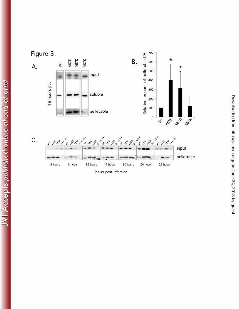

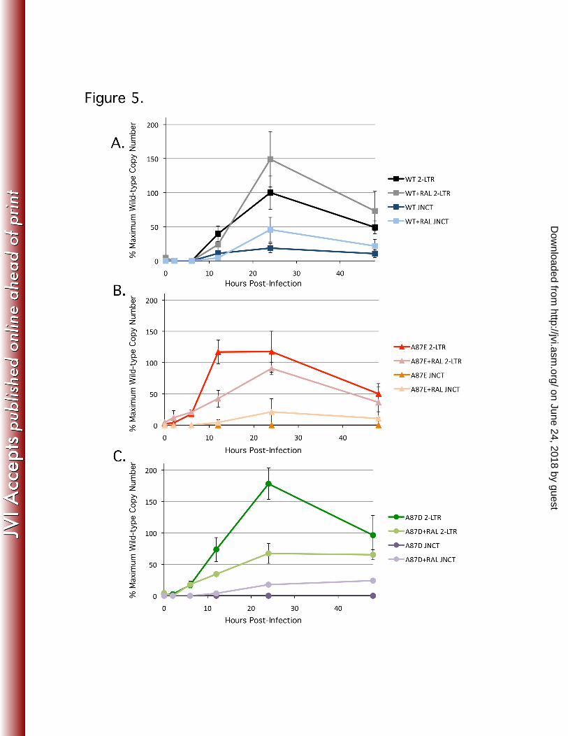
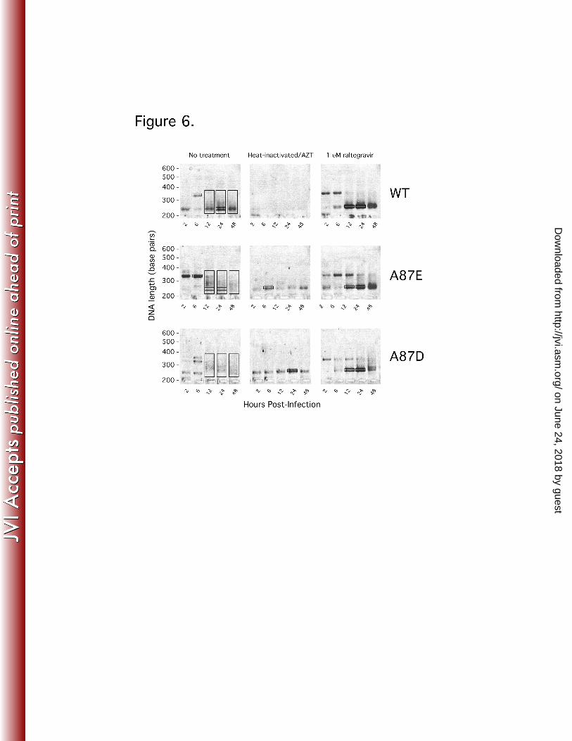


Table 1. Sequence-based classification of 2-LTR junctions.a
Virus WT SIVmac 239 A87E A87D
Time after
infection (h)
12
24
24
48
12+2
4
12
24
12
24
48
12+2
4
12
24
48
12+2
4
Raltegravir
(1μM)
treatment
No
No
No
No
Yes
No
No
No
No
No
Yes
No
No
No
Yes
Fragment
size (bp)
smea
r
(230-
330)
~21
0
smea
r
(230-
330)
smea
r
(230-
330)
~210
~21
0
~21
0
smea
r
(230-
330)
smea
r
(230-
330)
smea
r
(230-
330)
~210
smea
r
(230-
330)
smea
r
(230-
330)
smea
r
(230-
330)
~210
Class I
unprocesse
d
12/40
7/10
17/43
9/34
6/7
8/17
3/4
1/97
0/45
3/34
7/8
5/53
0/43
4/30
21/23
Class II
3’processed
9/40
1/10
19/43
14/34
0/7
8/17
1/4
59/97
31/45
26/34
1/8
18/53
29/43
18/30
0/23
Class III
5’processed
7/40
0/10
5/43
3/34
0/7
1/17
0/4
19/97
11/45
2/34
0/8
22/53
13/43
7/30
0/23
Class IV
Unknown
12/40
2/10
2/43
8/34
1/7
0/17
0/4
21/97
3/45
3/34
0/8
8/53
1/43
1/30
2/23
a The indicated PCR-amplified products from the experiment shown in Fig. 6 were excised from the gel, cloned and sequenced. The number of sequenced products in each class divided by the total number of sequenced products is shown.
on June 24, 2018 by guesthttp://jvi.asm
.org/D
ownloaded from