kaposi' s sarcoma of the small intestine after renal … · kaposi' s sarcoma is a rare...
TRANSCRIPT
J Korean Radiol Soc 1997; 36: 469-472
Kaposi' s Sarcoma of the Small Intestine After Renal Transplantation: Radiological and Endoscopic Findings
l
Hyun Kim, M.D. , Ha Hun Song, M.D. , Si Won Kang, M.D.
Kyung Sub Shinn, M.D. , Byung Min Ahn, M.D. 2
A case of Kaposi' s sarcoma involving the small bowel two years after receiving a re nal transplant is described. Immunosuppression had been achieved using cyclosporine A and prednisolone. Lesions extended from the duodenum to the ileum; radiologically, they were demonstrated on small bowel follow-through study and computed tomography as multip!e small nodu!ar intra!umina! masses with or without central umbilication, and endoscopically, were seen as intramural mucosal elevations with a central crater-like ulceration.
Index Words : Intestinal neoplasms, CT Intestines , radiography
Kaposi' s sarcoma is a rare malignant vascular tumor of unknown etiology, characteristically affecting the skin but sometimes involving other parts ofthe body. Patients with acquired immunodeficiency syndrome (AIDS) and AIDS-related disorders are predisposed to its development (1); it may also occur in organ trans plant recipients who undergo prolonged or intensive immunosuppressive therapy (2 - 4)
The radiological manifestations of AIDS-related Kaposi' s sarcoma of the intestinal tract have been infrequently demonstrated and reported (5 - 9) , while the radiological findings of intestinal Kaposi ’s sarcoma after renal transplantation have not been previously reported. We report the occurrence of Kaposi' s sarcoma of the small intestine, including radiological and endoscopic findings , in a patient who received renal transplantation.
Case Report
Two years prior to consultation, a 31-year-old female received a renal allograft because of chronic renal
1 Department of Rad io logy, Taejon 5 t. Mary’ s HospitaL the Catholic Univer
sity of Korea 1 Departme nt of Departmen t of Internal Med icine, Taejon 5t. Mary ’ s Hospi
la 1. t he Catholi c Un iversity 0 1" Korea Received November 25, 1996; Accepted Jan uary 9, 1997
Address re print requests to : Hyun Ki m M.D., Department of Ra diology , Taejon 5t. Marys Hospita1. ~ 520-2 Taehung-dong , Chung- ku , Taejo n,
30 1-0 12, Korea . Tel. 82-42-220-9625 Fax.82-42-252-6807
- 469
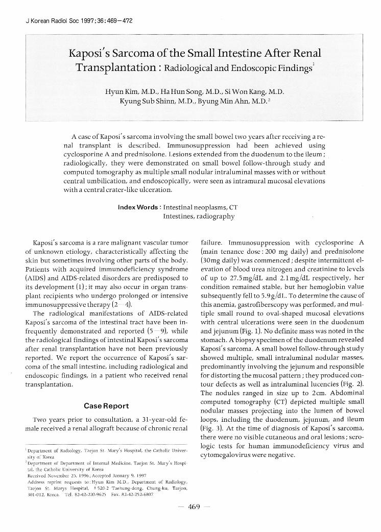
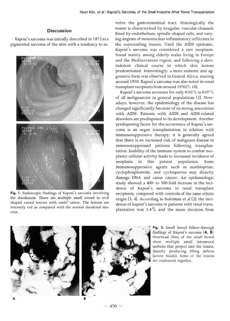
failure . Immunosuppression with cyclosporine A (main tenance dose: 200 mg dail y) and prednisolone (30mg daily) was commenced; despite intermittent elevation of blood urea nitrogen and creatinine to levels of up to 27 .5mg/dL and 2 , lmg /dL respectively , her condition remained stable, but her hemoglobin value subsequently fell to 5.9 g /dL. To determine the cause of this anemia, gastrofiberscopy was performed, and multiple small round to oval-shaped mucosal elevations with central ulcerations were seen in the duodenum and jejunum (Fig , 1). No definite mass was noted in the stomach. A biopsy specimen ofthe duodenum revealed Kaposi’s sarcoma. A small bowel follow-through study showed mu!tiple, small intraluminal nodular masses, predominantly involving the jejunum and responsible for distorting the mucosal pattern; they produced contour defects as well as intraluminallucencies (Fig , 2) ,
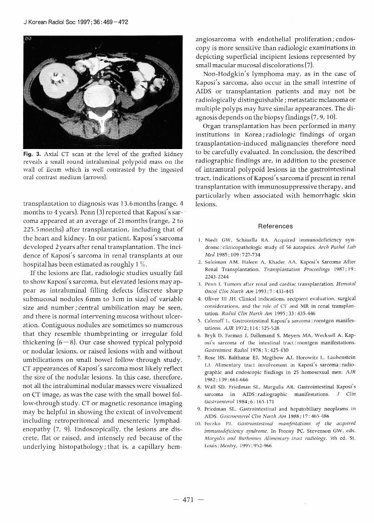
The nodules ranged in size up to 2cm , Abdominal computed tomography (CT) depicted multiple small nodular masses projecting into the lumen of bowel loops , including the duodenum, jejunum, and ileum (Fig. 3) , At the time of diagnosis of Kaposi ’s sarcoma, there were no visible cutaneous and orallesions; serologic tests for human immunodeficiency virus and cytomegalovirus were negative ,
Hyun Kim , et al : Kaposi’s Sarcoma, of the Smalllntestine After Renal Transpantation
Discussion
Kaposi ’s sarcoma was initially described in 1872 as a pigmented sarcoma of the skin with a tendency to in-
례 Fig. 1. Endoscopic findings of Kaposi' s sarcoma involving the duodenum. There are multiple small round to oval shaped raised lesions with umbi" .:::ation. The lesions are intensely red as compared with the normal duodenal mucosa.
철· 빠 ........
A B
volve the gastrointestinal tract . Histologically the tumor is characterized by irregular, vascular channels lined by endothelium, spindle-shaped cells, and varying degrees of mononuclear inflammatory infiltrates in the surrounding tissues. Until the AIDS epidemic, Kaposi ’s sarcoma was considered a rare neoplasm, found mainly among elderly males living in Europe and the Mediterranean region, and following a slow,
indolent clinical course in which skin lesions predominated . Interestingly, a more endemic and aggressive form was observed in Central Africa, starting around 1950. Kaposi' s sarcoma was also noted in renal transplant recipients 삼om around 1970 (7, 10)
Kaposi' s sarcoma accounts for only 0.02 % to 0.07 % of all malignancies in general populations (3). Now adays, however, the epidemiology of the disease has changed significantly because of its strong association with AIDS. Patients with AIDS and AIDS-related disorders are predisposed to its development. Another predisposing factor for the occurrence of Kaposi' s sarcoma is an organ transplantation in relation with immunosuppressive therapy; it is generally agreed that there is an increased risk of malignant disease in immunosuppressed patients following transplantation. Inability of the immune system to combat neoplastic cellular activity leads to increased incidence of neoplasia in this patient population. Some immunosuppressive agents such as azathioprine, cyclophosphamide, and cyclosporine may directly damage DNA and cause cancer. An epidemiologic study showed a 400- to 500-fold increase in the incidence of Kaposi ’s sarcoma in renal transplant recipients, compared with controls of the same ethnic origin (3, 4). According to Suleiman et al (2), the incidence of kaposi ’s sarcoma in patients with renal transplantation was 3.4 % and the mean duration from
Fig. 2. Small bowel follow-through findings of Kaposi ’s sarcoma (A, B). Overhead films of the small bowel show multiple small intramural nodules that project into the lumen,
thereby producing filling defects (arrow heads). Some of the lesions are coalescent together
- 470 -
J Korean Radiol Soc 1997: 36: 469-472
Fig. 3. Axial CT scan at the level of the grafted kidney reveals a sma!! round intraluminal polypoid mass on the wall of ileum which is well contrasted by the ingested oral contrast medium (arrows)
transp1antation to diagnosis was 13.6months (range, 4 months to 4 years) . Penn (3) reported that Kaposi ’ s sarcoma appeared at an average of 21 months (range, 2 to 225.5months) after transp1antation, including that of the heart and kidney. In our patient, Kaposi ’ s sarcoma
deve10ped 2years after rena1 transp1antation. The incidence of Kaposi' s sarcoma in rena1 transp1ants at our hospita1 has been estimated as rough1 y 1 %.
If the 1esions are flat, radio1ogic studies usually fai1
to show Kaposi ’ s sarcoma, but e1evated 1esions may appear as intra1umina1 filling defects (discrete sharp submucosa1 nodu1es 6mm to 3 cm in size) of variab1e size and number; centra1 umbilication may be seen, and there is norma1 intervening mucosa without ulcer ation. Contiguous nodu1es are sometimes so numerous
that they resemb1e thumbprinting or irregu1ar fo1d thickening (6 - 8). Our case showed typica1 po1ypoid or nodu1ar 1esions, or raised 1esions with and without
umbi1ications on sma11 bowel follow-through study. CT appearances of Kaposi ’ s sarcoma most like1y reflect the size of the nodu1ar 1esions. In this case, therefore, not a11 the intra1umina1 nodu1ar masses were visualized on CT image, as was the case with the sma11 bowel fo1-10w-through study. CT or magnetic resonance imaging may be he1pfu1 in showing the extent of invo1vement
including retroperitonea1 and mesenteric 1ymphadenopathy (7, 9). Endoscopica11y , the 1esions are discrete, flat or raised , and intense1y red because of the underlying histopatho1ogy ; that is, a capillary hem-
깎
angiosarcoma with endothelia1 proliferation; endoscopy is more sensitive than radio1ogic examinations in
depicting superficia1 incipient 1esions represented by small macu1ar mucosa1 disco1orations (7)
Non-Hodgkin ’ s 1ymphoma may, as in the case of Kaposi' s sarcoma, a1so occur in the sma11 intestine of
AIDS or transp1antation patients and may not be radio1ogica11y distinguishab1e; metastatic me1anoma or multip1e polyps may have similar appearances. The diagnosis depends on the biopsy findings (7 , 9, 10).
Organ transp1antation has been performed in many institutions in Korea; radio1ogic findings of organ
transp1antation-induced malignancies therefore need to be carefully eva1uated . In conclusion, the described
radiographic findings are, in addition to the presence
of intramura1 po1ypoid 1esions in the gastrointestina1 tract, indications ofKaposi' s sarcoma ifpresent in rena1 transplantation with immunosuppressive therapy, and
particularly when associated with hemorrhagic skin lesions.
References
l. Niedt GW , Schinella RA. Acquired immunodefici ency syndrome: clinicopathologic study of 56 autopsies. Arch Patho/ Lab
Med 1985; 109 :727-734
2. Suleiman AM, Haleen A, Khader AA. Kaposi’ S Sarcoma A fter Renal Transplantation. Transp/anta tion pmceedings 1987; 19 2243-2244
3. Penn 1. Tumors after renal and cardiac transplantation. Hemato/
Onco/ Clin N orth Am 1993; 7: 431-445 4. Oliver III JH. Clinical indications, recipient evaluation, surgical
considerations, and the role of CT and MR in renal transplan tation. Radio/ C/in North Am 1995; 33: 435-446
5. Calenoff L. Gastrointestinal Kaposi’ S sarcoma: roentgen manifestations. A JR 1972; 114: 525-5 28
6. Bryk D, Farman J, Dallemand S, Meyers MA , Wecksell A. KapOSI’S sarcoma of the intestinal tract: roentgen manifestations. Gastrointest Radio/ 1978; 3 : 42 5-430
7. Rose HS, Balthazar EJ, Megibow AJ, Horowitz L, Laubenstein LJ. Alimentary tract involvement in Kaposi’s sarcoma: radiographic and endoscopic findings in 25 homosexual men. AJR
1982 ; 139: 661-666 8‘ Wall SD, Friedman S1. Margulis AR. Gastrointestinal Kaposi ' s
sarcoma in AIDS: radiographic manifestations. J C/in
Gastroentero/ 1984; 6 : 165-171 9. Friedman SL. Gastrointestinal and hepatobiliary neoplasms in
AIDS. Gastroentero/ C/in Nort h A m 1988 ; 17 ‘ 465-486 10. Feczko P J. Gastrointestina/ manifestations of the acquired
Iml
Mar쟁guω띠il씨l“IS an띠d Bu r.깨hennes A/“lmentπtmηηy t ract rad이10이lωog강y . 5th ed. St.
Louis : Mosby, 1995: 952-966

Hyun Kim , et al : Kaposi’s Sarcoma, of the Smalllntestine Ater Renal Transplantation
대한밤사선의학호|지 1997; 36 : 469-472
신장이식수술후 발생한 소장의 Kaposi 육종:방사선학적 빛 내시경적 소견1
1 가톨릭대학교 의과대학 대전성모병원 방사선과 2가톨릭대학교 의과대학 대전성모병원 내과
검 현 · 송하헌 · 강사원 · 신경섭 · 안병민2
신장이식수술을 받은지 2년 후에 발생한 소장의 Kaposi 육종에 대해 보고한다. 신장이식후 사용된 면역억제
제는 cyclosporine A와 prednisolone이였다. Kaposi 육종은 섭이지장에서부터 회장까지 분포하였다. 이 병변은
저긴장성 십이지장조영솔, 소장조영술 및 전산화단층촬영 영상에서 소장내벽에 다수의 작은 결절성 종괴로 관
찰되었으며 , 일부 종괴에서는종괴의 중앙부위가함몰되어 보였다. 내시경상병변은중앙부위에 분화구같은 궤
양을 갖는 장내 점막이 융기된 종괴의 소견을 보였다.
” “