kenmed jan09 template - university of albertajmo/images/ped/curative.pdf · clínica sagrada...
TRANSCRIPT

632 | VOLUME 64 | NUMBER 4 | APRIL 2009 www.neurosurgery-online.com
CLINICAL STUDIESPedro Lylyk, M.D.Departments of Neurosurgery and Interventional Neuroradiology,Clínica Sagrada Familia;Clínica Adventista Belgrano; andFundación Lucha contra las EnfermedadesNeurológicas de la Infancia,Buenos Aires, Argentina
Carlos Miranda, M.D.Departments of Neurosurgery and Interventional Neuroradiology,Clínica Sagrada Familia;Clínica Adventista Belgrano; andFundación Lucha contra las EnfermedadesNeurológicas de la Infancia,Buenos Aires, Argentina
Rosana Ceratto, M.D.Departments of Neurosurgery and Interventional Neuroradiology,Clínica Sagrada Familia;Clínica Adventista Belgrano; andFundación Lucha contra las EnfermedadesNeurológicas de la Infancia,Buenos Aires, Argentina
Angel Ferrario, M.D.Departments of Neurosurgery and Interventional Neuroradiology,Clínica Sagrada Familia;Clínica Adventista Belgrano; andFundación Lucha contra las EnfermedadesNeurológicas de la Infancia,Buenos Aires, Argentina
Esteban Scrivano, M.D.Departments of Neurosurgery andInterventional Neuroradiology,Clínica Sagrada Familia;Clínica Adventista Belgrano; andFundación Lucha contra las EnfermedadesNeurológicas de la Infancia,Buenos Aires, Argentina
Hugh Ramirez Luna, M.D.Departments of Neurosurgery andInterventional Neuroradiology,Clínica Sagrada Familia;Clínica Adventista Belgrano; andFundación Lucha contra las EnfermedadesNeurológicas de la Infancia,Buenos Aires, Argentina
Aaron L. Berez, M.D.Chestnut Medical Technologies, Inc.,Menlo Park, California
Quang Tran, B.S.M.E., M.B.A.Chestnut Medical Technologies, Inc.,Menlo Park, California
Peter K. Nelson, M.D.Departments of Neurosurgery andNeuroradiology,New York University,New York, New York
David Fiorella, M.D., Ph.D.Departments of Neurosurgery and Neuroradiology,Barrow Neurological Institute,Phoenix, Arizona
Reprint requests:David Fiorella, M.D., Ph.D.,Department of Endovascular Neurosurgery,Barrow Neurological Institute,2910 North 3rd AvenuePhoenix, AZ 85013.Email: [email protected]
Received, September 9, 2008.Accepted, October 9, 2008.
Copyright © 2009 by theCongress of Neurological Surgeons Endovascular treatment of intracranial aneu -
rysms has evolved substantially over thepast 2 decades, transitioning from an inves-
tigational therapy into routine clinical practiceand ultimately emerging as the treatment of
ABBREVIATIONS: ISS, in- stent stenosis; mRS,modified Rankin Scale; PED, Pipeline emboliza-tion device; PITA, Pipeline for the IntracranialTreatment of Aneurysms
CURATIVE ENDOVASCULAR RECONSTRUCTION OFCEREBRAL ANEURYSMS WITH THE PIPELINE EMBOLIZATIONDEVICE: THE BUENOS AIRES EXPERIENCE
OBJECTIVES: The Pipeline embolization device (PED) (Chestnut Medical Technologies,Inc., Menlo Park, CA) is a new microcatheter- delivered endovascular construct designedto achieve the curative reconstruction of the parent arteries giving rise to wide- neckedand fusiform intracranial aneurysms. We present our initial periprocedural experiencewith the PED and midterm follow- up results for a series of 53 patients.METHODS: Patients harboring large and giant wide- necked, nonsaccular, and recurrentintracranial aneurysms were selected for treatment. All patients were pretreated with dualantiplatelet medications for at least 72 hours before surgery and continued taking bothagents for at least 6 months after treatment. A control digital subtraction angiogram wastypically performed at 3, 6, and 12 months.RESULTS: Fifty- three patients (age range, 11–77 years; average age, 55.2 years; 48female) with 63 intracranial aneurysms were treated with the PED. Small (n � 33), large(n � 22), and giant (n � 8) wide- necked aneurysms were included. A total of 72 PEDswere used. Treatment was achieved with a single PED in 44 aneurysms, with 2 overlap-ping PEDs in 17 aneurysms, and with 3 overlapping PEDs in 2 aneurysms. The mean timebetween the treatment and last follow- up digital subtraction angiogram was 5.9 months(range, 1–22 months). Complete angiographic occlusion was achieved in 56%, 93%,and 95% of aneurysms at 3 (n � 42), 6 (n � 28), and 12 (n � 18) months, respectively.The only aneurysm that remained patent at the time of the 12-month follow- up exam-ination had been treated previously with stent- supported coiling. The presence of apreexisting endoluminal stent may have limited the efficacy of the PED reconstructionin this aneurysm. No aneurysms demonstrated a deterioration of angiographic occlu-sion during the follow- up period (i.e., no recanalizations). No major complications(stroke or death) were encountered during the study period. Three patients (5%), allwith giant aneurysms, experienced transient exacerbations of preexisting cranial neu-ropathies and headache after the PED treatment. All 3 were treated with corticosteroids,and these symptoms resolved within 1 month.CONCLUSION: Endovascular reconstruction with the PED represents a safe, durable,and curative treatment of selected wide- necked, large and giant cerebral aneurysms.The rate of complete occlusion at the time of the 12-month follow- up examinationapproached 100% in the present study. To date, no angiographic recurrences have beenobserved during serial angiographic follow-up.
KEY WORDS: Aneurysm, Endovascular, Pipeline embolization device, Segmental arterial disease
Neurosurgery 64:632–643, 2009 DOI: 10.1227/01.NEU.0000339109.98070.65 www.neurosurgery- online.com

choice for many lesions. Randomized clinical trials such as theInternational Study of Subarachnoid Aneurysm Treatment (18)and the Barrow Ruptured Aneurysm Trial (17) have estab-lished the advantages of endovascular treatment in selectedclinical scenarios.
Despite this tremendous evolution in endovascular therapy,some important limitations remain, particularly in the treatmentof wide- necked, large and giant, or “nonsaccular” fusiformaneurysms. These lesions can frequently be difficult to recon-struct with coils, even when they are used with the commerciallyavailable self- expanding intracranial stents (Neuroform; BostonScientific, Natick, MA; Enterprise; Cordis Neurovascular,Warren, NJ; LEO and LEO Plus; Balt Extrusion, Montmorency,France). Endovascular treatments of such lesions frequently failto produce complete aneurysm occlusion. Even when completeor near- complete occlusion has been achieved after the initialembolization, these aneurysms remain prone to coil compactionand recanalization, and they frequently recur, requiring 1 ormore retreatments (8, 18, 19, 29).
To date, endovascular therapy has been almost exclusivelyfocused on filling the aneurysm sac with embolic material, i.e.,“endosaccular” treatment. This strategy is very effective for thetreatment of most narrow- necked aneurysms that arise from a“focal defect” in the parent artery wall (involving �25% of theparent artery circumference). However, endosaccular occlusiondoes not address the remaining circumference of the diseasedparent artery that gives rise to the aneurysm. In larger, more dysplastic- appearing aneurysms, the demarcation between thenormal artery and diseased vessel becomes less distinct. Forthis reason, the endosaccular strategy is often ineffective in treat-ing wide- necked or fusiform aneurysms that arise from a larger,more diffuse, “segmental defect” in the parent vessel.
These segmental defects are only addressed with an “endo-luminal” strategy that achieves circumferential parent vesselreconstruction. This effect has been achieved to some extentusing the commercially available balloon- expandable (14, 15,33) and self- expanding stents (2–4, 9, 11, 13, 16). However, theexisting intracranial stents have very limited metal surface areacoverage (6.5%–9% for self- expanding stents and 12%–16% forballoon mounted stents) and, thus, their ability to elicit remod-eling of the parent artery is limited. In most aneurysms, thesedevices are inadequate to achieve occlusion by themselves, andaggressive endosaccular coil embolization in concert withendoluminal reconstruction is required to reliably achieve adurable result.
The Pipeline embolization device (PED) (Chestnut MedicalTechnologies, Inc., Menlo Park, CA) represents the first endo -vascular construct specifically engineered to function as astand-alone device for the endovascular reconstruction of asegmentally diseased parent vessel. The PED is a self- expanding, microcatheter- delivered, cylindrical mesh devicecomposed of 48 individual cobalt chromium and platinumstrands. The device has 30% to 35% metal surface area coveragewhen fully deployed (7).
The initial experience with the PED has shown it to be effec-tive in achieving the curative anatomic reconstruction of large
NEUROSURGERY VOLUME 64 | NUMBER 4 | APRIL 2009 | 633
BUENOS AIRES PIPELINE EXPERIENCE
segmental vascular defects, which give rise to the wide- necked,large and giant, or nonsaccular aneurysms that have tradition-ally presented the greatest challenge to existing endovascularand open vascular neurosurgical treatment strategies (5–7, 20).We present periprocedural outcomes and midterm angio-graphic follow- up results for a series of 53 patients with 63 wide- necked aneurysms that were treated with the PED.
PATIENTS AND METHODS
Patient Population and SelectionThe present study is a prospective, all- inclusive case series of patients
undergoing treatment with the PED for wide- necked (defined asaneurysms with a dome- to- neck ratio of �2 or a neck size �4 mm) sac-cular aneurysms, nonsaccular aneurysms, large and giant aneurysms,and aneurysms for which previous treatment attempts failed. BetweenMarch 2006 and June 2008, 53 patients (mean age, 55.2 years; age range,11–77 years; 48 female and 5 male) harboring 63 aneurysms were treated.
Because the PED is an investigational device, its application in thisseries of patients was always prospectively approved either on a case- by- case basis as compassionate use (initial experience), within the con-text of the Pipeline for the Intracranial Treatment of Aneurysms (PITA)trial (a multicenter, single arm, nonrandomized trial) (20), or within thecontext of the post- PITA Buenos Aires registry (single- center registry)by our institutional ethics committees in accord with local regulations.Written informed consent was obtained from every patient. Allaneurysms were treated electively after the appropriate institutionalregulatory clearance had been secured, and the informed consent wasobtained. Six of the patients (with 6 treated aneurysms) included in thepresent series were also included in the PITA trial.
Antiplatelet Medication RegimenPatients were pretreated with 75 mg of clopidogrel and 325 mg of
aspirin at least 72 hours before PED treatment. Dual antiplatelet medica-tion was maintained for at least 6 months after the procedure.Intravenous heparin was administered during the procedure to maintainan activated clotting time between 250 to 300 seconds. Heparinizationwas not reversed at the conclusion of the procedure.
PEDThe PED is a flexible, microcatheter- delivered, self- expanding,
endovascular “stent- like” construct engineered specifically for the treat-ment of cerebral aneurysms (Fig. 1). The device consists of a braidedmesh cylinder composed of 48 individual platinum and cobaltchromium microfilaments. The stent is mounted in a recess on a flexi-ble delivery wire and is front- loaded via an introducer and deliveredthrough a standard 0.027-inch internal diameter microcatheter (MassTransit; Cordis Neurovascular, Warren NJ; Renegade Hi- Flo; BostonScientific).
ProcedureAll treatments were performed under general anesthesia and via
the transfemoral approach. In those procedures in which coils wereintroduced into the aneurysm, either the coiling was done before place-ment of the PED, or the microcatheter was placed within the saccularcomponent of the aneurysm and “jailed” by placing the PED constructacross the aneurysmal segment. With this jailing or parallel technique,coiling is subsequently performed through the jailed microcatheterafter the PED construct has been placed. After coiling is completed, the

634 | VOLUME 64 | NUMBER 4 | APRIL 2009 www.neurosurgery-online.com
LYLYK ET AL.
microcatheter is easily removed by gently retracting it from theaneurysm. The removal of the microcatheter did not disrupt the PEDconstruct in any of the aneurysms.
All PEDs were deployed following a standard procedure. First, themicrocatheter was manipulated under high- magnification fluoroscopicroadmap control across the aneurysm neck. The PED, mounted on adelivery wire and constrained within a sheath, was then inserted intothe rotating hemostatic valve and introduced into the hub of the micro-catheter. By pushing the delivery wire, the PED was advanced throughthe length of the microcatheter and into position for deployment. ThePED delivery wire was then held in place while the microcatheter wascarefully retracted to initiate deployment. Through a combination offorward pressure on the delivery wire and retraction of the micro-catheter, the device was deployed, expanding to come free of the deliv-ery microwire. When constrained within a microcatheter, the PED iselongated 2.5 times its maximally expanded deployed configuration.This foreshortening must be taken into account during the positioningand deployment of the construct.
Procedural Assessment and Follow- up ExaminationTechnical success was defined as PED deployment with complete
coverage of the aneurysm neck, preserved patency of the parent artery,and no clinically evident adverse events. Posttreatment clinical follow- up was performed at the time of discharge. Concurrent clinical andangiographic follow- up was performed at 1, 3, 6, and 12 months after
the treatment. Neurological examinations were performed by an inde-pendent neurologist.
Aneurysm sizes are provided as the single greatest dimension. Onlythe portion of the aneurysm opacified by contrast agent was measured.Regions of the aneurysm that were occluded by preexisting emboliza-tion coils or intraluminal thrombus were not included in the largestdimensional measurement. Vessel wall defects were classified as focal ifless than 25% of the circumference of the parent artery was involved bythe aneurysm neck and segmental if more than 25% of the circumfer-ence of the parent vessel wall was involved.
Follow- up angiography was performed in the standard projections aswell as in the working angle for PED placement. The primary angio-graphic end point was complete aneurysm occlusion. Any residual fill-ing of the aneu rysm was characterized as incomplete occlusion.
RESULTS
Patient CharacteristicsOver a 26-month study period (March 2006 to May 2008), 53
patients (average age, 55.2 years; age range, 11–77 years) with 63aneurysms were treated with the PED. The clinical presenta-tions of the pa tients are documented in Table 1. At the time oftreatment, 30 patients (56%) had a modified Rankin Scale (mRS)score of 0, 12 patients (23%) had a score of 1, and 11 patients
FIGURE 1. A 58-year-old woman with a giant right cavernous segmentinternal carotid artery (ICA) aneurysm presented with a right sixth nervepalsy. Postcontrast coronal T1-(A) and T2-weighted (B) magnetic resonanceimaging (MRI) scans demonstrating the giant aneurysm arising from theright ICA, impressing upon the medial aspect of the right temporal lobe.Shaded surface display from a three- dimensional (3D) rotational angiogram(C) and conventional angiogram in the lateral projection (D) demonstratinga long segmental defect arising from the posterior wall of the ICA. E,angiogram immediately after reconstruction with the Pipeline embolization
device (PED) showing that contrast medium flow has been diverted from theaneurysm sac into the cerebrovasculature. Contrast material within theaneurysm is static, forming a dependent contrast level or “eclipse sign.” Follow- up angiograms at 1 month (F) and 6 months (G) showing anatomicremodeling of the ICA with complete occlusion of the aneurysm. CoronalT2-weighted (H) and precontrast T1-weighted (I) MRI scans showing com-plete resolution of the mass effect from the aneurysm. Dotted circles in A, B,H, and I indicate the location of the aneurysm.
A B C D E
F G H I

NEUROSURGERY VOLUME 64 | NUMBER 4 | APRIL 2009 | 635
BUENOS AIRES PIPELINE EXPERIENCE
(21%) had a score of 2. Among the 11 patients with an mRS of2, 3 presented with previous subarachnoid hemorrhage, 3 withmass effect, 3 with cranial nerve palsy, and 2 with visual deficits(Fig. 1).
Lesion Characteristics: Location and SizeThe locations of the aneurysms treated are listed in Table 2; 55
(87.3%) involved the anterior circulation and 8 (12.7%) involvedthe posterior circulation. According to the International Study ofUnruptured Intracranial Aneurysms (32) size classification, 33(52%) aneurysms were small (�10 mm), 22 (35%) were large(10–25 mm), and 8 (13%) were giant (�25 mm) (32). The meananeurysm size was 11.1 mm (range, 3.5–30 mm). Fifty- five (87%)aneurysms were saccular, and 8 others (13%) were nonsaccular(circumferential, fusiform, or dissecting morphology). Accordingto our classification system, 94% of the aneurysms arose fromsegmental defects of the artery, whereas 6% arose from focaldefects. The 4 aneurysms arising from focal defects were eitherlarge (n � 3) or giant (n � 1) and were located within the ante-rior circulation.
Lesion Characteristics: Previous TreatmentOf the 63 lesions treated, 40 (63%) were de novo unruptured
aneurysms (Fig. 1), whereas 23 (37%) were previously treatedand subsequently recanalized (Fig. 2). Of the previously treatedaneurysms, 16 were unruptured and 7 had previously rup-tured. Previous treatments included coiling alone in 14 patients, stent- supported coiling in 6 patients, surgical clipping in 2patients, and stent monotherapy in 1 patient. The indicationsfor PED placement (Table 3) were categorized as saccularaneurysm with a dome/neck ratio of less than 2 (n � 32 [51%]),large or giant size (n � 4, 6%), fusiform/dissecting morphology(n � 8 [12%]), and failure of previous treatment (endovascularor surgical; n � 19, [31%]).
PED Treatment Forty- four aneurysms (70%) were treated with a single PED,
17 (27%) were treated with 2 PEDs, and 2 (3%) were treatedwith 3 PEDs. In 4 of the aneurysms (6%) in which a single PEDwas used for treatment, embolization coils were also used.During treatment of the remaining 59 lesions (94%), the PEDwas used as a stand- alone device without embolization coils.All aneurysms arising from focal defects in the parent arterywere treated with a single device.
PED deployment was technically successful 97% of the time(70 of 72 devices deployed). In 1 procedure, the proximal aspectof the PED was inadvertently deployed into the aneurysm, andan Alligator retrieval device (Chestnut Medical Technologies,Inc.) was used to retract and reposition the device across theaneurysm neck. In a second procedure, the distal tip of thePED delivery wire became engaged within the deployed PEDand fractured. The fractured distal aspect of the wire wassecured into a stable position against the vessel wall by thedeployed PED. Neither of these technical complicationsresulted in a clinically evident complication. In all patients(100%), the PEDs were ultimately deployed in an acceptableposition across the targeted aneurysm.
No major (stroke or death) clinically evident periprocedural(within 30 days) complications were encountered during thestudy period. Minor complications occurred in 6 of 53 patients(11%). Five patients developed hematomas at the femoralpuncture site. One patient developed a rash from a reaction tothe contrast material. Three patients (5%) initially presentingwith IIIrd and VIth cranial nerve palsies owing to giant carotidcavernous aneurysms developed headache and exacerbationof their cranial nerve palsies during the first postoperativeweek. All 3 were treated with a course of steroids. Two recov-ered to their pretreatment baseline over the next month, andthe third ultimately improved in comparison to the pretreat-ment status.
Angiographic Results: Immediate and Follow- upAt the conclusion of the treatment, only 5 of 63 (8%)
aneurysms showed complete angiographic occlusion. Alllesions that were completely occluded immediately after PEDplacement were small (�10 mm) aneurysms for which previous
TABLE 2. Aneurysm location
Aneurysm location No. %
Internal carotid artery
Cavernous 11 17.4
Carotid cave 5 7.9
Parophthalmic 9 14.2
Superior hypophyseal 5 7.9
Ophthalmic 13 20.6
Posterior communicating 10 15.8
Anterior choroidal 1 1.5
Carotid terminus 1 1.5
Posterior circulation
Posterior inferior coronary artery 1 1.5
Vertebral 4 6.3
Vertebrobasilar junction 1 1.5
Basilar 2 3.1
Total 63 100.0
TABLE 1. Clinical presentation
Presentation No. %
Incidental 25 47.2
Headache 6 11.3
Previous subarachnoid hemorrhage 7 13.2
Mass effect 4 7.5
Visual deficit 5 9.4
Cranial nerve palsy 6 11.3
Total 53 100.0

636 | VOLUME 64 | NUMBER 4 | APRIL 2009 www.neurosurgery-online.com
LYLYK ET AL.
therapy had not failed (i.e., denovo lesions). Although resid-ual filling was noted in the re -maining aneurysms, the tran-sit of contrast material intoand out of, the aneurysm wasmarkedly slowed, and the ini-tial inflow jet was disrupted.During the capillary andvenous phases of the angio -gram, the newly reconstructedparent artery could often bevisualized as a negative defectsurrounded by contrast mate-rial. This negative defect iscreated as unopacified inflowquickly clears the contrastmaterial from the lumen of thereconstructed parent artery,which then stands out in reliefagainst the more static con-trast material that is retainedwithin the aneurysm sac (Fig.3). Contrast material couldoften be seen layering withinthe dependant portion of thelarger aneurysms, forming aneclipse sign on subtractedimages, which typically per-sisted into the late venousphase (Fig. 4).
One-, 3-, 6-, and 12-monthangiographic follow- up re sultswere available for 51, 42, 28,and 18 aneurysms, respec-tively. The average angio-graphic follow- up period was5.9 months. By 6 months, 93%(26 of 28) of the aneurysms
had progressed to complete occlusion. Of the 18 aneurysmsstudied at 12 months, 17 (94.4%) had progressed to completeocclusion (Figs. 5 and 6; Table 4). The sole aneurysm with resid-ual filling after 12 months was a giant, circumferential, fusiformbasilar aneurysm treated with 2 PEDs. This lesion had recurredafter stent- supported coil embolization before PED treatment. Itis possible that the preexisting stent may have impaired the wallapposition of the PED construct in this patient.
Of the 38 vessels with 3-month angiographic follow- up, 3(8%) showed mild (25%–50%) in- stent stenosis (ISS), 2 (5%)showed moderate (50%–70%) ISS, and 2 (5%) showed severe(�70%) ISS. Three of these cases of ISS re solved to some extentby the 6-month follow- up angiogram, with 1 of the mild casesre solving completely, 1 of the mod erate stenoses regressing tomild ISS, and 1 case of the severe stenoses regressing to mod-erate ISS. All cases of ISS were asymptomatic and, thus, nonewere treated.
FIGURE 2. A 59-year-old woman initially presented with a symptomatic unruptured large right carotid- ophthalmicartery aneurysm arising from a segmental defect in the vessel. The aneurysm was initially treated with coil emboliza-tion. A, axial T2-weighted MRI showing the signal void corresponding to the dome of the aneurysm with mass effectupon the inferior medial aspect of the right frontal lobe. B, lateral angiogram obtained 1 year after the original treat-ment showing coil compaction and a large amount of residual filling of the aneurysm. C, native image immediatelyafter PED reconstruction showing the construct in place across the aneurysm neck (“stent,” arrow), the 0.027-inchinternal diameter (ID) delivery catheter (microcatheter) within the proximal cavernous segment of the internalcarotid artery and the PED delivery wire within the proximal middle cerebral artery more distally. Subtractedangiogram (D) and native (E) and reconstructed (F) 3D rotational angiograms at the 3-month follow- up examina-tion showing minimal residual filling in the region of the aneurysm neck only. Subtracted (G) and native (H)angiograms in the working projection at the 12-month follow- up examination demonstrating anatomic reconstruc-tion of the parent artery and complete aneurysm occlusion. This case demonstrates the rate at which progressivethrombosis and vascular remodeling occur after PED reconstruction.
A B C D
E F G H
TABLE 3. Indications for stent placement
No. (%) of No. ofIndication
aneurysms stents
De novo aneurysms
Saccular, dome/neck ratio >2 32 (51%) 33
Saccular (large/giant), 4 (6%) 4dome/neck ratio <2
Nonsaccular 4 (6%) 6
Recurrent aneurysms(retreatments)
Nonsaccular 4 (6%) 7
Saccular 19 (31%) 22
Total 63 (100%) 72
NEUROSURGERY VOLUME 64 | NUMBER 4 | APRIL 2009 | 637
BUENOS AIRES PIPELINE EXPERIENCE
Clinical Results Thirty- nine patients have
had at least 3 months of clinical follow- up, and 17 have had 1full year of follow- up. No pa -tients have experienced de -layed deterioration in theirclinical status after the 30-dayperiprocedural period. Two ofthe 12 patients with an initialmRS score of 2 improved to ascore of 1 at the 6-month clini-cal follow- up. Scores for allpatients with an initial mRSscore of 1 or 0 were unchangedat 3 to 6 months of follow- up.
DISCUSSIONThe most important find-
ings of the present study arethe following: 1) the PEDreproducibly elicits curativeendovascular reconstruction ofselected intracranial aneu -rysms; 2) aneurysm treatmentwith the PED is safe; 3) aneu -rysm treatment with the PEDis durable; 4) preexisting endo-luminal constructs can poten-tially limit the efficacy of thePED; and 5) primary endovas-cular reconstruction representsa fundamental paradigm shiftin the technique of endovascu-lar aneurysm treatment.
Endovascular therapy hasemerged as an accepted and, insome cases, preferred treat-ment for cerebral aneurysms.However, the technique hasthe major shortcomings ofincomplete treatment andquestionable long- term dura-bility. These shortcomingshave led to persisting reserva-tions about the technologydespite the results of large, ran-domized, multicenter trialsdemonstrating its superiorityto surgical clipping in selectedpatients (23).
In most reported series,only a minority of aneurysmstreated by coil embolizationare ultimately cured angio-
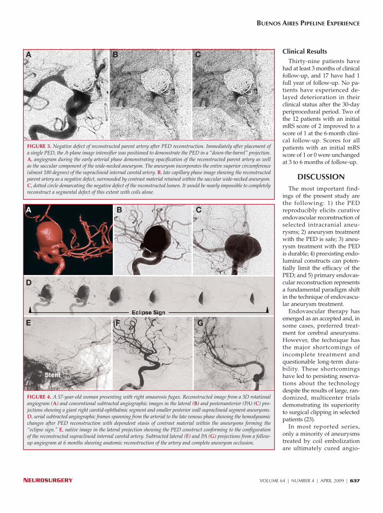
FIGURE 3. Negative defect of reconstructed parent artery after PED reconstruction. Immediately after placement ofa single PED, the A- plane image intensifier was positioned to demonstrate the PED in a “down- the- barrel” projection.A, angiogram during the early arterial phase demonstrating opacification of the reconstructed parent artery as wellas the saccular component of the wide- necked aneurysm. The aneurysm incorporates the entire superior circumference(almost 180 degrees) of the supraclinoid internal carotid artery. B, late capillary phase image showing the reconstructedparent artery as a negative defect, surrounded by contrast material retained within the saccular wide- necked aneurysm.C, dotted circle demarcating the negative defect of the reconstructed lumen. It would be nearly impossible to completelyreconstruct a segmental defect of this extent with coils alone.
A B C
FIGURE 4. A 57-year-old woman presenting with right amaurosis fugax. Reconstructed image from a 3D rotationalangiogram (A) and conventional subtracted angiographic images in the lateral (B) and posteroanterior (PA) (C) pro-jections showing a giant right carotid- ophthalmic segment and smaller posterior wall supraclinoid segment aneurysms.D, serial subtracted angiographic frames spanning from the arterial to the late venous phase showing the hemodynamicchanges after PED reconstruction with dependent stasis of contrast material within the aneurysms forming the“eclipse sign.” E, native image in the lateral projection showing the PED construct conforming to the configurationof the reconstructed supraclinoid internal carotid artery. Subtracted lateral (F) and PA (G) projections from a follow- up angiogram at 6 months showing anatomic reconstruction of the artery and complete aneurysm occlusion.
A B C
D
E F G

638 | VOLUME 64 | NUMBER 4 | APRIL 2009 www.neurosurgery-online.com
LYLYK ET AL.
graphically. Raymond et al. (24) reported a 38.3% rate of com-plete angiographic occlusion at the 12 month follow- up evalu-ation in a series of 353 consecutive coiled aneurysms. Kole et al.(12) reported a 19% rate of complete occlusion in a series of 131coiled aneu rysms with long- term angiographic follow- up(mean, 18 months). In the International SubarachnoidAneurysm Trial, a 66% rate of complete angiographic occlusionwas observed in a cohort largely (91%) composed of smallaneurysms (18). These rates of occlusion are even lower inselected subgroups such as large, giant, wide- necked, and non-saccular aneurysms.
The recent Cerebral Aneurysm Rerupture After Treatmentstudy (10) provides evidence that, at least for rupturedaneurysms, it is critical to achieve complete angiographic oblit-eration to provide adequate protection from subsequent hem-orrhage. In that study, ruptured aneurysms were followed aftereither surgical or endovascular treatment to assess the inci-dence of rehemorrhage. Although the overall hemorrhage riskwas very low after coiling (1.3 rehemorrhages per 100 person- years), the risk of rebleeding increased drastically with de -creasing levels of aneurysm occlusion (0.6 rehemorrhages per100 person- years for completely occluded aneurysms versus 15
FIGURE 5. A 55-year-old woman presenting with a VIth nerve palsy.Reconstructed image from a 3D rotational angiogram (A) and lateral sub-tracted angiogram (B) showing the giant left carotid- ophthalmic segmentand small superior hypophyseal aneurysms. C, subtracted image in the lat-eral projection showing markedly reduced flow into both aneurysms. D, native transorbital oblique image of the PED in position within the left supr-aclinoid internal carotid artery spanning the origins of both carotidaneurysms. Follow- up subtracted angiograms in the transorbital (E) and lat-eral (F) projections at 20 days showing complete occlusion of the giant
carotid- ophthalmic aneurysm with persistent patency of the smaller, more dis-tal superior hypophyseal artery aneurysm. Three- month follow- up digitalsubtraction angiograms in the transorbital (G) and lateral (H) projectionsshowing anatomic remodeling of the parent artery with complete occlusion ofboth aneurysms. This patient demonstrates that the anatomic location, con-figuration, and regional flow phenomena of the parent artery- aneurysm com-plex (more than aneurysm size) may dictate the rate at which thrombosisoccurs in some instances.
A B C D
E F G H

NEUROSURGERY VOLUME 64 | NUMBER 4 | APRIL 2009 | 639
BUENOS AIRES PIPELINE EXPERIENCE
the present series were also incorporated into the PITA data set.Thus, the PED establishes a new benchmark for the treatment ofcerebral aneurysms—complete aneurysm occlusion—that hasnot been reliably achieved by prior endovascular therapies.
Aneurysm Treatment With the PED Is SafeIn a series of 246 patients with unruptured aneurysms treated
with Guglielmi detachable coils, Murayama et al. (19) reporteda 5.3% rate (13 of 246) of procedural morbidity and mortality.The vast majority of the lesions included in the series ofMurayama et al. were amenable to coiling without the use of anadjunctive balloon (self- ex panding in tracranial stents were notavail able during this time). In contrast, the majority of an eu rysmstreated in the present series would have required the use of 1 ormore adjunctive devices to accomplish an en dosaccular embo -lization. Thus, one would ex pect that treatment with con -ventional en dovascular techniques would result in a much higherrate of periprocedural morbidity and mortality than that reportedby Mura yama et al. However, de spite the complexity of thelesions included in the present series, no major clinical ad verseev ents were encountered during either PED reconstruction or thesubsequent clinical follow- up period. These data provide prelim-inary support for the hypothesis that the PED presents not onlya more definitive treatment of selected complex intracranial aneu -rysms, but also a potentially safer treatment as well.
The safety profile of the PED may be attributed to severalaspects of the treatment strategy. First, the delicate saccularcomponent of the an eurysm does not have to be catheterized,and no coils or other materials are directly introduced into theaneurysm during treatment. Thus, the risk of procedural perfo-ration is much lower. Second, the PED can be used as a stand-alone therapy (as it was in 70% of the patients in the currentseries), considerably simplifying the entire procedure, particu-larly for the types of complex aneurysms included in the pres-ent series. In the majority of the present patients, definitiveaneurysm occlusion was accomplished in one step with thedeployment of a single PED across the aneurysmal segment. Ifconventional devices had been used, these large and complexaneurysms would typically have required not only the intro-duction of numerous embolization coils, but also periodic repo-sitioning of the microcatheter and coils within the aneurysm, aswell as the manipulation of 1 or more adjunctive devices (tem-porary occlusion balloons or stents) within the parent artery.
Aneurysm Treatment With the PED Is DurableOwing to the random distribution of coils within an
aneurysm and the tendency of the individual coil strands tobreak the aneurysm up into multiple small compartments, thebest packing densities that can be routinely achieved with con-ventional embolization coils (with or without adjunctivedevices) ranges between 30% and 40% in experimental siliconemodels and between 20% and 30% in clinical human aneurysmtreatments (21, 22, 25–28). The rates are much lower for largeand giant aneurysms and for aneurysms with wide necks (31).Thus, the majority (70%–80%) of the volume within coiledaneurysms is not filled with embolic material.
rehemorrhages per 100 person- years for partially occludedlesions). Moreover, several investigators have de monstrated thatincomplete aneurysm obliteration with coils contributes to insta-bility of treatment, with progressive coil compaction andaneurysm recurrence with time that can be associated with aneed for repeated treatments, bleeding, or death (1, 12, 24).
The PED differs fundamentally from these predicate endovas-cular technologies in that PED reconstruction reproducibly elic-its an angiogra phic cure of selected intra cranial aneurysms. Inthe pre sent series, more than 90% of the lesions treated withPED progressed to complete occlusion by the 6-month follow- upevaluation (Figs. 1–5). This level of efficacy is even more remark-able when one considers the types of lesions comprising thepresent series—large, giant, wide- necked, and nonsaccularaneurysms and aneurysms for which previous treatment hadfailed.
Similar results were achieved during the multicenter, single- arm PITA study, in which a series of 31 aneurysms (averagesize, 11.5 mm; average neck size, 5.8 mm) were treated with thePED with a 93% rate of complete occlusion at the 6-month follow- up examination (20). Six of the aneurysms included in
FIGURE 6. Rate of complete angiographic occlusion during the follow- upperiod (ordinate: percentage of aneurysms with follow- up showing com-plete angiographic occlusion; abscissa: time in months). Whereas fewaneurysm occluded immediately after the placement of the PED construct,nearly all lesions progressed to angiographic cure over the following 6 to12 months. This progression should be taken into account during PEDreconstruction procedures, in that the placement of multiple overlappingdevices to achieve immediate angiographic occlusion is not necessary in themajority of patients and, depending on the anatomic location, may lead toincreased complications.
TABLE 4. Rate of complete occlusion at angiographic follow-upexamination
Complete occlusion rateAneurysm size
Immediate 1 mo 3 mo 6 mo 12 mo
Small 5/33 11/30 14/22 8/14 9/9
Large 0/22 3/15 6/14 8/9 7/7
Giant 0/8 4/6 3/5 4/5 1/2

Not only are aneurysms difficult to pack densely with coils,but contiguously bridging the entire aneurysm neck with coilsis also extremely difficult, particularly if the aneurysm is wide- necked, incorporating a significant percentage of the circumfer-ence of the parent vessel (i.e., a segmental defect). In these sit-uations, even with the most meticulous technique, there areinvariably gaps between coils in the region of the defect. Thesegaps allow persistent inflow and impair the endothelializationand neointimal growth over the aneurysm neck, which are ulti-mately required to achieve complete angiographic occlusionand durable, curative embolization.
These technical limitations of coiling are manifest as poordurability of the immediate posttreatment result. Raymondet al. (24) observed a recanalization rate of 33.6% for all treatedaneurysms. This recurrence rate was considerably higher forlarge (50.6%) and wide- necked (52.3%) aneurysms. Similarly,Murayama et al. (19) reported recanalization rates of 35.3%and 59.1% for large and giant aneurysms, respectively. In addi-tion, once aneurysms recur and require retreatment, they fre-quently recur a second time, with a repeat recurrence rate of48.6% (24). For these reasons, patients who undergo aneurysmtherapy are consigned to a schedule of serial imaging follow- up. Although much of this follow- up can now be performednoninvasively with magnetic resonance imaging (30), follow- up angiography is often required. In many patients, one ormore retreatments may be necessary to maintain adequateaneurysm occlusion. When considered cumulatively, theseserial imaging evaluations and retreatments add significantlyto patient inconvenience and the overall cost associated withendovascular treatment.
In the present series, no treated lesions demonstratedrecanalization during an average of 5.9 months of follow- up.Moreover, no patient treated with a PED to date has demon-strated any deterioration in the angiographic appearance dur-ing serial follow- up (PKN, personal communication, 2008).Considering the mechanism by which aneurysm occlusionoccurs with the PED, it is difficult to hypothesize a mechanismby which recanalization or recurrence could occur after endo-luminal reconstruction of the parent artery and completeaneurysm occlusion have been successfully achieved.
Preexisting Endoluminal Constructs Can PotentiallyLimit the Efficacy of the PED
The only aneurysm in the current series that did not progressto complete occlusion at 1 year of follow- up had been previ-ously coiled with an adjunctive self- expanding stent. Indwellingendoluminal constructs (e.g., Neuroform and Enterprise) repre-sent important potential impediments to the efficacy of the PED.These devices may impair the apposition of the PED constructto the wall of the parent artery, setting up the potential for“endoleaks” around the outside of the construct, which canmaintain patency of the aneurysm sac and disrupt the over-growth of a homogeneous, contiguous layer of neointima andneoendothelium over the surface of the construct. In addition,the presence of these devices can significantly complicate thenavigation of the delivery catheter into position and the actual
640 | VOLUME 64 | NUMBER 4 | APRIL 2009 www.neurosurgery-online.com
LYLYK ET AL.
deployment of the PEDs, potentially increasing the technicaldifficulty and risks associated with the reconstruction.
Given the availability of the PED in the near future, operatorsmay take these issues into consideration before electively treat-ing complex aneurysms with 1 or more conventional self- expanding intracranial stents. This caution is particularly truefor unruptured, asymptomatic, or minimally symptomaticextradural aneurysms that are not likely to be cured with con-ventional endovascular procedures (e.g., large, giant, wide- necked, and circumferential aneurysms) and for those aneu -rysms that pose significant technical challenges to treatment withconventional devices (e.g., nonsaccular aneurysms). Treatingthese patients with conventional devices may preclude the abil-ity to achieve a curative constructive treatment with the PED inthe future. In addition, the complexity of the endovascular pro-cedures using conventional devices may expose the patients tosignificantly higher procedural risks than would PED recon-struction. The same consideration should be given before electivedeconstruction of a parent artery- aneurysm complex that may beamenable to constructive treatment with the PED.
Primary Endovascular Reconstruction Representsa Paradigm Shift in Endovascular Therapy
During endosaccular aneurysm coiling, the operator is obli-gated to achieve as dense a filling of the aneurysm as possiblewith embolic material with the goal of achieving completeaneurysm occlusion at the time of the initial procedure.Although some aneurysms can improve angiographically (e.g.,progressively thrombosis) after coil embolization, a significantproportion (as discussed above) recur, and, thus, the operatortypically views the immediate postembolization result as theangiographic baseline that will either remain stable or progres-sively deteriorate with time. In addition, a number of studieshave demonstrated that aneurysm packing density is inverselyrelated to the risk of future aneurysm recurrence (27, 28).
The technique of PED reconstruction differs fundamentallyfrom the operator’s perspective. The curative reconstruction thatis induced by the PED construct occurs over a period of weeks tomonths. Thus, the actual procedural technique and expectedangiographic findings are different from those for traditionalendosaccular aneurysm occlusion techniques. Residual filling atthe conclusion of the reconstruction procedure is the rule,although the pattern of inflow is usually dramatically differentafter PED placement. In particular, the transit of contrast materialinto the aneurysm is usually transformed from an organizedinflow jet to a disorganized “wash in” of contrast material duringthe arterial and early capillary phase of angiography. The contrastmaterial in the aneurysm becomes static and typically persistsinto the late venous phase of angiography. This retained contrastmaterial within the aneurysm often surrounds the reconstructedparent artery, which demonstrates normal arterial phase wash outof contrast material. This reconstructed neo- artery then appearsas a negative defect (Fig. 3) during the capillary phase of angiog-raphy, surrounded by retained intra- aneurysmal contrast mate-rial. In larger aneurysms this intra- aneurysmal stasis is also evi-denced by a persistent dependant layering of contrast material

NEUROSURGERY VOLUME 64 | NUMBER 4 | APRIL 2009 | 641
BUENOS AIRES PIPELINE EXPERIENCE
within the aneurysm sac (the “eclipse sign”) (Figs. 1 and 4). Theseangiographic findings indicate a marked disruption of aneurysminflow and predict the progression of these lesions to angio-graphic occlusion (Figs. 1 and 4).
It is important for the operator to recognize these signs andexpect residual filling at the immediate conclusion of the pro-cedure. Attempts to completely obliterate flow at the time ofthe original procedure by placing a number of telescoping PEDdevices could hypothetically result in an unnecessary compro-mise of the luminal diameter of the parent artery, an increase inthe thromboembolic risk associated with an increased volumeof foreign material within the parent artery or occlusion of elo-quent regional perforating or branch arteries. In addition, it isimportant to recognize that if aneurysm patency persists into follow- up, placement of an additional telescoping PED as partof a staged treatment represents a straightforward procedure.
PED LimitationsMany aneurysms that are among the most technically chal-
lenging to treat and most resistant to standard endovascularapproaches with the highest rates of immediate treatment fail-ure, incomplete treatment, and recanalization could hypothet-ically be easily treated and ultimately constructively cured withthe PED. This principle applies to many large, giant, wide- necked, nonsaccular, and recurrent aneurysms.
At the same time, there are anatomic locations and clinicalscenarios that pose significant challenges to PED reconstruc-tion. The PED, as an endoluminal construct, requires dualantiplatelet prophylaxis to maintain patency. For this reason,acute subarachnoid hemorrhage represents a relative con-traindication to PED reconstruction. The efficacy of a flow- diverting construct for the treatment of bifurcation aneurysmshas not, to date, been evaluated. It is not known whether recon-struction of a single limb of a major vascular bifurcation wouldprovide flow redirection that is sufficient to elicit aneurysmocclusion without creating physiologically significant flowcompromise within the contralateral (nonreconstructed) limb.Aneurysms arising from vascular segments with eloquent per-forators or branch vessels represent a potential limitation; how-ever, when applied judiciously in these locations, the existingexperience suggests that the patency of these vessels can bepreserved (5). However, in the setting of baseline perforatorcompromise, e.g., in atheromatous, dolichoectatic vessels, PEDreconstruction would probably not be as well tolerated. Finally,as mentioned above, preexisting intraluminal constructs, mayimpair PED reconstruction.
CONCLUSIONSEndovascular reconstruction with the PED represents a safe,
durable, and curative treatment of selected wide- necked, large,and giant cerebral aneurysms. Although there are limitationswith respect to the clinical scenarios and anatomic locations inwhich the device can be effectively used, for those aneurysmsamenable to treatment, PED reconstruction appears to repre-sent an optimal treatment modality.
DisclosuresAaron L. Berez, M.D., and Quang Tran, B.S.M.E., M.B.A., are stockholders in
and employees of Chestnut Medical Technologies, Inc. Peter K. Nelson, M.D., isa stockholder in Chestnut Medical Technologies, Inc. The other authors have nopersonal financial or institutional interest in any of the drugs, materials, ordevices described in this article.
REFERENCES1. Campi A, Ramzi N, Molyneux AJ, Summers PE, Kerr RS, Sneade M, Yarnold
JA, Rischmiller J, Byrne JV: Retreatment of ruptured cerebral aneurysms inpatients randomized by coiling or clipping in the International SubarachnoidAneurysm Trial (ISAT). Stroke 38:1538–1544, 2007.
2. Fiorella D, Albuquerque FC, Deshmukh VR, McDougall CG: Usefulness ofthe Neuroform stent for the treatment of cerebral aneurysms: Results at ini-tial (3–6-mo) follow- up. Neurosurgery 56:1191–1202, 2005.
3. Fiorella D, Albuquerque FC, Deshmukh VR, Woo HH, Rasmussen PA,Masaryk TJ, McDougall CG: Endovascular reconstruction with theNeuroform stent as monotherapy for the treatment of uncoilable intraduralpseudoaneurysms. Neurosurgery 59:291–300, 2006.
4. Fiorella D, Albuquerque FC, Han P, McDougall CG: Preliminary experienceusing the Neuroform stent for the treatment of cerebral aneurysms.Neurosurgery 54:6–17, 2004.
5. Fiorella D, Kelly ME, Albuquerque FC, Nelson PK: Curative reconstruction ofa giant mid- basilar trunk aneurysm with the Pipeline embolization device.Neurosurgery (in press).
6. Fiorella DK, Kelly ME, Turner RD, Lylyk P: Endovascular treatment of cere-bral aneurysms. Endovascular Today June, 53–65, 2008.
7. Fiorella D, Woo HH, Albuquerque FC, Nelson PK: Definitive reconstructionof circumferential, fusiform intracranial aneurysms with the pipelineembolization device. Neurosurgery 62:1115–1121, 2008.
8. Friedman JA, Nichols DA, Meyer FB, Pichelmann MA, McIver JI, ToussaintLG 3rd, Axley PL, Brown RD Jr: Guglielmi detachable coil treatment of rup-tured saccular cerebral aneurysms: Retrospective review of a 10-year single- center experience. AJNR Am J Neuroradiol 24:526–533, 2003.
9. Higashida RT, Halbach VV, Dowd CF, Juravsky L, Meagher S: Initial clinicalexperience with a new self- expanding nitinol stent for the treatment ofintracranial cerebral aneurysms: The Cordis Enterprise stent. AJNR Am JNeuroradiol 26:1751–1756, 2005.
10. Johnston SC, Dowd CF, Higashida RT, Lawton MT, Duckwiler GR, Gress DR:Predictors of rehemorrhage after treatment of ruptured intracranialaneurysms: The Cerebral Aneurysm Rerupture After Treatment (CARAT)study. Stroke 39:120–125, 2008.
11. Kis B, Weber W, Berlit P, Kühne D: Elective treatment of saccular and broad- necked intracranial aneurysms using a closed- cell nitinol stent (Leo).Neurosurgery 58:443–450, 2006.
12. Kole MK, Pelz DM, Kalapos P, Lee DH, Gulka IB, Lownie SP: Endovascularcoil embolization of intracranial aneurysms: Important factors related to ratesand outcomes of incomplete occlusion. J Neurosurg 102:607–615, 2005.
13. Lee YJ, Kim DJ, Suh SH, Lee SK, Kim J, Kim DI: Stent- assisted coil emboliza-tion of intracranial wide- necked aneurysms. Neuroradiology 47:680–689, 2005.
14. Lylyk P, Ceratto R, Hurvitz D, Basso A: Treatment of a vertebral dissectinganeurysm with stents and coils: Technical case report. Neurosurgery43:385–388, 1998.
15. Lylyk P, Cohen JE, Ceratto R, Ferrario A, Miranda C: Endovascular recon-struction of intracranial arteries by stent placement and combined techniques.J Neurosurg 97:1306–1313, 2002.
16. Lylyk P, Ferrario A, Pasbón B, Miranda C, Doroszuk G: Buenos Aires experi-ence with the Neuroform self- expanding stent for the treatment of intracra-nial aneurysms. J Neurosurg 102:235–241, 2005.
17. McDougall CG: Barrow ruptured aneurysm trial: One year results. Presentedat the American Association of Neurological Surgeons Annual Meeting,Chicago, Illinois, April 26–May 1, 2008.
18. Molyneux A, Kerr R, Stratton I, Sandercock P, Clarke M, Shrimpton J, HolmanR: International Subarachnoid Aneurysm Trial (ISAT) of neurosurgical clip-ping versus endovascular coiling in 2143 patients with ruptured intracranialaneurysms: A randomized trial. J Stroke Cerebrovasc Dis 11:304–314, 2002.

642 | VOLUME 64 | NUMBER 4 | APRIL 2009 www.neurosurgery-online.com
LYLYK ET AL.
19. Murayama Y, Nien YL, Duckwiler G, Gobin YP, Jahan R, Frazee J, Martin N,Viñuela F: Guglielmi detachable coil embolization of cerebral aneurysms: 11years’ experience. J Neurosurg 98:959–966, 2003.
20. Nelson PK: Pipeline for the intracranial treatment of aneurysms (PITA) trial.Presented at the International Stroke Conference, New Orleans, Louisiana,February 20–22, 2008.
21. Piotin M, Iijima A, Wada H, Moret J: Increasing the packing of smallaneurysms with complex- shaped coils: An in vitro study. AJNR Am JNeuroradiol 24:1446–1448, 2003.
22. Piotin M, Mandai S, Murphy KJ, Sugiu K, Gailloud P, Martin JB, RüfenachtDA: Dense packing of cerebral aneurysms: An in vitro study with detachableplatinum coils. AJNR Am J Neuroradiol 21:757–760, 2000.
23. Raja PV, Huang J, Germanwala AV, Gailloud P, Murphy KP, Tamargo RJ:Microsurgical clipping and endovascular coiling of intracranial aneurysms: Acritical review of the literature. Neurosurgery 62:1187–1203, 2008.
24. Raymond J, Guilbert F, Weill A, Georganos SA, Juravsky L, Lambert A,Lamoureux J, Chagnon M, Roy D: Long- term angiographic recurrences afterselective endovascular treatment of aneurysms with detachable coils. Stroke34:1398–1403, 2003.
25. Slob MJ, van Rooij WJ, Sluzewski M: Coil thickness and packing of cerebralaneurysms: A comparative study of two types of coils. AJNR Am JNeuroradiol 26:901–903, 2005.
26. Slob MJ, van Rooij WJ, Sluzewski M: Influence of coil thickness on packing, re- opening and retreatment of intracranial aneurysms: A comparative studybetween two types of coils. Neurol Res 27 [Suppl 1]:S116–S119, 2005.
27. Sluzewski M, van Rooij WJ, Slob MJ, Bescós JO, Slump CH, Wijnalda D:Relation between aneurysm volume, packing, and compaction in 145 cerebralaneurysms treated with coils. Radiology 231:653–658, 2004.
28. Tamatani S, Ito Y, Abe H, Koike T, Takeuchi S, Tanaka R: Evaluation of the sta-bility of aneurysms after embolization using detachable coils: Correlationbetween stability of aneurysms and embolized volume of aneurysms. AJNRAm J Neuroradiol 23:762–767, 2002.
29. Vallee JN, Aymard A, Vicaut E, Reis M, Merland JJ: Endovascular treatmentof basilar tip aneurysms with Guglielmi detachable coils: Predictors of imme-diate and long- term results with multivariate analysis 6-year experience.Radiology 226:867–879, 2003.
30. Wallace RC, Karis JP, Partovi S, Fiorella D: Noninvasive imaging of treatedcerebral aneurysms, part I: MR angiographic follow- up of coiled aneurysms.AJNR Am J Neuroradiol 28:1001–1008, 2007.
31. Wehman JC, Hanel RA, Levy EI, Hopkins LN: Giant cerebral aneurysms:Endovascular challenges. Neurosurgery 59 [Suppl 3]:S125–S138, S3–S13, 2006.
32. Wiebers DO, Whisnant JP, Huston J 3rd, Meissner I, Brown RD Jr, PiepgrasDG, Forbes GS, Thielen K, Nichols D, O’Fallon WM, Peacock J, Jaeger L,Kassell NF, Kongable- Beckman GL, Torner JC: Unruptured intracranialaneurysms: Natural history, clinical outcome, and risks of surgical andendovascular treatment. Lancet 362:103–110, 2003.
33. Zenteno MA, Murillo- Bonilla LM, Guinto G, Gomez CR, Martinez SR, Higuera- Calleja J, Lee A, Gomez- Llata S: Sole stenting bypass for the treat-ment of vertebral artery aneurysms: Technical case report. Neurosurgery 57[Suppl 1]:E208, 2005.
COMMENTS
Lylyk et al. are to be recognized for a significant advancement inendovascular aneurysm treatment. They describe the advantages of
their design in increasing metal contact with the parent vessel andreconstructing a lumen in both the coronal and sagittal planes.Traditional stents have a metal surface of 6% to 9%, or 12% to 16% forself-expanding and balloon-expanding stents, as compared with thePipeline embolization device (PED) (Chestnut Medical Technologies,Inc., Menlo Park, CA), which has a 30% to 35% metal surface coverage.
The authors describe progressive obliteration of the aneurysmthrough subsequent parent vessel remodeling. In their series of 53patients, this occurred with complete cure in 52 cases over a period of12 months. In only 1 patient did a failure of aneurysm occlusion occur;this was attributed to the prior placement of a self-expanding stent.
This resulted in an “endoleak” with persistent filling of the aneurysm,owing to lack of apposition and occlusion of the region between thestent and the parent artery. It is important that these cases representedthe most challenging of aneurysms, with 22 large, 8 giant, and only 33small aneurysms. Of note, 19 patients required multiple stents; 17patients received 2 stents, and 2 patients received 3 stents. No anterioror middle cerebral artery aneurysms were treated in this series, and thebasilar artery aneurysm that was treated was in the basilar trunk. Ofthe 38 vessels with angiographic follow-up at 3 months, 7 vesselsdemonstrated variable in-stent stenosis (3 mild, 2 moderate, and 2severe, with �70% luminal compromise). All cases of in-stent stenosiswere asymptomatic, and 3 of the 7 cases resolved over 6 months, withonly 1 severe case persisting at the 6-month follow-up.
The immediate angiographic findings show that the PED, as a stand-alone device, produces progressive flow redirection, not occlusion. Thisresults in decreased inflow, from a jet to a to-and-fro eddy current, thateventually leads to aneurysm thrombosis. Therefore, delayed occlu-sion occurs at 3, 6, and 12 months, with a clot build-up in theaneurysm. Immediate angiographic findings include an “ellipse” ofsubtracted stagnant contrast medium at the aneurysm neck entry zone.No reported cases of excessive clot burden were reported.
This work represent a significant paradigm shift in the future treat-ment of complex aneurysms. More time and experience will berequired to identify the as yet unidentified long-term effect of thesestents, yet the transition of a stand-alone stent has evolved. No longeris a stent being considered as a buttress for coils but as a scaffold toreshape the diseased parent artery. This addresses the inherent problemwith coils, which is filling a hole rather than closing it. It is hoped thatthis device will add durability to endovascular treatments and avoidrecurrence, regrowth, and the delayed compaction of coils into a throm-bus or pseudocapsule. Important limitations include antiplatelet ther-apy, delayed aneurysm occlusion, and limited experience at branchingvessels. The test of time will be the final arbiter of success, as the pre-liminary results of this group are very encouraging.
Rocco A. ArmondaBethesda, Maryland
The authors report the Buenos Aires experience in treating 53patients (63 aneurysms) with the PED. Not only are the results
impressive (95% complete angiographic occlusion at 12 months and nomajor complications), but this represents a paradigm shift in the treat-ment philosophy for endovascular therapy for aneurysms. Previousand current endovascular treatments for aneurysms have aimed atendosaccular occlusion, and this study represents a shift toward flowredirection and parent vessel reconstruction. The experience, skill, andexpertise of the operators of this group may be a factor in the impres-sive results reported here; however, the concept that a flow-redirectingstent would lead to curative vessel reconstruction is substantiated byprevious animal studies (1).
The PED represents a true advancement in the endovascular treat-ment of aneurysms, however, it is not a panacea. The authors had nostrokes in their experience, but perforating or other branch vessels thatmay be incorporated at the neck of an aneurysm may be at risk forocclusion as the aneurysm thromboses after placement of the device.Flow redirection may not be sufficient in some cases. Four of theaneurysms required coiling in addition to the PED. The PED may notbe appropriate in patients with acutely ruptured aneurysms, because ofthe risks of antiplatelet therapy in the setting of subarachnoid hemor-rhage, which have been documented by other groups (2). There may becases that do not occlude after placement of 1 or more PEDs, or theremay be aneurysm recurrences, requiring further therapy. If this occurs,

NEUROSURGERY VOLUME 64 | NUMBER 4 | APRIL 2009 | 643
BUENOS AIRES PIPELINE EXPERIENCE
the tight mesh of the PED will block access to the aneurysm for any fur-ther endovascular therapy, such as coiling or liquid embolics, if needed.
This Buenos Aires experience with the PED is an important contri-bution to our field. I believe that the change in treatment philosophywill lead to the introduction of further flow directors and other devicesthat will advance our treatment of aneurysms.
Brian L. HohGainesville, Florida
1. Kallmes DF, Ding YH, Dai D, Kadirvel R, Lewis DA, Cloft HJ: A new endolu-minal, flow-disrupting device for treatment of saccular aneurysms. Stroke38:2346–2352, 2007.
2. Tumialán LM, Zhang YJ, Cawley CM, Dion JE, Tong FC, Barrow DL:Intracranial hemorrhage associated with stent-assisted coil embolization ofcerebral aneurysms: A cautionary report. J Neurosurg 108:1122–1129, 2008.
In this study, Lylyk et al. report their preliminary experience in 53patients harboring 63 intracranial aneurysms treated with the
Pipeline device. All of the treated aneurysms had a wide neck.Although the majority were small aneurysms, several large aneu -rysms and a few giant aneurysms were treated as well. Thirty-sevenpercent of the treated aneurysms had recanalized after prior endovas-cular treatment. The authors’ results show that placement of a PEDacross the aneurysm’s neck is both feasible and safe. No major com-plications directly related to deployment of the device were encoun-tered. More importantly, after deployment of the device, progressiveobliteration of the target aneurysm was noted, and no recanalizationswere observed. Midterm angiographic follow-up revealed a 10% inci-dence of moderate or severe stenosis at the level of the stented seg-ment. All of the stenoses were asymptomatic, and some improvedover time.
This is a landmark study that outlines the potential of this new gen-eration of endovascular devices. Over the years, it has been exciting tofollow the development of these devices from a theoretical concept (1,4), to animal studies (2, 3, 6), and eventually to clinical application. Inthe early 1990s, Wakhloo et al. (6) and Geremia et al. (2) theorized thatsole stent placement across an intracranial aneurysm could modifyintra-aneurysmal hemodynamics and promote intraluminal aneurysmthrombosis, and they showed the potential of this approach in animalstudies. However, early intracranial stents were difficult to navigateand, because of their low porosity, did not result in significant modifi-cation of the intra-aneurysmal hemodynamics (4, 5). The PED representsa further evolution of intracranial stents, and, by virtue of the high den-sity of its struts, it induces hemodynamic changes, eventually leading tointra-aneurysmal thrombosis.
The results reported by Lylyk et al. are preliminary. Only a fewpatients had a follow-up of 1 year or longer. Several questions remainunanswered: Is this a definitive treatment? Is there a risk of long-termrecurrences? What is the long-term effect on vessel patency after place-ment of the Pipeline device? Which aneurysms are best suited for thisapproach? What is the fate of perforating vessels crossed by the device?It is hoped that data from the Pipeline for the Intracranial Treatment ofAneurysms trial will answer most of these questions. In the meantime,
I share with caution the authors’ enthusiasm for this device, which,undoubtedly, can open a new chapter in the evolution of endovasculartreatment of intracranial aneurysms.
Giuseppe LanzinoRochester, Minnesota
1. Aenis M, Stancampiano AP, Wakhloo AW, Lieber BB: Modeling of flow in astraight stented and nonstented side wall aneurysm model. J Biochem Eng119:206–212, 1997.
2. Geremia G, Haklin G, Brennecke L: Embolization of experimentally createdaneurysms with intravascular stent devices. AJNR Am J Neuroradiol15:1223–1231, 1994.
3. Kallmes DF, Ding YH, Dai D, Kadirvel R, Lewis DA, Cloft HJ: A new endolu-minal, flow-disrupting device for treatment of saccular aneurysms. Stroke38:2346–2352, 2007.
4. Lieber BB, Stancampiano AP, Wakhloo AK: Alteration of hemodynamics inaneurysm models by stenting: Influence of stent porosity. Ann Biomed Eng25:460–469, 1997.
5. Wakhloo AK, Lanzino G, Lieber BB, Hopkins LN: Stents for intracranialaneurysms: The beginning of a new endovascular era? Neurosurgery 43:377–379, 1998.
6. Wakhloo AK, Schellhammer F, de Vries J, Haberstroh J, Schumacher M: Self-expanding and balloon-expandable stents in the treatment of carotid aneurysms:An experimental study in a canine model. AJNR Am J Neuroradiol 15:493–502,1994.
The main goal of traditional endovascular treatment for intracranialaneurysms has been endosaccular aneurysm embolization with
parent vessel preservation. The success of this strategy is limited if theaneurysm is fusiform, large/giant, or wide-necked. Therefore, theseaneurysms have remained as “primary surgical” aneurysms treatedwith either direct surgical reconstruction or bypass. In contrast to thetraditional endovascular approach, the new PED aims to cureaneurysms by endovascular reconstruction of the parent vessel, evenwithout endosaccular embolization. Thus, the PED could be thought ofalmost as an “endovascular equivalent” to the surgical clip (“extravas-cular” parent vessel reconstruction).
In the current study, Lylyk et al. report their initial periproceduralexperience with the PED. Data were collected prospectively. Theauthors treated 63 intracranial aneurysms, 30 of which were large (22aneurysms) or giant (8 aneurysms). Mean time to last follow-upangiogram was 5.9 months. Complete angiographic occlusion wasachieved in 56% of aneurysms at 3 months, 93% at 6 months, and 95%at 12 months, suggesting progressive aneurysm thrombosis to be ulti-mately the mechanism of “cure.” There were no major complications.Three patients (5%) experienced a transient worsening of their pre-existing symptoms.
Lylyk et al. achieved impressive success with the PED, which isapparently a breakthrough in aneurysm therapy. We look forward tofurther reports of short- and long-term results with the PED in thenear future on a worldwide basis.
Erik F. HauckL. Nelson HopkinsBuffalo, New York