kinesiology ch 4 anatomy body puzzle.ppt · oculi vertebral column back region kinesiology books...
TRANSCRIPT
1
CHAPTER 4The Pieces of the Body Puzzle:
A Regional Approach
Kinesiology Books Publisher 1
Axial Skeleton • Head and neck Region • Back Region
Appendicular Skeleton• Pectoral Girdle• Scapulohumeral Region• Upper Limb• Pelvic Girdle• Lower Limb
TABLE OF CONTENTS
Kinesiology Books Publisher 2
• Head and Neck Region• Back Region
AXIAL SKELETON
Kinesiology Books Publisher 3
2
Skull
THE HEAD AND NECK REGION
Kinesiology Books Publisher 4
Facial Muscles
Kinesiology Books Publisher 5
Frontal
SKULL: CALVARIA
• Formed by 5 curved flat bones
• Protect the brain and the brain stem
• May be fractured by blows to the skull
• E.g. As a result of hitting the skull on the ice when playing hockey
Temporal
Parietal
WRONG
Why Wear A Helmet?
• To prevent a concussion•Brain injury due to shaking or jarring of the head
•Brain bounces against the inside of the skull
• Approximately 20% of reported concussions occur in organized sports
Kinesiology Books Publisher 6
3
Kinesiology Books Publisher 7
SKULL: FACIAL BONES
• Formed by 5 irregular bones
• Give it its individuality
• Provide protection for the eyes and air passages
• Allow chewing and entry of food
Nasal
Lacrimal
Maxilla
Mandible
Kinesiology Books Publisher 8
FACIAL MUSCLES
Allow us to:
• Change expression
• Display emotion
• Form words
• Close eyes to keep them moist and prevent discomfort
• Close mouth to chew
Orbicularisoris
Orbicularisoculi
Vertebral Column
BACK REGION
Kinesiology Books Publisher 9
Neck and Back Muscles
Ribs and Sternum
Abdominal Muscles
4
Kinesiology Books Publisher 10
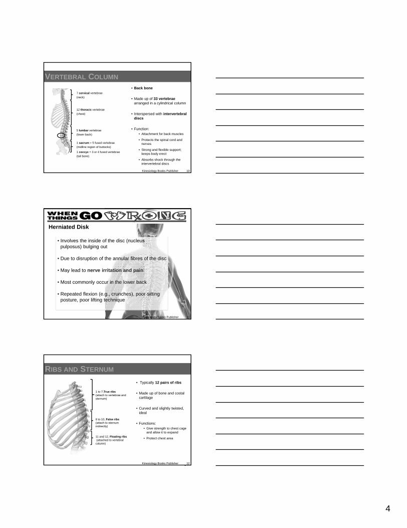
VERTEBRAL COLUMN
• Back bone
• Made up of 33 vertebrae arranged in a cylindrical column
• Interspersed with intervertebraldiscs
• Function:• Attachment for back muscles
• Protects the spinal cord and nerves
• Strong and flexible support; keeps body erect
• Absorbs shock through the intervertebral discs
7 cervical vertebrae
(neck)
12 thoracic vertebrae
(chest)
5 lumbar vertebrae
(lower back)
1 sacrum = 5 fused vertebrae
(midline region of buttocks)
1 coccyx = 3 or 4 fused vertebrae
(tail bone)
Kinesiology Books Publisher 11
• Involves the inside of the disc (nucleus pulposus) bulging out
• Due to disruption of the annular fibres of the disc
• May lead to nerve irritation and pain
• Most commonly occur in the lower back
• Repeated flexion (e.g., crunches), poor sitting posture, poor lifting technique
Herniated Disk
Kinesiology Books Publisher 12
RIBS AND STERNUM
• Typically 12 pairs of ribs
• Made up of bone and costal cartilage
• Curved and slightly twisted, ideal
• Functions: • Give strength to chest cage
and allow it to expand
• Protect chest area
1 to 7,True ribs(attach to vertebrae and sternum)
8 to 10, False ribs (attach to sternum indirectly)
11 and 12, Floating ribs(attached to vertebral column)

5
Kinesiology Books Publisher 13
RIBS AND STERNUM
• Sternum = midline breastbone
• 3 parts
• Provides direct attachments to clavicles and 1-7 rib pairs
Manubrium
SternalBody
Xiphoid Process
Kinesiology Books Publisher 14
NECK AND BACK MUSCLES
• Head sits on the first cervical vertebra (C1) = atlas
• To maintain this position there are muscles posterior, lateral, and anterior to the neck or cervical region
• Permit a wide range of movement and allow you to hold up your head
Posterior
Anterior
Lateral
ATLAS
Kinesiology Books Publisher 15
NECK AND BACK MUSCLES: ANTERIOR
• The most important pair = sternocleidomastoids
• Allow to:
• Flex head towards chest
• Get up from a supine position

6
Kinesiology Books Publisher 16
NECK AND BACK MUSCLES: POSTERIOR
• Large muscle mass = erector spinae
• Reaching in segments from sacrum (inferiorly) to skull (superiorly)
• Maintain erect position
• Anti-gravity muscles
• Stop working due to fainting
• Body falls face forward when not working
Kinesiology Books Publisher 17
ABDOMINAL MUSCLES
• Plywood-like muscular wall
• Attach
• Posteriorly: vertebral column, ribs, and hip bone
• Anteriorly: linea alba (translates as white line)
• Trilaminar
• 3 layers positioned on top of each other
Linea Alba
Kinesiology Books Publisher 18
Layer 1
External Oblique
Layer 2
Internal Oblique
Layer 3
Rectus Abdominis
Transversus abdominis
7
Kinesiology Books Publisher 19
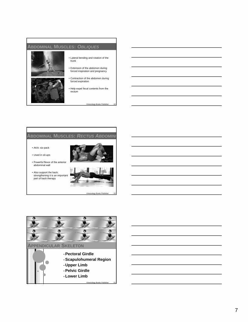
ABDOMINAL MUSCLES: OBLIQUES
• Lateral bending and rotation of the trunk
• Extension of the abdomen during forced inspiration and pregnancy
• Contraction of the abdomen during forced expiration
• Help expel fecal contents from the rectum
Kinesiology Books Publisher 20
ABDOMINAL MUSCLES: RECTUS ABDOMINI
• AKA: six-pack
• Used in sit-ups
• Powerful flexor of the anterior abdominal wall
• Also support the back; strengthening it is an important part of back therapy
• Pectoral Girdle• Scapulohumeral Region• Upper Limb• Pelvic Girdle
• Lower Limb
APPENDICULAR SKELETON
Kinesiology Books Publisher 21
8
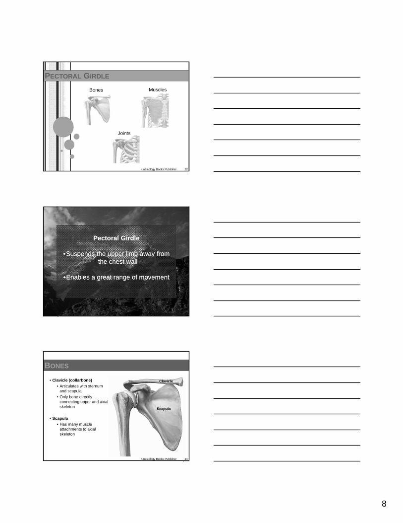
PECTORAL GIRDLE
Kinesiology Books Publisher 22
Bones Muscles
Joints
Kinesiology Books Publisher 23
Pectoral Girdle
•Suspends the upper limb away from the chest wall
•Enables a great range of movement
Pectoral Girdle
•Suspends the upper limb away from the chest wall
•Enables a great range of movement
Kinesiology Books Publisher 24
BONES
• Clavicle (collarbone)
• Articulates with sternum and scapula
• Only bone directly connecting upper and axial skeleton
• Scapula
• Has many muscle attachments to axial skeleton
Clavicle
Scapula

9
Kinesiology Books Publisher 25
MUSCLES
Anterior Group Posterior Group
Kinesiology Books Publisher 26
MUSCLES: ANTERIOR
Pectoralis major
Two heads:
1) Clavicular head flexes and medially rotates shoulder joint
2) Sternal head extends from flexed position and medially rotates shoulder joint
1
2
Pectoralis minor
• Attachments
• Ribs 3-5
• Coracoid process of the scapula
• Depresses and stabilizes scapula
Serratus anterior
• Steadies and holds scapula forward against chest wall (protracts it)
• Actions:
• Rope climbing
• Butterfly stroke
Kinesiology Books Publisher 27
MUSCLES: POSTERIOR
TRAPEZIUS
• Upper fibres
• From skull to scapula
• Elevate scapula
• Shrugging your shoulders
• Middle (transverse) fibres
• From ligamentum nuchae of cervical vertebrae to scapula
• Retract the scapula
• Lower fibres
• From C7 – T12 to scapula
• Depress the scapula

10
Kinesiology Books Publisher 28
MUSCLES: POSTERIOR
Latissmus dorsi
• From lower thoracic vertebrae, iliac crest, and thoracolumbar fascia to intertuberculargroove of the humerus
• Medially rotates, adducts, and extends humerus
Teres major
• Medially rotates and adducts humerus
Levator scapulae
• Rotates and elevates scapula
Rhomboid muscles
• Helps with scapula retraction and holding against thoracic wall
Kinesiology Books Publisher 29
JOINTS
• Sternoclavicular
• Only joint connecting pectoral girdle (clavicle ) and axial skeleton (sternum)
• Synovial joint strengthened by intracapsular disc and extrinsic ligaments
• Absorbs forces along clavicle
• Acromioclavicular Joint
• Connects clavicle’s lateral end and scapula’s acromion process
• Permits shoulder separations (e.g., in hockey, baseball, and football )
Lateral Muscles
SCAPULOHUMERAL REGION
Kinesiology Books Publisher 30
Superior and Posterior Muscles
Anterior Muscles
11
Kinesiology Books Publisher 31
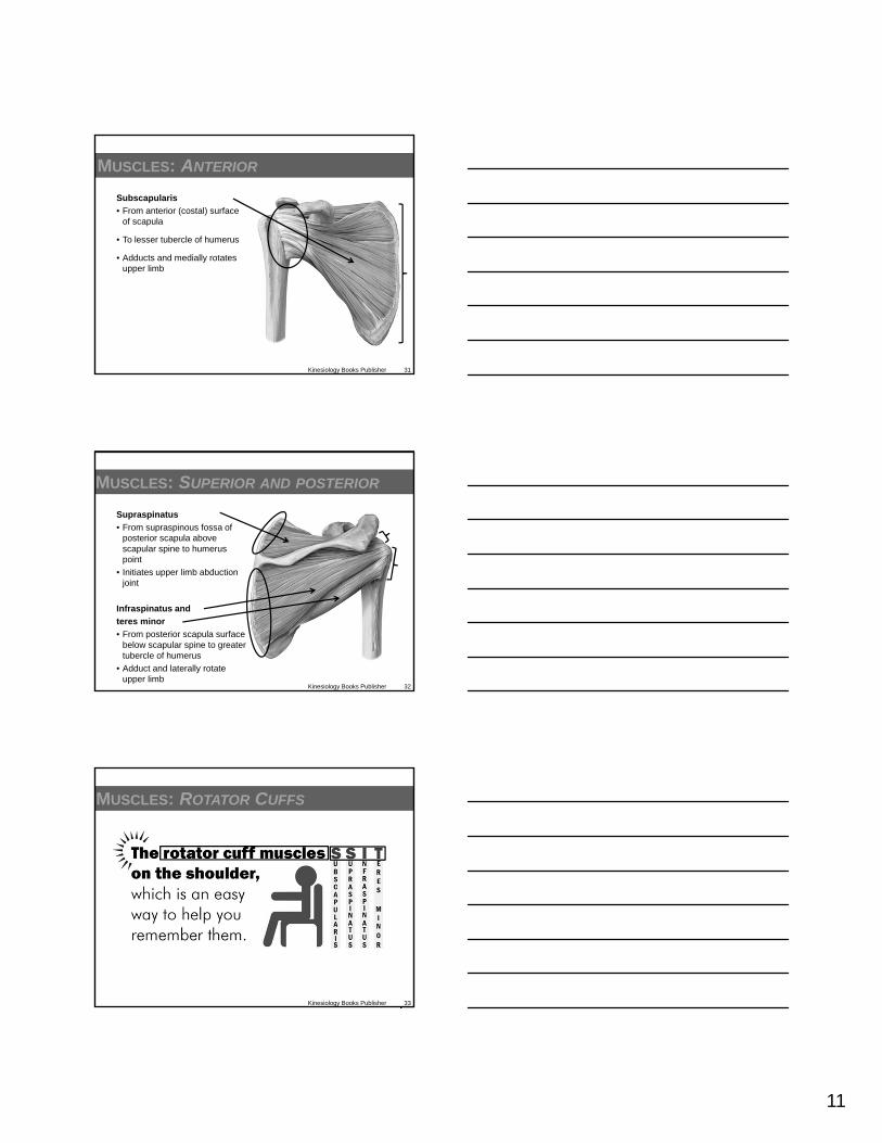
MUSCLES: ANTERIOR
Subscapularis
• From anterior (costal) surface of scapula
• To lesser tubercle of humerus
• Adducts and medially rotates upper limb
Kinesiology Books Publisher 32
MUSCLES: SUPERIOR AND POSTERIOR
Supraspinatus
• From supraspinous fossa of posterior scapula above scapular spine to humeruspoint
• Initiates upper limb abduction joint
Infraspinatus and
teres minor
• From posterior scapula surface below scapular spine to greater tubercle of humerus
• Adduct and laterally rotate upper limb
Kinesiology Books Publisher 33
MUSCLES: ROTATOR CUFFS
12
Kinesiology Books Publisher 34
• Shoulder joint is the most mobile and unstable joint
• Allows dynamic and powerful movements
• Requires to keep head of humeruscentralized in its socket
• Common injury = rotator cuffs tear
Rotator Cuffs Tear
Kinesiology Books Publisher 35
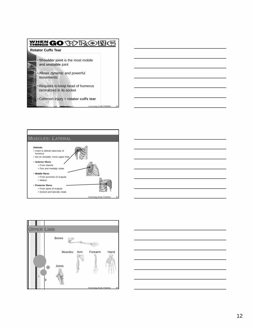
MUSCLES: LATERAL
Deltoids
• Insert to deltoid tuberosity of humerus
• Act on shoulder, move upper limb
• Anterior fibres
• From clavicle
• Flex and medially rotate
• Middle fibres
• From acromion of scapula
• Abduct
• Posterior fibres
• From spine of scapula
• Extend and laterally rotate
UPPER LIMB
Kinesiology Books Publisher 36
Bones
Muscles: Arm Forearm Hand
Joints

13
Kinesiology Books Publisher 37
Upper limb
Arm: shoulder to elbow+
Forearm: elbow to wrist+
Wrist+
Hand
Upper limb
Arm: shoulder to elbow+
Forearm: elbow to wrist+
Wrist+
Hand
Kinesiology Books Publisher 38
BONES
Arm (shoulder to elbow)
• Humerus
Forearm (elbow to wrist)
• Joined by a sheet of fibrous tissue (interosseous membrane)
• Radius
• Ulna
BONES
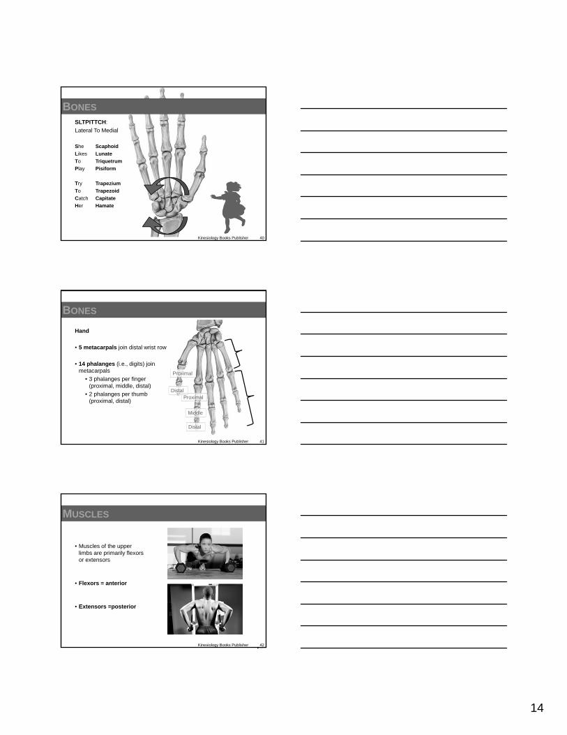
Wrist (carpus)
• 2 rows x 4 bones
• 8 carpals
Distal row: • Trapezium
• Trapezoid
• Capitate
• Hamate
Proximal row: • Scaphoid
• Lunate
• Triquetrum
• Pisiform
Kinesiology Books Publisher 39
14
BONES
SLTPITTCH:
Lateral To Medial
She Scaphoid
Likes Lunate
To Triquetrum
Play Pisiform
Try Trapezium
To Trapezoid
Catch Capitate
Her Hamate
Kinesiology Books Publisher 40
Kinesiology Books Publisher 41
BONES
Hand
• 5 metacarpals join distal wrist row
• 14 phalanges (i.e., digits) join metacarpals
• 3 phalanges per finger (proximal, middle, distal)
• 2 phalanges per thumb (proximal, distal)
Proximal
Middle
Distal
Proximal
Distal
Kinesiology Books Publisher 42
MUSCLES
• Muscles of the upper limbs are primarily flexors or extensors
• Flexors = anterior
• Extensors =posterior

15
Kinesiology Books Publisher 43
ARM MUSCLES: ANTERIOR
Coracobrachialis
• From coracoid process of scapula to humerus shaft
• Arm (shoulder) flexion
Kinesiology Books Publisher 44
ARM MUSCLES: ANTERIOR
Biceps brachii
• Long head
• Short head
• From scapula to radial tuberosity
• Elbow flexor and forearm supinator
Brachialis
• From anterior surface of humerusto ulna’s coronoid process
• Elbow flexor along with biceps
Kinesiology Books Publisher 45
ARM MUSCLES: POSTERIOR
Triceps brachii
• Medial head
• Lateral head
• Long head
• Medial and lateral head: from humerus posterior shaft
• Long head: from scapula’s inferior glenoid tubercle
• To ulna’s olecranon process
• Elbow extensor
16
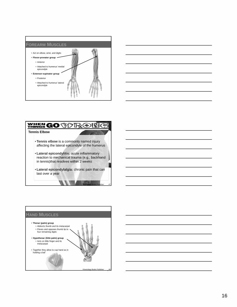
FOREARM MUSCLES
• Act on elbow, wrist, and digits
• Flexor-pronator group
• Anterior
• Attached to humerus’ medial epicondyle
• Extensor-supinator group
• Posterior
• Attached to humerus’ lateral epicondyle
Kinesiology Books Publisher 47
• Tennis elbow is a commonly named injury affecting the lateral epicondyle of the humerus
• Lateral epicondylitis: acute inflammatory reaction to mechanical trauma (e.g., backhand in tennis)that resolves within 2 weeks
• Lateral epicondylalgia: chronic pain that can last over a year
Tennis Elbow
HAND MUSCLES
• Thenar (palm) group
• Abducts thumb and its metacarpal
• Flexes and opposes thumb tip to four remaining digits
• Hypothenar (little palm) group
• Acts on little finger and its metacarpal
• Together they allow to cup hand as in holding a ball
Kinesiology Books Publisher 48

17
HAND MUSCLES
• Interossei muscles (between bones)
• Lumbrical muscles (earthworm)
• Collectively known as intrinsic (within) hand muscles
• Flex, extend, abduct, and adduct the fingers
• Position digits for fine movements
Kinesiology Books Publisher 49
Kinesiology Books Publisher 50
JOINTS
Shoulder (enohumeral) joint
• Between upper limb (humerushead) and scapula (glenoidfossa)
• Wide range of movements; compromise – lack of stability
• Large head articulating with shallow cup
Kinesiology Books Publisher 51
JOINTS
Elbow joints
• Humeroradial
• Flexion-extension
• Humeroulnar
• Flexion-extension
• Radioulnar
• Pronation-supination
18
Kinesiology Books Publisher 52
JOINTS
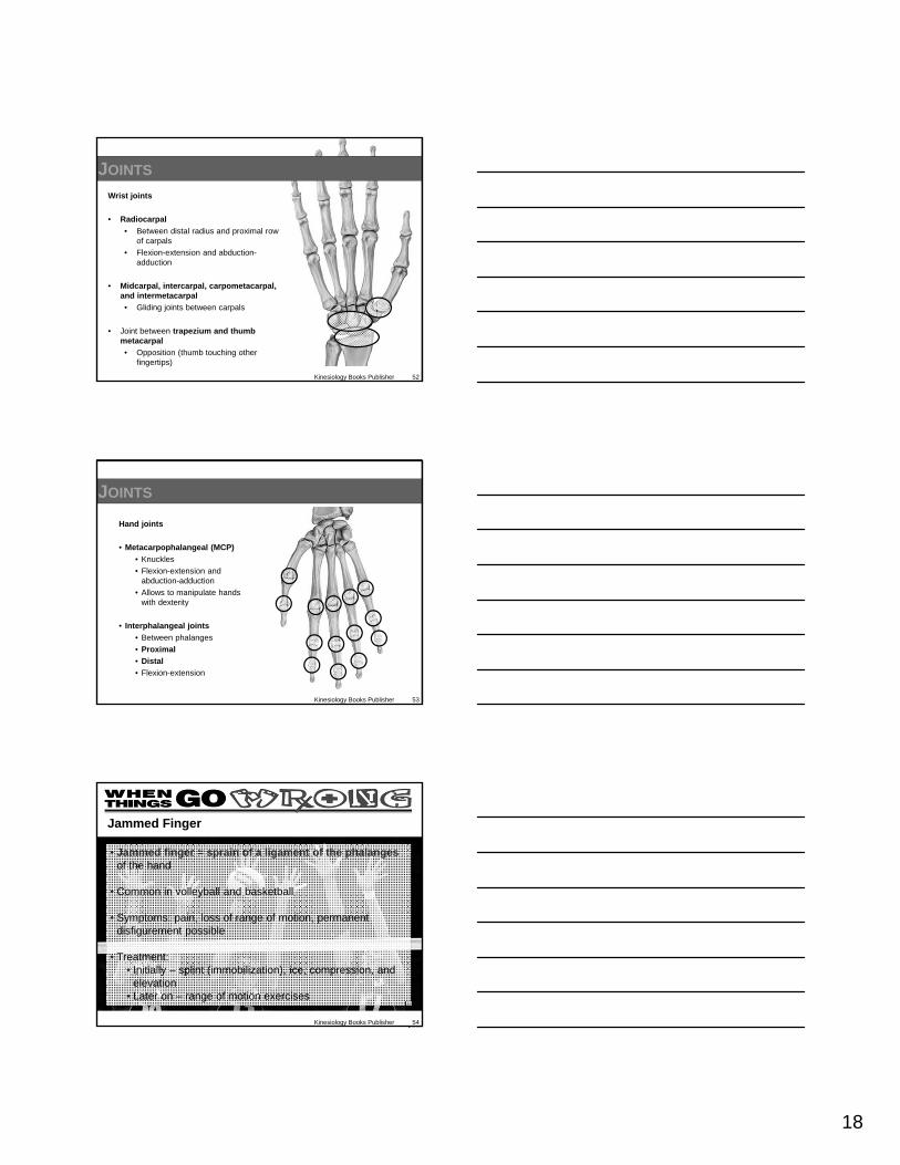
Wrist joints
• Radiocarpal
• Between distal radius and proximal row of carpals
• Flexion-extension and abduction-adduction
• Midcarpal, intercarpal, carpometacarpal, and intermetacarpal
• Gliding joints between carpals
• Joint between trapezium and thumb metacarpal
• Opposition (thumb touching other fingertips)
Kinesiology Books Publisher 53
JOINTS
Hand joints
• Metacarpophalangeal (MCP)
• Knuckles
• Flexion-extension and abduction-adduction
• Allows to manipulate hands with dexterity
• Interphalangeal joints
• Between phalanges
• Proximal
• Distal
• Flexion-extension
Kinesiology Books Publisher 54
• Jammed finger = sprain of a ligament of the phalanges of the hand
• Common in volleyball and basketball
• Symptoms: pain, loss of range of motion, permanent disfigurement possible
• Treatment: • Initially – splint (immobilization), ice, compression, and
elevation• Later on – range of motion exercises
Jammed Finger

19
Joints
PELVIC GIRDLE
Kinesiology Books Publisher 55
MusclesBones
Kinesiology Books Publisher 56
Pelvic Girdle
•Weight bearer
•Supports bladderand abdominal contents
•Sacrifices mobility for stability and strength
Pelvic Girdle
•Weight bearer
•Supports bladderand abdominal contents
•Sacrifices mobility for stability and strength
Kinesiology Books Publisher 57
BONES
• Os coxae – paired hip bones
• Each made up of:
• Ilium
• Pubis
• Ischium
• On the lateral surface is acetabulum – cup-shaped groove for femur’s head
Ilium

20
Kinesiology Books Publisher 58
MUSCLES
• Permit a wide range of movement in the lower limb
• Hip = ball and socket joint
• Prime focus = stability and transfer of weight for walking
• More limited than at the shoulder joint
Kinesiology Books Publisher 59
MUSCLES: ANTERIOR
Iliopsoas
• Formed by:
• Psoas major
• Iliacus
• Primary hip flexor
• Allows to bring:
• Thighs to chest
• Chest to knees
Psoas minor
• Weak hip flexor
• Missing 50% population
MUSCLES: POSTERIOR AND LATERAL
Gluteal muscles
• Gluteus maximus
• Largest and most superficial
• From ilium and sacrum to femur through illiotibial band
• Primary hip extensor
• Knee stabilizer in full extension
• Gluteus medius and minimus
• From ilium; lie deep and lateral to maximus
• Hip abductors
• Important for normal gait
Kinesiology Books Publisher 60

21
Kinesiology Books Publisher 61
JOINTS
Pubic Symphysis
• Fibrocartilaginous
• Unites 2 pubic bones
• Can soften right before birth for a wider opening
Sacroiliac
• Fibrous and synovial
• Unites sacrum 2 iliabones
• Stabilises ligaments that tie the sacrum to the hip bone
LOWER LIMB
Kinesiology Books Publisher 62
Bones
Muscles: Thigh Leg Foot
Joints
Kinesiology Books Publisher 63
Lower Limb
Thigh: hip to knee+
Leg: knee to ankle+
Ankle+
Foot
Lower Limb
Thigh: hip to knee+
Leg: knee to ankle+
Ankle+
Foot
22
Thigh
• Femur
• From hip to knee
• Largest bone
• Patella
• Kneecap
• Sesamoid bone
• In quadriceps muscles’ tendon
Kinesiology Books Publisher 64
BONES
Leg
• Tibia
• Fibula
• From knee to ankle
• Interosseous membrane
• Holds together firmly
• Provides stability
• Muscle attachment area
• Medial malleolus
• Distal tibia end
• Lateral malleolus
• Distal fibia end
Kinesiology Books Publisher 65
BONES
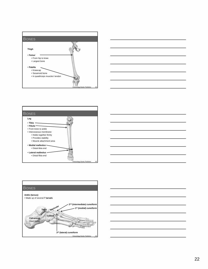
BONES
Ankle (tarsus)
• Made up of several 7 tarsals
Calcaneus
(heel bone)
Talus
Cuboid
1st (medial) cuneiform
2nd (intermediate) cuneiform
3rd (lateral) cuneiform
Kinesiology Books Publisher 66

23
BONES
Mid
dle
Pro
xim
al D
ista
l
Pro
xim
al
Dis
tal
Foot
• 5 metatarsals
• 14 phalanges (i.e., toes / digits) join metatarsals
• 3 phalanges per toe (proximal, middle, distal)
• 2 phalanges per big toe (proximal, distal)
Kinesiology Books Publisher 67
BONES
Hands (upper limb)
Carpus (wrist)
Carpals (8)
Metacarpals (5)
Phalanges (digits)
Thumb = 2 phalanges
“Clap your hands”
Feet (lower limb)
Tarsus (ankle)
Tarsals (7)
Metatarsals (5)
Phalanges (5 digits)
Big toe = 2 phalanges
“Tap your toes”
Kinesiology Books Publisher 68
Kinesiology Books Publisher 69
• Stress fracture = special type of fracture due to repeated low-magnitude forces (i.e., overtraining)
• Disruption of the continuity of the outer layer of cortical bone
• Painful especially during impact
• Common at tibia, metatarsals, femoral neck and pubis
• Unlike shin splints that occur at tibia: inflammation and pain without cortical bone disruption
Stress Fracture
24
Kinesiology Books Publisher 70
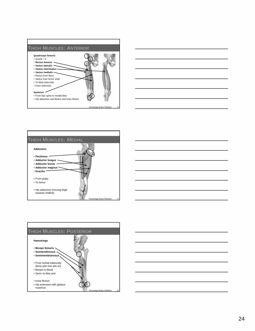
THIGH MUSCLES: ANTERIOR
Quadriceps femoris
• Quads = 4
• Rectus femoris
• Vastus lateralis
• Vastus intermedius
• Vastus medialis
• Rectus from illium
• Vastus from femur shaft
• To tibial tuberosity
• Knee extension
Sartorius
• From iliac spine to medial tibia
• Hip abduction and flexion and knee flexion
Kinesiology Books Publisher 71
THIGH MUSCLES: MEDIAL
Adductors
• Pectineus
• Adductor longus
• Adductor brevis
• Adductor magnus
• Gracilis
• From pubis
• To femur
• Hip adduction (moving thigh towards midline)
Kinesiology Books Publisher 72
Hamstrings
• Biceps femoris
• Semitendinosus
• Semimembranosus
• From ischial tuberosity(bony part one sits on)
• Biceps to fibula
• Semi- to tibia and
• Knee flexion
• Hip extension with gluteus maximus
THIGH MUSCLES: POSTERIOR
25
Kinesiology Books Publisher 73
• Jumper’s knee = pain affecting the infrapatellar ligament and/or patellar tendon
• Patellar tendonitis – acute
• Patellar tendonosis – chronic • More common• Due to muscular imbalances between the,
quadriceps and hamstrings muscles, poor landing and jumping technique, too much jumping
• Caused by repetitive eccentric knee actions
Jumper’s Knee
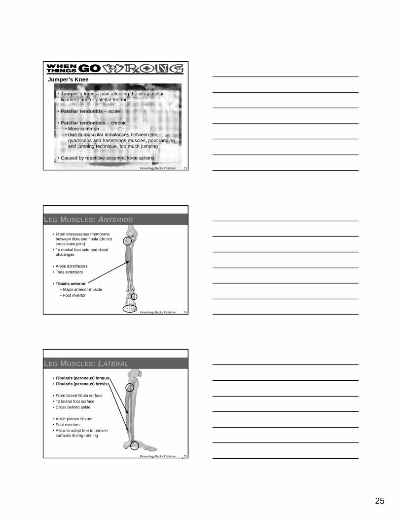
LEG MUSCLES: ANTERIOR
• From interosseous membrane between tibia and fibula (do not cross knee joint)
• To medial foot side and distal phalanges
• Ankle dorsiflexors
• Toes extensors
• Tibialis anterior
• Major anterior muscle
• Foot invertor
Kinesiology Books Publisher 74
LEG MUSCLES: LATERAL
• Fibularis (peroneus) longus
• Fibularis (peroneus) brevis
• From lateral fibula surface
• To lateral foot surface
• Cross behind ankle
• Ankle plantar flexors
• Foot evertors
• Allow to adapt feet to uneven surfaces during running
Kinesiology Books Publisher 75
26
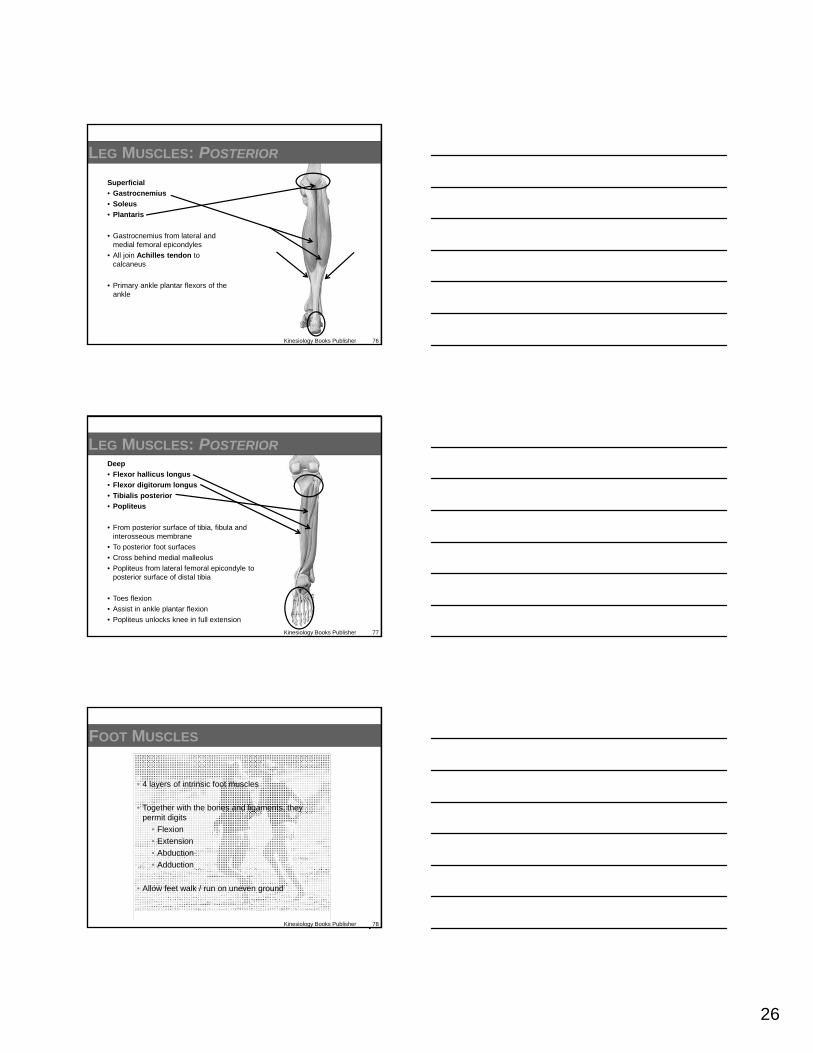
Superficial
• Gastrocnemius
• Soleus
• Plantaris
• Gastrocnemius from lateral and medial femoral epicondyles
• All join Achilles tendon to calcaneus
• Primary ankle plantar flexors of the ankle
Kinesiology Books Publisher 76
LEG MUSCLES: POSTERIOR
Deep
• Flexor hallicus longus
• Flexor digitorum longus
• Tibialis posterior
• Popliteus
• From posterior surface of tibia, fibula and interosseous membrane
• To posterior foot surfaces
• Cross behind medial malleolus
• Popliteus from lateral femoral epicondyle to posterior surface of distal tibia
• Toes flexion
• Assist in ankle plantar flexion
• Popliteus unlocks knee in full extension
Kinesiology Books Publisher 77
LEG MUSCLES: POSTERIOR
FOOT MUSCLES
Kinesiology Books Publisher 78
• 4 layers of intrinsic foot muscles
• Together with the bones and ligaments, they permit digits
• Flexion
• Extension
• Abduction
• Adduction
• Allow feet walk / run on uneven ground

27
JOINTS: HIP
Iliofemoral joint
• Between femur’s head and hip bone cup (acetabulum)
• Flexion-extension
• Abduction-adduction
• Circumduction
• Most stable synovial joint
• Deepened socket
• Intrinsic and very strong extrinsic ligaments
• Unlike shoulder joint, dislocation is rare
Kinesiology Books Publisher 79
JOINTS: KNEE
• Stable joint with incredible range of movement
• Flexion-extension (primary)
• Medial and lateral rotation in flexion
• Structural support
• Fibrocartilaginous discs
• Intrinsic ligaments
• Extrinsic ligaments
• Surrounding musculature
Kinesiology Books Publisher 80
Lateral
meniscus
Anterior and posterior cruciate
ligaments
Medial
meniscus
Medial collateral ligament
Lateral collateral ligament
JOINTS: ANKLE
Talocrural joint
• Many bones involved:
• Medial and lateral malleoli of tibia and fibula
• Talus
• Calcaneus
• Dorsiflexion
= most stable position
• Plantarflexion “En pointe” = least stable position
Kinesiology Books Publisher 81
28
JOINTS: FEET AND TOES
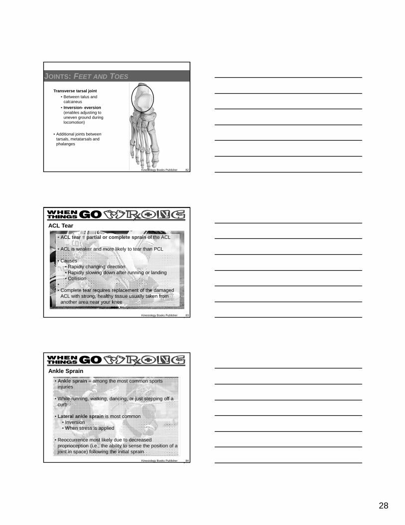
Transverse tarsal joint
• Between talus and calcaneus
• Inversion- eversion(enables adjusting to uneven ground during locomotion)
• Additional joints between tarsals, metatarsals and phalanges
Kinesiology Books Publisher 82
Kinesiology Books Publisher 83
• ACL tear = partial or complete sprain of the ACL
• ACL is weaker and more likely to tear than PCL
• Causes• Rapidly changing direction• Rapidly slowing down after running or landing• Collision
•• Complete tear requires replacement of the damaged
ACL with strong, healthy tissue usually taken from another area near your knee
ACL Tear
Kinesiology Books Publisher 84
• Ankle sprain = among the most common sports injuries
• While running, walking, dancing, or just stepping off a curb
• Lateral ankle sprain is most common • Inversion • When stress is applied
• Reoccurrence most likely due to decreased proprioception (i.e., the ability to sense the position of a joint in space) following the initial sprain
Ankle Sprain

29
• Human anatomy deals with the structures that make up the human body and how these various structures are related to one another
• Bones provide the structural framework necessary for support, muscles supply the power, and joints supply the mechanism that allows human movement to occur
• Our ability to move and perform an almost limitless number of skills can be enhanced with knowledge of anatomy
PUTTING IT ALL TOGETHER
Kinesiology Books Publisher 85