kinetics and mechanism of polyamide ("peptide") nucleic acid
TRANSCRIPT
Proc. Natl. Acad. Sci. USAVol. 92, pp. 2637-2641, March 1995Biochemistry
Kinetics and mechanism of polyamide ("peptide") nucleic acidbinding to duplex DNAVADIM V. DEMIDOVtt§, MICHAEL V. YAVNILOVICHt¶, BORIS P. BELOTSERKOVSKIHtII,MAxIM D. FRANK-KAMENETSKIIft4t, AND PETER E. NIELSENt:#tInstitute of Molecular Genetics, Russian Academy of Sciences, Kurchatov Square, 123182 Moscow, Russia; tCenter for Biomolecular Recognition, Department ofMedical Biochemistry and Genetics, Laboratory B, The Panum Institute, Blegdamsvej 3c, DK-2200 Copenhagen N, Denmark; and ttCenter for AdvancedBiotechnology, Department of Biomedical Engineering, Boston University, 36 Cummington Street, Boston, MA 02215
Communicated by Charles R Cantor, Boston University, Boston, MA, December 20, 1994
ABSTRACT To elucidate the mechanism of recognition ofdouble-stranded DNA (dsDNA) by homopyrimidine poly-amide ("peptide") nucleic acid (PNA) leading to the strand-displacement, the kinetics of the sequence-specific PNA/DNAbinding have been studied. The binding was monitored withtime by the gel retardation and nuclease S1 cleavage assays.The experimental kinetic curves obey pseudo-first-order ki-netics and the dependence of the pseudo-first-order rate con-stant, kps, on PNA concentration, P, obeys a power law kps
P' with 2 < y < 3. The kps values for binding of decamerPNA to dsDNA target sites with one mismatch are hundredsof times slower than for the correct site. A detailed kineticscheme for PNA/DNA binding is proposed that includes twomajor steps of the reaction of strand invasion: (i) a transientpartial opening of the PNA binding site on dsDNA and incor-poration of one PNA molecule with the formation of anintermediate PNA/DNA duplex and (ii) formation of a verystable PNA2/DNA triplex. A simple theoretical treatment ofthe proposed kinetic scheme is performed. The interpretationof our experimental data in the framework of the proposedkinetic scheme leads to the following conclusions. The se-quence specificity of the recognition is essentially provided atthe "search" step of the process, which consists in the highlyreversible transient formation of duplex between one PNAmolecule and the complementary strand of duplex DNA whilethe other DNA strand is displaced. This search step is followedby virtually irreversible "locking" step via PNA2/DNA triplexformation. The proposed mechanism explains how the bindingof homopyrimidine PNA to dsDNA meets two apparentlymutually contradictory features: high sequence specificity ofbinding and remarkable stability of both correct and mis-matched PNA/DNA complexes.
A new type of DNA analogue, polyamide ("peptide") nucleicacid (PNA), was described in 1991 (1). This sequence-specificDNA binding reagent is believed to be a very promising drug(2, 3) with numerous potential applications (4). For homopy-rimidine PNAs a unique type of duplex DNA/drug interactionis observed. It consists of PNA binding to one of the DNAstrands through formation of stable PNA2/DNA triplex whilethe noncomplementary DNA strand is left in single-strandedstate (1, 5, 6) thus forming a structure that we call the P loop.P-loop formation leads to selective inhibition of protein bind-ing to DNA (7, 8), results in transcription elongation arrest (2,7, 9, 10), creates an artificial transcription promoter (11),makes it possible to convert single-strand-specific nucleasesinto sequence-selective cutters (12), and, if PNA is biotiny-lated, to place electron-microscopy markers on double-stranded DNA (dsDNA) (13).
For biomedical and molecular biological applications ofPNA it is essential to understand the factors controlling PNA/DNA binding and its sequence specificity. The data indicatethat under conditions in which the PNA/DNA complexes arenormally studied, the binding is virtually irreversible (5), thusimplying a crucial role of kinetic factors in the strand-displacement reaction. Here we present a kinetic study ofhomopyrimidine PNAs binding to dsDNA and propose akinetic model for PNA/DNA sequence-specific recognition.
MATERIALS AND METHODSPNAs H-T1o-Lys-NH2, H-T5CT4-Lys-NH2, and H-T2CT2CT4-Lys-NH2 were synthesized as described (1, 14). The bis-PNAH-T8-NH2-R-NH2-T8-H consisted of two octathymidyl PNAscoupled in parallel orientation with a flexible N-acetamido-N,N'-bis(5-aminopentamethylenecarbonyl)ethylenediaminelinker (R).
Target plasmids were constructed by cloning the appropri-ate oligonucleotides into the polylinker of plasmid pUC19 andwere described in detail elsewhere (8, 12). Plasmid pTlO con-tains the A1o-Tlo insert cloned into the BamHI site, pT9C hasA5GA4T4CT5 cloned into the Sal I site, and pA8G2 hasT4CT2CT2*A2GA2GA4 cloned into the Pst I site. 32P-labeledDNA fragments were prepared by cutting the plasmids withrestriction enzymesEcoRI andHindlll (orPvu II) and labelingwith [a-32P]dNTP by the Klenow fragment of Escherichia coliDNA polymerase. PNA binding to dsDNA fragments wasperformed at 37°C in TE buffer (10 mM Tris.HCl/1 mMEDTA, pH 8.0) containing 10 mM NaCl. Some experimentswere performed at higher salt concentrations in a physiologicalbuffer (150 mM KCl/10 mM NaCl/15 mM MgCl2/1 mMCaCl2/1 mM spermidine/10 mM Tris HCI, pH 6.5). The pHvalues in concentrated PNA solutions were carefully con-trolled at 37°C by a micro pH meter. In low-salt kineticexperiments the binding was stopped after the desired times byaddition of NaCl up to 100 mM (5) and cooling. Then thesamples were analyzed by electrophoresis in 6-10% polyacryl-amide gels run in TAE (Tris acetate/EDTA) or TBE (Trisborate/EDTA) buffers. Quantitative analysis (we estimate theaccuracy of our experiments to be about 15%) was done byscanning of autoradiographs on a Molecular Dynamics com-puting densitometer or by direct counting of radioactive bandscut out from dried gels.
Abbreviations: PNA, polyamide ("peptide") nucleic acid; dsDNA,double-stranded DNA.§Present address: Center for Advanced Biotechnology, Boston Uni-versity, 36 Cummington Street, Boston, MA 02215.Present address: Department of Structural Biology, Weizmann In-stitute of Science, Rehovot, Israel.
IPresent address: Cell and Molecular Biology Laboratory, SRI Inter-national 205-19, 333 Ravenswood Avenue, Menlo Park, CA 94025.t4To whom reprint requests should be addressed.
2637
The publication costs of this article were defrayed in part by page chargepayment. This article must therefore be hereby marked "advertisement" inaccordance with 18 U.S.C. §1734 solely to indicate this fact.
2638 Biochemistry: Demidov et al.
The time course of PNA binding to dsDNA was quantita-tively analyzed in plots of ln(D/Do) versus time t, where D isthe yield of free DNA fragment (not bound to PNA) and Dois the total DNA quantity determined by the sum of thequantities of DNA fragments bound and not bound to PNA.
Pseudo-first-order kinetics was assumed (which proved tobe the case; see Results):
-ln(D/Do) = kpst, [1]
where kps is the pseudo-first-order rate constant. The depen-dence of kps on the PNA concentration (P) was approximatedas
k . Py. [2]
Thus, the exponent y was determined as the slope of linearcurves in coordinates ln ln(Do/D) versus In P for the sameperiod of time in the range of PNA concentrations we workedat. The limitations of the method we used for PNA/DNAcomplex detection did not allow us to investigate much higheror much lower PNA concentration than we did.Another method to determine y was also used. We deter-
mined the yield of the fragments, Dfr, obtained as a result ofdigestion of PNA/DNA complexes with nuclease Si (12). Forthe initial stage of the kinetics, when the fraction of complexesis small (Dfr/Do << 1),
Dfr/DO kpst >>Pet. [3]
Therefore, by plotting In Dfr versus InP for the same initial DNAquantities and the same periods of time, one can estimate y.
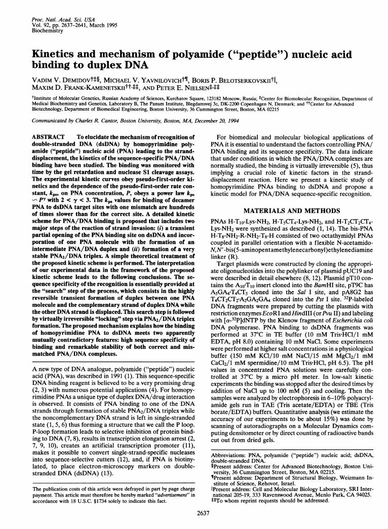
RESULTSTo study the kinetics of the binding of homopyrimidine PNAsto dsDNA, we at first used a gel retardation assay. The radio-labeled DNA fragments were complexed with PNA and thePNA/DNA complex was separated from free DNA by elec-trophoresis in a polyacrylamide gel with subsequent autora-diographic quantification of the intensities of both bands. Fig.1A shows a typical pattern. The intensity of the faster migrat-ing band gives the quantity of free DNA, whereas the slowermigrating band corresponds to the complex, which is retarded
B 1.0'
ATime 0 15 30 45 60 120(min)
C 0.8'x
E 0.6'0
c0 4
._2-
0.2'
0 100 200Time (min)
due to P-loop formation (7-9). By measuring the intensities ofthese two bands, normalized with their integral intensity, onecan follow the kinetics of the PNA binding to DNA.
Fig. 1B shows the kinetic data on the binding of 10 ,uM PNAH-Tio-Lys-NH2 with the perfectly matched (A1loT1o) andmismatched (A5GA4-T4CT5) sites on the corresponding linearDNA fragments. Fig. 1C demonstrates that the binding ofhomopyrimidine PNAs to DNA sites follows the pseudo-first-order kinetics described by Eq. 1. One observes a dramaticdifference in the values of the pseudo-first-order rate constant,kps, for the perfect and the mismatched sites. With PNAH-Tio-Lys-NH2, kps = 4.2 x 10⁻⁴ min-1 for the mismatchedsite, 100 times lower than for the perfect one (kps = 3.6 x 10⁻²min-).With PNA H-T5CT4-Lys-NH2 this difference is even greater:
we could not detect any binding of this PNA at 10 ,uM for 3days of incubation with plasmid pT10, carrying a single-mis-match insert, A1o-Tlo, while binding to pT9C, carrying theperfect target, A5GA4-T4CT5, was completed within an hour.This corresponds to a >1000-fold decrease of the rate ofbinding to the mismatched target as compared with the rate ofbinding to the perfect target. Only upon increasing theH-T5CT4-Lys-NH2 concentration by a factor of 10 did weobserve binding of this PNA to pT10, with kps = 5 x 10⁻³min-1 (Fig. 1 C). Proceeding from the value kps = 0.11 min-1for the binding of this PNA at 10 ,tM to the true target (Fig.1C) and taking the nonlinear, approximately quadratic, de-pendence of kp, on PNA concentration (see below) into ac-count, we can estimate that for the binding of 100 ,uMH-T5CT4-Lys-NH2 to the true site, kps = 10 min-1, a value 2000times higher than for the mismatched site. This estimation ofthe sequence selectivity ofPNA binding is very close to the onearrived at above. Note that in spite of a very low rate of PNAbinding to mismatched DNA sites, complete PNA/DNA bind-ing can be obtained by incubating for a long time at a high PNAconcentration. Thus these mismatched complexes are suffi-ciently thermodynamically stable.By employing gel retardation analysis as in Fig. 1A and
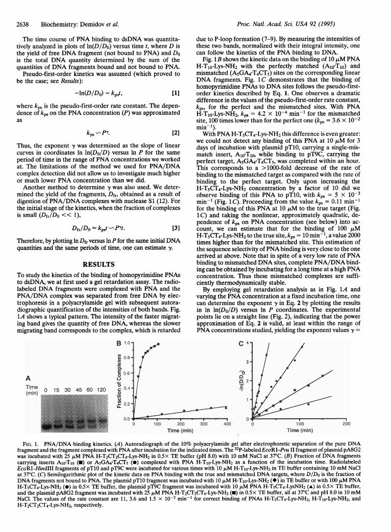
varying the PNA concentration at a fixed incubation time, onecan determine the exponent y in Eq. 2 by plotting the resultsin ln ln(Do/D) versus ln P coordinates. The experimentalpoints lie on a straight line (Fig. 2), indicating that the powerapproximation of Eq. 2 is valid, at least within the range ofPNA concentrations studied, yielding the exponent values y =
C 4-
3
0
403;0 400 0 200100
Time (min)
FIG. 1. PNA/DNA binding kinetics. (A) Autoradiograph of the 10% polyacrylamide gel after electrophoretic separation of the pure DNAfragment and the fragment complexed with PNA after incubation for the indicated times. The 32P-labeledEcoRI-Pvu II fragment of plasmid pA8G2was incubated with 25 ,uM PNA H-T2CT2CT4-Lys-NH2 in 0.5x TE buffer (pH 8.0) with 10 mM NaCl at 37°C. (B) Fraction of DNA fragmentscarrying inserts Alo-T1o (-) or A5GA4-T4CT5 (-) complexed with PNA H-TIo-Lys-NH2 as a function of the incubation time. RadiolabeledEcoRI-HindIII fragments of pTlO and pT9C were incubated for various times with 10 ,uM H-Tio-Lys-NH2 in TE buffer containing 10 mM NaClat 37°C. (C) Semilogarithmic plot of the kinetic data on PNA binding with the true and mismatched DNA targets, where DIDo is the fraction ofDNA fragments not bound to PNA. The plasmid pTlO fragment was incubated with 10 ,uM H-Tio-Lys-NH2 (*) in TE buffer or with 100 ALM PNAH-T5CT4-Lys-NH2 (-) in 0.5x TE buffer, the plasmid pT9C fragment was incubated with 10 ,uM PNA H-T5CT4-LysNH2 (-) in 0.5x TE buffer,and the plasmid pA8G2 fragment was incubated with 25 ,uM PNA H-T2CT2CT4-Lys-NH2 (-) in 0.5x TE buffer, all at 37°C and pH 8.0 in 10 mMNaCl. The values of the rate constant are 11, 3.6 and 1.5 x 10⁻² min-' for correct binding of PNAs H-T5CT4-Lys-NH2, H-Tio-Lys-NH2, andH-T2CT2CT4-Lys-NH2, respectively.
Proc. NatL Acad Sci. USA 92 (1995)
Proc. NatL Acad Sci USA 92 (1995) 2639
.--10s -12
.S -2
2 3 4InP
FIG. 2. Double logarithmic plot of the fraction DIDo of plasmidDNAs, carrying the appropriate inserts, not bound to the PNAs as afunction of PNA concentration (ILM) at 37°C in TE buffer at pH 8.0in 10 mM NaCl. The plasmid pTlO fragment was incubated for 30 minwith "monomeric" PNA H-Tio-Lys-NH2 (0) or with "dimeric" PNA(H-T8-NH2-R-NH2-T8-H) (0) and the plasmid pA8G2 fragment wasincubated for 15 min with PNA H-T2CT2CT4-Lys-NH2 (M)-
2.5 and 2.2 for H-T1o-Lys-NH2 and H-T2CT2CT4-Lys-NH2,respectively. In a similar experiment for a bis PNA consistingof two PNA T8 moieties connected by a flexible linker, fy = 1.5,lower by 1 unit than in the case of the "monomeric" PNAH-T1o-Lys-NH2 (Fig. 2).We also used nuclease S1 probing of the strand-displaced
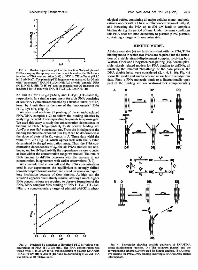
PNA/DNA complex (12) to follow the binding kinetics byanalyzing the yield of corresponding fragments in agarose gels.We used this assay to study the concentration dependence ofbinding of PNA H-TIo-Lys-NH2 to its perfect binding siteA1joTjO at two Na+ concentrations. From the initial part of thebinding kinetics the exponent y in Eq. 2 can be determined asthe slope of plots of ln Dfr versus ln P. These data yield thevalue -y = 2.7 (Fig. 3), which agrees well with the -y valuedetermined by the gel retardation assay. Thus, the PNA con-centration dependences of kps for all PNAs studied are non-linear, and for H-T1o-Lys-NH2 the dependence is close to cubic,at least within the concentration range we studied. The rate ofPNA binding to dsDNA decreases with the increase in saltconcentration, in agreement with earlier observations (5, 9).We conclude that at low salt and the PNA concentrations
used in our experiments the equilibrium is strongly shiftedtoward complex formation but that strand invasion can requirelong incubation because of slow kinetics. At high salt thesituation appears qualitatively similar, although much higherPNA concentrations are required to observe formation of thePNA/DNA complex: 50% binding of PNA H-T2CT2CT4-Lys-NH2 to a complementary target of plasmid pA8G2 in physi-
3.
ological buffer, containing all major cellular mono- and poly-cations, occurs within 1 hr at a PNA concentration of 350 ,uM,and increasing the PNA up to 500 ,uM leads to completebinding during this period of time. Under the same conditionsthis PNA does not bind detectably to plasmid pT9C plasmid,containing a target with one mismatch.
KINETIC MODEL
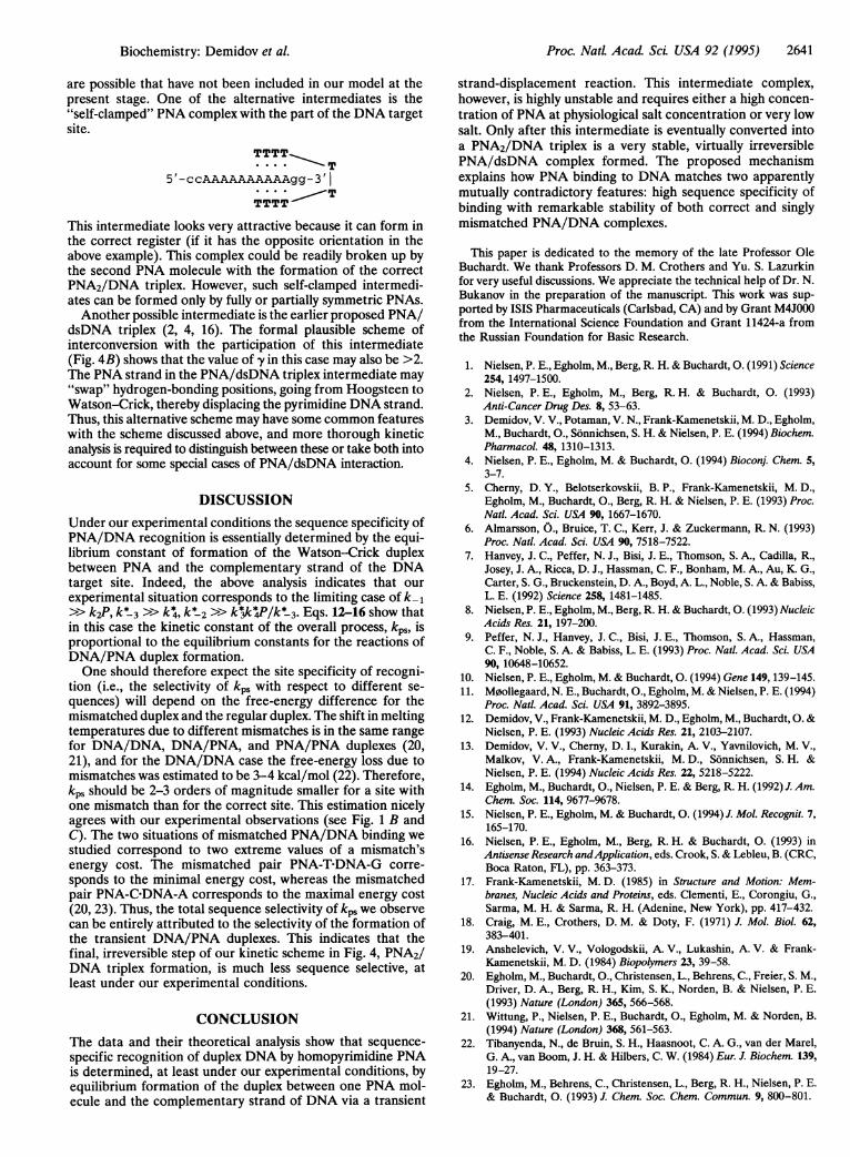
All data available (4) are fully consistent with the PNA/DNAbinding mode in which two PNAs are required for the forma-tion of a stable strand-displacement complex involving bothWatson-Crick and Hoogsteen base pairing (15). Several plau-sible, closely related models for PNA binding to dsDNA, allinvolving the inherent "breathing" of the base pairs in theDNA double helix, were considered (2, 4, 6, 8, 16). Fig. 4Ashows the model and kinetic scheme we use here to analyze ourdata. First, a PNA molecule binds to a fluctuationally openpart of the binding site via Watson-Crick complementary
A D DP DP2
. PNA-
DEDFr
TE
:1'DPNDP2
1 hfPkZ,DD __kP_N DP k DP2k1
kl4Jk~ * 1k43
DPr -w R i0
2k4D PDP
B
0-
-1.2.2 2.4 2.6 2.8 3.0 3.2 3.4
InP
FIG. 3. Nuclease S1 digestion of linearized pTlO at various con-centrations of PNA H-Tio-Lys-NH2. The PNA concentration was
varied from 10 to 25 ALM for 20 min incubation of plasmid DNA withPNA at 10mM (m) or 20mM (0) NaCl. Dfr for binding of 25 ;M PNAwas taken as 10 relative units.
rDP.DT
FIG. 4. Schematics showing possible pathways of PNA/DNAstrand-displacement reaction. (A) The pathways (Upper) and thecorresponding scheme (Lower) used for kinetic analysis. (B) Alterna-tive scheme for PNA/DNA binding involving a PNA/dsDNA triplexintermediate.
Biochemistry: Demidov et aL

2640 Biochemistry: Demidov et al.
pairing. For simplicity, we consider only two types of theseintermediate complexes: one, designated DP, with a correctlypositioned full-length PNA/DNA duplex and one, DP*, witha partial duplex, in which the PNA molecule is incorrectlypositioned with respect to its binding site. We can illustrate twopossible examples of such "shifted" complexes for DNAtargets in plasmids pTlO and pA8G2.
TTTTTTTTT TTTTCTTCTT
5' -ccAAAAAAAAAAgg-3' 5'-agAAGAAGAAAAcc-3'
The sequences of PNA-TIo and PNA-T2CT2CT4 are shown inbold type and DNA target sequences are capitalized. Inpractice, these partial duplexes are numerous. All of the aboveintermediates, however, are very unstable. We assume thatonly after the DP duplex is trapped by the second PNAmolecule, forming a triplex, is the very stable, virtually irre-versible, complex DP2 formed. The "shifted" intermediateDP* can also form the more stable transient complex (DP*) asa result of addition of the second PNA strand. We assume thatonce such a shifted triplex is formed it intensifies "breathings"of the adjacent DNA regions, thus greatly enhancing accessi-bility of the parts of the DNA site which remain in the duplexstate to the next PNA molecule. This third PNA moleculeforms the correct duplex with the same site, pushing out theshifted duplex, thus making intermediate DP**. The effect ofenhanced accessibility of the regions adjacent to the DNAopen site is well known from kinetic studies of the reaction ofDNA with formaldehyde (17). Finally, the transient DP2**complex is also converted into the stable DP2 triplex when yetanother PNA molecule pushes out the shifted second PNAstrand in the triplex.Note that we consider the final stage of the formation of the
"correct" triplex as irreversible, whereas we consider theformation of "incorrect" triplexes (like DP* species) as re-versible. Of course, the final stage is also reversible but thereverse kinetic constant is very small when the conditions favorPNA2/DNA triplex formation. Under such conditions thekinetic constant of dissociation of the complex is known todepend exponentially on the length of the complex (18, 19).This explains why the "incorrect" triplexes are predominantlyshort-lived whereas the "correct" triplex is very long-lived. Wealso do not consider for the first approximation all possibletransitions between shifted and correct intermediates, becausethis greatly simplifies the calculations and evidently missesnothing of principal importance.
Thus, let us designate by D the concentration of free DNA;by P the concentration of free PNA; by DP and DP* theconcentrations of complete and incomplete DNA complexeswith one PNA molecule, respectively; by DP* and DP2** theconcentrations of transitional complexes with two PNA mol-ecules; and by DP2 the concentration of the resulting PNA2/DNA triple complex. The following kinetic equations can bederived from the kinetic scheme presented in Fig. 4A:
dD/dt = -(k, + k*)(D)(P) + k-1DP + k*LDP* [4]
dDP/dt = kl(D)(P) - k-1DP - k2(DP)(P) [5]
dDP*/dt = kV(D)(P) + k* 2DP - k* IDP* - k*(DP*)(P) [6]
dDP /dt = k (DP*)(P) + k* 3(DP*2*)(P)-k*2DP* - k*(DP*)(P) [7]
dDP*2 */dt = k*(DP2)(P) - (k 3 + k4)(DP*2 *)(P) [8]
dDP2/dt = k2(DP)(P) + kA(DP2*)(P). [9]
In practice, one needs PNA at =10 ,uM to observe thestrand-displacement reaction within -1 hr. At the same time,
the usual DNA concentration-i.e., the concentration of thePNA binding sites-is -0.05 uM. This means that the numberof PNA molecules in our kinetic experiments exceeds thenumber of DNA molecules by a factor of 200. Therefore, thefree PNA concentration does not decrease during the reac-tion-i.e.,P is a constant. As indicated above, all intermediatesare so unstable that we can assume that their concentration islow and does not change significantly during the reaction-i.e.,dDP/dt = 0, dDP*/dt = 0, dDPI/dt = 0, and dDPP/dt = 0.From Eqs. 4-9, under the above quasistationary assumptionsand for the initial conditions D(O) = Do, DP2(0) = 0, we obtain
D = Doexp(-kpst)DP2 = Do[l - exp(-kpst)]kPs =Klk2P2 + K*K*K*k:Pl,
[10]
[11]
[12]
where
K1 = k1/(k-l + k2P)
K1* = k*/[k* 1 + (k2- k* 2K2)P]
K* = k2/[k*-2 + k3k4 P/(k* 3 + k4)]
K3* = k*/(k* 3 + k4).
[13]
[14]
[15][16]
Thus, we arrive at the conclusion that under the aboveassumption our complicated many-step kinetic scheme obeysthe pseudo-first-order kinetics (Eqs. 10 and 11) with complexnonlinear dependence of the pseudo-first-order rate constanton the PNA concentration, P (Eqs. 12-16). If we assume thatthe transient complexes are very unstable so that the kineticconstants and the PNA concentration correspond to the caseof k-1 »> k2P, k* 3 >> k4, and k* 2 >>k3 k4P/k* 3, then all Kivalues become independent ofP (K1 = kl/k-1, KT =klk*K2 = k*/k* 2, K: = k3/k* 3), and Eq. 12 is simplified to yielda simple polynomial dependence of kp, on P as a sum ofquadratic and cubic terms. This results in a curve of ln kpsversus lnP with the slope varying from 2 to 3. The latter meansthat for this case the dependence of the pseudo-first-order rateconstant onP can be approximated, at least in a limited rangeof variation of P, as the polynomial function 1 (Eq. 2) with 2< -y < 3. The y values we observe in our experiments lie withinthese limits. The fact that y> 2 indicates that the second term inEq. 12 is significant, which indicates that the unstable "shifted"intermediates play an important role in the overall kinetics. It istempting to interpret the fact that -y is larger for the regularPNA H-Tio-Lys-NH2 than for the less regularPNA H-T2CI2CT4-Lys-NH2 in terms of a larger role of the "shifted" intermediatesin the former case. More accurate measurements of the ry valuesfor PNAs with different sequences are necessary to establish sucha correlation between the y value and regularity of the sequence.
In the case of bis PNAs, the final triplex is formed byclamping of bis PNA and its formation does not depend on thePNA concentration. As a result, we have to substitute theproducts k2P and kVP in Eqs. 12, 13, and 15 for k2 and k4,respectively, to obtain the modified equation for kp.
kbis = K1k2P + K*,KjKjk*42, [17]
where K1 and K*2 are described by the modified Eq. 13 and Eq.15, respectively. Therefore, for bis PNA the exponent -y in Eq.2 should be 1 less than in the case of monomeric PNA, whichagrees perfectly with our experimental findings. Again, thefact that in this case exponent y > 1 indicates that for bis PNAthe shifted DP* intermediates also play an important role atthe initial step of binding.Our kinetic scheme is the simplest one that explains the
major features of our experimental data. Other intermediates
Proc. Natl Acad ScL USA 92 (1995)
Proc. NatL Acad Sci USA 92 (1995) 2641
are possible that have not been included in our model at thepresent stage. One of the alternative intermediates is the"self-clamped" PNA complex with the part of the DNA targetsite.
TTTT* ....T
5'-ccAAAAAAAAAAgg-3'
TTTT
This intermediate looks very attractive because it can form inthe correct register (if it has the opposite orientation in theabove example). This complex could be readily broken up bythe second PNA molecule with the formation of the correctPNA2/DNA triplex. However, such self-clamped intermedi-ates can be formed only by fully or partially symmetric PNAs.Another possible intermediate is the earlier proposed PNA/
dsDNA triplex (2, 4, 16). The formal plausible scheme ofinterconversion with the participation of this intermediate(Fig. 4B) shows that the value of -y in this case may also be >2.The PNA strand in the PNA/dsDNA triplex intermediate may"swap" hydrogen-bonding positions, going from Hoogsteen toWatson-Crick, thereby displacing the pyrimidine DNA strand.Thus, this alternative scheme may have some common featureswith the scheme discussed above, and more thorough kineticanalysis is required to distinguish between these or take both intoaccount for some special cases of PNA/dsDNA interaction.
DISCUSSIONUnder our experimental conditions the sequence specificity ofPNA/DNA recognition is essentially determined by the equi-librium constant of formation of the Watson-Crick duplexbetween PNA and the complementary strand of the DNAtarget site. Indeed, the above analysis indicates that ourexperimental situation corresponds to the limiting case of k-1>> k2P, k*3 >> k4, k*2 >> kkV*/k* 3. Eqs. 12-16 show thatin this case the kinetic constant of the overall process, kps, isproportional to the equilibrium constants for the reactions ofDNA/PNA duplex formation.One should therefore expect the site specificity of recogni-
tion (i.e., the selectivity of kps with respect to different se-quences) will depend on the free-energy difference for themismatched duplex and the regular duplex. The shift in meltingtemperatures due to different mismatches is in the same rangefor DNA/DNA, DNA/PNA, and PNA/PNA duplexes (20,21), and for the DNA/DNA case the free-energy loss due tomismatches was estimated to be 3-4 kcal/mol (22). Therefore,kps should be 2-3 orders of magnitude smaller for a site withone mismatch than for the correct site. This estimation nicelyagrees with our experimental observations (see Fig. 1 B andC). The two situations of mismatched PNA/DNA binding westudied correspond to two extreme values of a mismatch'senergy cost. The mismatched pair PNA-T-DNA-G corre-sponds to the minimal energy cost, whereas the mismatchedpair PNA-C-DNA-A corresponds to the maximal energy cost(20, 23). Thus, the total sequence selectivity of kps we observecan be entirely attributed to the selectivity of the formation ofthe transient DNA/PNA duplexes. This indicates that thefinal, irreversible step of our kinetic scheme in Fig. 4, PNA2/DNA triplex formation, is much less sequence selective, atleast under our experimental conditions.
CONCLUSIONThe data and their theoretical analysis show that sequence-specific recognition of duplex DNA by homopyrimidine PNAis determined, at least under our experimental conditions, byequilibrium formation of the duplex between one PNA mol-ecule and the complementary strand of DNA via a transient
strand-displacement reaction. This intermediate complex,however, is highly unstable and requires either a high concen-tration of PNA at physiological salt concentration or very lowsalt. Only after this intermediate is eventually converted intoa PNA2/DNA triplex is a very stable, virtually irreversiblePNA/dsDNA complex formed. The proposed mechanismexplains how PNA binding to DNA matches two apparentlymutually contradictory features: high sequence specificity ofbinding with remarkable stability of both correct and singlymismatched PNA/DNA complexes.
This paper is dedicated to the memory of the late Professor OleBuchardt. We thank Professors D. M. Crothers and Yu. S. Lazurkinfor very useful discussions. We appreciate the technical help of Dr. N.Bukanov in the preparation of the manuscript. This work was sup-ported by ISIS Pharmaceuticals (Carlsbad, CA) and by Grant M4JO00from the International Science Foundation and Grant 11424-a fromthe Russian Foundation for Basic Research.
1. Nielsen, P. E., Egholm, M., Berg, R. H. & Buchardt, 0. (1991) Science254, 1497-1500.
2. Nielsen, P. E., Egholm, M., Berg, R. H. & Buchardt, 0. (1993)Anti-Cancer Drug Des. 8, 53-63.
3. Demidov, V. V., Potaman, V. N., Frank-Kamenetskii, M. D., Egholm,M., Buchardt, O., Sonnichsen, S. H. & Nielsen, P. E. (1994) Biochem.Pharmacol. 48, 1310-1313.
4. Nielsen, P. E., Egholm, M. & Buchardt, 0. (1994) Bioconj. Chem. 5,3-7.
5. Cherny, D. Y., Belotserkovskii, B. P., Frank-Kamenetskii, M. D.,Egholm, M., Buchardt, O., Berg, R. H. & Nielsen, P. E. (1993) Proc.Natl. Acad. Sci. USA 90, 1667-1670.
6. Almarsson, O., Bruice, T. C., Kerr, J. & Zuckermann, R. N. (1993)Proc. Natl. Acad. Sci. USA 90, 7518-7522.
7. Hanvey, J. C., Peffer, N. J., Bisi, J. E., Thomson, S. A., Cadilla, R.,Josey, J. A., Ricca, D. J., Hassman, C. F., Bonham, M. A., Au, K. G.,Carter, S. G., Bruckenstein, D. A., Boyd, A. L., Noble, S. A. & Babiss,L. E. (1992) Science 258, 1481-1485.
8. Nielsen, P. E., Egholm, M., Berg, R. H. & Buchardt, 0. (1993) NucleicAcids Res. 21, 197-200.
9. Peffer, N. J., Hanvey, J. C., Bisi, J. E., Thomson, S. A., Hassman,C. F., Noble, S. A. & Babiss, L. E. (1993) Proc. Natl. Acad. Sci. USA90, 10648-10652.
10. Nielsen, P. E., Egholm, M. & Buchardt, 0. (1994) Gene 149,139-145.11. M0ollegaard, N. E., Buchardt, O., Egholm, M. & Nielsen, P. E. (1994)
Proc. Natl. Acad. Sci. USA 91, 3892-3895.12. Demidov, V., Frank-Kamenetskii, M. D., Egholm, M., Buchardt, 0. &
Nielsen, P. E. (1993) Nucleic Acids Res. 21, 2103-2107.13. Demidov, V. V., Cherny, D. I., Kurakin, A. V., Yavnilovich, M. V.,
Malkov, V. A., Frank-Kamenetskii, M. D., Sonnichsen, S. H. &Nielsen, P. E. (1994) Nucleic Acids Res. 22, 5218-5222.
14. Egholm, M., Buchardt, O., Nielsen, P. E. & Berg, R. H. (1992) J. Am.Chem. Soc. 114, 9677-9678.
15. Nielsen, P. E., Egholm, M. & Buchardt, 0. (1994) J. Mol. Recognit. 7,165-170.
16. Nielsen, P. E., Egholm, M., Berg, R. H. & Buchardt, 0. (1993) inAntisense Research andApplication, eds. Crook, S. & Lebleu, B. (CRC,Boca Raton, FL), pp. 363-373.
17. Frank-Kamenetskii, M. D. (1985) in Structure and Motion: Mem-branes, Nucleic Acids and Proteins, eds. Clementi, E., Corongiu, G.,Sarma, M. H. & Sarma, R. H. (Adenine, New York), pp. 417-432.
18. Craig, M. E., Crothers, D. M. & Doty, F. (1971) J. Mol. Biol. 62,383-401.
19. Anshelevich, V. V., Vologodskii, A. V., Lukashin, A. V. & Frank-Kamenetskii, M. D. (1984) Biopolymers 23, 39-58.
20. Egholm, M., Buchardt, O., Christensen, L., Behrens, C., Freier, S. M.,Driver, D. A., Berg, R. H., Kim, S. K., Norden, B. & Nielsen, P. E.(1993) Nature (London) 365, 566-568.
21. Wittung, P., Nielsen, P. E., Buchardt, O., Egholm, M. & Norden, B.(1994) Nature (London) 368, 561-563.
22. Tibanyenda, N., de Bruin, S. H., Haasnoot, C. A. G., van der Marel,G. A., van Boom, J. H. & Hilbers, C. W. (1984) Eur. J. Biochem. 139,19-27.
23. Egholm, M., Behrens, C., Christensen, L., Berg, R. H., Nielsen, P. E.& Buchardt, 0. (1993) J. Chem. Soc. Chem. Commun. 9, 800-801.
Biochemistry: Demidov et al.