king s research portal fileoligomerization requirements for mx2-mediated suppression of hiv-1...
TRANSCRIPT

King’s Research Portal
DOI:10.1128/JVI.02247-15
Document VersionPublisher's PDF, also known as Version of record
Link to publication record in King's Research Portal
Citation for published version (APA):Dicks, M. D. J., Goujon, C., Pollpeter, D., Betancor Quintana, G. J., Apolonia, L. F. S., Bergeron, J., & Malim, M.H. (2016). Oligomerization requirements for MX2 mediated suppression of HIV-1 infection. Journal of Virology,90(1), 22-32 . https://doi.org/10.1128/JVI.02247-15
Citing this paperPlease note that where the full-text provided on King's Research Portal is the Author Accepted Manuscript or Post-Print version this maydiffer from the final Published version. If citing, it is advised that you check and use the publisher's definitive version for pagination,volume/issue, and date of publication details. And where the final published version is provided on the Research Portal, if citing you areagain advised to check the publisher's website for any subsequent corrections.
General rightsCopyright and moral rights for the publications made accessible in the Research Portal are retained by the authors and/or other copyrightowners and it is a condition of accessing publications that users recognize and abide by the legal requirements associated with these rights.
•Users may download and print one copy of any publication from the Research Portal for the purpose of private study or research.•You may not further distribute the material or use it for any profit-making activity or commercial gain•You may freely distribute the URL identifying the publication in the Research Portal
Take down policyIf you believe that this document breaches copyright please contact [email protected] providing details, and we will remove access tothe work immediately and investigate your claim.
Download date: 29. Dec. 2019

Oligomerization Requirements for MX2-Mediated Suppression ofHIV-1 Infection
Matthew D. J. Dicks,a Caroline Goujon,b Darja Pollpeter,a Gilberto Betancor,a Luis Apolonia,a Julien R. C. Bergeron,c
Michael H. Malima
King’s College London, Department of Infectious Diseases, London, United Kingdoma; Centre d’Études d’Agents Pathogènes et Biotechnologies pour la Santé (CPBS),Montpellier, Franceb; University of British Columbia, Department of Biochemistry and Molecular Biology, Vancouver, Canadac
ABSTRACT
Human myxovirus resistance 2 (MX2/MXB) is an interferon-stimulated gene (ISG) and was recently identified as a late postentrysuppressor of human immunodeficiency virus type 1 (HIV-1) infection, inhibiting the nuclear accumulation of viral cDNAs. Al-though the HIV-1 capsid (CA) protein is believed to be the viral determinant of MX2-mediated inhibition, the precise mecha-nism of antiviral action remains unclear. The MX family of dynamin-like GTPases also includes MX1/MXA, a well-studied in-hibitor of a range of RNA and DNA viruses, including influenza A virus (FLUAV) and hepatitis B virus but not retroviruses. MX1and MX2 are closely related and share similar domain architectures and structures. However, MX2 possesses an extended N ter-minus that is essential for antiviral function and confers anti-HIV-1 activity on MX1 [MX1(NMX2)]. Higher-order oligomeriza-tion is required for the antiviral activity of MX1 against FLUAV, with current models proposing that MX1 forms ring structuresthat constrict around viral nucleoprotein complexes. Here, we performed structure-function studies to investigate the require-ments for oligomerization of both MX2 and chimeric MX1(NMX2) for the inhibition of HIV-1 infection. The oligomerizationstate of mutated proteins with amino acid substitutions at multiple putative oligomerization interfaces was assessed using acombination of covalent cross-linking and coimmunoprecipitation. We show that while monomeric MX2 and MX1(NMX2) mu-tants are not antiviral, higher-order oligomerization does not appear to be required for full antiviral activity of either protein.We propose that lower-order oligomerization of MX2 is sufficient for the effective inhibition of HIV-1.
IMPORTANCE
Interferon plays an important role in the control of virus replication during acute infection in vivo. Recently, cultured cell exper-iments identified human MX2 as a key effector in the interferon-mediated postentry block to HIV-1 infection. MX2 is a memberof a family of large dynamin-like GTPases that includes MX1/MXA, a closely related interferon-inducible inhibitor of severalviruses, including FLUAV, but not HIV-1. MX GTPases form higher-order oligomeric structures, and the oligomerization ofMX1 is required for inhibitory activity against many of its viral targets. Through structure-function studies, we report that mo-nomeric mutants of MX2 do not inhibit HIV-1. However, in contrast to MX1, oligomerization beyond dimer assembly does notseem to be required for the antiviral activity of MX2, implying that fundamental differences exist between the antiviral mecha-nisms employed by these closely related proteins.
Type 1 interferons (IFNs) are key cytokine mediators of innateimmunity and promote an antiviral state in response to acute
viral infection through the upregulation of interferon-stimulatedgenes (ISGs) (1, 2). Human myxovirus resistance 2 (MX2/MXB)recently has been identified as an interferon-inducible late posten-try inhibitor of human immunodeficiency virus type 1 (HIV-1)infection (3–5). MX2 imposes a block to viral replication thatoccurs after reverse transcription but prior to nuclear import, pre-venting the accumulation of nascent viral cDNAs in the nucleus(3, 4). The HIV-1 capsid (CA) protein is believed to be the viraldeterminant of inhibition (3–5); indeed, MX2 has been shown tointeract with recombinant capsid-nucleocapsid nanotubes invitro. However, the precise mechanism of antiviral action remainsunclear, not least because point mutations in CA that permit viralescape from MX2-mediated suppression do not appear to inhibitbinding of MX2 (6–8).
Human MX2 is a member of a family of large dynamin-likeGTPases that includes human MX1/MXA, a long-established in-terferon-induced inhibitor of a broad range of RNA and DNAviruses (9). Viruses inhibited by MX1 include influenza A virus(FLUAV), La Crosse virus, Thogoto virus, measles virus, and hep-
atitis B virus but not retroviruses such as HIV-1 (3, 9). While theprecise mechanism of action may differ for different viruses,GTPase activity and oligomerization are required for antiviralactivity of MX1 against FLUAV, the prototypical target of MX1restriction (10–12). Current models propose that MX1 forms oli-gomeric rings that interact with viral nucleoprotein complexes(13–15) and constrict around their target upon GTP hydrolysis (9,
Received 2 September 2015 Accepted 29 September 2015
Accepted manuscript posted online 7 October 2015
Citation Dicks MDJ, Goujon C, Pollpeter D, Betancor G, Apolonia L, Bergeron JRC,Malim MH. 2016. Oligomerization requirements for MX2-mediated suppression ofHIV-1 infection. J Virol 90:22–32. doi:10.1128/JVI.02247-15.
Editor: W. I. Sundquist
Address correspondence to Michael H. Malim, [email protected].
Copyright © 2015 Dicks et al. This is an open-access article distributed under theterms of the Creative Commons Attribution-Noncommercial-ShareAlike 3.0Unported license, which permits unrestricted noncommercial use, distribution,and reproduction in any medium, provided the original author and source arecredited.
crossmark
22 jvi.asm.org January 2016 Volume 90 Number 1Journal of Virology
on March 16, 2016 by K
ING
'S C
OLLE
GE
LON
DO
Nhttp://jvi.asm
.org/D
ownloaded from

16, 17). In contrast, GTPase activity is not essential for MX2 func-tion (3, 4, 18, 19), and the importance of oligomerization beyonddimer formation for antiviral activity has been questioned (6, 20),pointing to substantial differences in mechanisms of action.
Human MX1 and MX2 share 63% sequence identity and the samedomain architecture, and the structures of the two proteins are verysimilar, with a root-mean-square deviation of 6.4 Å for backboneatoms (6, 16, 21). Both proteins comprise an amino-terminal GTPasedomain (G domain) and a carboxy-terminal stalk domain that areconnected by a tripartite bundle signaling element (BSE) (Fig. 1).Comprehensive structural and biochemical studies with MX1 haveshown that the stalk domain is critical for oligomerization (10, 17),while the BSE transmits conformational changes between the G do-main and stalk upon GTP binding and hydrolysis (16).
MX2 possesses an extended N terminus compared to that of
MX1; indeed, this region has been shown to be essential for anti-HIV-1 activity (22, 23). MX2 also exists as two isoforms (24) dueto the presence of an alternative initiation methionine codon atposition 26. The longer 78-kDa form is antiviral and is partlyassociated with the nuclear envelope, while the shorter 76-kDaisoform is not antiviral and is cytoplasmic owing to the absenceof a nuclear envelope targeting sequence. The precise role ofthe MX2 N-terminal region presently is unclear, but it doescontain essential functional determinants other than a nuclearenvelope targeting sequence (22, 23, 25). More specifically, atriple-arginine motif at positions 11 to 13 is required foranti-HIV-1 activity (20) and recently has been proposed as anHIV-1 CA binding motif (26). Furthermore, the N-terminal 91amino acids of MX2 are sufficient to confer anti-HIV-1 activity onMX1 [MX1(NMX2)] and, remarkably, on heterologous protein scaf-
FIG 1 Structure and predicted oligomerization interfaces of human MX2. (A) Crystal structure of the MX2 dimer (protomers in green and blue) from Fribourghet al. (6) (PDB entry 4WHJ), in cartoon representation. The G domain, bundle signaling element (BSE), hinge region, stalk domain, and L1, L2, and L4 loops areindicated. The dimer interface (interface 2) between stalk domains is also shown (dashed line). (B) Predicted oligomerization interfaces of MX2. A dimer of stalkdimers (green and blue; purple and orange) is shown with dimer interface 2 and putative oligomerization interfaces 1 and 3 indicated. The latter two interfacescorrespond to crystallographic symmetry-related interfaces. Residues predicted to stabilize each interface by homology to MX1 are shown as sticks. All figureswere generated using the PyMol Molecular Graphics System (Schrödinger).
MX2 Oligomerization
January 2016 Volume 90 Number 1 jvi.asm.org 23Journal of Virology
on March 16, 2016 by K
ING
'S C
OLLE
GE
LON
DO
Nhttp://jvi.asm
.org/D
ownloaded from

folds (20, 22, 23, 25). Importantly, chimeric MX1(NMX2) recapitu-lates many features and specificities of MX2-mediated antiviral activ-ity (23); therefore, it serves as a valuable tool in dissecting itsmechanism of action.
Here, we performed site-directed mutagenesis studies to inves-tigate in detail the importance of oligomerization for the inhibi-tion of HIV-1 infection in the context of both human MX2 and theMX1(NMX2) chimera. Basing our work on previous studies per-formed with MX1 and FLUAV (10, 16) and recently publishedstructural data for an amino-terminally truncated form of MX2(6), we generated a series of point mutants at the various oli-gomerization interfaces within the stalk domain and determinedtheir anti-HIV-1 activity (Table 1). Protein cross-linking and co-immunoprecipitation assays were used to assess the oligomeriza-tion state of the mutant proteins. We show that monomeric formsof MX2 and MX1(NMX2) lack antiviral function, but in contrast toMX1 inhibition of FLUAV, oligomerization beyond dimer assem-bly does not appear to be required for anti-HIV-1 activity.
MATERIALS AND METHODSCell culture and plasmid constructs. Human 293T cells and U87-MGCD4� CXCR4� (3) cells were cultured in Dulbecco’s modified Eagle me-dium (DMEM) supplemented with fetal bovine serum (10%), L-glu-tamine, and penicillin-streptomycin. Site-directed mutagenesis was per-formed on human MX2 or MX1(NMX2) (residues 1 to 91 of MX2appended to residues 44 to 662 of human MX1 [23]) constructs contain-ing a C-terminal FLAG tag using standard PCR amplification techniques.For the loop 4 (L4) deletion mutants, overlapping PCR was used to deleteresidues 580 to 608 of MX2, and the sequence corresponding to residues533 to 561 of native human MX1 (10) was deleted in the context of theMX1(NMX2) chimera. Mutant constructs were cloned into EasiLV-MCS(3) using BamHI and XhoI restriction sites. FLAG-tagged mutants thenwere subcloned into pCAGGS (Addgene) using BclI and XhoI restrictionsites. GFP, MX1, and MX2 were subcloned into pCMV4.HA using Acc65Iand XbaI to introduce a triple-hemagglutinin (HA) tag at the C terminusbefore further subcloning into EasiLV-MCS or pCAGGS. An MX1(NMX2)construct with a C-terminal triple-HA tag was generated by overlappingPCR using HA-tagged MX1 as a template and cloned into pCAGGS usingNotI and XhoI restriction sites.
HIV-1 vector infectivity assays. HIV-1 infectivity assays were per-formed as described previously (3, 20, 23). Briefly, U87-MG CD4� CXCR4�
cells were transduced with C-terminally FLAG-tagged CD8, MX1, MX2, orMX1(NMX2) gene constructs using the doxycycline-inducible EasiLV lentivi-
ral vector system (3). After 6 h, transgene expression was induced for �72 hprior to challenge by the addition of doxycycline [0.05 �g/ml for MX1(NMX2)constructs, 0.5 �g/ml for all other constructs]. EasiLV transduction efficiencytypically was above 85% and was assessed by measuring the percentage of cellsexpressing E2-crimson (coexpressed via an internal ribosome entry site[IRES]) by flow cytometry (FACSCanto II; BD Biosciences). Protein expres-sion was assessed by immunoblotting; cell pellets were lysed in sample buffer(200 mM Tris-HCl, pH 6.8, 5.2% SDS, 20% glycerol, 0.1% bromophenolblue, 5% �-mercaptoethanol), resolved by SDS-PAGE, and analyzed usinganti-FLAG (mouse monoclonal M2; Sigma-Aldrich) and anti-Hsp90 (rabbit;Santa Cruz Biotechnology) antibodies by chemiluminescence. For viral infec-tion, �2.5 � 104 to 5 � 104 cells were seeded in 96-well plates and challengedwith a vesicular stomatitis virus G protein (VSV G)-pseudotyped 8.91 HIV-1Gag-Pol-based cytomegalovirus (CMV) immediate early-enhanced greenfluorescent protein lentiviral vector (HIV-1/GFP) at a multiplicity of infec-tion (MOI) of 0.2. Productive infection was enumerated by flow cytometry asthe percentage of E2-crimson-positive cells expressing GFP at �48 h postin-fection by flow cytometry. EasiLV particles and challenge HIV-1/GFP vectorstocks were prepared as described previously (3, 23).
Protein cross-linking. U87-MG CD4� CXCR4� cells (3) were trans-duced in 6-well plates with EasiLV vectors carrying MX1, MX2, orMX1(NMX2) FLAG-tagged constructs, and expression was induced withdoxycycline as described previously. After �72 h, cells were harvested inphosphate-buffered saline (PBS; Life Technologies), and cell pellets wereresuspended in cell lysis buffer (1� PBS, 0.5% Triton X-100, 1� proteaseinhibitor cocktail [Roche]) and lysed by brief sonication. Lysate wascleared by centrifugation at 1,500 � g for 10 min. The covalent cross-linking agent disuccinimidyl suberate (DSS; Thermo Scientific), a non-cleavable, amine-reactive N-hydroxysuccinimide (NHS) ester, was dis-solved in dimethyl sulfoxide (DMSO) at a stock solution of 10 mg/ml andadded to cell lysates at a final concentration of 100 �g/ml or 25 �g/ml.Samples containing 1% DMSO only were included as a control. Cross-linking reaction mixtures were incubated at room temperature for 1 hbefore the addition of protein sample buffer and resolved by SDS-PAGEon a 6% acrylamide gel. Immunoblotting was performed using a horse-radish peroxidase (HRP)-conjugated anti-FLAG antibody (mouse mono-clonal M2; Sigma-Aldrich) and chemiluminescence.
For cross-linking followed by immunoprecipitation, 293T cells werecotransfected with pCAGGS carrying FLAG-tagged MX2 or MX1(NMX2)and pCAGGS carrying triple-HA-tagged MX2 or MX1(NMX2) using Tran-sIT-2020 reagent (Mirus). After �30 h, cells were harvested and DSScross-linking performed as described previously. After 1 h, cross-linkerwas quenched with 50 mM Tris-HCl for 15 min, and cross-linked proteinwas immunoprecipitated in modified radioimmunoprecipitation assay(RIPA) buffer (50 mM Tris-HCl, pH 7.6, 150 mM NaCl, 1% Triton X-100,
TABLE 1 Summary of oligomerization mutants used in this studya
MX1 mutant RegionMX1oligomerization
MX1 antiviralactivityb (FLUAV)
CorrespondingMX2 mutation
WT Tetramer YesL617D Stalk interface 1 Dimer No M666DI376D Stalk interface 1 Dimer — I423DM527D Stalk interface 2 Monomer No M574DF602D Stalk interface 2 Monomer — Y651DYRGR440-AAAA443 Stalk interface 3 Dimer — YRGK487-AAAA490
R408D Stalk interface 3 Dimer No R455DG392D Stalk interface 3 Dimer — G439DE632A BSE hinge Dimer Partial E681AR640A BSE hinge Dimer No R689A�L4 �L4 loop Dimer No �L4a Shown is an overview of mutations previously shown to disrupt MX1 oligomerization (10, 16), their location within MX1, oligomerization state in sedimentation equilibriumassays with recombinant protein (10, 16), and consequences for anti-FLUAV activity (10, 16). Mutations generated in MX2 for this study, corresponding to those previouslycharacterized for MX1, are shown. The same MX1 mutations shown here also were introduced in the context of the MX1(NMX2) chimera in this study.b A dash indicates that the anti-FLUAV activity of the respective mutant was not tested.
Dicks et al.
24 jvi.asm.org January 2016 Volume 90 Number 1Journal of Virology
on March 16, 2016 by K
ING
'S C
OLLE
GE
LON
DO
Nhttp://jvi.asm
.org/D
ownloaded from

1% sodium deoxycholate, 0.5% SDS) using anti-HA magnetic beads(Pierce) for 2 h at 4°C. Beads were washed 4 times in modified RIPA bufferwith the NaCl concentration raised to 500 mM prior to the addition ofsample buffer, resolution by SDS-PAGE, and detection of HA- or FLAG-tagged MX2 or MX1(NMX2) by immunoblotting using HRP-conjugatedanti-HA (rat monoclonal 3F10; Sigma-Aldrich) or anti-FLAG antibodies.
Coimmunoprecipitation. 293T cells were seeded in 6-well plates andcotransfected with pCMV4- and pCAGGS-based plasmids encoding tri-ple HA-tagged and FLAG-tagged constructs, respectively, using polyeth-ylenimine. After �30 h, cells were harvested, resuspended in hypotoniclysis buffer (10 mM Tris-HCl, pH 8.0, 10 mM KCl, 1� protease inhibitorcocktail [Roche]), and lysed by Dounce homogenization. Lysates werecleared by centrifugation at 1,500 � g for 10 min, and KCl and TritonX-100 were added to cleared lysates at final concentrations of 100 mM and0.3%, respectively. HA-tagged proteins were immunoprecipitated usinganti-HA magnetic beads (Pierce) for 2 h at 4°C, and beads were washed afurther 4 times in wash buffer (10 mM Tris-HCl, pH 8.0, 200 mM KCl,0.3% Triton X-100) before the addition of sample buffer. HA- and FLAG-tagged proteins were resolved on 10% acrylamide gels by SDS-PAGE anddetected by immunoblotting using HRP-conjugated anti-HA or anti-FLAG antibody.
RESULTSImportance of the stalk oligomerization interfaces, BSE hingeregion, and L4 loop for the antiviral activity of MX2. The crystalstructure of the human MX2 dimer (residues 84 to 715) is shownin Fig. 1A (6). Human MX2 previously has been shown to formhigher-order oligomers (27), and oligomerization via the stalkdomain has been predicted to proceed in a manner similar to thatof the model described for human MX1 (10). Figure 1B shows twocrystallographic symmetry-related MX2 stalk dimers, highlight-ing the interaction interface involved in dimerization (interface 2)and putative higher-order oligomerization interfaces (interfaces 1and 3) corresponding to those described and characterized in de-tail for MX1 (10) and the related GTPase dynamin (28). For MX1,the dimer has been proposed as the basic structural unit (10),with interfaces 1 and 3 enabling tetramerization and subse-quent formation of the higher-order ring structures that havebeen observed in vitro (13). Recombinantly expressed wild-type MX1 forms stable tetramers in solution, while proteinswith mutations in stalk interface 1 or 3 are dimeric, and pro-teins with mutations in interface 2 are monomeric (10) (Table1). Mutation at the BSE hinge region of MX1 (E632A andR640A) and deletion of the L4 loop (which acts as a viral spec-ificity determinant in the context of MX1 [29, 30]) also disruptMX1 tetramer formation (10, 16). Importantly, disruption ateach of these interfaces led to the abrogation of MX1’s anti-FLUAV activity (10, 16) (Table 1).
In the current study, mutations corresponding to those char-acterized previously for MX1 (10, 16) were generated at the pre-dicted stalk interfaces, BSE hinge region, and L4 loop of MX2, asoutlined in Table 1. The locations of targeted residues within theMX2 stalk are shown in Fig. 1B. Putative dimer interface residuesV578 and F647 were identified from the MX2 crystal structurealone (6). The MX2 L2 loop mutation YRGK487-AAAA490, corre-sponding to YRGR440-AAAA443 in MX1, facilitated crystallizationof both proteins (6, 10) and previously has been shown to disruptstalk interface 3 in the context of MX1 (10).
The ability of these MX2 stalk mutants to inhibit HIV-1 infec-tion was tested (Fig. 2). Wild-type and mutated FLAG-tagged con-structs were expressed in U87-MG CD4� CXCR4� cells using thedoxycycline-inducible EasiLV lentiviral vector system (3), as con-
firmed by immunoblot analysis of cell lysates (Fig. 2, lower).Transduced cells subsequently were challenged with an HIV-1-based lentiviral vector (HIV-1/GFP), and transduction effi-ciency was assessed 48 h postchallenge by flow cytometry. Inagreement with previous studies, wild-type human MX2 inhib-ited HIV-1 infection by �90% relative to that of the CD8 neg-ative control, whereas the sole expression of the MX226 –715
short isoform had no antiviral effect (Fig. 2, upper) (3, 20, 23).Dimer interface mutants M574D, Y651D, and F647D all exhib-ited an essentially complete loss of antiviral activity, whileV578D exhibited a partial loss of activity (�80% inhibitionrelative to that of the CD8-negative control). Mutations at stalkinterface 1 (M666D and I423D) had no effect on antiviral ac-tivity (�90% inhibition), whereas mutations at interface 3yielded variable results, with R455D retaining modest activity(�50% inhibition) but YRGK487-AAAA490 and G439D bothretaining full activity. BSE-stalk hinge region mutants E681Aand R689A also exhibited a significant reduction in antiviralactivity (�55 to 65% inhibition), and deletion of the L4 loopmoderately affected function (�L4; �80% inhibition). Thedimer interface mutants M574D, Y651D, and F647D were con-sistently expressed at slightly lower levels than V578D (Fig. 2,lower), perhaps contributing to the severity of the impairmentto antiviral activity observed for these mutants. However, all
FIG 2 Mutations at the dimer interface, oligomerization interface 3, and hingeregion impair anti-HIV-1 activity of MX2. (Upper) U87-MG CD4� CXCR4�
cells were transduced with EasiLV vectors expressing FLAG-tagged CD8 (negativecontrol), MX226–715 (Short), wild-type MX2 (WT), or a series of MX2 constructsthat were mutated at predicted stalk oligomerization interfaces 1, 2, and 3 or theBSE-hinge (H) region or that were deleted of the L4 loop (�L4). Cells were treatedwith doxycycline (0.5 �g/ml) for �72 h prior to challenge with an HIV-1-basedlentiviral vector expressing GFP (HIV-1/GFP) at an MOI of 0.2. After �48 hpostchallenge, HIV-1/GFP transduction efficiency was assessed by flow cytometry.Mean percentages of transduced cells from three independent experiments withstandard deviations are shown. (Lower) Immunoblot analysis of parallel samplesfrom the upper panel. Levels of FLAG-tagged proteins were determined, andHsp90 was included as a loading control.
MX2 Oligomerization
January 2016 Volume 90 Number 1 jvi.asm.org 25Journal of Virology
on March 16, 2016 by K
ING
'S C
OLLE
GE
LON
DO
Nhttp://jvi.asm
.org/D
ownloaded from

other mutant proteins tested exhibited similar or higher ex-pression levels than wild-type MX2.
Characterizing the oligomerization state of MX2 stalk mu-tants. To investigate further the relationship between MX2 oli-gomerization and antiviral activity, the oligomerization statesof MX2 stalk mutants were addressed. Chemical cross-linkingof protein from mammalian cell extracts with disuccinimidyl
suberate (DSS), a noncleavable, amine-reactive N-hydroxysuc-cinimide (NHS) ester, enabled the identification of lower-or-der and higher-order oligomers for wild-type MX1 and MX2,chimeric MX1(NMX2), and the MX2 stalk variants (Fig. 3).U87-MG CD4� CXCR4� cells were transduced with EasiLV vec-tors expressing FLAG-tagged constructs, and expression wasinduced with doxycycline. DSS cross-linking of cell lysates re-
FIG 3 Chemical cross-linking reveals oligomerization state of MX2 stalk mutants. (A) U87-MG CD4� CXCR4� cells were transduced with EasiLV vectorsexpressing FLAG-tagged MX1, MX2, or MX1(NMX2), and protein expression was induced by the addition of doxycycline for �72 h. Cells were harvested andlysed, and disuccinimidyl suberate (DSS) was added to lysates at a concentration of 25 or 100 �g/ml (0 indicates DMSO-only control). After 1 h the reaction wasquenched, and FLAG-tagged protein was resolved by SDS-PAGE and detected by immunoblotting. (B) Cross-linking followed by immunoprecipitation. 293Tcells were cotransfected with FLAG-tagged MX2 WT or MX1(NMX2) WT and HA-tagged MX2 WT or MX1(NMX2) WT. After �30 h cells were lysed, proteincross-linked with DSS, and HA-tagged protein immunoprecipitated with anti-HA antibody. Cells transfected with FLAG-tagged constructs alone were includedas a negative control. Immunoblots of immunoprecipitated protein were probed with both anti-HA and anti-FLAG antibodies. (C) Cross-linking profiles ofFLAG-tagged MX2 stalk mutants. U87-MG CD4� CXCR4� cells were transduced, the expression of FLAG-tagged constructs was induced, and DSS cross-linkingwas performed as described for panel A. FLAG-tagged protein was resolved by SDS-PAGE and detected by immunoblotting. MX2 WT was included on eachmembrane to enable the direct comparison of the mutants to the wild type. The HIV-1 inhibition phenotype of each mutant from Fig. 2 is indicated (calculatedas percent inhibition relative to that of the CD8 control); , 50%; �, 50 to 85%; ��, �85%.
Dicks et al.
26 jvi.asm.org January 2016 Volume 90 Number 1Journal of Virology
on March 16, 2016 by K
ING
'S C
OLLE
GE
LON
DO
Nhttp://jvi.asm
.org/D
ownloaded from

vealed a similar concentration-dependent distribution of lower-and higher-order oligomers for MX1, MX2, and MX1(NMX2)(Fig. 3A). Monomeric proteins were observed at �80-kDa (76-kDa MX1 and 78-kDa and 76-kDa MX2 isoforms), with furtherspecies migration between the 136-kDa and 190-kDa markers cor-responding to lower-order oligomers. The approximate molecu-lar masses of these species are broadly consistent with those ofdimers and trimers, respectively, although this has not been formallyproven. The presence of more than two discrete species within thisregion of the gel indicates the presence of additional cross-linked (asyet unidentified) MX binding partners. Discrete high-molecular-mass bands most likely corresponding to tetramers and/or higher-order oligomers also can be identified for MX1, MX2, andMX1(NMX2), with an increase in abundance of these species observedwith the higher DSS concentration (Fig. 3A).
As a control to confirm that slower-migrating cross-linked com-plexes represented adducts containing multiple MX protein mole-cules rather than complexes with other cellular proteins, FLAG-tagged and HA-tagged wild-type MX2 or chimeric MX1(NMX2)were coexpressed in 293T cells (data not shown), lysates cross-linked, and HA-tagged protein immunoprecipitated (Fig. 3B). Asshown by subsequent immunoblotting, complexes containingFLAG-tagged MX proteins were readily isolated with the HA-spe-cific antibody, comparable distributions of the higher-molecular-mass cross-linked species were observed when probing for HA- orFLAG-tagged proteins, and the banding patterns were similar tothat observed previously (Fig. 3A). Together, these data show thatour cross-linking approach measures the oligomerization of MXproteins, as opposed to the formation of complexes with addi-tional cellular proteins.
MX2 stalk mutants varied considerably in their ability to oli-gomerize (Fig. 3C), but in general lower-order oligomer forma-tion corresponded with antiviral activity. Dimer interface (inter-face 2) mutants M574D, Y651D, and F647D lost the ability toform oligomers, with monomeric protein being the only speciesdetected in this assay. This result is consistent with these residuesbeing important for maintaining the dimer interface, supportingrecent structural studies (6). Importantly, each of these three mu-tants exhibited a complete loss of antiviral activity (Fig. 2). V578D,also located at the predicted dimer interface, formed lower-orderoligomers inefficiently with undetectable higher-order oligomer-ization (Fig. 3C) and had partial antiviral activity (Fig. 2).
In contrast, stalk interface 1 mutants M666D and I423D didnot affect MX2 oligomerization, with a distribution and abun-dance of lower- and higher-order oligomeric species comparableto that of wild-type MX2 (Fig. 3C). These data call into questionthe physiological relevance of predicted stalk interface 1, and cer-tainly these residues are not required either for oligomerization orthe antiviral activity of MX2 (Fig. 2). Stalk interface 3 mutantG439D had no oligomerization defect, while L2 loop mutantYRGK487-AAAA490 formed lower-order oligomers but exhibitedsignificantly impaired higher-order oligomerization (Fig. 3C).Both mutants, however, retained full anti-HIV-1 activity (Fig. 2).R455D, however, exhibited barely detectable lower-order oli-gomerization, no detectable higher-order oligomerization (Fig.3C), and significantly reduced antiviral activity (Fig. 2). The BSEhinge region mutants E681A and R689A both formed lower-orderand higher-order oligomers, albeit at reduced abundance com-pared to that of wild-type MX2 (Fig. 3C). Interestingly, both mu-tants demonstrated only modest antiviral activity (Fig. 2), perhaps
due to the reduction in overall efficiency of oligomerization. De-letion of the L4 loop also incurred a significant oligomerizationdefect, with lower-order species barely being detected. Taken to-gether, the infectivity and cross-linking data presented thus farimply a strict requirement for efficient lower-order oligomeriza-tion for full antiviral activity of MX2.
To validate our observations regarding oligomerization ofMX2 via an alternative approach, coimmunoprecipitation studieswere performed with HA-tagged wild-type MX2 and FLAG-tagged MX2 stalk mutants expressed in 293T cells, HA-specificimmunoprecipitation, and detection of associated FLAG-taggedproteins (Fig. 4). The assessment of coimmunoprecipitation effi-ciency between wild-type and mutant proteins parallels the ap-proach previously used in the context of MX1 (10). HA-taggedGFP was included as a negative control, and HA-tagged wild-typeMX1 also was included, since MX1 and MX2 do not significantlycolocalize and are not believed to form hetero-oligomers (27). AllHA-tagged and FLAG-tagged constructs were well expressed (Fig. 4,lower), and HA-tagged proteins were efficiently immunoprecipitatedin all samples (Fig. 4, upper). As expected, FLAG-tagged wild-typeMX2 was efficiently coimmunoprecipitated with HA-tagged wild-type MX2 but not with the GFP-HA control, and only a very weakinteraction was observed between MX1 and MX2 (Fig. 4, upper).
In concordance with our cross-linking data, dimer interfacemutants M574D, Y651D, and F647D exhibited only very weakinteractions with wild-type MX2 (comparable to that observedbetween MX1 and MX2), while stronger interactions were ob-served with V578D. BSE hinge mutants E681A and R689A andstalk interface 3 mutant R455D, all of which showed reduced an-tiviral activity (Fig. 2), exhibited weak coimmunoprecipitationwith wild-type MX2 (Fig. 4). Stalk interface 1 mutants M666D andI423D and interface 3 mutant G392D coprecipitated with an effi-
FIG 4 Coimmunoprecipitation of MX2 stalk mutants with wild-type MX2.293T cells were cotransfected with HA-tagged MX2 WT and FLAG-taggedMX2 WT or stalk mutants. Cells were lysed and HA-tagged protein immuno-precipitated with anti-HA antibody. The cotransfection of HA-tagged GFP orMX1 with FLAG-tagged MX2 WT also were included as negative controls.(Upper) Immunoblots of immunoprecipitated protein (IP) were probed withanti-FLAG or anti-HA antibodies. (Lower) Immunoblots of cell lysate prior toimmunoprecipitation (INPUT) were probed with anti-FLAG or anti-HA an-tibody as a control for protein expression.
MX2 Oligomerization
January 2016 Volume 90 Number 1 jvi.asm.org 27Journal of Virology
on March 16, 2016 by K
ING
'S C
OLLE
GE
LON
DO
Nhttp://jvi.asm
.org/D
ownloaded from

ciency comparable to that of wild-type MX2, again correlatingwith the cross-linking data and their full antiviral activity (Fig. 2).The L2 loop mutant YRGK487-AAAA490 also exhibited efficientcoimmunoprecipitation (Fig. 4) despite a clear defect in higher-order oligomerization (Fig. 3). Since YRGK487-AAAA490 also re-tained full antiviral activity, this result implies that higher-orderoligomerization is dispensable for MX2 function, provided thatlower-order oligomerization is sufficiently robust.
The dimer interface is important for anti-HIV-1 activity ofMX1(NMX2). We next sought to determine whether oligomeriza-tion also is required for the anti-HIV-1 activity of chimericMX1(NMX2). MX1(NMX2), a fusion of residues 1 to 91 of MX2 and44 to 662 of MX1, which therefore replaces the native N terminusof MX1 with the extended N-terminal region of MX2, can inhibitHIV-1 with potency comparable to that of wild-type MX2 (23).MX1(NMX2) recapitulates many of the features associated withMX2-mediated inhibition: infection is blocked prior to nuclearcDNA import, GTPase activity is dispensable for function, andviral substrate specificity is comparable (23). In this study, thebiochemical characterization of mutated proteins carrying previ-ously described MX1 oligomerization deficiencies (10, 16) wasexploited to investigate the role of MX1(NMX2) oligomerization inanti-HIV-1 activity. Mutations in the stalk region, BSE hinge re-gion, and L4 loop previously shown to disrupt MX1 oligomeriza-tion (Table 1) were introduced in the context of chimericMX1(NMX2). Note that, for ease of reference to previous studies,residue numbers assigned to these mutants represent the positionin the native MX1 sequence.
The ability of FLAG-tagged mutant constructs to inhibit HIV-1/GFP vector infection was tested and compared to that of wild-type MX1(NMX2) in a series of experiments performed in the sameway as those described for Fig. 2. All FLAG-tagged constructs werewell expressed in cell lysates (Fig. 5, lower). Note that chimericMX1(NMX2) also exists as two isoforms due to the alternative ini-tiation codon within the N terminus of MX2. As previously de-scribed (3, 23), MX1(NMX2) expression inhibited HIV-1 transduc-tion by over 90%, at least equivalent to the inhibition conferred byMX2, while MX1 had no antiviral activity against HIV-1 (Fig. 5, up-per). Strikingly, the only mutant protein that incurred any significantreduction in antiviral activity was the dimer interface mutantM527D, which exhibited a complete loss of activity.
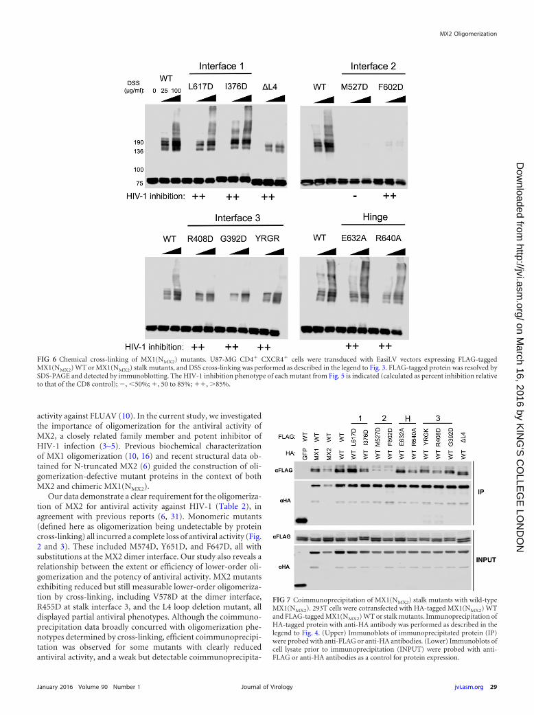
Characterizing the oligomerization state of MX1(NMX2)stalk mutants. The oligomerization state of FLAG-taggedMX1(NMX2) stalk mutants was assessed by protein cross-linkingas shown in Fig. 3. The dimer interface mutant M527D, the onlyMX1(NMX2) stalk mutant analyzed here to lose antiviral activity(Fig. 5), also was the only mutant for which monomeric proteinwas the only species detected (Fig. 6). While the other dimer in-terface mutant, F602D, also incurred a significant oligomerizationdefect, faint lower-order oligomeric species still were detectable,and this presumably was sufficient to support antiviral function(Fig. 5). All other mutants tested exhibited an efficiency of lower-oligomer formation comparable to that of wild-type MX1(NMX2)(Fig. 6). Stalk interface 3 mutants and the L4 loop deletion exhib-ited no or barely detectable higher-order oligomerization, inagreement with a previous biochemical characterization of MX1(10). The observation that these mutants, together with F602D,retain full antiviral activity provides strong evidence that higher-order oligomerization is not required for the inhibition of HIV-1infection by MX1(NMX2). Stalk interface 1 mutants and BSE hinge
mutants in the context of MX1(NMX2) incurred no observableoligomerization defect in this assay.
Coimmunoprecipitation studies also were performed withHA-tagged wild-type MX1(NMX2) and FLAG-tagged MX1(NMX2)stalk mutants, similar to those described for Fig. 4. Again, all HA-tagged and FLAG-tagged constructs were well expressed in trans-fected cells (Fig. 7, lower). As expected, FLAG-tagged wild-typeMX1(NMX2) failed to coprecipitate with the GFP-HA negativecontrol, but efficient coimmunoprecipitation was seen with theHA-tagged wild-type MX1(NMX2) positive control (Fig. 7, upper).Robust coimmunoprecipitation also was observed between MX1and MX1(NMX2). A far weaker interaction was observed betweenMX2 and MX1(NMX2), but interestingly, the longer isoform ofMX1(NMX2) coprecipitated more efficiently than the short iso-form, suggesting some role for the MX2 N terminus in self-assemblyor protein complex formation. In concordance with the cross-linkingdata, each of the MX1(NMX2) stalk and BSE hinge mutants testedcoprecipitated efficiently with wild-type MX1(NMX2), with the ex-ception of the dimer interface mutants M527D and F602D. No de-tectable interaction was observed with the short isoforms of eithermutant, while a weak interaction was observed with the long isoform.
DISCUSSION
The ability to form structured higher-order oligomers is a com-mon feature of dynamin-like GTPases (21), and, in the case ofhuman MX1, higher-order oligomerization is critical for antiviral
FIG 5 Point mutation at the dimer interface abrogates anti-HIV-1 activity ofMX1(NMX2). (Upper) U87-MG CD4� CXCR4� cells were transduced withEasiLV vectors expressing FLAG-tagged CD8, MX1, MX2, and MX1(NMX2)WT or a series of MX1(NMX2) variants with mutations at predicted stalk in-terfaces 1, 2, and 3 and the BSE-hinge (H) region or with a deletion of the L4loop (�L4). After doxycycline treatment, cells were challenged with the HIV/GFP lentiviral vector, and transduction efficiency was assessed as described inthe legend to Fig. 2. Mean percentages of transduced cells from three indepen-dent experiments with standard deviations are shown. (Lower) Immunoblotanalysis of parallel samples from the upper panel. Levels of FLAG-tagged pro-teins were determined, and Hsp90 was included as a loading control.
Dicks et al.
28 jvi.asm.org January 2016 Volume 90 Number 1Journal of Virology
on March 16, 2016 by K
ING
'S C
OLLE
GE
LON
DO
Nhttp://jvi.asm
.org/D
ownloaded from
activity against FLUAV (10). In the current study, we investigatedthe importance of oligomerization for the antiviral activity ofMX2, a closely related family member and potent inhibitor ofHIV-1 infection (3–5). Previous biochemical characterizationof MX1 oligomerization (10, 16) and recent structural data ob-tained for N-truncated MX2 (6) guided the construction of oli-gomerization-defective mutant proteins in the context of bothMX2 and chimeric MX1(NMX2).
Our data demonstrate a clear requirement for the oligomeriza-tion of MX2 for antiviral activity against HIV-1 (Table 2), inagreement with previous reports (6, 31). Monomeric mutants(defined here as oligomerization being undetectable by proteincross-linking) all incurred a complete loss of antiviral activity (Fig.2 and 3). These included M574D, Y651D, and F647D, all withsubstitutions at the MX2 dimer interface. Our study also reveals arelationship between the extent or efficiency of lower-order oli-gomerization and the potency of antiviral activity. MX2 mutantsexhibiting reduced but still measurable lower-order oligomeriza-tion by cross-linking, including V578D at the dimer interface,R455D at stalk interface 3, and the L4 loop deletion mutant, alldisplayed partial antiviral phenotypes. Although the coimmuno-precipitation data broadly concurred with oligomerization phe-notypes determined by cross-linking, efficient coimmunoprecipi-tation was observed for some mutants with clearly reducedantiviral activity, and a weak but detectable coimmunoprecipita-
FIG 6 Chemical cross-linking of MX1(NMX2) mutants. U87-MG CD4� CXCR4� cells were transduced with EasiLV vectors expressing FLAG-taggedMX1(NMX2) WT or MX1(NMX2) stalk mutants, and DSS cross-linking was performed as described in the legend to Fig. 3. FLAG-tagged protein was resolved bySDS-PAGE and detected by immunoblotting. The HIV-1 inhibition phenotype of each mutant from Fig. 5 is indicated (calculated as percent inhibition relativeto that of the CD8 control); , 50%; �, 50 to 85%; ��, �85%.
FIG 7 Coimmunoprecipitation of MX1(NMX2) stalk mutants with wild-typeMX1(NMX2). 293T cells were cotransfected with HA-tagged MX1(NMX2) WTand FLAG-tagged MX1(NMX2) WT or stalk mutants. Immunoprecipitation ofHA-tagged protein with anti-HA antibody was performed as described in thelegend to Fig. 4. (Upper) Immunoblots of immunoprecipitated protein (IP)were probed with anti-FLAG or anti-HA antibodies. (Lower) Immunoblots ofcell lysate prior to immunoprecipitation (INPUT) were probed with anti-FLAG or anti-HA antibodies as a control for protein expression.
MX2 Oligomerization
January 2016 Volume 90 Number 1 jvi.asm.org 29Journal of Virology
on March 16, 2016 by K
ING
'S C
OLLE
GE
LON
DO
Nhttp://jvi.asm
.org/D
ownloaded from

tion was noted with dimer interface mutants shown to be mono-meric by cross-linking (Fig. 4). Since coimmunoprecipitation wasperformed with cell lysates and is not dependent upon the prox-imity of interacting proteins (the distance constraint for DSS pro-tein cross-linking has been estimated to be 26 to 30 Å betweenalpha carbon atoms of cross-linked lysine residues [32]), it is pos-sible that some coimmunoprecipitation reflects the isolation ofMX protein-containing multiprotein complexes rather than directMX-MX interactions. Alternatively, the stability of interactions be-tween wild-type and mutant proteins, when assessed by coimmuno-precipitation, may appear greater than the stability of homotypic mu-tant protein interactions detected by protein cross-linking.
We observed no role for putative stalk interface 1 in either oli-gomerization or antiviral function of MX2, arguing against its biolog-ical relevance. Two previous studies, with the same M666D andI423D mutants, also have concluded that this purported interface isnot required for antiviral activity (6, 31), although the lack of an effecton oligomerization is in agreement with one study (31) but not theother (6). The latter study was performed with recombinant, malt-ose-binding protein (MBP)-tagged protein expressed in Escherichiacoli, which has the potential to behave differently than proteinsexpressed in mammalian cells. However, since our study assessedonly single point mutants at interface 1, we cannot exclude thepossibility that the mutations introduced here were insufficient todisrupt the interface.
The HIV-1 inhibition phenotypes observed for stalk interface 3mutants R455D and G439D and the L4 loop deletion were in agree-
ment with those observed previously (25), but here we show thatantiviral activity correlated with the relative ability of these mutants toform lower-order oligomers. Here, we also demonstrated a role forthe BSE hinge region in the antiviral activity of MX2, with mutantsE681A and R689A being significantly impaired for HIV-1 inhibition,although a previous study found no effect of the E681A mutation onfunction (6). The impact of these mutations on the oligomerizationof MX2 had not been tested previously, but here we show by proteincross-linking and coimmunoprecipitation that both mutations incura moderate oligomerization defect that likely explains their func-tional impairment. That mutant R689A retained any antiviral activitystands in contrast to corresponding mutant R640A in MX1, whichlost inhibitory function (16). Since the BSE hinge region has beenascribed a role in conformational coupling between the GTPase do-main and the stalk, its lesser importance for MX2 activity may have beenanticipated given the GTPase independence of this protein (3, 4).
Extending these studies, we also demonstrate the requirementfor lower-order oligomerization in the context of the MX1(NMX2)chimera, implying that this attribute enables the MX2 N-terminaldomain to mediate antiviral activity. Dimer interface mutantM527D, which failed to form any detectable oligomeric species byprotein cross-linking, exhibited a complete loss of antiviral activ-ity (Fig. 5 and 6). However, mutant F602D, which is also markedlydefective for oligomerization but still showed very faint lower-order cross-linking, was fully antiviral. Perhaps HIV-1 is more sensi-tive to inhibition by MX1(NMX2) than MX2, such that only a smallproportion of the active form is required. Indeed, the HIV-1 suppres-
TABLE 2 Oligomerization and HIV-1 inhibition data for MX2 and MX1(NMX2) mutantsa
Mutant SiteInhibitionof HIV-1b
Oligomerization bycross-linking Coimmunoprecipitation
MX2WT �� Higher order YesM666D Stalk 1 �� Higher order YesI423D Stalk 1 �� Higher order YesM574D Stalk 2 � Monomeric Very weakY651D Stalk 2 � Monomeric Very weakV578D Stalk 2 � Limited oligomerization YesF647D Stalk 2 � Monomeric Very weakE681A Hinge � Higher order WeakR689A Hinge � Higher order WeakYRGK-AAAA487–490 Stalk 3 �� Lower order YesR455D Stalk 3 � Limited oligomerization WeakG439D Stalk 3 �� Higher order Yes�L4 L4 loop � Lower order Yes
MX1(NMX2)WT �� Higher order YesL617D Stalk 1 �� Higher order YesI376D Stalk 1 �� Higher order YesM527D Stalk 2 � Monomeric WeakF602D Stalk 2 �� Limited oligomerization WeakE632A Hinge �� Higher order YesR640A Hinge �� Higher order YesYRGR-AAAA440–443 Stalk 3 �� Higher order YesR408D Stalk 3 �� Lower order YesG392D Stalk 3 �� Lower order Yes�L4 L4 loop �� Lower order Yes
a Summary of HIV-1 inhibition and protein oligomerization data shown in Fig. 2 to 7. The site, antiviral activity against HIV-1, oligomerization phenotype by protein cross-linking,and coimmunoprecipitation efficiency with WT or mutant proteins described in this study are shown.b The HIV-1 inhibition phenotype indicates percent inhibition calculated relative to that of the CD8 control (from Fig. 2 and 5): , 0 to 50%; �, 50 to 85%; ��, 85 to 100%.Mutants exhibiting a complete loss of antiviral activity are highlighted in boldface.
Dicks et al.
30 jvi.asm.org January 2016 Volume 90 Number 1Journal of Virology
on March 16, 2016 by K
ING
'S C
OLLE
GE
LON
DO
Nhttp://jvi.asm
.org/D
ownloaded from

sion phenotype observed upon the expression of MX1(NMX2) typi-cally is stronger than that for MX2 (23).
The data presented here corroborate previous observations us-ing constructs in which the amino-terminal 91 residues of MX2were fused to monomeric, dimeric, or trimeric versions of theyeast GCN4 leucine zipper domain (20). Dimeric and trimericconstructs retained �80% antiviral activity, while the monomericconstruct was not antiviral, implying that the dimerization of theMX2 N terminus is sufficient for antiviral activity to be elicited(20). Extending this observation, we now conclude that higher-order oligomerization is dispensable for full antiviral activity, par-ticularly in the context of MX1(NMX2). Dimer interface mutantF602D, stalk interface 3 mutants R408D, G392D, and YRGR440-AAAA443, and the L4 loop deletion all exhibited no (or barely)detectable higher-order oligomerization yet retained full antiviralactivity in this context. The dependence of MX2 activity upondimerization is reminiscent of fusions between the murine leuke-mia virus restriction factor Fv1 and cyclophilin A; these chimericproteins also suppress infection by inhibiting viral cDNA nuclear im-port, perhaps indicating commonalities in mechanism (33, 34).
Notably, wild-type MX2 and MX1(NMX2) both exhibited pro-files of lower-order and higher-order oligomeric forms by cross-linking that were similar to those of MX1 (Fig. 3A). However, anddespite the similarities in structure and propensity to form higher-order oligomers, current data support the conclusion that thereare a number of fundamental differences between the antiviralmechanisms of MX1 and MX2.
The mechanism underpinning the requirement for MX2 oli-gomerization is not presently understood. Previous studies haveshown that monomeric MX2 mutants fail to bind HIV-1 capsid-nucleocapsid nanotubes in vitro (6, 31). However, further inves-tigation is required to determine the precise nature and relevanceof the interaction between MX2 and CA, since mutations in CAthat permit viral escape from MX2-mediated inhibition do notblock MX2 binding in vitro (7, 8). The potential involvement ofadditional cellular factors, as well as the importance of MX2 oli-gomerization for their recruitment, will require future exploration.
ACKNOWLEDGMENTS
This work was supported by the U.K. Medical Research Council, the Well-come Trust, the European Commission’s Seventh Framework Pro-gramme (FP7/2007-2013) under grant agreements no. PIEF-GA-2009-237501 (to CG) and no. PIIF-GA-2012-329679 (to DP), and theDepartment of Health via a National Institute for Health Research Com-prehensive Biomedical Research Centre award to Guy’s and St. Thomas’NHS Foundation Trust in partnership with King’s College London andKing’s College Hospital NHS Foundation Trust.
FUNDING INFORMATIONWellcome Trust provided funding to Darja Pollpeter and Michael H Ma-lim under grant number 106223/Z/14/Z. Medical Research Council(MRC) provided funding to Matthew D.J. Dicks, Caroline Goujon, Gil-berto Betancor, Luis Apolonia, and Michael H Malim under grant num-ber G1000196. European Commission (EC) provided funding to CarolineGoujon and Darja Pollpeter under grant numbers GA-2009-237501 andGA-2012-329679.
DH | National Institute for Health Research (NIHR) provided fundingunder grant number guysbrc-2012-1.
REFERENCES1. Doyle T, Goujon C, Malim MH. 2015. HIV-1 and interferons: who’s
interfering with whom? Nat Rev Microbiol 13:403– 413. http://dx.doi.org/10.1038/nrmicro3449.
2. Schoggins JW, Rice CM. 2011. Interferon-stimulated genes and theirantiviral effector functions. Curr Opin Virol 1:519 –525. http://dx.doi.org/10.1016/j.coviro.2011.10.008.
3. Goujon C, Moncorgé O, Bauby H, Doyle T, Ward CC, Schaller T, HuéS, Barclay WS, Schulz R, Malim MH. 2013. Human MX2 is an interfer-on-induced post-entry inhibitor of HIV-1 infection. Nature 502:559 –562.http://dx.doi.org/10.1038/nature12542.
4. Kane M, Yadav SS, Bitzegeio J, Kutluay SB, Zang T, Wilson SJ, Schog-gins JW, Rice CM, Yamashita M, Hatziioannou T, Bieniasz PD. 2013.MX2 is an interferon-induced inhibitor of HIV-1 infection. Nature 502:563–566. http://dx.doi.org/10.1038/nature12653.
5. Liu Z, Pan Q, Ding S, Qian J, Xu F, Zhou J, Cen S, Guo F, Liang C. 2013.The interferon-inducible MxB protein inhibits HIV-1 infection. Cell HostMicrobe 14:398–410. http://dx.doi.org/10.1016/j.chom.2013.08.015.
6. Fribourgh JL, Nguyen HC, Matreyek KA, Alvarez FJ, Summers BJ,Dewdney TG, Aiken C, Zhang P, Engelman A, Xiong Y. 2014. Structuralinsight into HIV-1 restriction by MxB. Cell Host Microbe 16:627– 638.http://dx.doi.org/10.1016/j.chom.2014.09.021.
7. Fricke T, White TE, Schulte B, de Souza Aranha Vieira DA, Dharan A,Campbell EM, Brandariz-Nuñez A, Diaz-Griffero F. 2014. MxB binds tothe HIV-1 core and prevents the uncoating process of HIV-1. Retrovirol-ogy 11:68. http://dx.doi.org/10.1186/s12977-014-0068-x.
8. Liu Z, Pan Q, Liang Z, Qiao W, Cen S, Liang C. 2015. The highlypolymorphic cyclophilin A-binding loop in HIV-1 capsid modulates viralresistance to MxB. Retrovirology 12:1. http://dx.doi.org/10.1186/s12977-014-0129-1.
9. Haller O, Kochs G. 2011. Human MxA protein: an interferon-induceddynamin-like GTPase with broad antiviral activity. J Interferon CytokineRes 31:79 – 87. http://dx.doi.org/10.1089/jir.2010.0076.
10. Gao S, von der Malsburg A, Paeschke S, Behlke J, Haller O, Kochs G,Daumke O. 2010. Structural basis of oligomerization in the stalk region ofdynamin-like MxA. Nature 465:502–506. http://dx.doi.org/10.1038/nature08972.
11. Pitossi F, Blank A, Schröder A, Schwarz A, Hüssi P, Schwemmle M,Pavlovic J, Staeheli P. 1993. A functional GTP-binding motif is necessaryfor antiviral activity of Mx proteins. J Virol 67:6726 – 6732.
12. Ponten A, Sick C, Weeber M, Haller O, Kochs G. 1997. Dominant-negative mutants of human MxA protein: domains in the carboxy-terminal moiety are important for oligomerization and antiviral activity. JVirol 71:2591–2599.
13. Kochs G, Haener M, Aebi U, Haller O. 2002. Self-assembly of humanMxA GTPase into highly ordered dynamin-like oligomers. J Biol Chem277:14172–14176. http://dx.doi.org/10.1074/jbc.M200244200.
14. Kochs G, Janzen C, Hohenberg H, Haller O. 2002. Antivirally activeMxA protein sequesters La Crosse virus nucleocapsid protein into perinu-clear complexes. Proc Natl Acad Sci U S A 99:3153–3158. http://dx.doi.org/10.1073/pnas.052430399.
15. Turan K, Mibayashi M, Sugiyama K, Saito S, Numajiri A, Nagata K.2004. Nuclear MxA proteins form a complex with influenza virus NP andinhibit the transcription of the engineered influenza virus genome. Nu-cleic Acids Res 32:643– 652. http://dx.doi.org/10.1093/nar/gkh192.
16. Gao S, von der Malsburg A, Dick A, Faelber K, Schröder GF, Haller O,Kochs G, Daumke O. 2011. Structure of myxovirus resistance protein areveals intra- and intermolecular domain interactions required for theantiviral function. Immunity 35:514 –525. http://dx.doi.org/10.1016/j.immuni.2011.07.012.
17. Haller O, Gao S, von der Malsburg A, Daumke O, Kochs G. 2010.Dynamin-like MxA GTPase: structural insights into oligomerization andimplications for antiviral activity. J Biol Chem 285:28419 –28424. http://dx.doi.org/10.1074/jbc.R110.145839.
18. Dick A, Graf L, Olal D, von der Malsburg A, Gao S, Kochs G, DaumkeO. 2015. Role of nucleotide binding and GTPase domain dimerization indynamin-like myxovirus resistance protein A for GTPase activation andantiviral activity. J Biol Chem 290:12779 –12792. http://dx.doi.org/10.1074/jbc.M115.650325.
19. Rennie ML, McKelvie SA, Bulloch EM, Kingston RL. 2014. Transientdimerization of human MxA promotes GTP hydrolysis, resulting in a
MX2 Oligomerization
January 2016 Volume 90 Number 1 jvi.asm.org 31Journal of Virology
on March 16, 2016 by K
ING
'S C
OLLE
GE
LON
DO
Nhttp://jvi.asm
.org/D
ownloaded from

mechanical power stroke. Structure 22:1433–1445. http://dx.doi.org/10.1016/j.str.2014.08.015.
20. Goujon C, Greenbury RA, Papaioannou S, Doyle T, Malim MH. 2015.A triple-arginine motif in the amino-terminal domain and oligomeriza-tion are required for HIV-1 inhibition by human MX2. J Virol 89:4676 –4680. http://dx.doi.org/10.1128/JVI.00169-15.
21. Haller O, Staeheli P, Schwemmle M, Kochs G. 2015. Mx GTPases:dynamin-like antiviral machines of innate immunity. Trends Microbiol23:154 –163. http://dx.doi.org/10.1016/j.tim.2014.12.003.
22. Busnadiego I, Kane M, Rihn SJ, Preugschas HF, Hughes J, Blanco-MeloD, Strouvelle VP, Zang TM, Willett BJ, Boutell C, Bieniasz PD, WilsonSJ. 2014. Host and viral determinants of Mx2 antiretroviral activity. J Virol88:7738 –7752. http://dx.doi.org/10.1128/JVI.00214-14.
23. Goujon C, Moncorgé O, Bauby H, Doyle T, Barclay WS, Malim MH.2014. Transfer of the amino-terminal nuclear envelope targeting domainof human MX2 converts MX1 into an HIV-1 resistance factor. J Virol88:9017–9026. http://dx.doi.org/10.1128/JVI.01269-14.
24. Melén K, Keskinen P, Ronni T, Sareneva T, Lounatmaa K, Julkunen I.1996. Human MxB protein, an interferon-alpha-inducible GTPase, con-tains a nuclear targeting signal and is localized in the heterochromatinregion beneath the nuclear envelope. J Biol Chem 271:23478 –23486. http://dx.doi.org/10.1074/jbc.271.38.23478.
25. Matreyek KA, Wang W, Serrao E, Singh PK, Levin HL, Engelman A.2014. Host and viral determinants for MxB restriction of HIV-1 infection.Retrovirology 11:90. http://dx.doi.org/10.1186/s12977-014-0090-z.
26. Schulte B, Buffone C, Opp S, Di Nunzio F, Augusto De Souza AranhaVieira D, Brandariz-Nuñez A, Diaz-Griffero F. 2015. Restriction ofHIV-1 requires the N-terminal region of MxB/Mx2 as a capsid-bindingmotif but not as a nuclear localization signal. J Virol 89:8599 – 8610. http://dx.doi.org/10.1128/JVI.00753-15.
27. Melén K, Julkunen I. 1997. Nuclear cotransport mechanism of cytoplas-
mic human MxB protein. J Biol Chem 272:32353–32359. http://dx.doi.org/10.1074/jbc.272.51.32353.
28. Faelber K, Posor Y, Gao S, Held M, Roske Y, Schulze D, Haucke V, NoéF, Daumke O. 2011. Crystal structure of nucleotide-free dynamin. Nature477:556 –560. http://dx.doi.org/10.1038/nature10369.
29. Mitchell PS, Patzina C, Emerman M, Haller O, Malik HS, Kochs G.2012. Evolution-guided identification of antiviral specificity determinantsin the broadly acting interferon-induced innate immunity factor MxA.Cell Host Microbe 12:598 – 604. http://dx.doi.org/10.1016/j.chom.2012.09.005.
30. Patzina C, Haller O, Kochs G. 2014. Structural requirements for theantiviral activity of the human MxA protein against Thogoto and influ-enza A virus. J Biol Chem 289:6020 – 6027. http://dx.doi.org/10.1074/jbc.M113.543892.
31. Buffone C, Schulte B, Opp S, Diaz-Griffero F. 2015. Contribution ofMxB oligomerization to HIV-1 capsid binding and restriction. J Virol89:3285–3294. http://dx.doi.org/10.1128/JVI.03730-14.
32. Merkley ED, Rysavy S, Kahraman A, Hafen RP, Daggett V, AdkinsJN. 2014. Distance restraints from crosslinking mass spectrometry:mining a molecular dynamics simulation database to evaluate lysine-lysine distances. Protein Sci 23:747–759. http://dx.doi.org/10.1002/pro.2458.
33. Goldstone DC, Walker PA, Calder LJ, Coombs PJ, Kirkpatrick J, BallNJ, Hilditch L, Yap MW, Rosenthal PB, Stoye JP, Taylor IA. 2014.Structural studies of postentry restriction factors reveal antiparallel dimersthat enable avid binding to the HIV-1 capsid lattice. Proc Natl Acad SciU S A 111:9609 –9614. http://dx.doi.org/10.1073/pnas.1402448111.
34. Yap MW, Mortuza GB, Taylor IA, Stoye JP. 2007. The design of artificialretroviral restriction factors. Virology 365:302–314. http://dx.doi.org/10.1016/j.virol.2007.04.005.
Dicks et al.
32 jvi.asm.org January 2016 Volume 90 Number 1Journal of Virology
on March 16, 2016 by K
ING
'S C
OLLE
GE
LON
DO
Nhttp://jvi.asm
.org/D
ownloaded from