king s research portal · piergiorgio salvan ,1 j. donald tournier,1 dafnis batalle ,1 ......
TRANSCRIPT

King’s Research Portal
DOI:10.1002/hbm.2363210.1002/hbm.23632
Document VersionPublisher's PDF, also known as Version of record
Link to publication record in King's Research Portal
Citation for published version (APA):Salvan, P., Tournier, J. D., Batalle, D., Falconer, S., Chew, A., Kennea, N., ... Counsell, S. J. (2017). Languageability in preterm children is associated with arcuate fasciculi microstructure at term. Human Brain Mapping. DOI:10.1002/hbm.23632, 10.1002/hbm.23632
Citing this paperPlease note that where the full-text provided on King's Research Portal is the Author Accepted Manuscript or Post-Print version this maydiffer from the final Published version. If citing, it is advised that you check and use the publisher's definitive version for pagination,volume/issue, and date of publication details. And where the final published version is provided on the Research Portal, if citing you areagain advised to check the publisher's website for any subsequent corrections.
General rightsCopyright and moral rights for the publications made accessible in the Research Portal are retained by the authors and/or other copyrightowners and it is a condition of accessing publications that users recognize and abide by the legal requirements associated with these rights.
•Users may download and print one copy of any publication from the Research Portal for the purpose of private study or research.•You may not further distribute the material or use it for any profit-making activity or commercial gain•You may freely distribute the URL identifying the publication in the Research Portal
Take down policyIf you believe that this document breaches copyright please contact [email protected] providing details, and we will remove access tothe work immediately and investigate your claim.
Download date: 17. Jun. 2018

Language Ability in Preterm Children IsAssociated with Arcuate Fasciculi
Microstructure at Term
Piergiorgio Salvan ,1 J. Donald Tournier,1 Dafnis Batalle ,1
Shona Falconer,1 Andrew Chew,1 Nigel Kennea,2 Paul Aljabar,1
Ghislaine Dehaene-Lambertz,3 Tomoki Arichi,1,4 A. David Edwards,1,4* andSerena J. Counsell1
1Centre for the Developing Brain, Division of Imaging Sciences & Biomedical Engineering,King’s College London, United Kingdom
2Neonatal unit, St. George’s University Hospital NHS, London, United Kingdom3INSERM-CEA, Neurospin Center, Cognitive Neuroimaging Unit, Gif-Sur-Yvette, France
4Department of Bioengineering, Imperial College London, United Kingdom
r r
Abstract: In the mature human brain, the arcuate fasciculus mediates verbal working memory, wordlearning, and sublexical speech repetition. However, its contribution to early language acquisitionremains unclear. In this work, we aimed to evaluate the role of the direct segments of the arcuate fas-ciculi in the early acquisition of linguistic function. We imaged a cohort of 43 preterm born infants(median age at birth of 30 gestational weeks; median age at scan of 42 postmenstrual weeks) usinghigh b value high-angular resolution diffusion-weighted neuroimaging and assessed their linguisticperformance at 2 years of age. Using constrained spherical deconvolution tractography, we virtuallydissected the arcuate fasciculi and measured fractional anisotropy (FA) as a metric of white matterdevelopment. We found that term equivalent FA of the left and right arcuate fasciculi was significantlyassociated with individual differences in linguistic and cognitive abilities in early childhood, indepen-dent of the degree of prematurity. These findings suggest that differences in arcuate fasciculi micro-structure at the time of normal birth have a significant impact on language development and modulatethe first stages of language learning. Hum Brain Mapp 00:000–000, 2017. VC 2017 The Authors Human Brain
Mapping Published by Wiley Periodicals, Inc.
Key words: diffusion magnetic resonance imaging; infant; language development; brain; preterm birth
r r
VC 2017 The Authors Human Brain Mapping Published by Wiley Periodicals, Inc.This is an open access article under the terms of the Creative Commons Attribution License, which permits use, distribution andreproduction in any medium, provided the original work is properly cited.
Contract grant sponsor: National Institute for Health Research(NIHR) under its Programme Grants for Applied Research Pro-gramme; Contract grant number: RP-PG-0707-10154; Contractgrant sponsor: Academic Clinical Lectureship; Contract grantsponsor: Medical Research Council (UK); Contract grant number:MR/K006355/1 and MR/L011530/1; Contract grant sponsor: PhDstudentship.
*Correspondence to: Professor A.D. Edwards, Centre for theDeveloping Brain, Department of Perinatal Imaging and Health,
Division of Imaging Sciences and Biomedical Engineering, King’sCollege London, First Floor South Wing, St Thomas’ Hospital,London SE1 7EH, UK. E-mail: [email protected]
Received for publication 6 December 2016; Revised 31 March2017; Accepted 17 April 2017.
DOI: 10.1002/hbm.23632Published online 00 Month 2017 in Wiley Online Library(wileyonlinelibrary.com).
r Human Brain Mapping 00:00–00 (2017) r

INTRODUCTION
Comparative studies in humans and nonhuman pri-mates have shown that the evolution of language hasresulted from specific modifications of the cortical areasand pathways that mediate linguistic function [Rillinget al., 2008]. The arcuate fasciculus is a bilateral white-matter fiber tract linking the posterior superior temporalcortex (Wernicke’s area) to Brodmann area 44 in the fron-tal cortex (Broca’s area) via a dorsal projection that archesaround the Sylvian fissure [Catani et al., 2005; Rillinget al., 2008]. In the human brain, diffusion weighted imag-ing has shown that the organization and cortical termina-tions of this tract are strongly modified in comparison toprimates and has demonstrated that the auditory regionsof the temporal cortex have a higher probability of connec-tion via the dorsal pathway with the frontal cortices [Ril-ling et al., 2008, 2011; de Schotten et al., 2012]. In contrast,axonal tracing studies in monkeys have shown that thearcuate fasciculus connects to more dorsally locatedregions, such as the extrastriate visual cortex [Petrides andPandya, 1984; Schmahmann et al., 2007]. Taken together,these findings have generated the theory that theexpanded direct dorsal pathway may be a key structureresponsible for supporting the emergence of language inhumans.
In human adults, the arcuate fasciculus has been pro-posed to play an important role in the core syntactic com-putation of complex sentences [Berwick et al., 2013], inverbal short-term memory and in the perception of thephonetic structure of speech [Liberman and Mattingly,1985]. It is also hypothesized to play a distinctive role inspeech production via the integration of auditory andmotor representation. Predominantly at the syllable level,it is thought to map sensory targets in the auditory cortexto motor programs coded in Broca’s area [Hickok, 2012;Hickok and Poeppel, 2007]. Patients with injuries involv-ing either the left or right arcuate fasciculus have impairedability in phonological and word repetition tasks and inverbal short-term memory [Alexander et al., 1987; Bensonet al., 1973; Damasio and Damasio, 1980; Geschwind,1965]. With regard to learning, the microstructural proper-ties of the left direct segment of the arcuate fasciculushave been associated with the process of learning newwords in adulthood [L�opez-Barroso et al., 2013], whilstimproved performance in auditory verbal learning tasksare significantly associated with a less lateralized volumet-ric pattern of the direct pathways [Catani et al., 2007].Children with Angelman Syndrome in whom neither theleft nor right arcuate fasciculi can be identified on diffu-sion tractography have no oral language development,whereas when the left arcuate fasciculus cannot be identi-fied language difficulties are always observed [Paldinoet al., 2016; Wilson et al., 2011]. These observations con-firm the crucial role of the arcuate fasciculus in speechacquisition [Berwick et al., 2013; Hickok and Poeppel,2007].
As infants have impoverished language production andtheir reception abilities were thought to be limited to thesupra-segmental properties of speech, the role of the arcu-ate fasciculus has traditionally been considered to be sec-ondary during the first stages of language acquisition.Although inferior frontal regions are activated in severalfMRI studies in infants [Baldoli et al., 2015; Dehaene-Lambertz et al., 2006; Perani et al., 2011; Shultz et al.,2014], the ventral pathway, comprising the uncinate andthe inferior fronto-occipital fasciculus, has been proposedto initially be the main functional linguistic pathway con-necting temporal and frontal areas [Brauer et al., 2013;Dubois et al., 2015; Perani et al., 2011]. Recent advances indiffusion-weighted imaging have enabled the investigationof white-matter tracts thought to be involved in the acqui-sition of language and neurodevelopmental skills duringthe neonatal period [Brauer et al., 2013; Dubois et al., 2015;Perani et al., 2011]. These studies have suggested that, incontrast to the mature brain (where it terminates in Broca’sarea), the anterior direct segment of the arcuate fasciculuscannot be dissected after the premotor cortex [Duboiset al., 2015]. Whilst this finding may represent a genuinedevelopmental difference in the extent of the arcuate fas-ciculus [Brauer et al., 2013; Dubois et al., 2015; Peraniet al., 2011], it could also reflect the low angular resolutionused in the diffusion weighted sequences, which mayhave limited delineation of the arcuate fasciculus inregions where the fibers cross with the cortico-spinal tractsand the corpus callosum in the corona radiata [Duboiset al., 2015].
Premature birth is associated with verbal impairment,the severity of which increases with increasing prematu-rity at birth [Luu et al., 2009, 2011; van Noort-van derSpek et al., 2012]. Previous studies of infant brain develop-ment have shown that white-matter architecture is signifi-cantly altered following premature birth [Ball et al., 2014;Counsell et al., 2003; H€uppi et al., 1998; Rose et al., 2008]and the degree of this alteration is directly related to per-formance in specific neurodevelopmental domains [Bassiet al., 2008; Berman et al., 2009; Groppo et al., 2014]. It isthus possible that premature delivery also affects thewhite-matter structures that subserve language functionimpacting on later linguistic behavior.
To address the question of whether or not the arcuatefasciculus is a specific neurolinguistic precursor in earlyhuman infancy; and to assess whether the degree of pre-maturity affects arcuate fasciculus microstructure anddrives the relationship with later linguistic behavior, weused high b value high-angular resolution diffusion-weighted imaging (HARDI) in a cohort of 43 preterm borninfants at term equivalent age and assessed their linguisticdevelopmental performance at 2 years. We hypothesizedthat intersubject differences in composite linguistic skills at2 years would be associated with term equivalent frac-tional anisotropy (FA) of the left and right arcuate fascic-uli. To act as a control, we tested whether any relationship
r Salvan et al. r
r 2 r

between brain structure and language performance couldalso be associated with FA values of the cortico-spinaltracts and the superior longitudinal fasciculi.
MATERIALS AND METHODS
Infants
Preterm infants were recruited as part of the Evaluationof Preterm Imaging study (Eprime), and were imaged atterm equivalent age over a 3 year period (2010–2013) atQueen Charlotte’s and Chelsea Hospital, London. Thestudy was reviewed and approved by the NationalResearch Ethics Service, and all infants were studied fol-lowing written consent from their parents. A cohort of 43preterm born infants [median age at birth of 30.14 gesta-tional (GA) weeks; range 24–32; 18 females] with no evi-dence of focal abnormality on MRI were imaged usinghigh-angular resolution diffusion-weighted neuroimagingat 42.14 postmenstrual (PMA) weeks (range 39–46) (TableI), and followed up to around 22 months of age to assesstheir neurodevelopmental performance.
Acquisition of MRI Imaging Data at Term
Equivalent Age
All MRI studies were supervised by an experiencedpediatrician or nurse trained in neonatal resuscitation.Pulse oximetry, temperature, and heart rate were moni-tored throughout the period of image acquisition; hearingprotection in the form of silicone-based putty placed in theexternal ear (President Putty, Coltene; Whaledent) andMini-muffs (Natus Medical) was used for each infant.Sedation (25–50 mg/kg oral chloral hydrate) was adminis-tered to 33 infants. Imaging was acquired using an eight-channel phased array head coil on a 3-Tesla PhilipsAchieva MRI Scanner (Best, The Netherlands) located onthe Neonatal Intensive Care Unit. Whole-brain diffusion-weighted MRI data were acquired in 64 noncollinear direc-tions with b value of 2500 s/mm2 and 4 images withoutdiffusion weighting (isotropic voxel size of 2 mm; TE 5 62ms; TR 5 9000 ms). High-resolution anatomical imageswere acquired with pulse sequence parameters: T1weighted 3D MPRAGE: TR 5 17 ms, TE 5 4.6 ms, flipangle 138, slice thickness 0.8 mm, field-of-view 210 mm,matrix 256 3 256 (voxel size: 0.82 3 0.82 3 0.8 mm); andT2 weighted fast-spin echo: TR 5 8670 ms, TE 5 160 ms,flip angle 908, slice thickness 2 mm with 1 mm overlap,field-of-view 220 mm, matrix 256 3 256 (effective voxelsize: 0.86 3 0.86 3 1 mm).
Neurodevelopmental Assessment at 22 Months
Standardized neurodevelopmental assessment at amedian age of 22 months (range: 21–24 months; median of20 months corrected for prematurity) was carried out by
an experienced pediatrician or developmental psychologistwith the Bayley Scales of Infant and Toddler Development,Third Edition (BSID-III) [Bayley, 2006].
MRI Data Preprocessing
T2-weighted brain volumes were bias corrected, brainextracted and tissue segmented into white matter, graymatter, deep gray matter structures, and cerebrospinalfluid using a neonatal specific segmentation tool [Makro-poulos et al., 2014]. Diffusion MRI volumes were first visu-ally inspected to detect and exclude data with motionartifact. All subjects included in the study had 5 or fewervolumes excluded due to head-motion. B0 field inhomoge-neities, eddy currents, and intervolume motion were cor-rected using topup and eddy tools in FSL5 [Anderssonand Sotiropoulos, 2016; Andersson et al., 2003; Smith et al.,2004, 2015]. B1 field inhomogeneity was corrected usingITK-N4 [Tustison et al., 2010]. All rigid registrations innative subject space were estimated using FSL boundary-based registration optimized for neonatal tissue contrasts[Toulmin et al., 2015]; and nonlinear registrations to theT2-weighted template were estimated using AdvancedNormalization Tools [Avants et al., 2008]. All transforma-tion pairs were calculated independently and combinedinto a single transform to reduce interpolation error.
Tractography of the Arcuate Fasciculi
Estimation of fiber orientation distribution was com-puted through constrained spherical deconvolution [Tour-nier et al., 2004, 2007], with maximum spherical harmonicorder of 8. We used the MRtrix3 package to perform ana-tomically constrained probabilistic tractography (http://www.mrtrix.org) [Smith et al., 2012; Tournier et al., 2012].Fiber-tracking of the arcuate fasciculus was performed ineach subject’s native space independently for both hemi-spheres, using a two-region of interest approach. From theT2-weighted template, we back-projected two inclusionregions of interest and a seed-plane. To maximize thechances of virtually dissecting the arcuate fasciculus, the
TABLE I. Infant characteristics
Characteristic Value
Median (range) GA at birth (weeks) 30 (24 – 33)Median (range) birth weight (grams) 1205 (645 – 1990)Median (range) PMA at MRI (weeks) 42 (39 – 46)Female, no (%) 18 (42%)Chorioamnionitis, no (%) 1 (2%)Intrauterine growth restriction, no (%) 7 (16%)Median (range) mechanical
ventilation (days)0 (0 – 40)
Necrotizing enterocolitisrequiring surgery, no (%)
1 (2%)
Mean (6 SD) parental SES 17.4293 (68.0772)
AQ1 r Language Brain Organization in Preterm Neonates r
r 3 r
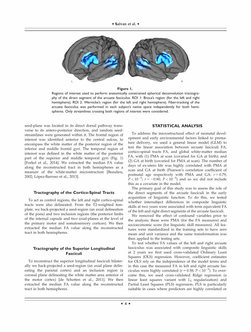
seed-plane was located in its direct dorsal pathway trans-verse to its antero-posterior direction, and random seed-streamlines were generated within it. The frontal region ofinterest was identified anterior to the central sulcus, toencompass the white matter of the posterior region of theinferior and middle frontal gyri. The temporal region ofinterest was defined in the white matter of the posteriorpart of the superior and middle temporal gyri (Fig. 1)[Forkel et al., 2014]. We extracted the median FA valuealong the reconstructed tract in both hemispheres as ameasure of the white-matter microstructure [Beaulieu,2002; L�opez-Barroso et al., 2013].
Tractography of the Cortico-Spinal Tracts
To act as control regions, the left and right cortico-spinaltracts were also delineated. From the T2-weighted tem-plate, we back-projected a seed-region (an axial delineationof the pons) and two inclusion regions (the posterior limbsof the internal capsule and two axial-planes at the level ofthe primary motor and somatosensory cortices). We thenextracted the median FA value along the reconstructedtract in both hemispheres.
Tractography of the Superior Longitudinal
Fasciculi
To reconstruct the superior longitudinal fasciculi bilater-ally we back-projected a seed-region (an axial plane delin-eating the parietal cortex) and an inclusion region (acoronal plane delineating the white matter area anterior ofthe motor cortex) [de Schotten et al., 2011]. We thenextracted the median FA value along the reconstructedtract in both hemispheres.
STATISTICAL ANALYSIS
To address the microstructural effect of neonatal devel-opment and early environmental factors linked to prema-ture delivery, we used a general linear model (GLM) totest the linear association between arcuate fasciculi FA,cortico-spinal tracts FA, and global white-matter medianFA, with (1) PMA at scan (covaried for GA at birth); and(2) GA at birth (covaried for PMA at scan). The number ofdays of ex-utero life was highly correlated with PMA atscan and GA at birth (Pearson’s correlation coefficient ofpostnatal age respectively with PMA and GA: r 5 0.79;P< 1025; r 5 20.90; P< 1025) and so we did not includethis as a covariate in the model.
The primary goal of this study was to assess the role ofthe direct segments of the arcuate fasciculi in the earlyacquisition of linguistic function. To do this, we testedwhether intersubject differences in composite linguisticskills at two years were associated with term equivalent FAof the left and right direct segments of the arcuate fasciculi.
We removed the effect of confound variables prior tothe analysis; these were PMA (for the FA measures) andsocioeconomic score (for linguistic skill measures). All fea-tures were standardized in the training sets to have zeromean and unit variance and the same transformation wasthen applied to the testing sets.
To test whether FA values of the left and right arcuatefasciculus was associated with composite linguistic skillsat 2 years we first used cross-validated Ordinary LeastSquares (OLS) regression. However, coefficient estimatesfor OLS rely on the independence of the model terms andin this case the measured FA in left and right arcuate fas-ciculus were highly correlated (r 5 0.58; P< 1025). To over-come this, we used cross-validated Ridge regression (alinear least squares variant with L2 regularization) andPartial Least Squares (PLS) regression. PLS is particularlysuitable in cases where predictors are highly correlated or
Figure 1.
Regions of interest used to perform anatomically constrained spherical deconvolution tractogra-
phy of the direct segment of the arcuate fasciculus. ROI 1: Broca’s region (for the left and right
hemisphere); ROI 2: Wernicke’s region (for the left and right hemisphere). Fiber-tracking of the
arcuate fasciculus was performed in each subject’s native space independently for both hemi-
spheres. Only streamlines crossing both regions of interest were considered.
r Salvan et al. r
r 4 r

even collinear, that is, where standard regression is notappropriate [Hotelling, 1936; Wegelin, 2000]. The approachidentifies linear combinations of the independent variablesthat optimally predict corresponding combinations of thedependent variables [Rosipal and Kr€amer, 2006]. Here, itwas applied with mode A and deflation mode canonical[Wegelin, 2000].
We used leave-one-out cross-validated PLS to assesswhether intersubject differences in linguistic abilities at 2years of age were associated with term equivalent FA ofthe left and right arcuate fasciculi. At each training itera-tion, the data for n 2 1 subjects were used to train a PLSmodel; the learnt link was then used to generate the lin-guistic score for the left-out subject. Following all itera-tions, the correlation between PLS FA scores and PLSlanguage scores was assessed. We then extracted the PLSrelative loadings of involvement averaged across all cross-validation folds; the mean and standard deviation (SD) ofthese parameters were extracted to assess model stability.
As a control, we also used the same cross-validated pipelineto test whether individual-differences in linguistic perfor-mance were associated with left and right cortico-spinal tractsFA, and left and right superior longitudinal fasciculus FA.
To test whether early environmental influences associatedwith preterm birth or global white-matter volume [Northamet al., 2012] were driving the identified brain-behavior link ina dose-dependent fashion, we calculated the partial correla-tion between PLS FA scores and PLS language scores adjust-ing for GA at birth, global white matter volume, and sex.
Statistical significance was determined with nonpara-metric permutation testing (10,000 permutations) with cor-rection for the Family wise error (FWE) rate [Winkleret al., 2014]. All analysis were performed using MATLAB(R2015b, The MathWorks, Natick, MA) and Scikit-learn[Pedregosa et al., 2011].
RESULTS
Neurodevelopmental Assessment
At 2 years of age, the mean scores of the BSID-III compos-ite language and cognitive abilities were respectively 90(SD 6 16.20) and 92 (SD 6 11.85), with a correlation betweenthe two of r 5 0.79; P 5 1025. No significant correlation wasfound between PMA at scan and composite language score.A trend toward significance was found between GA at birthand composite language score (r 5 0.21; P 5 0.09); a signifi-cant correlation was found between socioeconomic score(measured as the English Index of Multiple Deprivation)and composite language score (r 5 20.28; P 5 0.03).
Impact of Prematurity on the Arcuate Fasciculus
Microstructure
During the term equivalent period, prominent develop-ment occurred in the left and right arcuate fasciculi
microstructure (respectively, FWE corrected P-val-ues 5 0.0002 and 0.0013); in the cortico-spinal tracts(respectively left and right, FWE corrected P-values 5 0.0001 and 0.0016); and in the superior longitudinal fascicu-lus (respectively left and right, FWE corrected P-val-ues 5 0. 0007 and 0.0014). Increased prematurity at birthwas associated with significantly lower term equivalentFA of the left arcuate fasciculus (FWE corrected P-val-ue 5 0.0130) and a trend toward lower FA in right arcuatefasciculus (FWE corrected P-values 5 0.0612; Table II). Wealso found no significant difference between the left andright arcuate fasciculi in terms of term equivalent FA (Wil-coxon signed rank test: Zval 5 0.07; P 5 0.94) or tractlength (corrected for brain volume; Wilcoxon signed ranktest: Zval 5 0.01; P 5 0.99).
Term Equivalent Arcuate Fasciculus
Microstructure Is Associated with Intersubject
Differences in Linguistic Skills
Although all cross-validated regression analyses identi-fied statistically significant brain-behavior associations, thePLS regression model achieved greater nonparametric sta-tistical significance when compared with OLS and Ridgeregression (Table III). The cross-validated PLS analysishighlighted a statistically significant association betweenPLS FA scores and PLS language scores (r 5 0.36;FWE–corrected P-value 5 0.0110; Fig. 2). Across folds, thePLS mode accounted for 72% of variance in the arcuatefasciculi FA. The mean of the identified PLS loadings(0.6650 for left and 0.7736 for right) was two orders ofmagnitude higher than their SD (0.0059 and 0.0099),highlighting strong model stability. The overall strong
TABLE II. The impact of degree of prematurity on
white matter microstructure
PMA(cov GA)
GA(cov PMA)
Left arcuate FA 0.0002 0.0130Right arcuate FA 0.0013 0.0612Left cortico-spinal FA 0.0001 0.3448Right cortico-spinal FA 0.0016 0.8370Left superior longitudinal FA 0.0007 0.9995Right superior longitudinal FA 0.0004 0.5590
We assessed the effect of age at scan and gestational age at birthon arcuate fasciculi, cortico-spinal tracts, and superior longitudi-nal fasciculi FA. Showing FWE corrected P-values from GLM test-ing (10,000 permutations). A: Between 39 and 46 postmenstrualweeks, significant development (measured by PMA at scan covar-ied GA at birth) occurs in the arcuate fasciculi, cortico-spinal tract,and superior longitudinal fasciculi microstructure. B: Increasedprematurity at birth (measured by GA at birth covaried PMA atscan) is significantly associated with lower term equivalent FA ofleft arcuate fasciculus and a nonsignificant trend is seen in theright arcuate fasciculus.
r Language Brain Organization in Preterm Neonates r
r 5 r

positive PLS loadings indicates that children who devel-oped higher linguistic performance at two years werethose with higher FA along both the left and right arcuatefasciculi at term equivalent age.
The identified relationship remained significant whentested using partial correlation while adjusting for gesta-tional age at birth, sex, and global white matter volume(r 5 0.32, FWE–corrected P-value 5 0.0230). We then quan-tified the independent contribution of each variable in therelationship with language abilities (Table IV). This analy-sis confirmed that the only significant contribution wasarcuate fasciculi FA (FWE-corrected P-value 5 0.0226).
To determine whether efficient linguistic abilities at 2 yearswere related to higher FA in both arcuate fasciculi, we testedthe alternative hypothesis that lateralization in the arcuate
fasciculi FA would be associated with later linguistic abili-ties. We found no significant association between the degreeof asymmetry in the arcuate fasciculus microstructure [(LeftFA 2 Right FA)/(Left FA 1 Right FA)] and composite lin-guistic skills at 2 years (GLM testing with 10,000 permuta-tions: positive contrast FWE-corrected P-value 5 0.8918;negative contrast FWE-corrected P-value 5 0.1129).
When cognitive scores at 2 years were added to themodel as an additional response variable, intersubject dif-ferences in linguistic and cognitive abilities remained asso-ciated with term-equivalent FA of left and right arcuatefasciculus (r 5 0.37; FWE–corrected P-value 5 0.0148).Higher linguistic and cognitive performance at two yearsof age were linked with higher FA along both the left andright arcuate fasciculi at term equivalent age. Across folds,
Figure 2.
Intersubject differences in linguistic performance at two years
were associated with term equivalent FA of the left and right
arcuate fasciculus independently of degree of prematurity. A:
Visualization of an infant brain and the reconstructed arcuate
fasciculi from left-frontal; right-frontal; frontal and top view. The
tracts are colored by direction: green for anterior-posterior; red
for left-right; blue for superior-inferior. B: Using cross-validated
partial-least-square regression, one statistically significant mode
of brain-behavior covariation between PLS FA scores and PLS
language scores was identified (r 5 0.36; FWE–corrected P-val-
ue 5 0.0110). Term equivalent FA of the left and right arcuate
fasciculi was associated with individual differences in composite
linguistic skills in early childhood. This link was still present even
when controlling for degree of premature delivery measured by
GA at birth (r 5 0.32, FWE–corrected P-value 5 0.0230).
TABLE III. Relationship between linguistic skills at 2 years and FA of the left and right arcuate fasciculi at term
equivalent
Arcuate fasciculi FA Cortico-spinal tracts FA Superior Longitudinal fasciculi FA
r FWE-corrected P-value r FWE-corrected P-value r FWE-corrected P-value
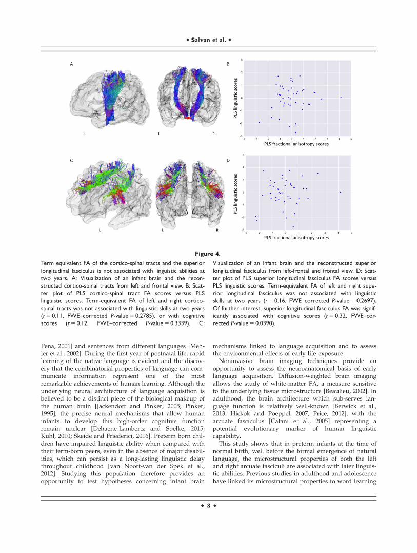
OLS 0.31 0.0275 20.06 0.4839 20.02 0.4325Ridge 0.29 0.0305 20.04 0.4556 20.01 0.4166PLS 0.36 0.0110 0.11 0.2785 0.16 0.2697
Showing rho correlation coefficient between FA at term equivalent and language scores at two years across different regression modelsand respective FWE-corrected P-values. The cross-validated PLS regression demonstrated greater nonparametric statistical significancevalues. However, no statistically significant association was found when testing the link between linguistic scores and FA of the cortico-spinal tracts or FA of the superior longitudinal fasciculus
r Salvan et al. r
r 6 r
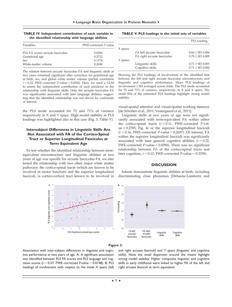
the PLS mode accounted for 72 and 71% of variancerespectively in X and Y space. High model stability in PLSloadings was highlighted also in this case (Fig. 3, Table V).
Intersubject Differences in Linguistic Skills Are
Not Associated with FA of the Cortico-Spinal
Tract or Superior Longitudinal Fasciculus at
Term Equivalent Age
To test whether the identified relationship between term-equivalent microstructure and linguistic abilities at twoyears of age was specific for arcuate fasciculus FA, we alsotested the relationship with two other major white matterpathways: the cortico-spinal tracts (which are known to beinvolved in motor function) and the superior longitudinalfasciculi, (a cortico-cortical tract known to be involved in
visual-spatial attention and visual-spatial working memory[de Schotten et al., 2011; Vestergaard et al., 2011].
Linguistic skills at two years of age were not signifi-cantly associated with term-equivalent FA within eitherthe cortico-spinal tracts (r 5 0.11, FWE–corrected P-val-ue 5 0.2785; Fig. 4) or the superior longitudinal fasciculi(r 5 0.16, FWE–corrected P-value 5 0.2697). Of interest, FAwithin the superior longitudinal fasciculi was significantlyassociated with later general cognitive abilities (r 5 0.32,FWE–corrected P-value 5 0.0390). There was no significantrelationship between FA of the cortico-spinal tracts andlater cognition; r 5 0.12, FWE–corrected P-value 5 0.3339).
DISCUSSION
Infants demonstrate linguistic abilities at birth, includingdiscriminating close phonemes [Dehaene-Lambertz and
Figure 3.
Association with inter-subject differences in linguistic and cogni-
tive performance at two years of age. A: A significant association
was identified between PLS FA scores and PLS language and cog-
nitive scores (r 5 0.37; FWE–corrected P-value 5 0.0148). B: PLS
loadings of involvement with respect to the initial X space (left
and right arcuate fasciculi) and Y space (linguistic and cognitive
skills). Note the small dispersion around the means highlight
strong model stability. Higher composite linguistic and cognitive
skills in early childhood were linked to higher FA of the left and
right arcuate fasciculi at term equivalent.
TABLE IV. Independent contribution of each variable in
the identified relationship with language abilities
Variables FWE-corrected P-value
PLS FA scores arcuate fasciculus 0.0226Gestational age 0.3712Sex 0.1174White-matter volume 0.2694
The relation between arcuate fasciculus FA and linguistic skills attwo years remained significant after correction for gestational ageat birth, sex, and global white matter volume (partial correlationr 5 0.32, FWE–corrected P-value 5 0.0230). Here, we used a GLMto assess the independent contribution of each predictor in therelationship with linguistic skills. Only the arcuate fasciculus FAwas significantly associated with later language abilities, sugges-ting that the identified relationship was not driven by confoundsof interest.
TABLE V. PLS loadings in the initial sets of variables
PLS loading
X space:FA left arcuate fasciculus 0.66 6 SD 0.006FA right arcuate fasciculus 0.78 6 SD 0.009
Y space:Linguistic skills 0.71 6 SD 0.003Cognitive skills 0.71 6 SD 0.002
Showing the PLS loadings of involvement in the identified linkbetween the left and right arcuate fasciculus microstructure, andlinguistic and cognitive performance. Mean PLS loadings ofinvolvement 6 SD averaged across folds. The PLS mode accountedfor 72 and 71% of variance, respectively, in X and Y space. Thesmall SDs of the estimated PLS loadings highlight strong modelstability
r Language Brain Organization in Preterm Neonates r
r 7 r
Pena, 2001] and sentences from different languages [Meh-ler et al., 2002]. During the first year of postnatal life, rapidlearning of the native language is evident and the discov-ery that the combinatorial properties of language can com-municate information represent one of the mostremarkable achievements of human learning. Although theunderlying neural architecture of language acquisition isbelieved to be a distinct piece of the biological makeup ofthe human brain [Jackendoff and Pinker, 2005; Pinker,1995], the precise neural mechanisms that allow humaninfants to develop this high-order cognitive functionremain unclear [Dehaene-Lambertz and Spelke, 2015;Kuhl, 2010; Skeide and Friederici, 2016]. Preterm born chil-dren have impaired linguistic ability when compared withtheir term-born peers, even in the absence of major disabil-ities, which can persist as a long-lasting linguistic delaythroughout childhood [van Noort-van der Spek et al.,2012]. Studying this population therefore provides anopportunity to test hypotheses concerning infant brain
mechanisms linked to language acquisition and to assessthe environmental effects of early life exposure.
Noninvasive brain imaging techniques provide anopportunity to assess the neuroanatomical basis of earlylanguage acquisition. Diffusion-weighted brain imagingallows the study of white-matter FA, a measure sensitiveto the underlying tissue microstructure [Beaulieu, 2002]. Inadulthood, the brain architecture which sub-serves lan-guage function is relatively well-known [Berwick et al.,2013; Hickok and Poeppel, 2007; Price, 2012], with thearcuate fasciculus [Catani et al., 2005] representing apotential evolutionary marker of human linguisticcapability.
This study shows that in preterm infants at the time ofnormal birth, well before the formal emergence of naturallanguage, the microstructural properties of both the leftand right arcuate fasciculi are associated with later linguis-tic abilities. Previous studies in adulthood and adolescencehave linked its microstructural properties to word learning
Figure 4.
Term equivalent FA of the cortico-spinal tracts and the superior
longitudinal fasciculus is not associated with linguistic abilities at
two years. A: Visualization of an infant brain and the recon-
structed cortico-spinal tracts from left and frontal view. B: Scat-
ter plot of PLS cortico-spinal tract FA scores versus PLS
linguistic scores. Term-equivalent FA of left and right cortico-
spinal tracts was not associated with linguistic skills at two years
(r 5 0.11, FWE–corrected P-value 5 0.2785), or with cognitive
scores (r 5 0.12, FWE–corrected P-value 5 0.3339). C:
Visualization of an infant brain and the reconstructed superior
longitudinal fasciculus from left-frontal and frontal view. D: Scat-
ter plot of PLS superior longitudinal fasciculus FA scores versus
PLS linguistic scores. Term-equivalent FA of left and right supe-
rior longitudinal fasciculus was not associated with linguistic
skills at two years (r 5 0.16, FWE–corrected P-value 5 0.2697).
Of further interest, superior longitudinal fasciculus FA was signif-
icantly associated with cognitive scores (r 5 0.32, FWE–cor-
rected P-value 5 0.0390).
r Salvan et al. r
r 8 r

[L�opez-Barroso et al., 2013]; the development of readingskills [Yeatman et al., 2011, 2012]; sentence comprehensionperformance [Skeide et al., 2015], and it has been shown tosupport syntactic processing of language [den Oudenet al., 2012]. We found no significant difference in termequivalent FA and tract-length between the left and rightarcuate fasciculi. We also found that symmetry in the leftand right arcuate fasciculi FA, rather than asymmetry, waslinked to later efficient linguistic abilities. This is in accor-dance with the language deficiencies reported after bothleft and right hemispheric lesions in infants [Bates andRoe, 2001] and with the observation that arcuate fasciculusvolumetric symmetry is linked to efficient auditory verballearning in adulthood [Catani et al., 2007]. Previous stud-ies in post-term infants have shown a left hemispheric lat-eralization for speech processing in the posterior part ofthe superior temporal region [Baldoli et al., 2015; Dehaene-Lambertz et al., 2002, 2006, 2010], but not for inferior fron-tal regions [Baldoli et al., 2015; Dehaene-Lambertz et al.,2006]. Indeed, left-lateralization in temporal areas increasesduring the first months of life [Baldoli et al., 2015; Peraniet al., 2011; Shultz et al., 2014]. This bilateral linkage mayalso be due to the involvement of right frontal regions,which are also activated when infants listen to speech[Dehaene-Lambertz et al., 2002, 2010], and are involved inattention, stimulus selection, and response to novelty.These are important processes for infants to comprehendsocial world requests, to communicate wants and needs,and to produce combinatorial-grammatical sentences bythe age of two years.
We may speculate on the role of the arcuate fasciculiduring the first stages of language acquisition. It providesa direct link between speech production and perceptionand an intracerebral mechanism for the ability at birth toimitate simple articulatory movements such as openingthe mouth or protusing the lips is evident from birth[Meltzoff and Moore, 1977]. Infants progressively convergetoward recognizable patterns of verbal production [Kuhland Meltzoff, 1996] and may benefit from the verbal bufferprovided by the dorsal pathway to memorize and analyzespeech segments [Dehaene-Lambertz et al., 2006]. The rela-tionship between linguistic skills at 2 years and its micro-structure confirm that the arcuate is a key element duringthe first stages of language learning.
Premature birth is associated with a long lasting signa-ture on whole-brain architecture [Ball et al., 2012, 2014;Counsell et al., 2003; Nosarti et al., 2002; Salvan et al.,2014] and later neurodevelopment [Delobel-Ayoub et al.,2009; Johnson et al., 2009; Marlow et al., 2005; Northamet al., 2012]. While the absence of a direct comparison withterm control infants limits our ability to assess the fullimpact of premature birth, we found that increasing pre-maturity at birth affects arcuate fasciculus microstructurebut, in the absence of severe neonatal brain injury, onlyminimally modulates the identified link with later linguis-tic skill. Although previous behavioral studies have
concluded that many of the language deficits in preterm-born children are more likely a result of general cognitiveproblems rather than a specific language impairment[Barre et al., 2011; Wolke and Meyer, 1999]; here, we showthat the linguistic impairment in preterm born childrenmay result from the microstructural alteration of a funda-mental brain language structure.
Of importance, our results support key specific involve-ment of the arcuate fasciculus in language acquisition aswe did not find a significant relationship with white mat-ter microstructure in either the superior longitudinal fas-ciculi or the cortico-spinal tracts. Furthermore, theidentified relationship between the arcuate fasciculus andboth language and cognition is in agreement with cogni-tive models of auditory-verbal working memory whichpredict the presence of an underlying, efficient workingmemory buffer for language learning and processing [Bad-deley, 2003; Baddeley et al., 1998]. At the age of two years,however, measures of complex linguistic skills stronglycorrelate to domain-general cognitive performance. There-fore, further investigations of specific cognitive domainsare needed in our subjects at an older age to distinguishmeasures of formal intelligence quotient, working-memory, and attention, from phonological, syntactic proc-essing, and semantics.
A potential limitation of this study is the use of FA as ameasure of underlying white-matter microstructure.Although we used high angular resolution diffusion-MRIdata and CSD based tractography to delineate the arcuatefasciculi, the observed relationship may be, at least in part,related to intersubject differences in the configuration ofcrossing fibers.
CONCLUSION
In summary, these results validate a neurolinguisticmodel in which arcuate fasciculus microstructure shortlyafter birth plays a role in early language acquisition. Wehave shown that a brain-behavior mode of covariationlinks linguistic performance in early childhood to a spe-cific structure in the infant brain, which is known to sup-port complex language function in adulthood. Themicrostructure of the arcuate fasciculus at around the timeof normal birth underpins linguistic development at 2years of age independent of the extreme environmentalinfluences caused by premature extrauterine life.
ACKNOWLEDGMENTS
The authors are grateful to the families, clinicians, andinvestigators who made the Eprime study possible, partic-ularly Denis Azzopardi, Mary Rutherford, and MaggieRedshaw. The views expressed are those of the authorsand not necessarily those of the NHS, the NIHR, or theDepartment of Health.
r Language Brain Organization in Preterm Neonates r
r 9 r

REFERENCES
Alexander MP, Naeser MA, Palumbo CL (1987): Correlations of
subcortical lesions sites and aphasia profiles. Brain 110:
961–988.Andersson JLR, Sotiropoulos SN (2016): An integrated approach
to correction for off-resonance effects and subject movement in
diffusion MR imaging. Neuroimage 125:1063–1078.Andersson JLR, Skare S, Ashburner J (2003): How to correct sus-
ceptibility distortions in spin-echo echo-planar images: Appli-
cation to diffusion tensor imaging. Neuroimage 20:870–888.Avants BB, Epstein CL, Grossman M, Gee JC (2008): Symmetric
diffeomorphic image registration with cross-correlation: Evalu-
ating automated labeling of elderly and neurodegenerative
brain. Med Image Anal 12:26–41.Baddeley A (2003): Working memory: Looking back and looking
forward. Nat Rev Neurosci 4:829–839.Baddeley A, Gathercole S, Papagno C (1998): The phonological
loop as a language learning device. Psychol Rev. 105:158–173.Baldoli C, Scola E, Della Rosa PA, Pontesilli S, Longaretti R,
Poloniato A, Scotti R, Blasi V, Cirillo S, Iadanza A, Rovelli R,
Barera G, Scifo P (2015): Maturation of preterm newborn
brains: A fMRI–DTI study of auditory processing of linguistic
stimuli and white matter development. Brain Struct Func 220:
3733–3751.Ball G, Boardman JP, Rueckert D, Aljabar P, Arichi T, Merchant
N, Gousias IS, Edwards AD, Counsell SJ (2012): The effect of
preterm birth on thalamic and cortical development. Cereb
Cortex 22:1016–1024.Ball G, Aljabar P, Zebari S, Tusor N, Arichi T, Merchant N,
Robinson EC, Ogundipe E, Rueckert D, Edwards AD, Counsell
SJ (2014): Rich-club organization of the newborn human brain.
Proc Natl Acad Sci U S A 111:7456–7461.Barre N, Morgan A, Doyle LW, Anderson PJ (2011): Language
abilities in children who were very preterm and/or very low
birth weight: A meta-analysis. J Pediatr 158:766–774.Bassi L, Ricci D, Volzone A, Allsop JM, Srinivasan L, Pai A,
Ribes C, Ramenghi LA, Mercuri E, Mosca F (2008): Probabil-
istic diffusion tractography of the optic radiations and visual
function in preterm infants at term equivalent age. Brain 131:
573–582.Bates E, Roe K (2001): A vJ Language Development in Children
with Unilateral Brain Injury. Handb Dev Cogn Neurosci 281.Bayley N (2006): Bayley Scales of Infant and Toddler Development: Tech-
nical Manual. 3rd ed. San Antonio TX: Harcourt Assessment.Beaulieu C (2002): The basis of anisotropic water diffusion in the
nervous system–a technical review. NMR Biomed 15:435–455.Benson DF, Sheremata WA, Bouchard R, Segarra JM, Price D,
Geschwind N (1973): Conduction aphasia: A clinicopathologi-
cal study. Arch Neurol 28:339.Berman JI, Glass HC, Miller SP, Mukherjee P, Ferriero DM,
Barkovich AJ, Vigneron DB, Henry RG (2009): Quantitative
fiber tracking analysis of the optic radiation correlated with
visual performance in premature newborns. Am J Neuroradiol
30:120–124.Berwick RC, Friederici AD, Chomsky N, Bolhuis JJ (2013): Evolution,
brain, and the nature of language. Trends Cogn Sci 17:89–98.Brauer J, Anwander A, Perani D, Friederici AD (2013): Dorsal and
ventral pathways in language development. Brain Lang 127:
289–295.Catani M, Jones DK, Ffytche DH (2005): Perisylvian language net-
works of the human brain. Ann Neurol 57:8–16.
Catani M, Allin MPG, Husain M, Pugliese L, Mesulam MM,
Murray RM, Jones DK (2007): Symmetries in human brain lan-
guage pathways correlate with verbal recall. Proc Natl Acad
Sci 104:17163–17168.Counsell SJ, Allsop JM, Harrison MC, Larkman DJ, Kennea NL,
Kapellou O, Cowan FM, Hajnal JV, Edwards AD, Rutherford
MA (2003): Diffusion-weighted imaging of the brain in preterm
infants with focal and diffuse white matter abnormality. Pedi-
atrics 112:1–7.Damasio H, Damasio AR (1980): The anatomical basis of conduc-
tion aphasia. Brain 103:337–350.Dehaene-Lambertz G, Pena M (2001): Electrophysiological evi-
dence for automatic phonetic processing in neonates. Neurore-
port. 14:3155–3158.Dehaene-Lambertz G, Spelke ES (2015): The Infancy of the Human
Brain. Neuron 88:93–109.Dehaene-Lambertz G, Dehaene S, Hertz-Pannier L (2002): Func-
tional neuroimaging of speech perception in infants. Science
298:2013–2015.Dehaene-Lambertz G, Hertz-Pannier L, Dubois J, M�eriaux S,
Roche A, Sigman M, Dehaene S (2006): Functional organization
of perisylvian activation during presentation of sentences in
preverbal infants. Proc Natl Acad Sci U S A 103:14240–14245.Dehaene-Lambertz G, Montavont A, Jobert A, Allirol L (2010):
Language or music, mother or Mozart? Structural and environ-
mental influences on infants’ language networks. Brain Lang.
2:53–65.Delobel-Ayoub M, Arnaud C, White-Koning M, Casper C, Pierrat
V, Garel M, Burguet A, Roze J-C, Matis J, Picaud J-C (2009):
Behavioral problems and cognitive performance at 5 years of
age after very preterm birth: the EPIPAGE Study. Pediatrics
123:1485–1492.Dubois J, Poupon C, Thirion B, Simonnet H, Kulikova S, Leroy F,
Hertz-Pannier L, Dehaene-Lambertz G (2015): Exploring the
early organization and maturation of linguistic pathways in
the human infant brain. Cereb Cortex bhv082.Forkel SJ, De Schotten MT, Dell’Acqua F, Kalra L, Murphy DGM,
Williams SCR, Catani M (2014): Anatomical predictors of apha-
sia recovery: A tractography study of bilateral perisylvian lan-
guage networks. Brain 137:2027–2039.Geschwind N (1965): Disconnexion syndromes in animals and
man. Brain 88:585.Groppo M, Ricci D, Bassi L, Merchant N, Doria V, Arichi T,
Allsop JM, Ramenghi L, Fox MJ, Cowan FM (2014): Develop-
ment of the optic radiations and visual function after prema-
ture birth. Cortex 56:30–37.Hickok G (2012): Computational neuroanatomy of speech produc-
tion. Nat Rev Neurosci 13:135–145.Hickok G, Poeppel D (2007): The cortical organization of speech
processing. Nat Rev Neurosci 8:393–402.Hotelling H (1936): Relations between two sets of variates. Biome-
trika 28:321–377.H€uppi PS, Maier SE, Peled S, Zientara GP, Barnes PD, Jolesz FA,
Volpe JJ, Huppi PS, Maier SE, Peled S, Zientara GP, Barnes PD,
Jolesz FA, Volpe JJ (1998): Microstructural development of
human newborn cerebral white matter assessed in vivo by diffu-
sion tensor magnetic resonance imaging. Pediatr Res 44:584–590.Jackendoff R, Pinker S (2005): The nature of the language faculty
and its implications for evolution of language (Reply to Fitch,
Hauser, and Chomsky). Cognition 97:211–225.Johnson S, Fawke J, Hennessy E, Rowell V, Thomas S, Wolke D,
Marlow N (2009): Neurodevelopmental disability through 11
r Salvan et al. r
r 10 r

years of age in children born before 26 weeks of gestation.
Pediatrics 124:e249–e257.Kuhl PK (2010): Brain Mechanisms in Early Language Acquisition.
Neuron 67:713–727.Kuhl P, Meltzoff A (1996): Infant vocalizations in response to
speech: Vocal imitation and developmental change. J Acoust
Soc. 100:2425–2438.Liberman AM, Mattingly IG (1985): The motor theory of speech
perception revised. Cognition 21:1–36.L�opez-Barroso D, Catani M, Ripoll�es P, Dell’Acqua F, Rodr�ıguez-
Fornells A, de Diego-Balaguer R (2013): Word learning is
mediated by the left arcuate fasciculus. Proc Natl Acad Sci U S
A 110:13168–13173.Luu TM, Vohr BR, Schneider KC, Katz KH, Tucker R, Allan WC,
Ment LR (2009): Trajectories of receptive language develop-
ment from 3 to 12 years of age for very preterm children. Pedi-
atrics 124:333–341.Luu TM, Vohr BR, Allan W, Schneider KC, Ment LR (2011): Evi-
dence for catch-up in cognition and receptive vocabulary
among adolescents born very preterm. Pediatrics 128:313–322.Makropoulos A, Gousias IS, Ledig C, Aljabar P, Serag A, Hajnal
JV, Edwards AD, Counsell SJ, Rueckert D (2014): Automatic
whole brain MRI segmentation of the developing neonatal
brain. IEEE Trans Med Imaging 33:1818–1831.Marlow N, Wolke D, Bracewell MA, Samara M (2005): Neurologic
and developmental disability at six years of age after
extremely preterm birth. N Engl J Med 352:9–19.Mehler J, Jusczyk P, Lambertz G, Halsted N, Bertoncini J, Amiel-
Tison C (2002): A precursor of language acquisition in young
infants. Psycholinguist Crit Concepts Psychol 4:25.Meltzoff A, Moore M (1977): Imitation of facial and manual ges-
tures by human neonates. Science. 198:75–78.van Noort-van der Spek IL, Franken M-CJP, Weisglas-Kuperus N
(2012): Language Functions in Preterm-Born Children: A Sys-
tematic Review and Meta-analysis. Pediatrics 129:745–754.Northam GB, Li�egeois F, Tournier J-D, Croft LJ, Johns PN, Chong
WK, Wyatt JS, Baldeweg T (2012): Interhemispheric temporal
lobe connectivity predicts language impairment in adolescents
born preterm. Brain 135:3781–3798.Nosarti C, Al-Asady MHS, Frangou S, Stewart AL, Rifkin L,
Murray RM (2002): Adolescents who were born very preterm
have decreased brain volumes. Brain 125:1616–1623.den Ouden D-B, Saur D, Mader W, Schelter B, Lukic S, Wali E,
Timmer J, Thompson CK (2012): Network modulation during
complex syntactic processing. Neuroimage 59:815–823.Paldino M, Hedges K, Golriz F (2016): The Arcuate Fasciculus and
Language Development in a Cohort of Pediatric Patients with
Malformations of Cortical Development. Am J. 37:169–175.Pedregosa F, Varoquaux G, Gramfort A, Michel V, Thirion B, Grisel O,
Blondel M, Prettenhofer P, Weiss R, Dubourg V (2011): Scikit-learn:
Machine learning in Python. J Mach Learn Res 12:2825–2830.Perani D, Saccuman MC, Scifo P, Anwander A, Spada D, Baldoli C,
Poloniato A, Lohmann G, Friederici AD (2011): Neural language
networks at birth. Proc Natl Acad Sci U S A 108:16056–16061.Petrides M, Pandya DN (1984): Projections to the frontal cortex
from the posterior parietal region in the rhesus monkey.
J Comp Neurol 228:105–116.Pinker S (1995): The language instinct: The new science of lan-
guage and mind, Vol. 7529. UK: Penguin.Price CJ (2012): A review and synthesis of the first 20years of PET
and fMRI studies of heard speech, spoken language and read-
ing. Neuroimage 62:816–847.
Rilling JK, Glasser MF, Preuss TM, Ma X, Zhao T, Hu X, BehrensTEJ (2008): The evolution of the arcuate fasciculus revealedwith comparative DTI. Nat Neurosci 11:426–428.
Rilling JK, Glasser MF, Jbabdi S, Andersson J, Preuss TM (2011):Continuity, divergence, and the evolution of brain languagepathways. Front Evol Neurosci 3:
Rose SE, Hatzigeorgiou X, Strudwick MW, Durbridge G, DaviesPSW, Colditz PB (2008): Altered white matter diffusion anisot-ropy in normal and preterm infants at term-equivalent age.Magn Reson Med 60:761–767.
Rosipal R, Kr€amer N (2006): Overview and recent advances inpartial least squares. In: Saunders C, Grobelnik M, Gunn S,Shawe-Taylor J, editors. Subspace, Latent Structure and FeatureSelection Techniques. New York: Springer. pp 34–51.
Salvan P, Froudist Walsh S, Allin MPG, Walshe M, Murray RM,Bhattacharyya S, Mcguire PK, Williams SCR, Nosarti C (2014):Road work on memory lane-Functional and structural altera-tions to the learning and memory circuit in adults born verypreterm. Neuroimage. 114:152–161.
Schmahmann JD, Pandya DN, Wang R, Dai G, D’Arceuil HE, deCrespigny AJ, Wedeen VJ (2007): Association fibre pathwaysof the brain: Parallel observations from diffusion spectrumimaging and autoradiography. Brain 130:630–653.
de Schotten MT, Dell’Acqua F, Forkel SJ, Simmons A, Vergani F,Murphy DGM, Catani M (2011): A lateralized brain networkfor visuospatial attention. Nat Neurosci 14:1245–1246.
de Schotten MT, Dell’Acqua F, Valabregue R, Catani M (2012):Monkey to human comparative anatomy of the frontal lobeassociation tracts. Cortex 48:82–96.
Shultz S, Vouloumanos A, Bennett R (2014): Neural specializationfor speech in the first months of life. Developmental. 17:766–774.
Skeide M, Friederici A (2016): The ontogeny of the cortical lan-guage network. Nat Rev Neurosci. 17:323–332.
Skeide MA, Brauer J, Friederici AD (2015): Brain functional andstructural predictors of language performance. Cereb Cortexbhv042.
Smith SM, Jenkinson M, Woolrich MW, Beckmann CF, BehrensTEJ, Johansen-Berg H, Bannister PR, De Luca M, Drobnjak I,Flitney DE (2004): Advances in functional and structural MRimage analysis and implementation as FSL. Neuroimage 23:S208–S219.
Smith RE, Tournier J-D, Calamante F, Connelly A (2012): Anatom-ically-constrained tractography: Improved diffusion MRIstreamlines tractography through effective use of anatomicalinformation. Neuroimage 62:1924–1938.
Smith RE, Tournier J-D, Calamante F, Connelly A (2015): SIFT2:Enabling dense quantitative assessment of brain white matterconnectivity using streamlines tractography. Neuroimage 119:338–351.
Toulmin H, Beckmann CF, O’Muircheartaigh J, Ball G, NongenaP, Makropoulos A, Ederies A, Counsell SJ, Kennea N, ArichiT, Tusor N, Rutherford MA, Azzopardi D, Gonzalez-Cinca N,Hajnal JV, Edwards AD (2015): Specialization and integrationof functional thalamocortical connectivity in the human infant.Proc Natl Acad Sci U S A 201422638.
Tournier J-D, Calamante F, Gadian DG, Connelly A (2004): Directestimation of the fiber orientation density function fromdiffusion-weighted MRI data using spherical deconvolution.Neuroimage 23:1176–1185.
Tournier J-D, Calamante F, Connelly A (2007): Robust determina-tion of the fibre orientation distribution in diffusion MRI: Non-negativity constrained super-resolved spherical deconvolution.Neuroimage 35:1459–1472.
r Language Brain Organization in Preterm Neonates r
r 11 r

Tournier JD, Calamante F, Connelly A (2012): MRtrix: Diffusiontractography in crossing fiber regions. Int J Imaging Syst Tech-nol 22:53–66.
Vestergaard M, Madsen KS, Baar�e WFC, Skimminge A, EjersboLR, Ramsøy TZ, Gerlach C, Akeson P, Paulson OB, JerniganTL (2011): White Matter Microstructure in Superior Longitudi-nal Fasciculus Associated with Spatial Working Memory Per-formance in Children. J Cogn Neurosci 23:2135–2146.
Wegelin JA (2000): “A Survey of Partial Least Squares (PLS) Meth-ods, with Emphasis on the Two-Block Case,” technical report,Dept. of Statistics, Univ. of Washington.
Wilson B, Sundaram S, Huq A, Jeong J (2011): Abnormal lan-guage pathway in children with Angelman syndrome. Pediat-ric. 44:350–356.
Winkler AMA, Ridgway GRG, Webster MA, Smith SSM, NicholsTE (2014): Permutation inference for the general linear model.Neuroimage 92:381–397.
Wolke D, Meyer R (1999): Cognitive status, language attainment,and prereading skills of 6-year-old very preterm children andtheir peers: The Bavarian Longitudinal Study. Dev Med ChildNeurol 41:94–109.
Yeatman JD, Dougherty RF, Rykhlevskaia E, Sherbondy AJ,Deutsch GK, Wandell BA, Ben-Shachar M (2011): Anatomicalproperties of the arcuate fasciculus predict phonological andreading skills in children. J Cogn Neurosci 23:3304–3317.
Yeatman JD, Dougherty RF, Ben-Shachar M, Wandell BA (2012):Development of white matter and reading skills. Proc NatlAcad Sci U S A 109:E3045–E3053.
r Salvan et al. r
r 12 r