knee disorders final - handout disorders - 2.pdfspecial tests • divide knee special tests into...
TRANSCRIPT

1
Bryant Walrod, MDAssistant Professor – Clinical
Department of Family MedicineDivision of Sports Medicine
The Ohio State University Wexner Medical Center
A Standardized Approach to the Knee Examination
Basic ApproachBasic Approach
• Inspection
• Palpation
• Strength Testing
• Range of Motion
• Special Tests

2
Knee ExaminationKnee Examination• It is important to begin with a standardized
approach to the knee exam so as to not miss anything.
• One also needs to ensure adequate exposure
‒ Have the patient get into shorts to fully expose the knee.
• Examine sitting up and supine
• Compare with contralateral side
InspectionInspection• Inspect patient sitting with
knees bent.
‒ Observe for an obvious dislocation of the patella or knee joint.
• Evaluate for an effusion versus a bursitis.
‒ It is also important to distinguish between intra-articular or extra articular swelling.

3
InspectionInspection• Observe also for erythema,
induration and rashes.
• Inspect for scars
‒ may indicate a previous surgery which will influence your physical exam.
• Look also for muscle atrophy of the quadriceps, hamstrings and gastrocnemius.
Functional ObservationFunctional Observation
• Observe for a J sign
• Picture of j sign and

4
Functional ObservationFunctional Observation• Genu varus
• Genu valgus
• Genu recurvatum
• Femoral anterversion
ObservationObservation
• Observe ambulation
‒ Pronation
‒ Pes planus
‒ Antalgic gait
‒ External rotation of leg

5
ObservationObservation
• Next have the patient stand on one leg.
• Observe for valgus deviation of the knee with single leg squatting
Palpation Palpation • Start with the patient sitting on the table to
knees bent to 90o.• Palpate initially for warmth• Then palpate specific structures:
‒ Patella tendon origin and its insertion on tibial tuberosity
‒ Quadriceps tendon‒ Pes anserine and Iliotibial bursal areas•Gerdy’s tubercle
‒ Medial and lateral joint line: •Assess for joint line tenderness

6
Palpation Palpation ‒ Medial and lateral femoral condyle
‒ Medial and lateral tibial plateau
‒ Proximal fibula and tibial/fibular articulation
‒ Patella and medial and lateral patella facet
‒ Medial and lateral patellofemoral retinaculum
‒ Medial and lateral collateral ligaments
‒ Popliteus
‒ Assess for crepitus with knee extension
PalpationPalpation• In the supine position:
• Palpate for intra-articular versus extra-articular effusion:
‒ Milking test
‒ Fluctuation test
‒ Ballotment

7
Strength Testing Strength Testing
• Strength Testing:
• With the patient still sitting and knee joint flexed to 90°perform isometric strength testing for leg extension and flexion.
• Grade this on a 5/5 scale.
Range of Motion Range of Motion
• Have the patient move to a supine and relaxed position:
‒ Active Range of motion: -3 to 135-140°
‒ Passive Range of motion: -3 to 135-140°
8
Special TestsSpecial Tests• There are a multitude of special tests with
a variety of different names.
• Endeavor to understand the concept of the tests that you are performing and the specific structure that you are testing instead of memorizing the name of the person that first described the test.
Special TestsSpecial Tests• Divide knee special tests into three
subsections
‒ Patellofemoral articulation/extensor mechanism
‒ Mensical and chondral evaluation
‒ Knee instability
•Medial and Lateral
•Anterior and Posterior

9
Patellofemoral Articulation/Extensor Mechanism
Patellofemoral Articulation/Extensor Mechanism
• Measuring the Q angle
‒ Measure q angle at 30° of flexion to move the patella into the proximal portion of the trochlea
• Males ≤ 10°
• Females ≤ 15°
Patellofemoral Articulation/Extensor Mechanism
Patellofemoral Articulation/Extensor Mechanism
• Then have the patient move to full extension
‒ Patella quadrant glide
‒ Patella tilt
‒ Patellar subluxation/apprehension test
‒ Assess for pain with patella palpation, compression test and also with quad activation

10
Meniscal and ChondralEvaluation
Meniscal and ChondralEvaluation
• Pain at the joint line with palpation and with passive flexion can be positive for a meniscal injury
• McMurray’s test ‒ Attempt to grind the torn meniscus with compression
and rotation‒ Assess for pain at the joint line of the affected
meniscus and also a popping sensation• Medial meniscus:
‒ The knee is flexed and a varus stress is applied while the leg is externally rotated. During extension, the patient will feel pain at the medial joint line and snapping or popping will be appreciated at the medial joint line.
• Lateral meniscus:‒ The knee is flexed and a valgus stress is applied while
the leg is internally rotated. During extension, the patient will feel pain at the lateral joint line and snapping or popping will be appreciated at the lateral joint line.
Meniscal and ChondralEvaluation
Meniscal and ChondralEvaluation
• Apley Test:‒ Patient is prone with the knee flexed to 90o. The knee
is then pushed anteriorly and twisted.‒ Then the knee is pulled while twisting it. ‒ If pain is felt with the compression portion of this and
not with the distraction portion, it is considered positive.• If there is no difference with compression and
distraction, consider an articular chondral lesion over a fibrocartilaginous menisical lesion
• Thessaly’s Test: ‒ Have the patient stand on one leg. (you may need to
assist with balance) ‒ Ask the patient to flex to 20o and then twist on the
knee. ‒ Pain at the joint line or a catching/popping sensation is
considered a positive test.

11
Knee InstabilityKnee Instability
• Anterior and posterior tibial/femoral
• Medial and lateral tibial/femoral
Medial and lateral Knee Instability
Medial and lateral Knee Instability
• Stabilize the knee joint with the assistance of your hip and arm:‒ Apply a varus stress the knee at 0o and 30o to
assess for laxity of the Lateral Collateral Ligament (LCL)
‒ Apply a valgus stress the knee at 0o and 30°to assess for laxity of the Medial Collateral Ligament (MCL)
‒ It is important to stress the knee in both full extension and also 30oof flexion. • At full extension, the cruciate ligaments, the
posterior capsule and the condyles can restrict motion and can give a false negative

12
Medial and lateral Knee Instability
Medial and lateral Knee Instability
‒ When performing the test, the examiner takes note of increased laxity when stressing the ligament:
• 0-5mm: grade 1
• 5-10mm with hard end point: grade 2
• >10 mm with soft endpoint: grade 3.
• Pain does not make this test positive but rather laxity.
• One may note pain at the contralateral joint line from which you are stressing for meniscal injury or advanced arthritis.
Anterior Instability Anterior Instability
• Lachman Test:‒ With the patient supine and the heel on the
examination table, flex the knee to 30o and slightly externally rotate the leg.
‒ Ask the patient to relax. Stabilize the distal femur with one hand and grab the proximal tibia with the other.
‒ Apply an anterior force to the tibia relative to the femur. • The heel should not rise up off of the table.
‒ The examiner will assess for a firm endpoint or a feeling of a rubber band snapping on itself.

13
Anterior Instability Anterior Instability
‒ Lachman can be graded as: • Negative: no increased translation• Grade 1: 0-5mm increased translation, • Grade 2: 5-10 mm increased translation• Grade 3: greater than 10 mm increased
translation or no endpoint noted. ‒ This test can be modified if the patient has
large thighs or if the examiner has small hands by placing a knee under the distal hamstring to move the knee into 30o of flexion
‒ Sensitivity = 96% for diagnosing complete tears
Anterior Instability Anterior Instability • Anterior drawer test:
‒ Place the patient supine and flex the hip to 45o and the knee to 90o.
‒ Stabilize the tibia by gently sitting on the foot. ‒ The tibia should be in neutral rotation and the
hamstrings should be relaxed. ‒ It is important to start with the knee in neutral position.
• A PCL tear may give a false positive anterior drawer test if the examiner does not start in neutral.
‒ The tibia is then stressed to move anterior to the femur. Anterior drawer test is graded similar to the Lachman test:• Negative: no increased translation• Grade 1: 0-5mm increased translation• Grade 2: 5-10 mm increased translation• Grade 3: greater than 10 mm increased translation
or no endpoint noted.

14
Anterior Instability Anterior Instability • Pivot shift test:
‒ Place the patient in the supine position and ask them to relax.
‒ The ankle is grasped with one hand and the proximal tibia with the other while the knee is held in flexion.
‒ Internally rotate the knee and apply a valgus stress as the knee is slowly extended.
‒ A positive test is posterior sliding (reduction) of the lateral tibia plateau at about 30o of flexion. • This is secondary to the iliotibial band
which has a posterior force on the knee in flexion and an anterior force on the knee in extension. • An ACL deficient knee is anterior laterally
unstable.
Posterior Instability Posterior Instability • Posterior drawer test:
‒ Place the patient in the supine position with the hip flexed to 45o and the knee flexed to 90°
‒ The tibia remains in neutral rotation.
‒ Stabilize the lower extremity by sitting on the patient’s foot.
‒ Attempt to translate the tibia posteriorly relative to the femur.
‒ A positive test is increased posterior translation.

15
Posterior Instability Posterior Instability • Sag sign:
‒ Patient is supine, the hips are flexed to 45o and the knees flexed to 90°
‒ Observe for posterior translation of the tibia tuberosity of the affected knee versus the contralateral knee.
‒ This test can be accentuated with quad activation.
Posterior Instability Posterior Instability • Gravity sign:
‒ Patient is supine with both the hips and knees flexed to 90o.
‒ Rest one hand under the knees and one hand gently supports the feet.
‒ Observe for a posterior translation of the tibia relative the contralateral side.
‒ An important point to also consider when evaluating a PCL injury is varus and valgus stress testing of the knee:• Increased laxity at both 0 and 30o may
indicated a collateral ligament injury with a concomitant PCL/ACL injury.

16
Clinton Hartz, MDAssistant Professor – Clinical
Department of Family MedicineDivision of Sports Medicine
The Ohio State University Wexner Medical Center
Common KneeDisorders
ObjectivesObjectives
• Discuss several most common knee disorders seen in primary care clinic.
• Give common presenting symptoms and exam findings
• Review some the recent literature on treatment guidelines for several knee disorders

17
Case #1Case #1• 32 yo female who is a
recreational runner presents with left knee pain. Pain worse with getting up from sitting position, going up and down steps. Denies any trauma or instability. Has occasional “clicking” and trace swelling.
Patellofemoral Syndrome/Chondromalacia PatellaePatellofemoral Syndrome/Chondromalacia Patellae
• Pain Location
• Non-specific or vague (medial, lateral or infra patellar)
• Aggravated by loading patellofemoral joint (PFJ)
• Going up and down steps
• Rising from sitting position

18
Patellofemoral SyndromePatellofemoral Syndrome• Physical Exam
• Positive patellar compression testing
• May have trace effusion
• Crepitus/clicking under patella with knee flexion
• Treatment
• Responds well to physical therapy
• Home exercises
• Anti-inflammatory medications
• Rarely need injections
Patellofemoral SyndromePatellofemoral Syndrome• Look beyond the knee for contributing
factors• Pes planus
• Weak hip muscles
• Glut medius • Glut minimus
• Valgus deviation
• Single leg squatting• Not balance

19
Patellofemoral SyndromePatellofemoral Syndrome
• Ferber et al. 2015 Strengthening of the Hip and Core Versus Knee Muscles for the Treatment of Patellofemoral Pain: A Multicenter Randomized Controlled Trial.
• HIP and KNEE rehabilitation protocols produced improvements (6 weeks).
• Although outcomes were similar, the HIP protocol resulted in earlier resolution of pain and greater overall gains in strength
Patellar TendinopathyPatellar Tendinopathy
• aka “Patellar Tendonitis”
• Pain location
• Inferior pole of patella most common
• Worse with jumping activities (i.e. Basketball,volleyball, climbing stairs)
• “Jumper’s Knee”

20
Patellar TendinopathyPatellar Tendinopathy• Physical Exam
• May see quadriceps wasting
• Tender inferior pole patella or distal tendon
• Unusual to see any swelling
Patellar TendinopathyPatellar Tendinopathy
• Treatment
• NSAID’s
• Formal PT to work on core, hips and quadriceps
• Failed conservative treatment can consider referral for prolotherapy, PRP (experimental) or ultrasound guided tenodesis.

21
Fat Pad ImpingementFat Pad Impingement• Hyperextension injury common
• Pain Location
• Pain inferior pole patella
• Exacerbated by stairs, prolonged standing, knee extension
Fat Pad ImpingementFat Pad Impingement
• Physical Exam
• Tender in fat pad region (inferior pole patella, deep to tendon)
• “Puffy” appearance
• Active extension may be painful

22
Fat Pad Impingement Fat Pad Impingement • Treatment
• Formal PT to work on muscle training
• Improve lower limb biomechanics
Patellar DislocationPatellar Dislocation
• Traumatic or Atraumatic
• Twisting or jumping with sensation of “popping out,” pain and immediate swelling
• Reduce spontaneously with knee extension and lateral pressure over patella

23
Patellar DislocationPatellar Dislocation• Physical Exam
• Knee effusion
• Positive lateral patellar apprehension
• Tenderness over medial border patella
• X-ray’s should be performed
Patellar DislocationPatellar Dislocation• Treatment
• Depend on presentation
• Atraumatic can be treated conservatively
–physical therapy, bracing as needed
• Acute Traumatic
• Conservative vs surgical
• Physical Therapy
–goal to reduce recurrence
–Lengthy rehab ~12 weeks

24
Patellar DislocationPatellar Dislocation• Bitar et al. 2012 Traumatic Patellar
Dislocation Nonoperative Treatment Compared With MPFL Reconstruction Using Patellar Tendon
• Treatment with MPFL reconstruction using the patellar tendon produced better results, based on the analyses of posttreatment recurrences and the better final results of the Kujala questionnaire after a minimum follow-up period of 2 years.
Case #2Case #2• A 21 yo male soccer player presents to
your office for evaluation of knee pain. He was trying to make a lateral cut to the ball when he felt a “pop” in his knee with significant pain. He developed swelling almost immediately, with some associated instability. On exam, he has notable knee effusion, a soft end point on Lachman testing, and opens with valgus stress. What is the diagnosis?

25
Anterior Cruciate Ligament InjuryAnterior Cruciate Ligament Injury
• Complete vs Partial tear
• Pain Location
• global
•most complete very painful initially
• unable to continue activity
• immediate swelling (hemarthrosis)
ACL InjuryACL Injury
• Physical Exam
• Restricted movement (esp loss extension)
• Lateral/medial joint line tenderness
• Positive Lachman (Compare to other side)
• Xray needed

26
ACL InjuryACL Injury• Treatment
• Conservative vs Surgical
• Age of patient
• Degree of instability
• associated injury (ie. MCL, meniscus)
• Type sports/activity level/occupation
• Physical therapy
ACL InjuryACL Injury• Frobell et al. BMJ 2013 Treatment for acute
anterior cruciate ligament tear: five year outcome of randomised trial• A strategy of rehabilitation plus early ACL
reconstruction did not provide better results at five years than a strategy of initial rehabilitation with the option of having a later ACL reconstruction. • Results did not differ between knees
surgically reconstructed early or late and those treated with rehabilitation alone.

27
Medial Collateral Ligament StrainMedial Collateral Ligament Strain
• Can be associated with ACL injury or alone
• Caused by valgus stress
• Pain Location
• mostly medial
• Soft tissue swelling
• Pain with flexion knee
MCL StrainMCL Strain• Physical Exam
• Valgus stress pain and instability (joint opening)
• Valgus stress at 0o and 30°• Compare with contra-lateral side• Feel for end point• May get laxity at 0o with a torn PCL/ACL• May also try Anterior drawer test with
external rotation to test the MCL

28
MCL StrainMCL Strain• 3 Types (I, II, III) depending on severity of joint line
opening
• Grade I: No increased instability, firm endpoint, pain with palpation at MCL, negativemeniscus tests (<5mm instability)
• Grade II: Increased instability, but end point isstill present, pain with provocative testing (6-10mm)
• Grade III: Gross laxity with mushy endpoint orno endpoint, (bony block)
• May not have pain as there are no fibers left tostress (>10mm)
MCL StrainMCL Strain• Treatment• Type I- RICE, NSAIDs, home exercises and
consider formal PT• Type II- relative rest (if pain with walking
consider crutches), early ROM and HEP, formal PT for ~8weeks, and hinged knee brace as needed
• Type III- same as type II but depending on activity/occupation may need surgery. • Unusual to have isolated Type III without ACL
or meniscal injury.

29
Case #3Case #3• A 54yo male presents to your office for
evaluation of right knee pain that has been getting progressively worse for the last 2-3 years. Denies any trauma, but has noticed some instability, and “popping” with going up and down steps. He also states his knee feels “swollen.” X-rays are pending. What’s your likely diagnosis?
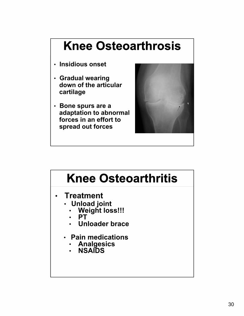
Knee OsteoarthrosisKnee Osteoarthrosis• Pain location
• joint line, most typically medial
• Night mostly
30
Knee OsteoarthrosisKnee Osteoarthrosis• Insidious onset
• Gradual wearingdown of the articularcartilage
• Bone spurs are aadaptation to abnormalforces in an effort tospread out forces
Knee OsteoarthritisKnee Osteoarthritis• Treatment
• Unload joint• Weight loss!!!• PT• Unloader brace
• Pain medications• Analgesics• NSAIDS

31
Knee OsetoarthrosisKnee Osetoarthrosis• Injections• Corticosteroids• Viscosupplementation• Pro-inflammatory • PRP
• Surgery • Total or hemiarthroplasty• No meniscectomy for partial tearing
or degenerative changes
Knee OsteoarthritisKnee Osteoarthritis• AAOS 2013
• Self-management programs, strengthening, low-impact aerobic exercises, and neuromuscular education and engage in physical activity consistent with national guidelines
• Weight loss for patients with symptomatic OA of the knee and a body mass index ≥25
• Unable to recommend for or against the use of a valgus-directing force brace (medial compartment unloader) for patients with symptomatic OA of the knee.

32
Knee OsteoarthritisKnee Osteoarthritis• We cannot recommend using glucosamine and
chondroitin for patients with symptomatic OA of the knee
• We recommend non-steroidal anti-inflammatory drugs (oral or topical) or tramadol for patients with symptomatic OA of the knee.
Knee OsteoarthritisKnee Osteoarthritis• We are unable to recommend for or against
the use of acetaminophen, opioids, or pain patches for patients with symptomatic OA of the knee
• We are unable to recommend for or against the use of intra-articular corticosteroids for patients with symptomatic OA of the knee.
• We cannot recommend using HA for patients with symptomatic OA of the knee
• We cannot recommend performing arthroscopy with lavage and/or débridement in patients with a primary diagnosis of symptomatic OA of the knee.

33
Knee OsteoarthritisKnee Osteoarthritis• We are unable to recommend for or against
arthroscopic partial meniscectomy in patients with OA of the knee with a torn meniscus.
• The practitioner might perform a valgus-producing proximal tibial osteotomy in patients with symptomatic medial compartment OA of the knee.
Knee OsteoarthrosisKnee Osteoarthrosis• R. Bannuru et al. 2014 Relative efficacy of
hyaluronic acid in comparison with NSAIDs for knee osteoarthritis: A systematic review and meta-analysis meta-analysis
• Suggests that IAHA is not significantly different from continuous oral NSAIDs at 4 and 12 weeks.
• Only a short follow-up duration. • Given the favorable safety profile of IAHA over
NSAIDs, this result suggests that IAHA might be a viable alternative to NSAIDs for knee OA, especially for older patients at greater risk for systemic adverse events

34
Meniscal TearsMeniscal Tears• Can be medial or lateral
• Typically a twisting injury
• Swelling occurs but usually 4-8 hours later • secondary to synovial effusion
• Occasional instability
• True locking (Mechanical symptoms)• Bucket handle flap tear• Rare• Inability to fully extend
Meniscal TearsMeniscal Tears• Physical Exam
• Pain at joint line
• Mechanical symptoms
• McMurray’s
• Pain with hyperextension
• Pain with hyper flexion
• Effusion

35
Mensical TearsMensical Tears• Physical Exam
• McMurray’s• May feel a click• Sensitivity: 26-58%• Specificity: 93-94%
• Apley’s Grind• Distraction: collateral ligament injury
• Thessaly’s• More accurate than McMurray • Probability of injury if positive: 81% )• Probability of injury if negative: 1%
• Caution with concomitant osteoarthrosis
Meniscal TearsMeniscal Tears• Treatment• Typically conservative• Rehabilitation for a month • Caution if mechanical symptoms• Locking• True Buckling
• Surgical intervention• Arthroscopy• Partial menisectomy• Meniscus Repair

36
Meniscal TearsMeniscal Tears• Herlin et al: 2013: • 96 patients: No difference in pain after
arthoscopic menisectomy plus therapy versus therapy alone – 5 years
• Yin 2013:• 102 patients: No significant differences
between arthroscopic meniscectomy and nonoperative mgt with strengthening exercises in terms of relief in knee pain, improved knee function, or increased satisfaction in patients at 2 years
Meniscal TearsMeniscal Tears• R. Sihvonen et al 2013 Arthroscopic Partial
Meniscectomy versus Sham Surgery for a Degenerative Meniscal Tear.
• 129 patients without knee osteoarthritis but with symptoms of a degenerative medial meniscus tear
• The outcomes after arthroscopic partial meniscectomy were no better than those after a sham surgical procedure.

37
Pes Anserine BursitisPes Anserine Bursitis• Pain Location• Bursa located under sartorius, gracilis,
semitendinosus tendons• Anteromedial knee pain, over bursa,
possible swelling• Treatment• Activity modification, anti-inflammatories
PT• Consider corticosteroid injection
Iliotibial band syndrome/bursitisIliotibial band syndrome/bursitis
• Pain Location
• Lateral knee pain, common in long-distance runners, cyclists
• Precipitated by downhill running

38
Iliotibial band syndrome/bursitis
Iliotibial band syndrome/bursitis
• Pain Proximal to lateral joint line
• Ober’s test positive
• Treatment
• Activity modification, anti-inflammatories, PT. Consider foot orthotics. Possible injection
ConclusionConclusion• Knee pain is common patient complaint
• History and physical and mechanism of injury very important in developing diagnosis
• Mechanical symptoms (locking, catching) or instability on exam requires further work up (MRI and/or referral)
• Many knee disorders can be treated conservatively with good physical therapy, short term NSAIDs and possible injection.