knowing me, knowing you: can a knowledge of risk factors for
TRANSCRIPT

Journal of Alzheimer’s Disease 25 (2011) 395–415DOI 10.3233/JAD-2011-110026IOS Press
395
Review
Knowing Me, Knowing You: Can aKnowledge of Risk Factors for Alzheimer’sDisease Prove Useful in Understanding thePathogenesis of Parkinson’s Disease?
Greg T. Sutherlanda,∗, Gerhard A. Siebertc, Jillian J. Krila,b and George D. Mellickc,d,e
aDiscipline of Pathology, Sydney Medical School, University of Sydney, Sydney, NSW, AustraliabDiscipline of Medicine, Sydney Medical School, University of Sydney, Sydney, NSW, AustraliacEskitis Institute for Cell and Molecular Therapies, School of Biomolecular and Physical Sciences,Griffith University, Brisbane, QLD, AustraliadDepartment of Neurology, Princess Alexandra Hospital, Brisbane, QLD, AustraliaeDepartment of Neurology, Royal Brisbane and Women’s Hospital, Brisbane, QLD, Australia
Accepted 18 February 2011
Abstract. Alzheimer’s disease (AD) and Parkinson’s disease (PD) are the two most common neurodegenerative disorders. Whysome individuals develop one disease rather than the other is not clear. Association studies with a case-control design are thetime-honored approach to identifying risk factors. Extensive association studies have been carried out in both diseases creatinga large knowledge database, however, reproducible risk factors remain rare. This general lack of knowledge of pathogenesisprevents us from reducing the worldwide burden of these diseases. Case-control studies are reductionist paradigms that assume,for maximum power, that the two populations being compared are exclusive and homogenous. The common occurrence ofincidental AD and PD-type pathology combined with ‘intermediate phenotypes’ such as dementia with Lewy bodies suggestthat aging itself, AD, and PD are part of a complex continuum characterized by variable amounts of amyloid-�, tau, and�-synuclein pathology. This heterogeneity may be a contributor to the lack of reproducibility in association studies to date.Here, we speculate on alternative experimental approaches to the case-control paradigm and consider how the association-studyliterature for AD and PD might be re-interpreted in terms of a disease spectrum.
Keywords: Alzheimer’s disease, association studies, dementia, neuropathology, Parkinson’s disease
INTRODUCTION
An ever-increasing world population, with a grow-ing proportion of elderly, poses an immense challenge
∗Correspondence to: Dr Greg T. Sutherland, Discipline of Pathol-ogy, Sydney Medical School, University of Sydney, Sydney, NSW2006, Australia. Tel.: +61 2 9036 7233; Fax: +61 2 9351 3429;E-mail: [email protected].
from the rising socio-economical burden of neurode-generative disease. An epidemic seems unavoidableunless therapies can be found to prevent, delay, orreverse these conditions. The two most common neu-rodegenerative disorders are Alzheimer’s disease (AD)and Parkinson’s disease (PD), both of which are thesubject of major research efforts, consistent with theirsocial and economic importance. Monogenic formsof these diseases are known and their identification
ISSN 1387-2877/11/$27.50 © 2011 – IOS Press and the authors. All rights reserved

396 G.T. Sutherland et al. / Alzheimer’s and Parkinson’s Disease Risk Factors
has been invaluable for elucidating aspects of thepathogenic mechanisms. However, such forms are rare,and the majority of PD and AD cases occur spo-radically, without familial or geographical clustering.These common forms are referred to as “idiopathic”or literally, of unknown pathogenesis. In the absenceof known causative factors, it is widely assumed thatthe majority of sporadic cases are caused by a com-plex interaction between common genetic variants andenvironmental factors on a background of aging [1, 2].
Risk factors are often assessed by association stud-ies, in a case-control setting. In more recent years,technological advances have greatly enhanced thebreadth of the questions that can be asked with globalanalyses such as genome wide association and tran-scriptomic studies applied to both diseases. However,in general, these genome-scale platforms have onlyconfirmed known risk factors and not identified thenovel targets needed to reduce the burden of thesediseases [3].
The underlying premise for this review is a con-sideration of the potential reasons behind this lack ofsuccess. Moreover, it is to discuss whether the com-bined research knowledge within AD and PD could beharnessed to gain greater research traction for eitherdisease. Importantly it builds on the previous work ofcolleagues who have considered AD and PD patholo-gies within a wider neurodegenerative spectrum [4–6].
PART I: EXCLUSIVITY ANDHOMOGENEITY
The case-control study is a reductionist paradigmthat assumes the two populations under considera-tion are exclusive and homogenous. However, a lackof homogeneity is recognized in both AD and PD.In PD, for example, there is a common divisionbetween tremor-dominant and bradykinetic (PIGD)sub-phenotypes [7], and these subtypes have differentpatterns of pathology [8, 9]. This suggests that differentpathophysiological mechanisms are operating [8], animportant consideration for the design of associationstudies. However such variation is often (pragmati-cally) dismissed to maximize statistical power. Lessappreciated is the possible lack of exclusivity betweencases and the “unaffected controls” used in the com-parison groups.
AD phenotype
AD manifests clinically as a loss of cognitive func-tions including memory, language, visuoconstructive,
and executive function. AD is the most common neu-rodegenerative disease, with a mean age at diseaseonset during the eighth decade. AD accounts forapproximately 60% of dementia cases [10, 11] andaffects approximately 1% of the population in Westerncountries [10, 12, 13].
The neuropathology of AD is characterized by twopathognomonic entities: the intraneuronal neurofibril-lary tangle (NFT) and the extracellular neuritic plaque.NFTs are chiefly composed of fibrillar forms of ahyperphosphorylated protein called microtubule asso-ciated protein tau (MAPT or tau), while plaques arepredominantly composed of fibrillated short peptidescollectively termed amyloid-� (A�). A staging schemefor the neuropathological progression of AD based onthe density and distribution of NFTs has been proposedby Braak and Braak (the ‘Braak staging scheme’) [14]and this has been incorporated into current diagnos-tic criteria [15]. For most individuals, there is a goodcorrelation between Braak stage (i.e., the spread ofNFT pathology from the medial temporal lobe, throughthe temporal and frontal neocortices, and into the pri-mary cortices) and the probability of dementia [14,16]. However in the hippocampus neuronal loss mayactually exceed NFT formation [17].
Neuritic plaques (‘plaques’) are an important partof the diagnostic criteria for AD, but the extracellularamyloid load does not correlate as well with dura-tion and severity of AD as the NFT count [18, 19].Nevertheless, the major working hypothesis for ADpathogenesis, the A� hypothesis, suggests that extra-cellular A� precipitates a cascade of events resultingin the formation of NFTs and neuronal loss [20].
PD phenotype
PD is clinically characterized by the presence of twoor more cardinal signs: tremor, postural instability, andbradykinesia. The mean age at disease onset is duringthe seventh decade of life [21]. The prevalence of PDin European, North American, and Australian popu-lations is around one third of AD, with estimates of0.3–0.4% of the population [22, 23]. The characteristicmotor signs of PD result from the loss of dopaminergicneurons in the substantia nigra pars compacta (SNc)located in the midbrain [24, 25]. It has long been con-sidered that at diagnosis PD patients have lost morethan 50% of their SNc dopaminergic neurons [26].However, a recent review suggests this figure is morelikely to be 30% with the additional loss of 50–60% ofthe terminal axons of these cells [27].
G.T. Sutherland et al. / Alzheimer’s and Parkinson’s Disease Risk Factors 397
Pathology in PD also affects the olfactory pathway,including the amygdala, the spinal cord, and dorsalcranial nuclei of the medulla [28–32]. However, rel-ative to the gross atrophy seen in AD, atrophy of theterminal PD brain is minimal (see [33]). PD is char-acterized by a pathognomonic entity, the Lewy body(LB), an intraneuronal inclusion composed of multi-ple proteins including fibrillar forms of �-synuclein[34]. LB pathology also incorporates the accumula-tion of insoluble �-synuclein immunopositive proteinin cell processes, called Lewy neurites. Current stag-ing of PD is based on �-synuclein immunoreactivity,although there is not a strong correlation between LBdensity and neuronal loss [35].
Normal aging
Association studies set out to identify differencesbetween groups of individuals with a clinical pheno-type (e.g., AD or PD) and those who do not expressthis phenotype (the control group). In so doing it ishoped to inform the related question as to why “cases”develop the AD-type or LB-associated pathologies thatlead to the clinical phenotype, while the majority of theaged population do not. However, the existence of inci-dental pathology and disease prodromes in clinicallyunaffected individuals has major implications for theinterpretation of most case-control findings.
Aging is associated with cerebral atrophy due todecreases in both grey and white matter, white mat-ter hyperintensities (WMH) [36], and cognitive decline[37]. Our work shows that white matter loss is moreextensive than grey matter loss [38, 39]. Consistentwith our findings and contrary to popular belief, thetotal number of neurons in the human adult brain doesnot change greatly with aging [40–43]. However, thereare areas of the brain that show greater decline in neu-ron numbers with age, such as the SNc [44]. In the caseof the SNc, the pattern of neuronal loss is distinct fromand less extensive than that seen in PD [44].
In contrast to neuronal loss, diagnostic entitiessuch as LBs, NFTs, and plaques are common in thepostmortem brain tissue of neurologically normal indi-viduals. An accurate quantification of the frequencyof these pathological entities is difficult to make astoday brain postmortem examinations of control indi-viduals is often under the auspices of specific researchprograms and may not be representative of the entirepopulation. Estimates suggest that senile plaques areseen in as many as 30% of all brains examined; herethe plaques are almost exclusively of the diffuse type,a likely predecessor to the neuritic plaques common
in AD [45, 46]. A similar percentage of cognitivelynormal individuals have tau-positive neuritic pathol-ogy [47–50], although tau pathology confined to justthe entorhinal-perirhinal cortex appears to be seen inmost, if not all, aged brains [51–53]. Studies varyconsiderably as to the proportion of neurologicallynormal persons harboring LBs at the time of theirdeath (8–31%), but they are less common than plaques[54–57]. The common occurrence of incidental ADand PD-type pathology may be an important factor inthe small effect sizes and lack of reproducibility inassociation studies to date.
A clinical threshold and the oldest old
The most obvious explanation for the relatively highprevalence of asymptomatic neuropathological pheno-types is threshold effects. That is pathology, in theabsence of lethal co-morbidities, would continue toaccumulate until it reached the threshold for onset ofclinical disease [58, 59]. However, there is increasingevidence that the association between increasing ADpathology and severity of dementia does not hold in theoldest old (usually defined as >90 years) [60]. There isconsiderable overlap in the density of AD pathologyin the oldest old patients with and without demen-tia [61–63], whereas brain atrophy and neuronal lossare strongly associated with dementia at all ages [64].In addition, there is an increased frequency of mixedpathology, and in particular AD with cerebrovasculardisease, in the oldest old with dementia [65–67], sug-gesting that AD pathology per se is not a good markerfor neurodegeneration in these individuals.
Similarly, �-synuclein positive structures werefound in 35% of centenarians and were not related tocognitive status [68]. The LB pathology was, however,mainly Lewy neurites with few or no LBs. The fre-quency of LB pathology was related to senile plaquedensity but interestingly, given the potential interac-tions between �-synuclein and tau discussed below,not to NFT staging. The distribution pattern of LBpathology was actually similar to that seen in PD butthe pigmented neurons in the SN were relatively wellpreserved.
Therefore, in the oldest old, additional factors appearto modify the impact of neuropathology [64]. Oneidea is that the oldest old rely more on compensatorymechanisms to offset the effects of neurodegenerativepathologies, thus avoiding the tipping point of any par-ticular disease threshold [69]. Here Liao et al. regardcompensation to result from a more facile use of alter-native brain circuits instead of local synaptic plasticity
398 G.T. Sutherland et al. / Alzheimer’s and Parkinson’s Disease Risk Factors
or direct interactions at a molecular level. How wellthe findings from these ‘special’ individuals can beextrapolated to moderately aged individuals remainsto be seen, but it raises the possibility that factors that‘drive’ pathology are less important than those thatconfer susceptibility or resistance [70].
The case-case paradigm
Given the common occurrence of incidentalpathology among aged but neurologically normal indi-viduals, we pose the following question: Would greaterprogress be made by directly comparing AD and PDcases in association studies? For example, wouldthe knowledge of AD specific risk factors improveour knowledge of why other individuals develop PD?Could case-case comparisons of AD and PD be moreinformative than parallel case-control studies? At theoutset such an approach might seem illogical given thedistinctive clinical and pathological features of thesediseases. However, these two proteinopathies share ageas their leading risk factor and are the first and sec-ond most common late-onset neurological conditions.In contrast to case-control studies, individuals can bealmost definitively classified on a clinical basis as hav-ing either AD or PD. By applying the same premises ofexclusivity and homogeneity, the case-case paradigmwould be most effective if incidental or mixed pathol-ogy was generally absent from the brains of sufferersof either disease.
Lewy body pathology in AD cases
Unfortunately for our proposed case-case paradigm,it appears that up to 50% of autopsy-proven AD casesshow some LB pathology [71, 72]. Notably, this highproportion might actually reflect the sampling bias ofresearch cohorts, as Schneider and colleagues wereable to show in a community-based cohort of olderpersons that only 12% of non-demented, 13% of mildcognitively impaired and 24% of AD cases had Lewybodies [73]. In a further study, Uchikado and col-leagues reported that 43% of their AD cases had�-synuclein-positive neuronal lesions that resembledLBs but they could divide these cases into those withtypical Lewy body disease (diffuse distribution includ-ing neocortex) and those (24% of all cases) whereLBs were confined to the amygdala [74]. The authorssuggested that these groups represent two differentsubtypes of AD. This finding also highlights the factthere are various forms of �-synucleinopathies, LBs
themselves are only one specific form. In the remain-der of this review we will use the term “�-synucleinpathology” to represent all forms of synucleinopathyincluding, but not restricted to, LBs. Other AD stud-ies have also reported �-synuclein pathology confinedto the amygdala and have come to the conclusion that�-synuclein pathology is common in AD without signsof parkinsonism [72]. The susceptibility of the amyg-dala to �-synuclein pathology is predictably unknown,but nigra-amygdala connections are known to beimportant for the enhancement of attention in asso-ciative learning that is modulated by the cholinergicsystem [75].
AD pathology in PD cases
The proportion of non-demented PD cases showingtypical AD pathology is difficult to assess from theliterature. Estimates of the number of cases that alsomeet diagnostic criteria for AD are as low as 3% [76].However it appears that approximately 50% of non-demented PD patients may have some Alzheimer’stype pathology [77, 78]. Furthermore in studies thathave included demented PD patients, the degree ofcognitive decline significantly correlated with ADpathology [76]. The occurrence of AD pathology inthe context of PD is further complicated by the pres-ence of a spectrum of clinical entities characterizedby both dementia and parkinsonism. These are largelycategorized in the clinic on the basis of primary symp-tom presentation and the temporal nature of thesesymptoms. Two common clinical designations on thisspectrum are Dementia with Lewy bodies (DLB) andParkinson’s disease dementia (PDD).
Dementia with Lewy bodies
Dementia with Lewy bodies (DLB) can have a clini-cal presentation very similar to AD [79, 80], includinga similar age at disease onset. DLB probably accountsfor 15% of total dementia cases making it almost ascommon as PD itself [10, 81]. DLB cases also incor-porate different mixes of the characteristic AD and�-synuclein pathology [82, 83] with neocortical A�deposition being particularly prominent [84].
PD with dementia
It is commonly quoted that approximately 30% ofPD cases develop dementia [85, 86] although recentdata from the longitudinal Sydney Multicentre Study

G.T. Sutherland et al. / Alzheimer’s and Parkinson’s Disease Risk Factors 399
of PD suggests that dementia in PD may actually beinevitable given sufficient longevity [87]. A diagnosisof PDD is made if dementia does not occur within ayear of a clinical diagnosis of PD, if it does then thealternative diagnosis of DLB is made [80].
Mixed pathology and cognitive decline?
In both DLB and PDD there remains debate overboth the regional and pathological basis for cognitivedecline. In DLB, both subcortical [88] and cortical�-synuclein pathology [89] have been suggested tobe the main determinant. It should be noted that LBsdo not correlate well with the degree of neuronal lossin the cortex of DLB patients and small �-synucleinaggregates in presynaptic terminals may be the major�-synuclein pathology [90]. These aggregates resultin axonal degeneration [91] and therefore emulate thefindings in PD [27]. However, others suggest that ADpathology is more important, particularly when the A�load can be similar to that seen in AD [92].
The situation is even less clear in PDD, althoughclassically �-synuclein pathology has been regardedas the primary pathologic substrate for dementia [93].In reality, the neuropathology of PDD patients isheterogeneous with variable amounts of A�, tau,�-synuclein and other pathological proteins [94]; thus aspecific clinopathological classification may be purelyacademic.
An alternative and increasingly popular view isthat these pathologies combine synergistically tocause neurodegeneration [94, 95]. In vivo interactionsbetween tau and �-synuclein are seen in double trans-genic mice [96] and are supported by genetic studies[97]. In non-demented PD cases, Uchikado et al. foundthat the amygdala LB density correlated with the den-sity of NFTs, but not plaques. However other studieshave suggested the A� was more likely to promote thedeposition of �-synuclein than tau [98, 99]. Certainlythe latter two studies would appear to reflect the situ-ation in most DLB cases. Recently a triple transgenicanimal model demonstrated the synergistic effects oftau, �-synuclein and A� pathologies in acceleratingneurodegeneration and cognitive decline [100]. Inter-estingly a decrease in �-synuclein solubility precededthe changes in tau solubility.
In summary AD, PD, DLB, PDD, and aging itselfappear to be part of a complex continuum character-ized by variable amounts of A�, tau, and �-synucleinpathology. Under these circumstances, our proposedAD-PD case-case design is unlikely to confer anysignificant advantage over the classic case-control
paradigm. The way forward for determining risk fac-tors in late-onset neurodegenerative diseases is notentirely clear but it is obviously important to tryand work the idea of a disease continuum into ourapproaches. In the next part we re-examine aspectsof the AD-PD association study literature with this inmind.
PART II: RISK FACTORS FORA DEMENTIA-PD SPECTRUM
Disease pathogenesis is a dynamic interplaybetween a causative factor(s) and the host (tissue).The development of disease will be a combination ofthe exposure dosage and the resistance of the indi-vidual to that exposure. Late-onset neurodegenerativediseases, with their long disease trajectories, make itdifficult to retrospectively determine factors that ini-tiate or result from the drivers of the disease process.Thus it is unclear whether the associated neuropatho-logical entities are representative of mechanisms thatrequire attenuation or bolstering.
The occurrence of these A�, tau, and �-synucleinpathologies in aged brains is far more common than theclinical neurodegenerative diseases themselves. Thisimplies that most aged individuals have been exposedto factors that promote or ‘drive’ the development ofAD or PD, but very few actually succumb to this dis-ease process by the time of their death. These findingsconfer limitations on case-control studies to find thefactors that ‘drive’ the disease, because many of thesefactors may also be operating in the “control” group.Nevertheless, the correlation between pathologicalload and clinical decline in most (young-old) individ-uals suggests that these entities are themselves delete-rious or closely associated with what is driving the dis-ease process. This lends itself to studies that substitutedisease status with pathological load as a continuousvariable. This experimental design is obviously limitedto postmortem cases. In contrast, the putative factorsthat protect brain tissue against, or compensate for,these pathologies may show greater disparity betweencases and controls than the pathology-promoting fac-tors. However, the caveat here is that real protectivefactors may remain hitherto unexplored because can-didate factors to date have been largely chosen basedon our knowledge of neuropathology and genetics.
In the previous section we described how mixturesof AD and PD pathology are common in both the agedbrain and even more so in the brains of those clinicallydiagnosed with either disease. This could represent
400 G.T. Sutherland et al. / Alzheimer’s and Parkinson’s Disease Risk Factors
individuals unfortunate enough to have risk factors forboth diseases but it also appears that the presence ofeither pathology increases the risk of the other develop-ing. We suggested that the clinical syndromes were partof a complex (non-linear) continuum of overlappingpathologies.
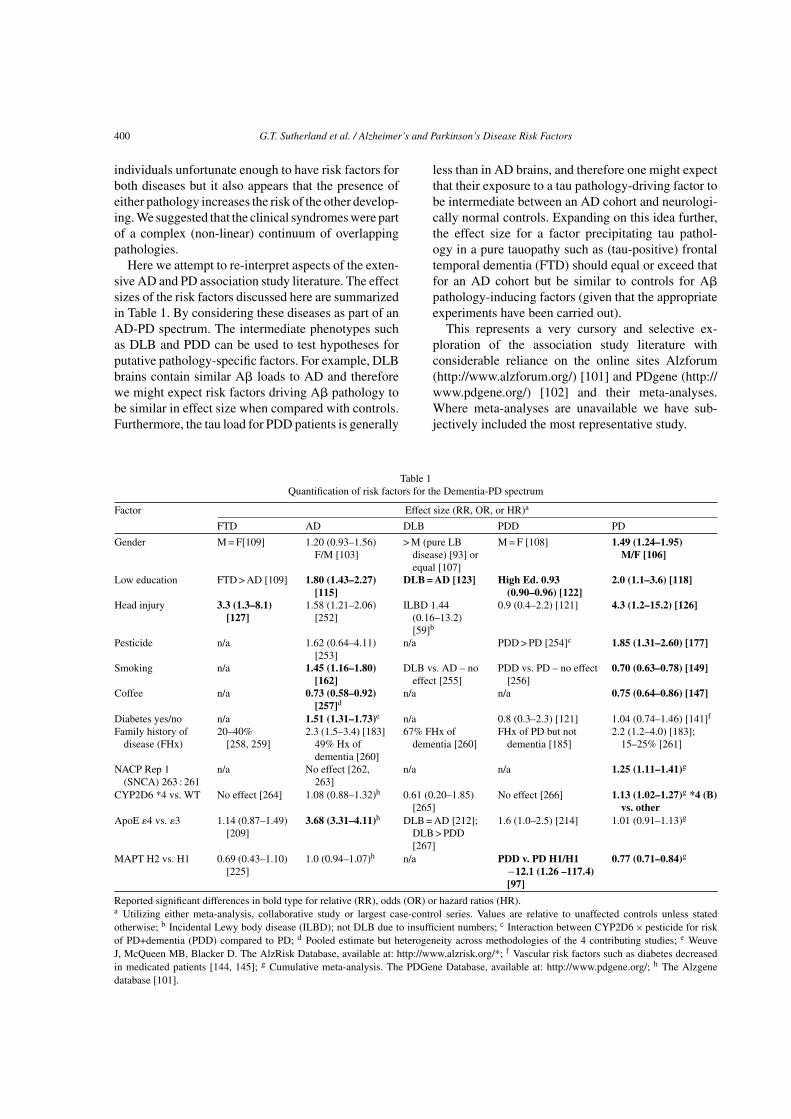
Here we attempt to re-interpret aspects of the exten-sive AD and PD association study literature. The effectsizes of the risk factors discussed here are summarizedin Table 1. By considering these diseases as part of anAD-PD spectrum. The intermediate phenotypes suchas DLB and PDD can be used to test hypotheses forputative pathology-specific factors. For example, DLBbrains contain similar A� loads to AD and thereforewe might expect risk factors driving A� pathology tobe similar in effect size when compared with controls.Furthermore, the tau load for PDD patients is generally
less than in AD brains, and therefore one might expectthat their exposure to a tau pathology-driving factor tobe intermediate between an AD cohort and neurologi-cally normal controls. Expanding on this idea further,the effect size for a factor precipitating tau pathol-ogy in a pure tauopathy such as (tau-positive) frontaltemporal dementia (FTD) should equal or exceed thatfor an AD cohort but be similar to controls for A�pathology-inducing factors (given that the appropriateexperiments have been carried out).
This represents a very cursory and selective ex-ploration of the association study literature withconsiderable reliance on the online sites Alzforum(http://www.alzforum.org/) [101] and PDgene (http://www.pdgene.org/) [102] and their meta-analyses.Where meta-analyses are unavailable we have sub-jectively included the most representative study.
Table 1Quantification of risk factors for the Dementia-PD spectrum
Factor Effect size (RR, OR, or HR)a
FTD AD DLB PDD PD
Gender M = F[109] 1.20 (0.93–1.56)F/M [103]
> M (pure LBdisease) [93] orequal [107]
M = F [108] 1.49 (1.24–1.95)M/F [106]
Low education FTD > AD [109] 1.80 (1.43–2.27)[115]
DLB = AD [123] High Ed. 0.93(0.90–0.96) [122]
2.0 (1.1–3.6) [118]
Head injury 3.3 (1.3–8.1)[127]
1.58 (1.21–2.06)[252]
ILBD 1.44(0.16–13.2)[59]b
0.9 (0.4–2.2) [121] 4.3 (1.2–15.2) [126]
Pesticide n/a 1.62 (0.64–4.11)[253]
n/a PDD > PD [254]c 1.85 (1.31–2.60) [177]
Smoking n/a 1.45 (1.16–1.80)[162]
DLB vs. AD – noeffect [255]
PDD vs. PD – no effect[256]
0.70 (0.63–0.78) [149]
Coffee n/a 0.73 (0.58–0.92)[257]d
n/a n/a 0.75 (0.64–0.86) [147]
Diabetes yes/no n/a 1.51 (1.31–1.73)e n/a 0.8 (0.3–2.3) [121] 1.04 (0.74–1.46) [141]f
Family history ofdisease (FHx)
20–40%[258, 259]
2.3 (1.5–3.4) [183]49% Hx ofdementia [260]
67% FHx ofdementia [260]
FHx of PD but notdementia [185]
2.2 (1.2–4.0) [183];15–25% [261]
NACP Rep 1(SNCA) 263 : 261
n/a No effect [262,263]
n/a n/a 1.25 (1.11–1.41)g
CYP2D6 *4 vs. WT No effect [264] 1.08 (0.88–1.32)h 0.61 (0.20–1.85)[265]
No effect [266] 1.13 (1.02–1.27)g *4 (B)vs. other
ApoE �4 vs. �3 1.14 (0.87–1.49)[209]
3.68 (3.31–4.11)h DLB = AD [212];DLB > PDD[267]
1.6 (1.0–2.5) [214] 1.01 (0.91–1.13)g
MAPT H2 vs. H1 0.69 (0.43–1.10)[225]
1.0 (0.94–1.07)h n/a PDD v. PD H1/H1−12.1 (1.26 –117.4)[97]
0.77 (0.71–0.84)g
Reported significant differences in bold type for relative (RR), odds (OR) or hazard ratios (HR).a Utilizing either meta-analysis, collaborative study or largest case-control series. Values are relative to unaffected controls unless statedotherwise; b Incidental Lewy body disease (ILBD); not DLB due to insufficient numbers; c Interaction between CYP2D6 × pesticide for riskof PD+dementia (PDD) compared to PD; d Pooled estimate but heterogeneity across methodologies of the 4 contributing studies; e WeuveJ, McQueen MB, Blacker D. The AlzRisk Database, available at: http://www.alzrisk.org/*; f Vascular risk factors such as diabetes decreasedin medicated patients [144, 145]; g Cumulative meta-analysis. The PDGene Database, available at: http://www.pdgene.org/; h The Alzgenedatabase [101].
G.T. Sutherland et al. / Alzheimer’s and Parkinson’s Disease Risk Factors 401
Gender
The majority of research points to a higher preva-lence of AD in females, but at least one large studyfound no difference [103]. It was originally thoughtthat longevity per se could account for the higherfemale prevalence but the effect remains after adjust-ment for age and education [104]. One hypothesis isthat AD pathology is more likely to manifest clinicallyas dementia in women than in men [105]. However thismay contradict findings in the oldest old and those ofRuitenberg et al. who found that the incidence of ADincreased in women over 90 years of age.
The opposite gender effect is seen in PD with a mod-erate relative risk effect in men (1.5 fold) [106]. Thelack of gender differences in the intermediate pheno-types of DLB [107] and PDD [108] seem consistentwith the contrasting effects in AD and PD if gender dif-ferences are associated with the pathology as opposedto resisting its effects. Similarly the lack of effect inFTD patients [109] could suggest that the female gen-der augments A� rather tau pathology in AD [110,111]. In terms of biological plausibility, higher lev-els of oligomeric A�1-42 and insoluble A� have beenfound in the brain tissue of women [112] as well as infemale mutant A�PP transgenic mice [113, 114].
Education
The prevalence of AD is inversely associated withyears of education [115], but increased education isalso directed related to the amount of AD pathologyand cerebral hypoperfusion on imaging studies [69,116]. This apparent paradox, where a factor appearsto augment and resist pathology simultaneously, hasbeen explained by education imparting a cognitivereserve that delays the onset of clinical symptoms[117]. It is worthwhile emphasizing that this postulatedprotective mechanism is acting mechanistically andspatially remote from the pathology itself. Thereforesuch an effect would not be detectable by sampling ofthe affected region and the increasingly commonplaceassay by the genome-scale platforms.
In contrast, higher levels of education appear tobe a risk factor for PD [118]. The hypothesis thatincreased education protects against cognitive declineis supported by comparisons between PDD and non-demented PD patients [119–122]. It is also consistentwith the equivalent education levels between AD andDLB patients [123], as the global severity of dementiais very similar between DLB and AD patients [123].
Head injury
Dementia pugilistica is a progressive memory dis-order common in ex-boxers associated with AD-likebrain pathology. Traumatic brain injury is also impli-cated in the development of AD [124, 125] and PD[126], but in seems likely to only account for only afew cases of both diseases. The relatively high risk ofhead trauma in FTD [127] suggests that head traumamay modify the risk of AD via a tau-driven mechanism,although less than half FTD are tau-positive, and dataare not stratified for tau-positive versus tau-negativeFTD.
High levels of cleaved tau are observed in the cere-brospinal fluid (CSF) and serum of patients with acutebrain injury [128]. The cleavage of tau is being increas-ingly seen as a pivotal step in NFT formation andneurodegeneration in AD [129–131]. In fact tau cleav-age events may even be the link between A� and tau inthe pathogenesis of AD according to recent work in atau transgenic mouse model [132]. Interestingly acutebrain trauma has also been associated with increasesin A�PP and A� [133, 134] and �-synuclein [57, 135].This suggests that the aberrant expression of all theproteins of interest could be general responses to braintrauma rather than disease specific [136]. This fits wellwith the development of mixed pathologies but doesnot explain the initial susceptibility of certain cell pop-ulations or the primary pathology that develops withinthem.
Cardiovascular disease risk factors
Cardiovascular risk factors have been of particu-lar interest in AD given the dual role of ApoE �4variant in AD and systemic dyslipidemia [137, 138].Perhaps surprisingly then, given the impact of ApoE �4on AD risk, it is only concurrent diabetes that isreplicable across multiple independent studies (http://www.alzforum.org). This may reflect defective insulinsignaling [139], a factor that appears to result in theabnormal phosphorylation of tau [140].
In contrast, no cardiovascular risk factors, includingdiabetes or ApoE �4 status, appear to be associatedwith PD [141–143]. The lack of association with PDmay be confounded by the sympathetic modulation ofthe cardiovascular system by PD medications [144,145] and systemic manifestations of PD. In termsof intermediate phenotypes diabetes does not appearto influence the development of dementia among PDpatients [121]. The artificial induction of diabetes mel-litus in a tau transgenic model of FTD was shown to
402 G.T. Sutherland et al. / Alzheimer’s and Parkinson’s Disease Risk Factors
cause a massive deposition of hyperphosphorylated,insoluble tau [146]. If diabetes is acting via tau pathol-ogy one might expect an association between diabetesand FTD, although we are not aware of any studies todate that support this idea.
Smoking
Cigarette smoking is one of the few environmen-tal exposures that has been consistently shown to beassociated with a reduced risk of PD [147–151]. Italso seems that this effect depends more on smok-ing duration than on intensity [152]. Nicotine, throughdirect effects on receptors, or indirect effects such asinhibition of monoamine oxidase activity or inductionof P450 enzymes, is the main suspect [153]. Workby ourselves, and others, has shown that both pas-sive smoking and other forms of tobacco provide thesame protective effect [154, 155], adding weight to adirect role of tobacco-related constituents in the asso-ciation. Furthermore the protective effect of smokingmay rely on interactions with xenobiotic metabolismgenes [156, 157] similar to pesticide exposure dis-cussed below. Other researchers have extended thesegene × environmental interactions to include variantsof genes involved in dopamine signaling [157] and theSNCA gene [158, 159]. SNCA encodes �-synuclein andis one of the few genes whose common variants arereproducibly shown to be risk factors in sporadic formsof PD [160]. The positive interactions with smoking[158, 159] and pesticides [159], hints at the mechanis-tic drivers of �-synuclein dysfunction.
In what appears to be a clear contrast with PD, smok-ing is a risk factor for AD [161–165], although otherstudies suggest that the effect is limited to ApoE �4 car-riers [166, 167]. The independent effects of smokingand ApoE genotype on cardiovascular disease (CVD)risk provide an obvious interactive mechanism for ADdevelopment, but as discussed above, diabetes seemsto be the only CVD factor with a robust associationwith AD. Smoking is positively associated with type2 diabetes but whether this is causal remains to bedetermined [168]. However, irrespective of the roleof smoking, ApoE �4 (discussed below) and diabetescombine to enhance the risk of AD [169].
Heavy smoking has been shown to reduce the riskof �-synuclein pathology in a community-based studywith the greatest reductions being seen in the SN[165]. So in comparison to education in AD, smokingappears to act directly to reduce the pathological load.A deduction from this work is that �-synuclein pathol-ogy is either deleterious itself or a reliable barometerof the disease process in PD. As an aside, there was no
concurrent increase in AD pathology in other regionssuggesting that the smoking effect in AD is not medi-ated by exacerbating A� or tau pathology [170].
Pesticides
The interest in pesticides and PD pathogenesis stemsfrom the chemical similarities of common pesticideslike paraquat, to the Demerol derivative, 1-methyl-4-phenyl-1,2,3,6-tetrahydropyridine (MPTP) [171].Accidental MPTP intoxication causes an acute formof parkinsonism [172, 173]. Like its toxic metabo-lite, MPP+ [174], the herbicide paraquat can alsoinduce parkinsonism in animal models [175]. Surpris-ingly paraquat does not appear to act as a substratefor the dopamine transporter, nor does it effectivelyinhibit mitochondrial complex I [176]. Studies investi-gating pesticide exposure in PD have produced variableresults, but a meta-analysis suggests that it is a mod-erate risk factor [177]. Evidence from our laboratorysuggests that the true risk associated with pesticidesmay well be higher but is effectively masked by hetero-geneity of xenobiotic metabolism gene variants withinthe case and control groups [178].
In contrast, there appears to be no pesticide effectwith AD or the intermediate phenotypes consistentwith the proposed selective effect on SNc dopamin-ergic neurons [176, 179]. Nevertheless one needs to becautious in emphasizing mitochondrial dysfunction asbeing PD-specific because one recent paper describesthe synergistically deleterious effects of tau and A�on murine respiratory complex 1 and IV respectively[180].
Family history
In looking at family history as a risk factor wemight appear to be asking the nonsensical questionof how much of the etiology of a sporadic disease(no familial clustering) can be attributed to genetics?PD was always regarded as the archetypal non-geneticdisease but the discovery of monogenic forms of thedisease combined with the re-evaluation of twin studieshas some researchers now suggesting that the spo-radic PD is entirely genetic [181]. This suggestionis further supported by recent evaluations of com-mon genetic variability in genome-wide associationstudies (GWAS), which have apportioned up to 25%of the population attributable risk in PD to small-effect sized common genetic variants [182]. Familyhistory as a risk factor appears to be similar betweenPD (OR = 2.2 (95% CI = 1.2–4.0)) and AD (OR = 2.3(95% CI = 1.5–3.4)) [183]. In terms of common genetic

G.T. Sutherland et al. / Alzheimer’s and Parkinson’s Disease Risk Factors 403
risk factors, there are conflicting studies supporting anincreased risk of dementia in PD relatives [184–186].A family history of dementia is however a risk factorfor DLB [187] and four-fold greater in magnitude thanfor PDD or PD [185, 187]. In contrast a family historyof PD is similar for PD, PDD, and DLB patients [185].
Monogenic forms of AD and PD are proof that thesediseases, or at least phenocopies of them, can be totallygenetic in nature. Autosomal dominant forms of ADare caused by mutations in the presenilin 1 (PS1) geneand more rarely in the PS2 and AβPP genes [188].Autosomal dominant forms of PD are caused by muta-tions in SNCA and the genes that encode the proteins,leucine-rich region Kinase 2 (LRRK2) and UbiquitinC-terminal hydrolase-1 (UCHL-1) while autosomal-recessive forms are caused by mutations in genes forparkin (PARK2), DJ-1 (PARK7) and PTEN-inducedputative kinase 1 (PINK1). More recently mutationshave also been found in the genes encoding HtrA ser-ine peptidase 2 (HTRA2), lysosomal ATPase type 13A2(ATP13A2) and Glucocerebrosidase (GBA) [189]. GBAmutations have also been linked to DLB and maybe a specific driver for �-synuclein pathology [190].Most of these mutations result in early disease onsetsalthough LRRK2 mutations are a notable exceptionin PD.
An obvious question seems to be how common vari-ants of these genes might modify the risk for sporadicdisease [191]. Monogenic disease gene variants havenot been detected in GWASs of AD, but PD GWASssuggest that SNCA [182, 192, 193] and LRRK2 [182,192] variants are risk factors. However, both have verymodest effect sizes, particularly when compared to theApoE �4 variant in AD [194, 195]. It has been sug-gested that ApoE �4, by itself, could account for asmuch as 50% of attributable risk in sporadic AD [196].
ApoE
GWASs in AD have universally detected the ApoE�4 risk effect [197–204]. However despite the effectsize it is still far from clear how ApoE �4 increasesthe risk of AD. An important observation may be thatplaques are more frequent in ApoE �4 allele carriers[205], but there is no difference in NFT load betweenthe different ApoE genotypes [206]. A mechanisticlink with A� pathology is certainly supported byanimal studies [207, 208] and seems consistent withthe lack of ApoE �4 effect in PD and FTD [209],while female gender could also to play an importantinterrelated role [112].
The frequency of ApoE �4 in DLB patients is similarto AD [210–212] with a lessened, but nevertheless sig-
nificant, risk effect on the development of dementiain PD [213–215]. Neuropathological [84] and neu-roimaging studies [216] studies both support increasedA� loads in DLB compared with PDD. A caveat hereis that Drzezga et al. and a second imaging study failedto show a link between ApoE genotype and brain atro-phy in AD patients despite the association with A�loads [205, 217]. This suggested to Vemuri and col-leagues that neurodegeneration in AD may be mediatedby tau pathology and sequential to amyloidosis [217],a course of events first suggested by the ‘amyloid cas-cade’ hypothesis [218].
MAPT
Until recently microtubule-associated protein tau(MAPT or tau) had largely played second fiddle toA� in efforts to unravel the pathogenesis of AD. Thepresence of tau-based inclusions across so many neu-rodegenerative diseases hints at a general mechanismof neurodegeneration but the protein itself remainsan enigma. No mutations in the MAPT gene, havebeen described in AD or PD patients, but they areresponsible for an inherited variant of frontotemporaldementia with parkinsonism (FTDP-17) [219]. A 900-kb inversion polymorphism around the tau gene hasled to two distinct lineages in the human population,H1 and H2 [220]. Surprisingly the variant H2 alleledoes not affect risk for the most common tauopathy,AD, but is protective in PD (Table 1). The commonH1 extended tau haplotype is also associated with pro-gressive supranuclear palsy, one of the parkinsoniansyndromes characterized by tau deposition [221].
Associations between single nucleotide polymor-phisms (SNPs) marking this haplotype and PD havebeen confirmed in two large GWASs of Caucasian pop-ulations [182, 193] but not, as expected, in a JapaneseGWAS where the H2 haplotype is largely absent [192].The modifying effect of tau in PD may be mediated viathe putative interactions with �-synuclein as describedabove. Interestingly patients harboring either SNCA(A53 T) [222] or LRRK2 (G2019S) mutations [223]can feature tau rather than �-synuclein-predominantpathology.
No studies have looked at whether the H1 allele hasan effect on DLB risk, but the H1/H1 genotype is a pre-dictor of PDD [97, 224]. This effect combined with theprotective trend of the H2 allele in FTD [225] suggeststhat increased tau expression [226] drives dementiaeither independently, or through an interaction with�-synuclein pathology. The A� load in AD and DLBmay be sufficient through direct [97] or indirect means[217] to precipitate tau aberrant expression or post-

404 G.T. Sutherland et al. / Alzheimer’s and Parkinson’s Disease Risk Factors
translational modification in the absence of geneticpredispositions.
However, if common variants of tau can drivedementia, then what is happening in the >50% of tau-negative cases of FTD? Some familial, and the majorityof sporadic FTLD patients without tau, have depositsof the TAR DNA binding protein 43 (TDP-43) [227].Interestingly, TDP-43 pathology is also commonlyfound in DLB and AD cases suggesting a general rolein neurodegeneration [228–231]. In contrast, studiesin PD suggest that TDP-43 pathology is either absence[232], or very uncommon [231] (Fig. 1). TDP-43 istransposed to the cytoplasm under pathological condi-tions, but the link between tau and TDP-43 pathologyis still unclear despite their indistinguishable end stagedegeneration in FTD.
In principle, the AD-PD risk factor literature doesseem to support the idea of a neurodegenerative dis-ease spectrum. We suggest that the risk factors for A�,tau, and �-synuclein pathologies occur commonly andoften together. How they might combine to form themyriad of individual disease phenotypes is speculativebut we have summarized our ideas in diagrammaticform to facilitate further discussion of this area (Fig. 1).Looking at the Fig. 1, the aging influence in neu-rodegeneration has been stylistically represented by anhourglass. Aging results in cellular deterioration but itis not entirely a haphazard process. Stochastic damageis a driving force but the rates of damage accumulationand decline relies on regulation by pathways that havebeen conserved during evolution [233, 234]. Further-more postulated aging mechanisms such as decreased
Fig. 1. A schematic of how risk factors might produce the dementia-PD spectrum. Using an hourglass analogy for ageing, this schematicsummarizes how risk factors might contribute to neurodegeneration across the dementia-PD spectrum. Risk factors, both deleterious andprotective, combine to produce the three pathological ‘drivers’: Amyloid-�, tau, and �-synuclein pathology represented by the yellow, red,and blue dendritic clusters respectively. The risk factor scale ranges from largely protective ‘white’ (e.g., coffee), through differential ‘grey’effects (e.g., smoking) to largely deleterious ‘black’ (e.g., head injury). Dendro-dendritic synapses represent putative competing or excludingrisk factors. Other factors will have their effects by acting directly on cellular resistance or via compensatory mechanisms (grey dendrites). Theperikaryon represents the temporal and spatial ‘mix’ of factors in a typical aged human brain (80 years). Axons lead to the final phenotypes withprotective or compensatory mechanisms (grey axons) continuing to minimize the impact of pathology and the clinical manifestations of disease(via axo-axonal synapses).
G.T. Sutherland et al. / Alzheimer’s and Parkinson’s Disease Risk Factors 405
ability to manage oxidative stress [235] and proteotox-icity [235–237] are remarkably similar to the workinghypotheses for AD and PD. Interestingly ApoE �4confers a four-fold decrease in longevity compared to�2 in persons over 85 years [238] although this mayreflect reduced morbidity from cardiovascular diseaserather than increased neurodegenerative disease bur-den [239]. Nevertheless it is not a simple process todelineate ‘cause’ and ‘effect’ issues between aging andneurodegeneration.
The other aspect of these late-onset diseases isthe time component itself. This allows the possibil-ity that chronic exposure to very subtle risk factorscould be sufficient for disease; a challenging sce-nario for association studies. The time componentalso increases the likelihood that multiple rounds ofcompensatory or protective host responses will maskprimary pathogenic clues from postmortem detection.Such secondary or incidental findings can misdirect thesubsequent research focus or result in therapeutics thatwill prove palliative at best. Lastly, the time compo-nent is difficult to accelerate accurately in cell cultureor laboratory animal models aimed at recapitulatingthe disease history.
The base of the hourglass encloses a reservoir ofthese known, putative, or as yet, unknown risk factors.We envisage that subsets of these factors, both delete-rious and protective interact to create ‘drivers’ for thethree pathologies. We have used yellow, red and blue‘dendrites’ to designate the A�, tau, and �-synucleinpathologies, respectively (color available in online ver-sion only; otherwise left to right respectively). In con-trast to drivers of ‘pathology’ other factors will modifyrisk by enhancing or attenuating cellular protection orcompensatory mechanisms (grey ‘dendrites’). In par-ticular we are referring to the effects of female genderand education on creating a ‘cognitive reserve’ that actsto postpone clinical manifestations of dementia.
The neuronal cell body is a mixer for the impactof individual ‘drivers’ against the general resolve ofthe human brain to maintain homeostasis or employcompensatory mechanisms in response to injury. Thismaintenance of homeostasis is what we have termedcellular resistance while compensation is likely to be‘multiscalar’, potentially operating on all levels froma molecular basis, through enhanced local plasticity tothe use of alternative circuitry [240].
Finally, ‘axons’ from the cell body carry the blendof A�, tau, and �-synuclein pathologies towards thefinal neuropathological and clinical phenotypes. Theaxo-axonal synapses (grey) reinforce the idea thatsome individuals enjoy a heightened resistance to, or
an inherent ability or compensate for, these patholo-gies. The top of the hourglass shows the dementia-PDphenotypes displayed according to their estimatedprevalence. We suggest that approximately 75% ofaged individuals die with some neuropathology butless than 10% are clinically affected. Notwithstandingdosage effects this suggests that nearly all individualsare exposed factors that drive these ‘pathologies’.
PART III: THE FUTURE DIRECTIONOF ASSOCIATION STUDIES
The original hypothesis of this essay was to exam-ine whether knowledge of disease-specific risk factorsfor AD could assist in focusing efforts into under-standing the pathogenesis of sporadic forms of PD,or and vice versa. ‘Intermediate’ diseases and inci-dental pathology suggested that there is actually adementia-PD spectrum involving various combina-tions of A�, tau, �-synuclein, and probably TDP-43pathology, in a non-linear pattern. Although not alto-gether explained by a threshold effect, the amount andextent of pathology generally reflects the degree ofneurological dysfunction. Given this continuum, wethought that our hypothesis might be better pursued interms of factors that appeared to promote or attenuateA�, tau, or �-synuclein pathology across the variousphenotypes. In the case of smoking in PD, this certainlyseemed to be the case [165], but other known risk fac-tors appeared to be acting in a compensatory capacitythat was physically remote from pathology itself.
In the introduction we also suggested that using ADcontrols for PD, or vice versa, may be a more effectivestrategy to finding risk factors than the classic case-control paradigm. This approach seems particularly aptgiven the prevalence of incidental pathology in unaf-fected controls. However the commonality of mixedpathology amongst AD and PD cases suggests that theutility of AD-PD case comparisons may be just as lim-ited, or effective, as the classic-case control paradigm.
Single disease entities are far easier to explore inexperimental paradigms but here they are not entirelyaccurate. The risk factors for A�, tau, and �-synucleinpathologies often occur together across the AD-PDspectrum and show strong interrelations [94, 95]. Per-haps thinking in terms of a neurodegenerative diseasecontinuum may be one way to advance our scien-tific investigation of these diseases. For example, onecould study pathologically confirmed cases and con-sider the “load” of specific pathologies as continuousdependent variables in relation to particular indepen-
406 G.T. Sutherland et al. / Alzheimer’s and Parkinson’s Disease Risk Factors
dent risk factors. Excitingly, the continuing progressof neuroimaging initiatives allowing a more accuratequantification of pathological (A�) load and regionalatrophy creates the possibility that the research findingscould actually benefit the patients involved [241].
Similarly, potential treatments in various stages ofpreclinical or clinical trials are largely aimed at theattenuation of pathology. The best known example isA� immunotherapy that, despite setbacks in humanclinical trials, still holds considerable promise [242].Similarly, strategies aimed at reducing NFTs [243] and�-synuclein pathology [244] or their formation [245,246] are also being developed.
The reduction of pathology is not the only treat-ment option. It appears equally prudent, to identify andsubsequently augment neuronal survival mechanisms.In the absence of pathology-attenuating therapeuticsthese will still reduce the burden of disease by extend-ing the quality of life and reducing the need forhigh-dependence care. In addition, it is important toconsider the role of inflammation in the overall diseaseprocesses as inflammatory correlates such as activatedmicroglia are seen to a variable extent in all diseasesacross the AD-PD spectrum [247]. However, a greaterexploration of this area, and particularly the researcheffort into finding effective modulators of neuroin-flammation, falls outside the scope of this review (see[248]).
Whatever the treatment rationale, success is likely tohinge on the concurrent discovery of pre-clinical mark-ers to identify at-risk individuals. This will facilitatethe utilization of a therapeutic window where neuronalnumbers remain consistent with adequate functional-ity. Luckily the discovery of a rational therapeutic tar-get and preclinical marker may be mutually inclusivetasks. They both require investigation of early diseaseparadigms to find the primary pathogenic mechanisms.In this respect, the advent of patient derived stem cellmodels looks very promising for the in vitro modelingof disease history on differentiable susceptible geneticbackgrounds [249]. Similarly, presymptomatic stagesof animal models and the prospective studies of pro-dromal conditions and asymptomatic mutation carriers[250] could be employed.
It is an exciting time to be involved in diseaseresearch with genome-scale technologies enablingthousands of parameters to be tested simultaneously.These multidimensional platforms, in combinationwith improving annotational algorithms, are moving ussteadily towards a systems biology approach to health,or as recently described, ‘network medicine’ [251].However these powerful approaches still rely on the
relevance of the question being asked and the contextit is being asked in. Case-control studies are reduc-tionist paradigms that assume, for maximum power,that the two populations being compared are exclu-sive and homogenous. We, and others [94, 95], suggestthat AD and PD are neither exclusive nor homoge-nous entities but rather part of a complex continuumcharacterized by variable amounts of A�, tau, and�-synuclein pathology. This heterogeneity may be animportant factor in the lack of reproducibility in case-control studies to date. The consideration of incidentaland mixed pathologies will make experimental designand interpretation more challenging but it should alsolead to greater clarity from our studies of late-onsetneurodegenerative diseases such as AD and PD.
ACKNOWLEDGMENTS
We would like to thank Mr Jeremy Newman for hisassistance with illustrations. G. Sutherland wishes tothank Dr Russell Snell for his fruitful discussions andcomments and the support of the Geriatric MedicalFoundation of Queensland.
Authors’ disclosures available online (http://www.j-alz.com/disclosures/view.php?id=782).
REFERENCES
[1] Le Couteur DG, Muller M, Yang MC, Mellick GD, McLeanAJ (2002) Age-environment and gene-environment interac-tions in the pathogenesis of Parkinson’s disease. Rev EnvironHealth 17, 51-64.
[2] Tol J, Roks G, Slooter AJ, van Duijn CM (1999) Genetic andenvironmental factors in Alzheimer’s disease. Rev Neurol(Paris) 155 (Suppl 4), S10-S16.
[3] Mellick GD, Silburn PA, Sutherland GT, Siebert GA (2010)Exploiting the potential of molecular profiling in Parkin-son’s disease: current practice and future probabilities.Expert Rev Mol Diagn 10, 1035-1050.
[4] Lippa CF, Smith TW, Swearer JM (1994) Alzheimer’sdisease and Lewy body disease: a comparative clinicopatho-logical study. Ann Neurol 35, 81-88.
[5] McKeith IG, Ince P, Jaros EB, Fairbairn A, Ballard C, GraceJ, Morris CM, Perry RH (1998) What are the relationsbetween Lewy body disease and AD? J Neural Transm Suppl54, 107-116.
[6] Strobel G (2009) The spectrum series: grappling with theoverlap between Alzheimer’s and Parkinson’s diseases. 9 thInternational Conference on Alzheimer’s and Parkinson’sDiseases, 11–15 March 2009, Prague, Czech Republic.J Alzheimers Dis 18, 625-640.
[7] Jankovic J, McDermott M, Carter J, Gauthier S, Goetz C,Golbe L, Huber S, Koller W, Olanow C, Shoulson I, et al.(1990) Variable expression of Parkinson’s disease: a base-line analysis of the DATATOP cohort. The Parkinson StudyGroup. Neurology 40, 1529-1534.
G.T. Sutherland et al. / Alzheimer’s and Parkinson’s Disease Risk Factors 407
[8] Jellinger KA (1999) Post mortem studies in Parkinson’sdisease – is it possible to detect brain areas for specificsymptoms? J Neural Transm Suppl 56, 1-29.
[9] Selikhova M, Williams DR, Kempster PA, Holton JL,Revesz T, Lees AJ (2009) A clinico-pathological study ofsubtypes in Parkinson’s disease. Brain 132, 2947-2957.
[10] Lobo A, Launer LJ, Fratiglioni L, Andersen K, Di CarloA, Breteler MM, Copeland JR, Dartigues JF, Jagger C,Martinez-Lage J, Soininen H, Hofman A (2000) Prevalenceof dementia and major subtypes in Europe: a collaborativestudy of population-based cohorts. Neurologic Diseases inthe Elderly Research Group. Neurology 54, S4-S9.
[11] Fratiglioni L, De Ronchi D, Aguero-Torres H (1999) World-wide prevalence and incidence of dementia. Drugs Aging15, 365-375.
[12] Jorm AF, Dear KB, Burgess NM (2005) Projections of futurenumbers of dementia cases in Australia with and withoutprevention. Aust N Z J Psychiatry 39, 959-963.
[13] Plassman BL, Langa KM, Fisher GG, Heeringa SG, WeirDR, Ofstedal MB, Burke JR, Hurd MD, Potter GG, RodgersWL, Steffens DC, Willis RJ, Wallace RB (2007) Prevalenceof dementia in the United States: the aging, demographics,and memory study. Neuroepidemiology 29, 125-132.
[14] Braak H, Braak E (1991) Neuropathological stageing ofAlzheimer-related changes. Acta Neuropathol (Berl) 82,239-259.
[15] NIA-Reagan Institute Working Group (1997) Consen-sus recommendations for the postmortem diagnosis ofAlzheimer’s disease. The National Institute on Aging, andReagan Institute Working Group on Diagnostic Criteria forthe Neuropathological Assessment of Alzheimer’s Disease.Neurobiol Aging 18, S1-S2.
[16] Newell KL, Hyman BT, Growdon JH, Hedley-Whyte ET(1999) Application of the National Institute on Aging(NIA)-Reagan Institute criteria for the neuropathologicaldiagnosis of Alzheimer disease. J Neuropathol Exp Neurol58, 1147-1155.
[17] Kril JJ, Patel S, Harding AJ, Halliday GM (2002) Neuronloss from the hippocampus of Alzheimer’s disease exceedsextracellular neurofibrillary tangle formation. Acta Neu-ropathol 103, 370-376.
[18] Arriagada PV, Growdon JH, Hedley-Whyte ET, Hyman BT(1992) Neurofibrillary tangles but not senile plaques parallelduration and severity of Alzheimer’s disease. Neurology 42,631-639.
[19] Bierer LM, Hof PR, Purohit DP, Carlin L, Schmeidler J,Davis KL, Perl DP (1995) Neocortical neurofibrillary tan-gles correlate with dementia severity in Alzheimer’s disease.Arch Neurol 52, 81-88.
[20] Hardy J, Selkoe DJ (2002) The amyloid hypothesis of Alz-heimer’s disease: progress and problems on the road to ther-apeutics. Science 297, 353-356.
[21] Mellick GD, Maraganore DM, Silburn PA (2005) Australiandata and meta-analysis lend support for alpha-synuclein(NACP-Rep1) as a risk factor for Parkinson’s disease. Neu-rosci Lett 375, 112-116.
[22] de Rijk MC, Launer LJ, Berger K, Breteler MM, DartiguesJF, Baldereschi M, Fratiglioni L, Lobo A, Martinez-Lage J,Trenkwalder C, Hofman A (2000) Prevalence of Parkinson’sdisease in Europe: a collaborative study of population-basedcohorts. Neurologic Diseases in the Elderly Research Group.Neurology 54, S21-S23.
[23] Weintraub D, Comella CL, Horn S (2008) Parkinson’sdisease – Part 1: Pathophysiology, symptoms, burden, diag-nosis, and assessment. Am J Manag Care 14, S40-S48.
[24] Hughes AJ (1997) Clinicopathological aspects of Parkin-son’s disease. Eur Neurol 38 (Suppl 2), 13-20.
[25] Hughes AJ, Daniel SE, Kilford L, Lees AJ (1992) Accu-racy of clinical diagnosis of idiopathic Parkinson’s disease:a clinico-pathological study of 100 cases. J Neurol Neuro-surg Psychiatry 55, 181-184.
[26] Bernheimer H, Birkmayer W, Hornykiewicz O, Jellinger K,Seitelberger F (1973) Brain dopamine and the syndromesof Parkinson and Huntington. Clinical, morphological andneurochemical correlations. J Neurol Sci 20, 415-455.
[27] Cheng HC, Ulane CM, Burke RE (2010) Clinical progres-sion in Parkinson disease and the neurobiology of axons.Ann Neurol 67, 715-725.
[28] Braak H, Del Tredici K, Rub U, de Vos RA, Jansen SteurEN, Braak E (2003) Staging of brain pathology related tosporadic Parkinson’s disease. Neurobiol Aging 24, 197-211.
[29] Braak H, Muller CM, Rub U, Ackermann H, Bratzke H, deVos RA, Del Tredici K (2006) Pathology associated withsporadic Parkinson’s disease – where does it end? J NeuralTransm Suppl 70, 89-97.
[30] Harding AJ, Stimson E, Henderson JM, Halliday GM (2002)Clinical correlates of selective pathology in the amygdala ofpatients with Parkinson’s disease. Brain 125, 2431-2445.
[31] Hubbard PS, Esiri MM, Reading M, McShane R, Nagy Z(2007) Alpha-synuclein pathology in the olfactory pathwaysof dementia patients. J Anat 211, 117-124.
[32] Kalaitzakis ME, Graeber MB, Gentleman SM, Pearce RK(2008) Controversies over the staging of alpha-synucleinpathology in Parkinson’s disease. Acta Neuropathol 116,125-128.
[33] Jellinger K, Mizuno H (2003) Parkinson’s disease. In Neu-rodegeneration: The Molecular Pathology of Dementia andMovement Disorders, Dickson D, ed. ISN Neuropath Press,Basel, pp. 159-187.
[34] Leverenz JB, Umar I, Wang Q, Montine TJ, McMillan PJ,Tsuang DW, Jin J, Pan C, Shin J, Zhu D, Zhang J (2007)Proteomic identification of novel proteins in cortical lewybodies. Brain Pathol 17, 139-145.
[35] Jellinger KA (2009) A critical evaluation of current stag-ing of alpha-synuclein pathology in Lewy body disorders.Biochim Biophys Acta 1792, 730-740.
[36] Young VG, Halliday GM, Kril JJ (2008) Neuropathologiccorrelates of white matter hyperintensities. Neurology 71,804-811.
[37] Ziegler DA, Piguet O, Salat DH, Prince K, Connally E,Corkin S (2010) Cognition in healthy aging is related toregional white matter integrity, but not cortical thickness.Neurobiol Aging 31, 1912-1926.
[38] Double KL, Halliday GM, Kril JJ, Harasty JA, Cullen K,Brooks WS, Creasey H, Broe GA (1996) Topography ofbrain atrophy during normal aging and Alzheimer’s disease.Neurobiol Aging 17, 513-521.
[39] Piguet O, Double KL, Kril JJ, Harasty J, Macdonald V,McRitchie DA, Halliday GM (2009) White matter loss inhealthy ageing: a postmortem analysis. Neurobiol Aging 30,1288-1295.
[40] Harding AJ, Halliday GM, Kril JJ (1998) Variation in hip-pocampal neuron number with age and brain volume. CerebCortex 8, 710-718.
[41] Terry RD, DeTeresa R, Hansen LA (1987) Neocortical cellcounts in normal human adult aging. Ann Neurol 21, 530-539.
[42] Pakkenberg B, Gundersen HJ (1997) Neocortical neuronnumber in humans: effect of sex and age. J Comp Neurol384, 312-320.
408 G.T. Sutherland et al. / Alzheimer’s and Parkinson’s Disease Risk Factors
[43] Kubis N, Faucheux BA, Ransmayr G, Damier P, DuyckaertsC, Henin D, Forette B, Le Charpentier Y, Hauw JJ, Agid Y,Hirsch EC (2000) Preservation of midbrain catecholamin-ergic neurons in very old human subjects. Brain 123(Pt 2),366-373.
[44] Fearnley JM, Lees AJ (1991) Ageing and Parkinson’s dis-ease: substantia nigra regional selectivity. Brain 114(Pt 5),2283-2301.
[45] Braak H, Braak E (1997) Frequency of stages of Alzheimer-related lesions in different age categories. Neurobiol Aging18, 351-357.
[46] Davis DG, Schmitt FA, Wekstein DR, Markesbery WR(1999) Alzheimer neuropathologic alterations in aged cog-nitively normal subjects. J Neuropathol Exp Neurol 58,376-388.
[47] Neuropathology Group of the Medical Research CouncilCognitive Function and Ageing Study (2001) Patholog-ical correlates of late-onset dementia in a multicentre,community-based population in England and Wales. Lancet357, 169-175.
[48] Knopman DS, Parisi JE, Salviati A, Floriach-Robert M,Boeve BF, Ivnik RJ, Smith GE, Dickson DW, JohnsonKA, Petersen LE, McDonald WC, Braak H, PetersenRC (2003) Neuropathology of cognitively normal elderly.J Neuropathol Exp Neurol 62, 1087-1095.
[49] Pikkarainen M, Kauppinen T, Alafuzoff I (2009) Hyper-phosphorylated tau in the occipital cortex in agednondemented subjects. J Neuropathol Exp Neurol 68, 653-660.
[50] McKee AC, Au R, Cabral HJ, Kowall NW, Seshadri S,Kubilus CA, Drake J, Wolf PA (2006) Visual associationpathology in preclinical Alzheimer disease. J NeuropatholExp Neurol 65, 621-630.
[51] Price JL, McKeel DW Jr, Buckles VD, Roe CM, Xiong C,Grundman M, Hansen LA, Petersen RC, Parisi JE, Dick-son DW, Smith CD, Davis DG, Schmitt FA, MarkesberyWR, Kaye J, Kurlan R, Hulette C, Kurland BF, Hig-don R, Kukull W, Morris JC (2009) Neuropathology ofnondemented aging: presumptive evidence for preclinicalAlzheimer disease. Neurobiol Aging 30, 1026-1036.
[52] Schmitt FA, Davis DG, Wekstein DR, Smith CD, AshfordJW, Markesbery WR (2000) “Preclinical” AD revisited:neuropathology of cognitively normal older adults. Neurol-ogy 55, 370-376.
[53] Troncoso JC, Martin LJ, Dal Forno G, Kawas CH (1996)Neuropathology in controls and demented subjects from theBaltimore Longitudinal Study of Aging. Neurobiol Aging17, 365-371.
[54] Jellinger KA (2004) Lewy body-related alpha-synucleino-pathy in the aged human brain. J Neural Transm 111, 1219-1235.
[55] Markesbery WR, Jicha GA, Liu H, Schmitt FA (2009) Lewybody pathology in normal elderly subjects. J NeuropatholExp Neurol 68, 816-822.
[56] Adler CH, Connor DJ, Hentz JG, Sabbagh MN, CavinessJN, Shill HA, Noble B, Beach TG (2010) Incidental Lewybody disease: clinical comparison to a control cohort. MovDisord 25, 642-646.
[57] Uryu K, Giasson BI, Longhi L, Martinez D, Murray I, ConteV, Nakamura M, Saatman K, Talbot K, Horiguchi T, McIn-tosh T, Lee VM, Trojanowski JQ (2003) Age-dependentsynuclein pathology following traumatic brain injury inmice. Exp Neurol 184, 214-224.
[58] Dickson DW, Fujishiro H, DelleDonne A, Menke J, AhmedZ, Klos KJ, Josephs KA, Frigerio R, Burnett M, Parisi JE,
Ahlskog JE (2008) Evidence that incidental Lewy bodydisease is pre-symptomatic Parkinson’s disease. Acta Neu-ropathol 115, 437-444.
[59] Frigerio R, Fujishiro H, Ahn TB, Josephs KA, MaraganoreDM, Delledonne A, Parisi JE, Klos KJ, Boeve BF,Dickson DW, Ahlskog JE (2009) Incidental Lewy bodydisease: Do some cases represent a preclinical stage ofdementia with Lewy bodies? Neurobiol Aging doi:10.1016/j.neurobiolaging.2009.05.019
[60] Kril JJ (2009) Alzheimer disease: Alzheimer disease neu-ropathology in the oldest old. Nat Rev Neurol 5, 411-412.
[61] Haroutunian V, Schnaider-Beeri M, Schmeidler J, WysockiM, Purohit DP, Perl DP, Libow LS, Lesser GT, MaroukianM, Grossman HT (2008) Role of the neuropathology ofAlzheimer disease in dementia in the oldest-old. ArchNeurol 65, 1211-1217.
[62] Polvikoski T, Sulkava R, Rastas S, Sutela A, Niinisto L,Notkola IL, Verkkoniemi A, Viramo P, Juva K, Haltia M(2006) Incidence of dementia in very elderly individuals:a clinical, neuropathological and molecular genetic study.Neuroepidemiology 26, 76-82.
[63] Prohovnik I, Perl DP, Davis KL, Libow L, Lesser G,Haroutunian V (2006) Dissociation of neuropathology fromseverity of dementia in late-onset Alzheimer disease. Neu-rology 66, 49-55.
[64] Savva GM, Wharton SB, Ince PG, Forster G, Matthews FE,Brayne C (2009) Age, neuropathology, and dementia. NEngl J Med 360, 2302-2309.
[65] Jellinger KA, Attems J (2010) Prevalence of dementia dis-orders in the oldest-old: an autopsy study. Acta Neuropathol119, 421-433.
[66] Laukka EJ, Fratiglioni L, Backman L (2010) The influenceof vascular disease on cognitive performance in the preclini-cal and early phases of Alzheimer’s disease. Dement GeriatrCogn Disord 29, 498-503.
[67] Strozyk D, Dickson DW, Lipton RB, Katz M, Derby CA,Lee S, Wang C, Verghese J (2010) Contribution of vascularpathology to the clinical expression of dementia. NeurobiolAging 31, 1710-1720.
[68] Ding ZT, Wang Y, Jiang YP, Hashizume Y, Yoshida M,Mimuro M, Inagaki T, Iwase T (2006) Characteristics ofalpha-synucleinopathy in centenarians. Acta Neuropathol(Berl) 111, 450-458.
[69] Liao YC, Liu RS, Teng EL, Lee YC, Wang PN, Lin KN,Chung CP, Liu HC (2005) Cognitive reserve: a SPECTstudy of 132 Alzheimer’s disease patients with an educa-tion range of 0–19 years. Dement Geriatr Cogn Disord 20,8-14.
[70] Frigerio R, Fujishiro H, Maraganore DM, Klos KJ, Delle-Donne A, Heckman MG, Crook JE, Josephs KA, Parisi JE,Boeve BF, Dickson DW, Ahlskog JE (2009) Comparisonof risk factor profiles in incidental Lewy body disease andParkinson disease. Arch Neurol 66, 1114-1119.
[71] Hamilton RL (2000) Lewy bodies in Alzheimer’s disease:a neuropathological review of 145 cases using alpha-synuclein immunohistochemistry. Brain Pathol 10, 378-384.
[72] Jellinger KA (2003) Alpha-synuclein pathology in Parkin-son’s and Alzheimer’s disease brain: incidence and topo-graphic distribution – a pilot study. Acta Neuropathol 106,191-201.
[73] Schneider JA, Aggarwal NT, Barnes L, Boyle P, BennettDA (2009) The neuropathology of older persons with andwithout dementia from community versus clinic cohorts.J Alzheimers Dis 18, 691-701.
G.T. Sutherland et al. / Alzheimer’s and Parkinson’s Disease Risk Factors 409
[74] Uchikado H, Lin WL, DeLucia MW, Dickson DW (2006)Alzheimer disease with amygdala Lewy bodies: a distinctform of alpha-synucleinopathy. J Neuropathol Exp Neurol65, 685-697.
[75] Braak H, Braak E, Yilmazer D, Bohl J (1996) Functionalanatomy of human hippocampal formation and relatedstructures. J Child Neurol 11, 265-275.
[76] Jellinger KA, Seppi K, Wenning GK, Poewe W (2002)Impact of coexistent Alzheimer pathology on the naturalhistory of Parkinson’s disease. J Neural Transm 109, 329-339.
[77] Mann DM, Jones D (1990) Deposition of amyloid (A4)protein within the brains of persons with dementing disor-ders other than Alzheimer’s disease and Down’s syndrome.Neurosci Lett 109, 68-75.
[78] Papapetropoulos S, Lieberman A, Gonzalez J, Mash DC(2005) Can Alzheimer’s type pathology influence the clin-ical phenotype of Parkinson’s disease? Acta Neurol Scand111, 353-359.
[79] McKeith IG, Dickson DW, Lowe J, Emre M, O’Brien JT,Feldman H, Cummings J, Duda JE, Lippa C, Perry EK, Aars-land D, Arai H, Ballard CG, Boeve B, Burn DJ, Costa D, DelSer T, Dubois B, Galasko D, Gauthier S, Goetz CG, Gomez-Tortosa E, Halliday G, Hansen LA, Hardy J, Iwatsubo T,Kalaria RN, Kaufer D, Kenny RA, Korczyn A, Kosaka K,Lee VM, Lees A, Litvan I, Londos E, Lopez OL, MinoshimaS, Mizuno Y, Molina JA, Mukaetova-Ladinska EB, PasquierF, Perry RH, Schulz JB, Trojanowski JQ, Yamada M (2005)Diagnosis and management of dementia with Lewy bod-ies: third report of the DLB Consortium. Neurology 65,1863-1872.
[80] McKeith IG, Galasko D, Kosaka K, Perry EK, DicksonDW, Hansen LA, Salmon DP, Lowe J, Mirra SS, Byrne EJ,Lennox G, Quinn NP, Edwardson JA, Ince PG, Bergeron C,Burns A, Miller BL, Lovestone S, Collerton D, Jansen EN,Ballard C, de Vos RA, Wilcock GK, Jellinger KA, Perry RH(1996) Consensus guidelines for the clinical and pathologicdiagnosis of dementia with Lewy bodies (DLB): report ofthe consortium on DLB international workshop. Neurology47, 1113-1124.
[81] Zaccai J, McCracken C, Brayne C (2005) A systematicreview of prevalence and incidence studies of dementia withLewy bodies. Age Ageing 34, 561-566.
[82] McKeith I (2007) Dementia with Lewy bodies and Parkin-son’s disease with dementia: where two worlds collide.Pract Neurol 7, 374-382.
[83] Halliday G, Hely M, Reid W, Morris J (2008) The progres-sion of pathology in longitudinally followed patients withParkinson’s disease. Acta Neuropathol 115, 409-415.
[84] Halliday GM, McCann H (2010) The progression of pathol-ogy in Parkinson’s disease. Ann N Y Acad Sci 1184, 188-195.
[85] Dodel R, Csoti I, Ebersbach G, Fuchs G, Hahne M, KuhnW, Oechsner M, Jost W, Reichmann H, Schulz JB (2008)Lewy body dementia and Parkinson’s disease with dementia.J Neurol 255 (Suppl 5), 39-47.
[86] Aarsland D, Zaccai J, Brayne C (2005) A systematic reviewof prevalence studies of dementia in Parkinson’s disease.Mov Disord 20, 1255-1263.
[87] Hely MA, Reid WG, Adena MA, Halliday GM, Morris JG(2008) The Sydney multicenter study of Parkinson’s disease:the inevitability of dementia at 20 years. Mov Disord 23,837-844.
[88] Kraybill ML, Larson EB, Tsuang DW, Teri L, McCormickWC, Bowen JD, Kukull WA, Leverenz JB, Cherrier MM(2005) Cognitive differences in dementia patients with
autopsy-verified AD, Lewy body pathology, or both. Neu-rology 64, 2069-2073.
[89] Aarsland D, Ballard C, Larsen JP, McKeith I (2001) Acomparative study of psychiatric symptoms in dementiawith Lewy bodies and Parkinson’s disease with and withoutdementia. Int J Geriatr Psychiatry 16, 528-536.
[90] Kramer ML, Schulz-Schaeffer WJ (2007) Presynapticalpha-synuclein aggregates, not Lewy bodies, cause neu-rodegeneration in dementia with Lewy bodies. J Neurosci27, 1405-1410.
[91] Siebert H, Kahle PJ, Kramer ML, Isik T, Schluter OM,Schulz-Schaeffer WJ, Bruck W (2010) Over-expression ofalpha-synuclein in the nervous system enhances axonaldegeneration after peripheral nerve lesion in a transgenicmouse strain. J Neurochem 114, 1007-1018.
[92] Deramecourt V, Bombois S, Maurage CA, Ghestem A,Drobecq H, Vanmechelen E, Lebert F, Pasquier F, Dela-courte A (2006) Biochemical staging of synucleinopathyand amyloid deposition in dementia with Lewy bodies.J Neuropathol Exp Neurol 65, 278-288.
[93] Barker WW, Luis CA, Kashuba A, Luis M, Harwood DG,Loewenstein D, Waters C, Jimison P, Shepherd E, SevushS, Graff-Radford N, Newland D, Todd M, Miller B, GoldM, Heilman K, Doty L, Goodman I, Robinson B, Pearl G,Dickson D, Duara R (2002) Relative frequencies ofAlzheimer disease, Lewy body, vascular and frontotem-poral dementia, and hippocampal sclerosis in the State ofFlorida Brain Bank. Alzheimer Dis Assoc Disord 16, 203-212.
[94] Jellinger KA (2011) Interaction between alpha-synucleinand tau in Parkinson’s disease comment on Wills et al.:elevated tauopathy and alpha-synuclein pathology in post-mortem Parkinson’s disease brains with and withoutdementia. Exp Neurol 227, 13-18.
[95] Jellinger KA (2010) Basic mechanisms of neurodegenera-tion: a critical update. J Cell Mol Med 14, 457-487.
[96] Giasson BI, Forman MS, Higuchi M, Golbe LI, GravesCL, Kotzbauer PT, Trojanowski JQ, Lee VM (2003) Initia-tion and synergistic fibrillization of tau and alpha-synuclein.Science 300, 636-640.
[97] Williams-Gray CH, Evans JR, Goris A, Foltynie T, BanM, Robbins TW, Brayne C, Kolachana BS, WeinbergerDR, Sawcer SJ, Barker RA (2009) The distinct cognitivesyndromes of Parkinson’s disease: 5 year follow-up of theCamPaIGN cohort. Brain 132(Pt 11), 2958-2969.
[98] Mikolaenko I, Pletnikova O, Kawas CH, O’Brien R, ResnickSM, Crain B, Troncoso JC (2005) Alpha-synuclein lesionsin normal aging, Parkinson disease, and Alzheimer disease:evidence from the Baltimore Longitudinal Study of Aging(BLSA). J Neuropathol Exp Neurol 64, 156-162.
[99] Pletnikova O, West N, Lee MK, Rudow GL, Skolasky RL,Dawson TM, Marsh L, Troncoso JC (2005) Abeta depositionis associated with enhanced cortical alpha-synuclein lesionsin Lewy body diseases. Neurobiol Aging 26, 1183-1192.
[100] Clinton LK, Blurton-Jones M, Myczek K, Trojanowski JQ,LaFerla FM (2010) Synergistic interactions between Abeta,tau, and alpha-synuclein: acceleration of neuropathologyand cognitive decline. J Neurosci 30, 7281-7289.
[101] Bertram L, McQueen MB, Mullin K, Blacker D, TanziRE (2007) Systematic meta-analyses of Alzheimer dis-ease genetic association studies: the AlzGene database. NatGenet 39, 17-23.
[102] Lill CM, Bagade S, McQueen, Bagade S, Kavvoura F,Schjeide BMM, Allen NC, Tanzi R, Khoury MJ, IoannidisJPA, Bertram L (2009) The PDGene Database. Alzheimer
410 G.T. Sutherland et al. / Alzheimer’s and Parkinson’s Disease Risk Factors
Research Forum, http://www.pdgene.org/, Accessed08/10/09.
[103] Ruitenberg A, Ott A, van Swieten JC, Hofman A, BretelerMM (2001) Incidence of dementia: does gender make adifference? Neurobiol Aging 22, 575-580.
[104] Yue X, Lu M, Lancaster T, Cao P, Honda S, Staufenbiel M,Harada N, Zhong Z, Shen Y, Li R (2005) Brain estrogen defi-ciency accelerates Abeta plaque formation in an Alzheimer’sdisease animal model. Proc Natl Acad Sci U S A 102, 19198-19203.
[105] Barnes LL, Wilson RS, Bienias JL, Schneider JA, Evans DA,Bennett DA (2005) Sex differences in the clinical manifesta-tions of Alzheimer disease pathology. Arch Gen Psychiatry62, 685-691.
[106] Wooten GF, Currie LJ, Bovbjerg VE, Lee JK, Patrie J (2004)Are men at greater risk for Parkinson’s disease than women?J Neurol Neurosurg Psychiatry 75, 637-639.
[107] Fujimi K, Sasaki K, Noda K, Wakisaka Y, Tanizaki Y, Mat-sui Y, Sekita A, Iida M, Kiyohara Y, Kanba S, Iwaki T(2008) Clinicopathological outline of dementia with Lewybodies applying the revised criteria: the Hisayama Study.Brain Pathol 18, 317-325.
[108] Riedel O, Klotsche J, Spottke A, Deuschl G, Forstl H,Henn F, Heuser I, Oertel W, Reichmann H, Riederer P,Trenkwalder C, Dodel R, Wittchen HU (2008) Cognitiveimpairment in 873 patients with idiopathic Parkinson’s dis-ease. Results from the German Study on Epidemiology ofParkinson’s Disease with Dementia (GEPAD). J Neurol 255,255-264.
[109] Borroni B, Alberici A, Agosti C, Premi E, Padovani A (2008)Education plays a different role in Frontotemporal Dementiaand Alzheimer’s disease. Int J Geriatr Psychiatry 23, 796-800.
[110] Kraszpulski M, Soininen H, Helisalmi S, Alafuzoff I (2001)The load and distribution of beta-amyloid in brain tissue ofpatients with Alzheimer’s disease. Acta Neurol Scand 103,88-92.
[111] Johnson JK, McCleary R, Oshita MH, Cotman CW (1998)Initiation and propagation stages of beta-amyloid are asso-ciated with distinctive apolipoprotein E, age, and genderprofiles. Brain Res 798, 18-24.
[112] van Helmond Z, Miners JS, Kehoe PG, Love S (2010)Oligomeric Abeta in Alzheimer’s disease: relationship toplaque and tangle pathology, APOE genotype and cerebralamyloid angiopathy. Brain Pathol 20, 468-480.
[113] Callahan MJ, Lipinski WJ, Bian F, Durham RA, Pack A,Walker LC (2001) Augmented senile plaque load in agedfemale beta-amyloid precursor protein-transgenic mice. AmJ Pathol 158, 1173-1177.
[114] Hirata-Fukae C, Li HF, Hoe HS, Gray AJ, Minami SS,Hamada K, Niikura T, Hua F, Tsukagoshi-Nagai H,Horikoshi-Sakuraba Y, Mughal M, Rebeck GW, LaFerlaFM, Mattson MP, Iwata N, Saido TC, Klein WL, DuffKE, Aisen PS, Matsuoka Y (2008) Females exhibit moreextensive amyloid, but not tau, pathology in an Alzheimertransgenic model. Brain Res 1216, 92-103.
[115] Caamano-Isorna F, Corral M, Montes-Martinez A, Tak-kouche B (2006) Education and dementia: a meta-analyticstudy. Neuroepidemiology 26, 226-232.
[116] Perneczky R, Drzezga A, Diehl-Schmid J, Schmid G,Wohlschlager A, Kars S, Grimmer T, Wagenpfeil S,Monsch A, Kurz A (2006) Schooling mediates brainreserve in Alzheimer’s disease: findings of fluoro-deoxy-glucose-positron emission tomography. J Neurol NeurosurgPsychiatry 77, 1060-1063.
[117] Stern Y, Gurland B, Tatemichi TK, Tang MX, Wilder D,Mayeux R (1994) Influence of education and occupationon the incidence of Alzheimer’s disease. JAMA 271, 1004-1010.
[118] Frigerio R, Elbaz A, Sanft KR, Peterson BJ, Bower JH,Ahlskog JE, Grossardt BR, de Andrade M, Maraganore DM,Rocca WA (2005) Education and occupations precedingParkinson disease: a population-based case-control study.Neurology 65, 1575-1583.
[119] Cohen OS, Vakil E, Tanne D, Nitsan Z, Schwartz R, Hassin-Baer S (2007) Educational level as a modulator of cognitiveperformance and neuropsychyatric features in Parkinsondisease. Cogn Behav Neurol 20, 68-72.
[120] Glatt SL, Hubble JP, Lyons K, Paolo A, Troster AI, Has-sanein RE, Koller WC (1996) Risk factors for dementia inParkinson’s disease: effect of education. Neuroepidemiology15, 20-25.
[121] Levy G, Tang MX, Cote LJ, Louis ED, Alfaro B,Mejia H, Stern Y, Marder K (2002) Do risk factors forAlzheimer’s disease predict dementia in Parkinson’s dis-ease? An exploratory study. Mov Disord 17, 250-257.
[122] Louis ED, Tang MX, Mayeux R (2004) Parkinsonian signsin older people in a community-based study: risk of incidentdementia. Arch Neurol 61, 1273-1276.
[123] Tiraboschi P, Salmon DP, Hansen LA, Hofstetter RC, ThalLJ, Corey-Bloom J (2006) What best differentiates Lewybody from Alzheimer’s disease in early-stage dementia?Brain 129, 729-735.
[124] Guo Z, Cupples LA, Kurz A, Auerbach SH, Volicer L, ChuiH, Green RC, Sadovnick AD, Duara R, DeCarli C, JohnsonK, Go RC, Growdon JH, Haines JL, Kukull WA, Farrer LA(2000) Head injury and the risk of AD in the MIRAGEstudy. Neurology 54, 1316-1323.
[125] Starkstein SE, Jorge R (2005) Dementia after traumatic braininjury. Int Psychogeriatr 17 (Suppl 1), S93-S107.
[126] Bower JH, Maraganore DM, Peterson BJ, McDonnell SK,Ahlskog JE, Rocca WA (2003) Head trauma preceding PD:a case-control study. Neurology 60, 1610-1615.
[127] Rosso SM, Landweer EJ, Houterman M, Donker Kaat L,van Duijn CM, van Swieten JC (2003) Medical and environ-mental risk factors for sporadic frontotemporal dementia:a retrospective case-control study. J Neurol Neurosurg Psy-chiatry 74, 1574-1576.
[128] Zemlan FP, Jauch EC, Mulchahey JJ, Gabbita SP, RosenbergWS, Speciale SG, Zuccarello M (2002) C-tau biomarker ofneuronal damage in severe brain injured patients: associa-tion with elevated intracranial pressure and clinical outcome.Brain Res 947, 131-139.
[129] de Calignon A, Fox LM, Pitstick R, Carlson GA, BacskaiBJ, Spires-Jones TL, Hyman BT (2010) Caspase activationprecedes and leads to tangles. Nature 464, 1201-1204.
[130] Gamblin TC, Chen F, Zambrano A, Abraha A, LagalwarS, Guillozet AL, Lu M, Fu Y, Garcia-Sierra F, LaPointeN, Miller R, Berry RW, Binder LI, Cryns VL (2003) Cas-pase cleavage of tau: linking amyloid and neurofibrillarytangles in Alzheimer’s disease. Proc Natl Acad Sci U S A100, 10032-10037.
[131] Kim W, Lee S, Hall GF (2010) Secretion of human taufragments resembling CSF-tau in Alzheimer’s disease ismodulated by the presence of the exon 2 insert. FEBS Lett584, 3085-3088.
[132] Ittner LM, Ke YD, Delerue F, Bi M, Gladbach A, vanEersel J, Wolfing H, Chieng BC, Christie MJ, Napier IA,Eckert A, Staufenbiel M, Hardeman E, Gotz J (2010)
G.T. Sutherland et al. / Alzheimer’s and Parkinson’s Disease Risk Factors 411
Dendritic function of Tau mediates amyloid-beta toxic-ity in Alzheimer’s disease mouse models. Cell 142, 387-397.
[133] Gentleman SM, Nash MJ, Sweeting CJ, Graham DI, RobertsGW (1993) Beta-amyloid precursor protein (beta APP) asa marker for axonal injury after head injury. Neurosci Lett160, 139-144.
[134] Roberts GW, Gentleman SM, Lynch A, Murray L, LandonM, Graham DI (1994) Beta amyloid protein deposition in thebrain after severe head injury: implications for the pathogen-esis of Alzheimer’s disease. J Neurol Neurosurg Psychiatry57, 419-425.
[135] Newell KL, Boyer P, Gomez-Tortosa E, Hobbs W, Hedley-Whyte ET, Vonsattel JP, Hyman BT (1999) Alpha-synucleinimmunoreactivity is present in axonal swellings in neuro-axonal dystrophy and acute traumatic brain injury. J Neu-ropathol Exp Neurol 58, 1263-1268.
[136] Uryu K, Chen XH, Martinez D, Browne KD, Johnson VE,Graham DI, Lee VM, Trojanowski JQ, Smith DH (2007)Multiple proteins implicated in neurodegenerative diseasesaccumulate in axons after brain trauma in humans. Exp Neu-rol 208, 185-192.
[137] Haan MN, Shemanski L, Jagust WJ, Manolio TA, Kuller L(1999) The role of APOE epsilon4 in modulating effects ofother risk factors for cognitive decline in elderly persons.JAMA 282, 40-46.
[138] Helzner EP, Luchsinger JA, Scarmeas N, Cosentino S,Brickman AM, Glymour MM, Stern Y (2009) Contribu-tion of vascular risk factors to the progression in Alzheimerdisease. Arch Neurol 66, 343-348.
[139] Rivera EJ, Goldin A, Fulmer N, Tavares R, Wands JR, dela Monte SM (2005) Insulin and insulin-like growth fac-tor expression and function deteriorate with progressionof Alzheimer’s disease: link to brain reductions in acetyl-choline. J Alzheimers Dis 8, 247-268.
[140] Hong M, Lee VM (1997) Insulin and insulin-like growthfactor-1 regulate tau phosphorylation in cultured humanneurons. J Biol Chem 272, 19547-19553.
[141] Driver JA, Smith A, Buring JE, Gaziano JM, Kurth T,Logroscino G (2008) Prospective cohort study of type 2diabetes and the risk of Parkinson’s disease. Diabetes Care31, 2003-2005.
[142] Haugarvoll K, Aarsland D, Wentzel-Larsen T, Larsen JP(2005) The influence of cerebrovascular risk factors on inci-dent dementia in patients with Parkinson’s disease. ActaNeurol Scand 112, 386-390.
[143] Scigliano G, Musicco M, Soliveri P, Piccolo I, Ronchetti G,Girotti F (2006) Reduced risk factors for vascular disordersin Parkinson disease patients: a case-control study. Stroke37, 1184-1188.
[144] Becker C, Brobert GP, Johansson S, Jick SS, Meier CR(2008) Diabetes in patients with idiopathic Parkinson’s dis-ease. Diabetes Care 31, 1808-1812.
[145] Scigliano G, Ronchetti G, Girotti F, Musicco M (2009)Sympathetic modulation by levodopa reduces vascular riskfactors in Parkinson disease. Parkinsonism Relat Disord 15,138-143.
[146] Ke YD, Delerue F, Gladbach A, Gotz J, Ittner LM (2009)Experimental diabetes mellitus exacerbates tau pathologyin a transgenic mouse model of Alzheimer’s disease. PLoSOne 4, e7917.
[147] Hernan MA, Takkouche B, Caamano-Isorna F, Gestal-OteroJJ (2002) A meta-analysis of coffee drinking, cigarettesmoking, and the risk of Parkinson’s disease. Ann Neurol52, 276-284.
[148] Quik M (2004) Smoking, nicotine and Parkinson’s disease.Trends Neurosci 27, 561-568.
[149] Ritz B, Ascherio A, Checkoway H, Marder KS, Nelson LM,Rocca WA, Ross GW, Strickland D, Van Den Eeden SK,Gorell J (2007) Pooled analysis of tobacco use and risk ofParkinson disease. Arch Neurol 64, 990-997.
[150] Sugita M, Izuno T, Tatemichi M, Otahara Y (2001) Meta-analysis for epidemiologic studies on the relationshipbetween smoking and Parkinson’s disease. J Epidemiol 11,87-94.
[151] Thacker EL, O’Reilly EJ, Weisskopf MG, Chen H,Schwarzschild MA, McCullough ML, Calle EE, Thun MJ,Ascherio A (2007) Temporal relationship between cigarettesmoking and risk of Parkinson disease. Neurology 68, 764-768.
[152] Chen H, Huang X, Guo X, Mailman RB, Park Y, Kamel F,Umbach DM, Xu Q, Hollenbeck A, Schatzkin A, Blair A(2010) Smoking duration, intensity, and risk of Parkinsondisease. Neurology 74, 878-884.
[153] Benedetti MD, Bower JH, Maraganore DM, McDonnellSK, Peterson BJ, Ahlskog JE, Schaid DJ, Rocca WA(2000) Smoking, alcohol, and coffee consumption preced-ing Parkinson’s disease: a case-control study. Neurology 55,1350-1358.
[154] Mellick GD, Gartner CE, Silburn PA, Battistutta D (2006)Passive smoking and Parkinson disease. Neurology 67, 179-180.
[155] O’Reilly EJ, McCullough ML, Chao A, Henley SJ, CalleEE, Thun MJ, Ascherio A (2005) Smokeless tobacco useand the risk of Parkinson’s disease mortality. Mov Disord20, 1383-1384.
[156] Deng Y, Newman B, Dunne MP, Silburn PA, Mellick GD(2004) Case-only study of interactions between geneticpolymorphisms of GSTM1, P1, T1 and Z1 and smokingin Parkinson’s disease. Neurosci Lett 366, 326-331.
[157] De Palma G, Dick FD, Calzetti S, Scott NW, Prescott GJ,Osborne A, Haites N, Mozzoni P, Negrotti A, Scaglioni A,Mutti A (2010) A case-control study of Parkinson’s diseaseand tobacco use: gene-tobacco interactions. Mov Disord 25,912-919.
[158] McCulloch CC, Kay DM, Factor SA, Samii A, Nutt JG, Hig-gins DS, Griffith A, Roberts JW, Leis BC, Montimurro JS,Zabetian CP, Payami H (2008) Exploring gene-environmentinteractions in Parkinson’s disease. Hum Genet 123, 257-265.
[159] Gatto NM, Rhodes SL, Manthripragada AD, Bronstein J,Cockburn M, Farrer M, Ritz B (2010) alpha-Synuclein genemay interact with environmental factors in increasing riskof Parkinson’s disease. Neuroepidemiology 35, 191-195.
[160] Saad M, Lesage S, Saint-Pierre A, Corvol JC, Zelenika D,Lambert JC, Vidailhet M, Mellick GD, Lohmann E, Durif F,Pollak P, Damier P, Tison F, Silburn PA, Tzourio C, ForlaniS, Loriot MA, Giroud M, Helmer C, Portet F, Amouyel P,Lathrop M, Elbaz A, Durr A, Martinez M, Brice A (2011)Genome-wide association study confirms BST1 and sug-gests a locus on 12 q24 as risk loci for Parkinson’s diseasein the European population. Hum Mol Genet 20, 615-627.
[161] Anstey KJ, von Sanden C, Salim A, O’Kearney R (2007)Smoking as a risk factor for dementia and cognitive decline:a meta-analysis of prospective studies. Am J Epidemiol 166,367-378.
[162] Cataldo JK, Prochaska JJ, Glantz SA (2009) Cigarette smok-ing is a risk factor for alzheimer’s disease: an analysiscontrolling for tobacco industry affiliation. J Alzheimers Dis19, 465-480.
412 G.T. Sutherland et al. / Alzheimer’s and Parkinson’s Disease Risk Factors
[163] Ott A, Slooter AJ, Hofman A, van Harskamp F, WittemanJC, Van Broeckhoven C, van Duijn CM, Breteler MM (1998)Smoking and risk of dementia and Alzheimer’s disease in apopulation-based cohort study: the Rotterdam Study. Lancet351, 1840-1843.
[164] Reitz C, den Heijer T, van Duijn C, Hofman A, BretelerMM (2007) Relation between smoking and risk of dementiaand Alzheimer disease: the Rotterdam Study. Neurology 69,998-1005.
[165] Tsuang D, Larson EB, Li G, Shofer JB, Montine KS, Thomp-son ML, Sonnen JA, Crane PK, Leverenz JB, Montine TJ(2010) Association between lifetime cigarette smoking andlewy body accumulation. Brain Pathol 20, 412-418.
[166] Kivipelto M, Rovio S, Ngandu T, Kareholt I, Eskelinen M,Winblad B, Hachinski V, Cedazo-Minguez A, Soininen H,Tuomilehto J, Nissinen A (2008) Apolipoprotein E epsilon4magnifies lifestyle risks for dementia: a population-basedstudy. J Cell Mol Med 12, 2762-2771.
[167] Rusanen M, Rovio S, Ngandu T, Nissinen A, Tuomile-hto J, Soininen H, Kivipelto M (2010) Midlife smoking,apolipoprotein E and risk of dementia and Alzheimer’s dis-ease: a population-based cardiovascular risk factors, agingand dementia study. Dement Geriatr Cogn Disord 30, 277-284.
[168] Willi C, Bodenmann P, Ghali WA, Faris PD, Cornuz J (2007)Active smoking and the risk of type 2 diabetes: a systematicreview and meta-analysis. JAMA 298, 2654-2664.
[169] Irie F, Fitzpatrick AL, Lopez OL, Kuller LH, Peila R, New-man AB, Launer LJ (2008) Enhanced risk for Alzheimerdisease in persons with type 2 diabetes and APOE epsilon4:the Cardiovascular Health Study Cognition Study. ArchNeurol 65, 89-93.
[170] Sabbagh MN, Tyas SL, Emery SC, Hansen LA, Alford MF,Reid RT, Tiraboschi P, Thal LJ (2005) Smoking affects thephenotype of Alzheimer disease. Neurology 64, 1301-1303.
[171] Barbeau A, Roy M, Cloutier T, Plasse L, Paris S (1987) Envi-ronmental and genetic factors in the etiology of Parkinson’sdisease. In Parkinson’s Disease. Advances in Neurology.Yahr MD, Bergmann KJ, eds. Raven Press, New York, pp.299-306.
[172] Davis GC, Williams AC, Markey SP, Ebert MH, Caine ED,Reichert CM, Kopin IJ (1979) Chronic Parkinsonism sec-ondary to intravenous injection of meperidine analogues.Psychiatry Res 1, 249-254.
[173] Langston JW, Ballard PA Jr (1983) Parkinson’s diseasein a chemist working with 1-methyl-4-phenyl-1,2,5,6-tetra-hydropyridine. N Engl J Med 309, 310.
[174] Langston JW, Irwin I, Langston EB, Forno LS (1984)1-Methyl-4-phenylpyridinium ion (MPP+): identification ofa metabolite of MPTP, a toxin selective to the substantianigra. Neurosci Lett 48, 87-92.
[175] Nicklas WJ, Youngster SK, Kindt MV, Heikkila RE (1987)MPTP, MPP+ and mitochondrial function. Life Sci 40, 721-729.
[176] Richardson JR, Quan Y, Sherer TB, Greenamyre JT, MillerGW (2005) Paraquat neurotoxicity is distinct from that ofMPTP and rotenone. Toxicol Sci 88, 193-201.
[177] Priyadarshi A, Khuder SA, Schaub EA, Priyadarshi SS(2001) Environmental risk factors and Parkinson’s disease:a metaanalysis. Environ Res 86, 122-127.
[178] Mellick GD (2006) CYP450, genetics and Parkinson’s dis-ease: gene × environment interactions hold the key. J NeuralTransm Suppl 70, 159-165.
[179] Choi WS, Kruse SE, Palmiter RD, Xia Z (2008) Mitochon-drial complex I inhibition is not required for dopaminergic
neuron death induced by rotenone, MPP+, or paraquat. ProcNatl Acad Sci U S A 105, 15136-15141.
[180] Rhein V, Song X, Wiesner A, Ittner LM, Baysang G, MeierF, Ozmen L, Bluethmann H, Drose S, Brandt U, Savaskan E,Czech C, Gotz J, Eckert A (2009) Amyloid-beta and tau syn-ergistically impair the oxidative phosphorylation system intriple transgenic Alzheimer’s disease mice. Proc Natl AcadSci U S A 106, 20057-20062.
[181] Hardy J (2006) No definitive evidence for a role for the envi-ronment in the etiology of Parkinson’s disease. Mov Disord21, 1790-1791.
[182] Simon-Sanchez J, Schulte C, Bras JM, Sharma M, Gibbs JR,Berg D, Paisan-Ruiz C, Lichtner P, Scholz SW, HernandezDG, Kruger R, Federoff M, Klein C, Goate A, Perlmut-ter J, Bonin M, Nalls MA, Illig T, Gieger C, Houlden H,Steffens M, Okun MS, Racette BA, Cookson MR, FooteKD, Fernandez HH, Traynor BJ, Schreiber S, Arepalli S,Zonozi R, Gwinn K, van der Brug M, Lopez G, Chanock SJ,Schatzkin A, Park Y, Hollenbeck A, Gao J, Huang X, WoodNW, Lorenz D, Deuschl G, Chen H, Riess O, Hardy JA,Singleton AB, Gasser T (2009) Genome-wide associationstudy reveals genetic risk underlying Parkinson’s disease.Nat Genet 41, 1308-1312.
[183] Rosen AR, Steenland NK, Hanfelt J, Factor SA, Lah JJ,Levey AI (2007) Evidence of shared risk for Alzheimer’sdisease and Parkinson’s disease using family history. Neu-rogenetics 8, 263-270.
[184] Rocca WA, Bower JH, Ahlskog JE, Elbaz A, Grossardt BR,McDonnell SK, Schaid DJ, Maraganore DM (2007) Riskof cognitive impairment or dementia in relatives of patientswith Parkinson disease. Arch Neurol 64, 1458-1464.
[185] Kurz MW, Larsen JP, Kvaloy JT, Aarsland D (2006) Asso-ciations between family history of Parkinson’s disease anddementia and risk of dementia in Parkinson’s disease:A community-based, longitudinal study. Mov Disord 21,2170-2174.
[186] Levy G, Louis ED, Mejia-Santana H, Cote L, Andrews H,Harris J, Waters C, Ford B, Frucht S, Fahn S, Ottman R,Marder K (2004) Lack of familial aggregation of Parkinsondisease and Alzheimer disease. Arch Neurol 61, 1033-1039.
[187] Papapetropoulos S, Lieberman A, Gonzalez J, Singer C,Laufer DZ, Mash DC (2006) Family history of dementia:dementia with lewy bodies and dementia in Parkinson’sdisease. J Neuropsychiatry Clin Neurosci 18, 113-116.
[188] Brouwers N, Sleegers K, Van Broeckhoven C (2008) Molec-ular genetics of Alzheimer’s disease: an update. Ann Med40, 562-583.
[189] Lesage S, Brice A (2009) Parkinson’s disease: from mono-genic forms to genetic susceptibility factors. Hum Mol Genet18, R48-R59.
[190] Clark LN, Kartsaklis LA, Wolf Gilbert R, Dorado B, RossBM, Kisselev S, Verbitsky M, Mejia-Santana H, Cote LJ,Andrews H, Vonsattel JP, Fahn S, Mayeux R, Honig LS,Marder K (2009) Association of glucocerebrosidase muta-tions with dementia with lewy bodies. Arch Neurol 66,578-583.
[191] Sutherland GT, Halliday GM, Silburn PA, Mastaglia FL,Rowe DB, Boyle RS, O’Sullivan JD, Ly T, Wilton SD,Mellick GD (2009) Do polymorphisms in the familialParkinsonism genes contribute to risk for sporadic Parkin-son’s disease? Mov Disord 24, 833-838.
[192] Satake W, Nakabayashi Y, Mizuta I, Hirota Y, Ito C,Kubo M, Kawaguchi T, Tsunoda T, Watanabe M, TakedaA, Tomiyama H, Nakashima K, Hasegawa K, Obata F,Yoshikawa T, Kawakami H, Sakoda S, Yamamoto M,

G.T. Sutherland et al. / Alzheimer’s and Parkinson’s Disease Risk Factors 413
Hattori N, Murata M, Nakamura Y, Toda T (2009) Genome-wide association study identifies common variants at fourloci as genetic risk factors for Parkinson’s disease. Nat Genet41, 1303-1307.
[193] Edwards TL, Scott WK, Almonte C, Burt A, Powell EH,Beecham GW, Wang L, Zuchner S, Konidari I, Wang G,Singer C, Nahab F, Scott B, Stajich JM, Pericak-Vance M,Haines J, Vance JM, Martin ER (2010) Genome-wide asso-ciation study confirms SNPs in SNCA and the MAPT regionas common risk factors for Parkinson disease. Ann HumGenet 74, 97-109.
[194] Corder EH, Saunders AM, Strittmatter WJ, Schmechel DE,Gaskell PC, Small GW, Roses AD, Haines JL, Pericak-Vance MA (1993) Gene dose of apolipoprotein E type 4allele and the risk of Alzheimer’s disease in late onset fam-ilies. Science 261, 921-923.
[195] Strittmatter WJ, Weisgraber KH, Huang DY, Dong LM,Salvesen GS, Pericak-Vance M, Schmechel D, SaundersAM, Goldgaber D, Roses AD (1993) Binding of humanapolipoprotein E to synthetic amyloid beta peptide: isoform-specific effects and implications for late-onset Alzheimerdisease. Proc Natl Acad Sci U S A 90, 8098-8102.
[196] Ashford JW (2004) APOE genotype effects on Alzheimer’sdisease onset and epidemiology. J Mol Neurosci 23, 157-165.
[197] Abraham R, Moskvina V, Sims R, Hollingworth P, Mor-gan A, Georgieva L, Dowzell K, Cichon S, Hillmer AM,O’Donovan MC, Williams J, Owen MJ, Kirov G (2008) Agenome-wide association study for late-onset Alzheimer’sdisease using DNA pooling. BMC Med Genomics 1, 44.
[198] Beecham GW, Martin ER, Li YJ, Slifer MA, Gilbert JR,Haines JL, Pericak-Vance MA (2009) Genome-wide asso-ciation study implicates a chromosome 12 risk locus forlate-onset Alzheimer disease. Am J Hum Genet 84, 35-43.
[199] Bertram L, Lange C, Mullin K, Parkinson M, Hsiao M,Hogan MF, Schjeide BM, Hooli B, Divito J, Ionita I, JiangH, Laird N, Moscarillo T, Ohlsen KL, Elliott K, Wang X,Hu-Lince D, Ryder M, Murphy A, Wagner SL, BlackerD, Becker KD, Tanzi RE (2008) Genome-wide associationanalysis reveals putative Alzheimer’s disease susceptibilityloci in addition to APOE. Am J Hum Genet 83, 623-632.
[200] Carrasquillo MM, Zou F, Pankratz VS, Wilcox SL, MaL, Walker LP, Younkin SG, Younkin CS, Younkin LH,Bisceglio GD, Ertekin-Taner N, Crook JE, Dickson DW,Petersen RC, Graff-Radford NR, Younkin SG (2009)Genetic variation in PCDH11X is associated with suscep-tibility to late-onset Alzheimer’s disease. Nat Genet 41,192-198.
[201] Coon KD, Myers AJ, Craig DW, Webster JA, Pearson JV,Lince DH, Zismann VL, Beach TG, Leung D, Bryden L,Halperin RF, Marlowe L, Kaleem M, Walker DG, Ravid R,Heward CB, Rogers J, Papassotiropoulos A, Reiman EM,Hardy J, Stephan DA (2007) A high-density whole-genomeassociation study reveals that APOE is the major suscep-tibility gene for sporadic late-onset Alzheimer’s disease. JClin Psychiatry 68, 613-618.
[202] Feulner TM, Laws SM, Friedrich P, Wagenpfeil S, Wurst SH,Riehle C, Kuhn KA, Krawczak M, Schreiber S, Nikolaus S,Forstl H, Kurz A, Riemenschneider M (2010) Examinationof the current top candidate genes for AD in a genome-wideassociation study. Mol Psychiatry 15, 756-766.
[203] Li H, Wetten S, Li L, St Jean PL, Upmanyu R, Surh L, Hos-ford D, Barnes MR, Briley JD, Borrie M, Coletta N, DelisleR, Dhalla D, Ehm MG, Feldman HH, Fornazzari L, GauthierS, Goodgame N, Guzman D, Hammond S, Hollingworth
P, Hsiung GY, Johnson J, Kelly DD, Keren R, Kertesz A,King KS, Lovestone S, Loy-English I, Matthews PM, OwenMJ, Plumpton M, Pryse-Phillips W, Prinjha RK, RichardsonJC, Saunders A, Slater AJ, St George-Hyslop PH, StinnettSW, Swartz JE, Taylor RL, Wherrett J, Williams J, YarnallDP, Gibson RA, Irizarry MC, Middleton LT, Roses AD(2008) Candidate single-nucleotide polymorphisms from agenomewide association study of Alzheimer disease. ArchNeurol 65, 45-53.
[204] Webster JA, Myers AJ, Pearson JV, Craig DW, Hu-LinceD, Coon KD, Zismann VL, Beach T, Leung D, Bryden L,Halperin RF, Marlowe L, Kaleem M, Huentelman MJ,Joshipura K, Walker D, Heward CB, Ravid R, Rogers J,Papassotiropoulos A, Hardy J, Reiman EM, Stephan DA(2008) Sorl1 as an Alzheimer’s disease predisposition gene?Neurodegener Dis 5, 60-64.
[205] Drzezga A, Grimmer T, Henriksen G, Muhlau M, PerneczkyR, Miederer I, Praus C, Sorg C, Wohlschlager A, Riemen-schneider M, Wester HJ, Foerstl H, Schwaiger M, Kurz A(2009) Effect of APOE genotype on amyloid plaque loadand gray matter volume in Alzheimer disease. Neurology72, 1487-1494.
[206] Kok E, Haikonen S, Luoto T, Huhtala H, Goebeler S, Haa-pasalo H, Karhunen PJ (2009) Apolipoprotein E-dependentaccumulation of Alzheimer disease-related lesions begins inmiddle age. Ann Neurol 65, 650-657.
[207] Holtzman DM, Bales KR, Tenkova T, Fagan AM, Parsada-nian M, Sartorius LJ, Mackey B, Olney J, McKeel D,Wozniak D, Paul SM (2000) Apolipoprotein E isoform-dependent amyloid deposition and neuritic degeneration ina mouse model of Alzheimer’s disease. Proc Natl Acad SciU S A 97, 2892-2897.
[208] Sadowski MJ, Pankiewicz J, Scholtzova H, Mehta PD,Prelli F, Quartermain D, Wisniewski T (2006) Blockingthe apolipoprotein E/amyloid-beta interaction as a poten-tial therapeutic approach for Alzheimer’s disease. Proc NatlAcad Sci U S A 103, 18787-18792.
[209] Verpillat P, Camuzat A, Hannequin D, Thomas-Anterion C,Puel M, Belliard S, Dubois B, Didic M, Lacomblez L, More-aud O, Golfier V, Campion D, Brice A, Clerget-Darpoux F(2002) Apolipoprotein E gene in frontotemporal dementia:an association study and meta-analysis. Eur J Hum Genet10, 399-405.
[210] Feher A, Juhasz A, Rimanoczy A, Kalman J, Janka Z (2009)Association between BDNF Val66Met polymorphism andAlzheimer disease, dementia with Lewy bodies, and Pickdisease. Alzheimer Dis Assoc Disord 23, 224-228.
[211] Nervi A, Reitz C, Tang MX, Santana V, Piriz A, Reyes-Dumeyer D, Lantigua R, Medrano M, Jimenez-VelazquezIZ, Lee JH, Mayeux R (2008) Comparison of clinical man-ifestations in Alzheimer disease and dementia with Lewybodies. Arch Neurol 65, 1634-1639.
[212] Singleton AB, Wharton A, O’Brien KK, Walker MP, McK-eith IG, Ballard CG, O’Brien J, Perry RH, Ince PG,Edwardson JA, Morris CM (2002) Clinical and neuropatho-logical correlates of apolipoprotein E genotype in dementiawith Lewy bodies. Dement Geriatr Cogn Disord 14, 167-175.
[213] Parsian A, Racette B, Goldsmith LJ, Perlmutter JS (2002)Parkinson’s disease and apolipoprotein E: possible asso-ciation with dementia but not age at onset. Genomics 79,458-461.
[214] Huang X, Chen P, Kaufer DI, Troster AI, Poole C (2006)Apolipoprotein E and dementia in Parkinson disease: ameta-analysis. Arch Neurol 63, 189-193.

414 G.T. Sutherland et al. / Alzheimer’s and Parkinson’s Disease Risk Factors
[215] Williams-Gray CH, Goris A, Saiki M, Foltynie T, Comp-ston DA, Sawcer SJ, Barker RA (2009) Apolipoprotein Egenotype as a risk factor for susceptibility to and dementiain Parkinson’s disease. J Neurol 256, 493-498.
[216] Gomperts SN, Rentz DM, Moran E, Becker JA, LocascioJJ, Klunk WE, Mathis CA, Elmaleh DR, Shoup T, FischmanAJ, Hyman BT, Growdon JH, Johnson KA (2008) Imagingamyloid deposition in Lewy body diseases. Neurology 71,903-910.
[217] Vemuri P, Wiste HJ, Weigand SD, Knopman DS, Shaw LM,Trojanowski JQ, Aisen PS, Weiner M, Petersen RC, JackCR Jr (2010) Effect of apolipoprotein E on biomarkers ofamyloid load and neuronal pathology in Alzheimer disease.Ann Neurol 67, 308-316.
[218] Hardy JA, Higgins GA (1992) Alzheimer’s disease: the amy-loid cascade hypothesis. Science 256, 184-185.
[219] Hutton M (2000) Molecular genetics of chromosome 17tauopathies. Ann N Y Acad Sci 920, 63-73.
[220] Stefansson H, Helgason A, Thorleifsson G, SteinthorsdottirV, Masson G, Barnard J, Baker A, Jonasdottir A, Inga-son A, Gudnadottir VG, Desnica N, Hicks A, Gylfason A,Gudbjartsson DF, Jonsdottir GM, Sainz J, Agnarsson K, Bir-gisdottir B, Ghosh S, Olafsdottir A, Cazier JB, KristjanssonK, Frigge ML, Thorgeirsson TE, Gulcher JR, Kong A, Ste-fansson K (2005) A common inversion under selection inEuropeans. Nat Genet 37, 129-137.
[221] Baker M, Litvan I, Houlden H, Adamson J, Dickson D,Perez-Tur J, Hardy J, Lynch T, Bigio E, Hutton M (1999)Association of an extended haplotype in the tau gene withprogressive supranuclear palsy. Hum Mol Genet 8, 711-715.
[222] Duda JE, Giasson BI, Mabon ME, Miller DC, Golbe LI,Lee VM, Trojanowski JQ (2002) Concurrence of alpha-synuclein and tau brain pathology in the Contursi kindred.Acta Neuropathol (Berl) 104, 7-11.
[223] Zimprich A, Biskup S, Leitner P, Lichtner P, Farrer M, Lin-coln S, Kachergus J, Hulihan M, Uitti RJ, Calne DB, StoesslAJ, Pfeiffer RF, Patenge N, Carbajal IC, Vieregge P, AsmusF, Muller-Myhsok B, Dickson DW, Meitinger T, StromTM, Wszolek ZK, Gasser T (2004) Mutations in LRRK2cause autosomal-dominant parkinsonism with pleomorphicpathology. Neuron 44, 601-607.
[224] Goris A, Williams-Gray CH, Clark GR, Foltynie T, LewisSJ, Brown J, Ban M, Spillantini MG, Compston A, Burn DJ,Chinnery PF, Barker RA, Sawcer SJ (2007) Tau and alpha-synuclein in susceptibility to, and dementia in, Parkinson’sdisease. Ann Neurol 62, 145-153.
[225] Verpillat P, Camuzat A, Hannequin D, Thomas-AnterionC, Puel M, Belliard S, Dubois B, Didic M, Michel BF,Lacomblez L, Moreaud O, Sellal F, Golfier V, Campion D,Clerget-Darpoux F, Brice A (2002) Association between theextended tau haplotype and frontotemporal dementia. ArchNeurol 59, 935-939.
[226] Kwok JB, Teber ET, Loy C, Hallupp M, Nicholson G, Mel-lick GD, Buchanan DD, Silburn PA, Schofield PR (2004)Tau haplotypes regulate transcription and are associatedwith Parkinson’s disease. Ann Neurol 55, 329-334.
[227] Burrell JR, Hodges JR (2010) From FUS to Fibs: what’s newin frontotemporal dementia? J Alzheimers Dis 21, 349-360.
[228] Arai T, Mackenzie IR, Hasegawa M, Nonoka T, Niizato K,Tsuchiya K, Iritani S, Onaya M, Akiyama H (2009) Phos-phorylated TDP-43 in Alzheimer’s disease and dementiawith Lewy bodies. Acta Neuropathol 117, 125-136.
[229] Jellinger KA (2008) Neuropathological aspects of Alzhei-mer disease, Parkinson disease and frontotemporal demen-tia. Neurodegener Dis 5, 118-121.
[230] Kadokura A, Yamazaki T, Lemere CA, Takatama M,Okamoto K (2009) Regional distribution of TDP-43 inclu-sions in Alzheimer disease (AD) brains: their relation to ADcommon pathology. Neuropathology 29, 566-573.
[231] Nakashima-Yasuda H, Uryu K, Robinson J, Xie SX, HurtigH, Duda JE, Arnold SE, Siderowf A, Grossman M, Lev-erenz JB, Woltjer R, Lopez OL, Hamilton R, Tsuang DW,Galasko D, Masliah E, Kaye J, Clark CM, Montine TJ, LeeVM, Trojanowski JQ (2007) Co-morbidity of TDP-43 pro-teinopathy in Lewy body related diseases. Acta Neuropathol114, 221-229.
[232] Higashi S, Iseki E, Yamamoto R, Minegishi M, Hino H,Fujisawa K, Togo T, Katsuse O, Uchikado H, Furukawa Y,Kosaka K, Arai H (2007) Concurrence of TDP-43, tau andalpha-synuclein pathology in brains of Alzheimer’s diseaseand dementia with Lewy bodies. Brain Res 1184, 284-294.
[233] Kenyon C (2005) The plasticity of aging: insights from long-lived mutants. Cell 120, 449-460.
[234] Niedernhofer LJ, Garinis GA, Raams A, Lalai AS, RobinsonAR, Appeldoorn E, Odijk H, Oostendorp R, Ahmad A, vanLeeuwen W, Theil AF, Vermeulen W, van der Horst GT,Meinecke P, Kleijer WJ, Vijg J, Jaspers NG, HoeijmakersJH (2006) A new progeroid syndrome reveals that genotoxicstress suppresses the somatotroph axis. Nature 444, 1038-1043.
[235] Kregel KC, Zhang HJ (2007) An integrated view of oxida-tive stress in aging: basic mechanisms, functional effects,and pathological considerations. Am J Physiol Regul IntegrComp Physiol 292, R18-R36.
[236] Bandyopadhyay U, Cuervo AM (2007) Chaperone-medi-ated autophagy in aging and neurodegeneration: Lessonsfrom alpha-synuclein. Exp Gerontol 42, 120-128.
[237] Cohen E, Bieschke J, Perciavalle RM, Kelly JW, Dillin A(2006) Opposing activities protect against age-onset pro-teotoxicity. Science 313, 1604-1610.
[238] Corder EH, Lannfelt L, Viitanen M, Corder LS, MantonKG, Winblad B, Basun H (1996) Apolipoprotein E genotypedetermines survival in the oldest old (85 years or older) whohave good cognition. Arch Neurol 53, 418-422.
[239] Christensen K, Johnson TE, Vaupel JW (2006) The questfor genetic determinants of human longevity: challengesand insights. Nat Rev Genet 7, 436-448.
[240] Valenzuela MJ, Breakspear M, Sachdev P (2007) Complexmental activity and the aging brain: molecular, cellular andcortical network mechanisms. Brain Res Rev 56, 198-213.
[241] Weiner MW, Aisen PS, Jack CR Jr, Jagust WJ, TrojanowskiJQ, Shaw L, Saykin AJ, Morris JC, Cairns N, Beckett LA,Toga A, Green R, Walter S, Soares H, Snyder P, SiemersE, Potter W, Cole PE, Schmidt M (2010) The Alzheimer’sdisease neuroimaging initiative: progress report and futureplans. Alzheimers Dement 6, 202-211, e207.
[242] Lemere CA, Masliah E (2010) Can Alzheimer disease beprevented by amyloid-beta immunotherapy? Nat Rev Neurol6, 108-119.
[243] Sigurdsson EM (2009) Tau-focused immunotherapy forAlzheimer’s disease and related tauopathies. Curr AlzheimerRes 6, 446-450.
[244] Masliah E, Rockenstein E, Adame A, Alford M, CrewsL, Hashimoto M, Seubert P, Lee M, Goldstein J, ChilcoteT, Games D, Schenk D (2005) Effects of alpha-synucleinimmunization in a mouse model of Parkinson’s disease.Neuron 46, 857-868.
[245] Schneider A, Mandelkow E (2008) Tau-based treatmentstrategies in neurodegenerative diseases. Neurotherapeutics5, 443-457.

G.T. Sutherland et al. / Alzheimer’s and Parkinson’s Disease Risk Factors 415
[246] Hashimoto M, Rockenstein E, Mante M, Crews L, Bar-OnP, Gage FH, Marr R, Masliah E (2004) An antiaggregationgene therapy strategy for Lewy body disease utilizing beta-synuclein lentivirus in a transgenic model. Gene Ther 11,1713-1723.
[247] Mrak RE, Griffin WS (2007) Common inflammatory mech-anisms in Lewy body disease and Alzheimer disease. JNeuropathol Exp Neurol 66, 683-686.
[248] Skaper SD (2007) The brain as a target for inflammatoryprocesses and neuroprotective strategies. Ann N Y Acad Sci1122, 23-34.
[249] Sutherland GT, Sidhu KS (2011) Pluripotent stem cellsfor (neurodegenerative) disease modelling. In Frontiers inPluripotent Stem Cells Research and Therapeutic Poten-tials; Bench-to-Bedside, Sidhu KS, ed. Bentham Press.
[250] Brice A (2010) Learning from genetic forms of neurodegen-eration. Nat Med 16, 1371.
[251] Barabasi AL, Gulbahce N, Loscalzo J (2011) Networkmedicine: a network-based approach to human disease. NatRev Genet 12, 56-68.
[252] Fleminger S, Oliver DL, Lovestone S, Rabe-Hesketh S,Giora A (2003) Head injury as a risk factor for Alzheimer’sdisease: the evidence 10 years on; a partial replication.J Neurol Neurosurg Psychiatry 74, 857-862.
[253] Gauthier E, Fortier I, Courchesne F, Pepin P, Mortimer J,Gauvreau D (2001) Environmental pesticide exposure asa risk factor for Alzheimer’s disease: a case-control study.Environ Res 86, 37-45.
[254] Hubble JP, Kurth JH, Glatt SL, Kurth MC, Schellenberg GD,Hassanein RE, Lieberman A, Koller WC (1998) Gene-toxininteraction as a putative risk factor for Parkinson’s diseasewith dementia. Neuroepidemiology 17, 96-104.
[255] Olichney JM, Murphy C, Hofstetter CR, Foster K, HansenLA, Thal LJ, Katzman R (2005) Anosmia is very commonin the Lewy body variant of Alzheimer’s disease. J NeurolNeurosurg Psychiatry 76, 1342-1347.
[256] Pankratz N, Byder L, Halter C, Rudolph A, Shults CW, Con-neally PM, Foroud T, Nichols WC (2006) Presence of anAPOE4 allele results in significantly earlier onset of Parkin-son’s disease and a higher risk with dementia. Mov Disord21, 45-49.
[257] Barranco Quintana JL, Allam MF, Serrano Del Castillo A,Fernandez-Crehuet Navajas R (2007) Alzheimer’s diseaseand coffee: a quantitative review. Neurol Res 29, 91-95.
[258] McKhann GM, Albert MS, Grossman M, Miller B, DicksonD, Trojanowski JQ (2001) Clinical and pathological diagno-sis of frontotemporal dementia: report of the Work Group onFrontotemporal Dementia and Pick’s Disease. Arch Neurol58, 1803-1809.
[259] Snowden JS, Neary D, Mann DM (2002) Frontotemporaldementia. Br J Psychiatry 180, 140-143.
[260] Woodruff BK, Graff-Radford NR, Ferman TJ, Dickson DW,DeLucia MW, Crook JE, Arvanitakis Z, Brassler S, WatersC, Barker W, Duara R (2006) Family history of dementiais a risk factor for Lewy body disease. Neurology 66, 1949-1950.
[261] Sellbach AN, Boyle RS, Silburn PA, Mellick GD (2006)Parkinson’s disease and family history. Parkinsonism RelatDisord 12, 399-409.
[262] Hellman NE, Grant EA, Goate AM (1998) Failure to repli-cate a protective effect of allele 2 of NACP/alpha-synucleinpolymorphism in Alzheimer’s disease: an association study.Ann Neurol 44, 278-281.
[263] Xia Y, Rohan de Silva HA, Rosi BL, Yamaoka LH, Rimm-ler JB, Pericak-Vance MA, Roses AD, Chen X, MasliahE, DeTeresa R, Iwai A, Sundsmo M, Thomas RG, Hof-stetter CR, Gregory E, Hansen LA, Katzman R, Thal LJ,Saitoh T (1996) Genetic studies in Alzheimer’s disease withan NACP/alpha-synuclein polymorphism. Ann Neurol 40,207-215.
[264] Siddons MA, Pickering-Brown SM, Mann DM, Owen F,Cooper PN (1996) Debrisoquine hydroxylase gene poly-morphism frequencies in patients with amyotrophic lateralsclerosis. Neurosci Lett 208, 65-68.
[265] Huckvale C, Richardson AM, Mann DM, Pickering-BrownSM (2003) Debrisoquine hydroxylase gene polymorphism(CYP2D6*4) in dementia with Lewy bodies. J Neurol Neu-rosurg Psychiatry 74, 135-136.
[266] Golab-Janowska M, Honczarenko K, Gawronska-Szklarz B,Potemkowski A (2007) CYP2D6 gene polymorphism as aprobable risk factor for Alzheimer’s disease and Parkinson’sdisease with dementia. Neurol Neurochir Pol 41, 113-121.
[267] Lane R, He Y, Morris C, Leverenz JB, Emre M, BallardC (2009) BuChE-K and APOE epsilon4 allele frequenciesin Lewy body dementias, and influence of genotype andhyperhomocysteinemia on cognitive decline. Mov Disord24, 392-400.