krp3a and krp3b: candidate motors in spermatid maturation ...€¦ · 843 biology of reproduction...
TRANSCRIPT

843
BIOLOGY OF REPRODUCTION 66, 843–855 (2002)
KRP3A and KRP3B: Candidate Motors in Spermatid Maturationin the Seminiferous Epithelium1
Yong Zou,3 Clarke F. Millette,4 and Ann O. Sperry2,3
Department of Anatomy and Cell Biology,3 Brody School of Medicine at East Carolina University,Greenville, North Carolina 27858Department of Cell Biology and Neuroscience,4 University of South Carolina School of Medicine,Columbia, South Carolina 29209
ABSTRACT
We have identified KRP3, a novel kinesin-related protein ex-pressed in the mammalian testis, and have examined the tissuedistribution and subcellular localization of isoforms of this protein.Isolation of KRP3 clones, using the head domain identified in aprevious PCR screen as probe, identified at least two KRP3 isoformsin the rat. We have isolated coding sequences of two highly relatedcDNAs from the rat testis that we have termed KRP3A and KRP3B(kinesin-related protein 3, A and B). Both cDNAs code for pre-dicted polypeptides with the three-domain structure typical of ki-nesin superfamily members; namely a conserved motor domain, aregion capable of forming a limited coiled-coil secondary structure,and a globular tail domain. Although almost identical in their headand stalk domains, these motors diverge in their tail domains. Thisgroup of motors is found in many tissues and cell types. The KRP3Bmotor contains DNA-binding motifs and an RCC1 (regulator ofchromosome condensation 1) consensus sequence in its tail do-main. Despite this similarity, KRP3B is not associated with the samestructures as RCC1. Instead, KRP3 isoforms localize with the nucleiof developing spermatids, and their immunolocalization in the tes-tis overlaps with that of the small GTPase Ran. Like Ran, KRP3motors are associated in a polarized fashion with the nucleus ofmaturing spermatids at various stages of elongation. Our findingssuggest a possible role for KRP3 motor isoforms in spermatid mat-uration mediated by possible interaction with the Ran GTPase.
gamete biology, gametogenesis, sperm maturation, spermatid,testis
INTRODUCTION
Successful spermatogenesis requires the participation oftwo principal cell types within the seminiferous epithelium;the gametogenic cells, and the supporting Sertoli cells.Each of these cell types contains an abundant and complexmicrotubule cytoskeleton that undergoes extensive rear-rangement during the cycle of spermatogenesis. The micro-tubule network of a spermatogenic cell reorganizes duringits lifetime to form the mitotic and meiotic spindles, thespermatid manchette, and the sperm flagella. The transfor-mation of the Sertoli cell microtubule cytoskeleton is, per-haps, less dramatic but is no less important to its functions,
1Supported in part by grants GM60628 (A.O.S.) and HD15269 andHD37280 (C.F.M.) from the National Institutes of Health.2Correspondence: Ann O. Sperry, Dept. of Anatomy and Cell Biology,Brody School of Medicine at East Carolina University, 600 Moye Blvd.,Greenville, NC 27858. FAX: 252 816 2850;e-mail: [email protected]
Received: 15 June 2001.First decision: 16 July 2001.Accepted: 31 October 2001.Q 2002 by the Society for the Study of Reproduction, Inc.ISSN: 0006-3363. http://www.biolreprod.org
including structural support of germ cells as well as posi-tioning spermatids within the epithelium.
Molecular motors perform important functions in manytypes of intracellular motility and are specialized to performtasks ranging from chromosome segregation to vesicletransport (reviewed in [1–3]). The cells of the seminiferousepithelium provide an excellent opportunity to examine thedifferent structural demands on these specialized proteinsin an array of microtubule complexes. Although microtu-bule-based movements are critical to spermatogenesis andSertoli cell function, only a handful of motor proteins havebeen identified and characterized from the seminiferous ep-ithelium [4–7].
To our knowledge, one class of kinesin-related molecularmotors that has not been identified so far in the testis arethose that associate with nuclei and/or DNA. Kinesins bindingDNA have been identified with functions in metaphase chro-mosome alignment and nuclear architecture [8, 9]. The chro-matin of spermatogenic cells undergoes extraordinary remod-eling during the transformation to mature spermatozoa. Thischange involves the substitution of somatic histones sequen-tially with testis-specific histone variants, transition proteins,and finally, protamines. Concomitant with nuclear condensa-tion is the radical reshaping of the spermatid nucleus. Themolecular and regulatory mechanisms underlying these pro-found transformations in chromatin structure and nuclearshape are not completely understood. As the spermatid nucle-ar content and shape are drastically modified, the spermatidsare transported through the epithelium by a proposed, micro-tubule-based mechanism [10].
The motor proteins described here are localized to the nu-cleus of developing male germ cells and are candidate motorsinvolved in the morphological changes associated with sper-matid maturation. In addition, we have localized RCC1 (reg-ulator of chromosome condensation 1) to the manchette ofcondensing spermatids. The chromatin-bound exchange factorfor the small GTPase Ran, RCC1 has been implicated alongwith Ran in such critical nuclear activities as nuclear-cyto-plasmic transport, spindle nucleation, and nuclear membraneformation [11–13]. Ran itself is closely associated with KRP3(kinesin-related protein 3) motors and the developing sper-matid nucleus in a pivotal location for regulation of the nu-clear transformations of spermatogenesis.
MATERIALS AND METHODS
Isolation and Characterization of cDNA Clonesand DNA Manipulations
Recombinant lambda gt10 clones containing KRP3-related se-quences were identified by hybridization screening of a rat testis li-brary (Clontech, Palo Alto, CA) according to established techniques[6]. The two unique cDNA clones, termed KRP3A and KRP3B, weresequenced in both directions with overlapping oligonucleotide primers

844 ZOU ET AL.
(Gibco-BRL, Rockville, MD; the sequencing facility at the Universityof Tennessee, Knoxville). Sequence analysis was performed usingDnasis (Eastman Kodak, Rochester, NY), and alignments were gener-ated against sequences in GenBank databases using the National Centerfor Biotechnology Information Blast server. The probability that re-gions of these proteins assume a coiled-coil structure was determinedusing the secondary-structure prediction programs Coils and Paircoils[14, 15].
In Situ Hybridization and Northern Blot AnalysisThe KRP3 messages were detected with in situ hybridization (ISH)
essentially as described previously [6] except using digoxigenin (DIG)-labeled probes instead of radioactive ones with modifications [16]. Testeswere dissected from adult, prepuberal, or puberal rats and immersion-fixedovernight in 4% (w/v) paraformaldehyde (PFA) in PBS (pH 7.4) afterpiercing of the capsule at the poles and orbit. In some cases, testes wereperfusion-fixed through the spermatic artery after removal from the ani-mal. The organs were then incubated overnight in 0.5 M sucrose in PBS,placed in cryoprotectant, cut into 15-mm sections, and transferred to Vec-tabond-coated slides (Vector Laboratories, Burlingame, CA). After dryingat 378C, the sections were washed with PBS and treated with 0.3% TritonX-100 for 15 min at room temperature. Sections were washed again withPBS, digested with 1 mg/ml of proteinase K for 15 min at room temper-ature, and postfixed with 4% PFA in PBS for 5 min at room temperature.Sections were then acetylated, dehydrated, and prehybridized in 43 SSC(13 SSC: 0.15 M sodium chloride and 0.015 M sodium citrate) in 50%(v/v) deionized formamide at 378C for 15 min.
Antisense RNA probes were prepared as described previously fromlinearized Bluescript vectors containing KRP fragments [6]. The followingplasmids were used as template for transcription: 1) pKRP2, containingapproximately 500 base pairs (bp) encoding the motor domain, 2) pKRP3,with approximately 500 bp encoding the KRP3 motor domain, 3)p72.1tail, containing 150 bp from the unique tail of KRP3A, and 4)p85.2tail, containing 450 bp from the unique tail of KRP3B. Plasmidswere linearized with the appropriate enzyme, purified, and used as tem-plate for run-off transcription in the presence of DIG-labeled uridine tri-phosphate. The probe yield and DIG incorporation were quantified usingtest and control strips from Boehringer Mannheim (Indianapolis, IN). Sec-tions were incubated with 5–10 ng of labeled probe in approximately 40ml of hybridization solution (40% [v/v] deionized formamide, 10% [w/v]dextran sulfate, 13 Denhardt solution, 43 SSC, 10 mM dithiothreitol, 1mg/ml of yeast tRNA, and 1 mg/ml of denatured and sheared salmonsperm DNA). Hybridization was conducted for a minimum of 16 h at508C under a coverglass sealed with rubber cement. After hybridization,sections were treated with RNase and rinsed with increasing stringency,concluding with two 10-min washes in 0.13 SSC at 458C.
Labeled probe was localized with an alkaline phosphatase-conjugated anti-DIG antibody (Boehringer Mannheim) diluted 1:1000 in blocking buffer (1%[v/v] Triton X-100 and 10% [v/v] normal sheep serum in PBS). Alkalinephosphatase activity was detected in tissues by colorimetric assay using ni-troblue tetrazolium (NBT)/5-bromo-4-chloro-3-indolyl-phosphate (BCIP) fol-lowed by mounting with Vectashield media containing 49,69-diamidino-2-phenylindole (DAPI; Vector Laboratories). When desired, microtubules werecolocalized with the in situ signal using an anti-a-tubulin antibody conjugatedto fluorescein isothiocyanate (FITC; 1:50 dilution; Sigma, St. Louis, MO).Controls in these experiments included hybridization in the absence of probeand hybridization with sense-strand RNA probe.
A multiple-tissue Northern blot (Clontech) was used to identify KRP3transcripts in different rat tissues using the three KRP3 DNA fragmentsdescribed above as probe (KRP3head, 72.1tail, and 85.2tail). Two differentblot preparations were used for these experiments and gave identical re-sults. The motor probe was labeled by mixed primer labeling using thekit from Boehringer Mannheim and [a32P]dATP. Blots were hybridized toprobe in ExpressHyb solution (Clontech) for 1 h at 688C. After hybrid-ization, blots were washed at high stringency and exposed to a MolecularDynamics PhosphorImager screen (Amersham Pharmacia Biotech, Pisca-taway, NJ) for 3 days (KRP3head), 7 days (KRP3A, 72.5tail), or 5 days(KRP3B, 85.2tail).
Cell PurificationSpermatogenic cells were isolated as described previously [17] except that
perfusion of the testicular arteries was omitted. Cells were released from theepithelium by sequential digestion with 0.5 mg/ml of collagenase and 0.5 mg/ml of trypsin and mechanical disruption. The resultant cell suspension wasfurther purified by sedimentation at unit gravity (Staput method) through a 2–
4% BSA gradient. Late pachytene spermatocytes and round spermatids wereobtained from adult animals, whereas spermatogonia were obtained from 8-day-old animals. Sertoli cells were isolated from the rat (strain CD; HarlanLaboratory, Indianapolis, IN) as described previously [18]. Immature Sertolicells were obtained from animals 17 days of age, whereas adult Sertoli cellswere obtained from animals older than 70 days postpartum. After dissociationfrom the tubules, the cells were cultured in Dulbecco modified Eagle medium/F-12 containing 5% FBS and antibiotics at a cell density of approximately 23 106 per 35-mm dish. After 2 days of incubation at 338C, the medium wasreplaced with fresh medium. Thereafter, the medium was changed every 2days for 6 days. Contaminating germ cells were lysed with hypotonic solutionon Day 3 as described previously [19]. Leydig cell contamination was mea-sured histochemically by assessing 3-b-steroid dehydrogenase, and after 6days, the culture was greater than 95% Sertoli cells.
Western Blot AnalysisProtein extracts were prepared from purified cell types and whole testis
using a modification of published protocols [17, 20]. Briefly, decapsulatedtestes from adult or prepuberal rats or frozen pellets of purified gameto-genic cells or Sertoli cells were homogenized in buffer containing proteaseinhibitors and centrifuged two times to remove cellular debris. For someexperiments, enriched fractions of microtubule-associated proteins wereprepared from crude lysates by microtubule affinity as described previ-ously [21]. In this case, endogenous microtubules were supplemented with250 mM bovine brain tubulin (Cytoskeleton, Denver, CO) stabilized with10 mM paclitaxel in the presence of 1 mM GTP and 1 mM AMP-PNP(Sigma) at 378C for 15 min followed by a 10-min incubation on ice.Kinesin-related proteins bind reversibly to microtubules in the presence ofAMP-PNP, therefore providing a method for enrichment. Microtubules andassociated proteins were pelleted by centrifugation through a 25% (w/v)sucrose cushion at 100 000 3 g for 20 min at room temperature and re-suspended in sample buffer for analysis.
Proteins were separated by PAGE through 10% (w/v) acrylamide gels,equilibrated in and electrophoretically transferred from the gel matrix topolyvinylidene fluoride membrane (BioRad Laboratories, Hercules, CA)in Towbin transfer buffer [22]. Proteins were detected on the membranewith the following primary antibodies: 1) KRP3head monoclonal antibodyraised to the peptide HQATTEEEALNL present in both KRP3A andKRP3B coding sequences (gift of S. Brady, UT Southwestern MedicalCenter, Dallas, TX), 2) anti-KRP3Atail affinity-purified polyclonal anti-body raised in rabbits against the unique peptide KLLNDKKTLENTDfound in the tail domain of KRP3A, and 3) anti-KRP3Btail affinity-puri-fied polyclonal antibody raised in rabbits against the unique peptideVSSGLSHVLPRSN found in the tail domain of KRP3B. Both peptideswere synthesized by the Protein Sequence Core Facility, University ofNorth Carolina at Chapel Hill, and were linked to keyhole limpet hemo-cyanin for antibody production or to an agarose support for affinity puri-fication using the Sulfolink kit (Pierce, Rockford, IL). Immune complexesbound to the membrane were detected with a horse radish peroxidase-conjugated donkey secondary antibody diluted 1:1000 in TBST (20 mMTris [pH 7.5], 154 mM NaCl, 2 mM EGTA, 2 mM MgCl2, and 0.1% [v/v] Triton X-100; Jackson ImmunoResearch, Inc., West Grove, PA) anddeveloped with enhanced chemiluminescence reagents from AmershamPharmacia Biotech as described by the manufacturer.
ImmunofluorescenceThe KRP3 isoforms were detected in tissue sections essentially as pre-
viously described for isolated cells [4]. Testes were obtained from prepu-beral or sexually mature rats (7, 14, 20, or 30 days or 8–10 wk; Sprague-Dawley; Harlan) and processed as described above for ISH except for anadditional acetone rinse after sectioning. The tissue was blocked in 2%(w/v) BSA in TBST and incubated sequentially with primary and second-ary antibodies.
Testis sections were incubated with affinity-purified KRP3Atail or un-purified KRP3Btail (the KRP3Btail polyclonal antibody was unstable afterpurification) isoform-specific polyclonal antibodies at 1:50 to 1:100 dilu-tion in immunofluorescence blocking buffer (2% BSA and 0.1% azide inTBST) for 1 h, then rinsed three times in TBST. The KRP3head mono-clonal antibody routinely gave much lower signal on tissue sections com-pared to that on Western blots and, therefore, was not used for immuno-fluorescence. The primary antibody was detected with donkey anti-rabbitimmunoglobulin G conjugated to Texas Red (1:200 dilution; JacksonImmunoResearch Laboratories) and nuclei stained with DAPI containedin the mounting media (Vectashield) or with Sytox green (MolecularProbes, Eugene, OR). The intracellular localization of KRP3-related anti-

845MOTOR ISOFORMS IN SPERMATID MATURATION
FIG. 1. Schematic and predicted polypeptide sequence of KRP3 cDNA clones. A) KRP3A and KRP3B are identical in the area shown by thewhite boxes and divergent in that represented by the filled boxes. Two white arrowheads indicate the possible start sites of translation in KRP3A,whereas the filled arrowhead indicates the stop codons in the KRP3B cDNA. The poly(A) tail present in the KRP3B clone is shown. The motordomain of these proteins is located at the amino terminus, as indicated, and the region in each polypeptide that is predicted to form a coiled-coil structure is denoted with a coil above the schematic [14]. B and C) Predicted amino acid sequence for KRP3A and KRP3B, respectively,with the downstream start site in KRP3A indicated by a white arrowhead. The divergent sequence of each polypeptide is shaded. Homology tothe signature 2 consensus of RCC1 is indicated in the KRP3B sequence by a solid underline (C), whereas homology to zinc finger DNA-bindingproteins is shown with a dashed underline. The KRP3A and KRP3B sequences are available from GenBank/EMBL/DDJB under accession numbersAY035402 and AY035403.
FIG. 2. Developmental expression of KRP3 mRNAs. The KRP3 mRNA was detected by ISH of Day 7, 14, 20, and 30 rat testis and epididymis(arrow, Day 7) using a probe to the KRP3head domain. Intense staining of pachytene spermatocytes is indicated by arrowheads (C and G).Staining of more basally located cells is indicated by arrows (C). The lower two photographs in each age group are a higher magnification ofthe same experiment. Magnification 350 (A–D), 3100 (G and H), and 3250 (E, F, and I–L).

846 ZOU ET AL.
FIG. 3. ISH analysis of motor messages in normal adult testis. Motor messages were detected using antisense RNA probes labeled with DIG asdescribed in Materials and Methods. Hybridization signals of KRP3head probe (A–F) and KRP2head probe (H) on testis sections developed withNBT/BCIP and visualized by bright-field illumination are shown. Hybridization with the sense probes for the respective messages are also shown(G and I). Magnification 350 (A, B, and G–I), 3100 (B and E), and 3250 (C and F).
gens was observed with a Nikon E600 fluorescence microscope (Melville,NY) fit with appropriate filters and images captured with an Orca II CCDcamera from Hamamatsu (Bridgewater, NJ) and analyzed with Metaviewimage analysis and acquisition software from Universal Imaging Corpo-ration (Downingtown, PA). Controls for these experiments included a non-specific antibody (anti-HIS6 monoclonal; Qiagen, Valencia, CA), omissionof primary antibody, normal rabbit sera, and preincubation of primaryantibody with the peptide antigen. Antibodies specific for Ran (rabbit anti-human Ran; Covance, Inc., Princeton, NJ) [23] and RCC1 (rabbit anti-Xenopus RCC1; gift of Dr. Yixian Zheng, Carnegie Institution of Wash-ington, Baltimore, MD) were used to localize these proteins in testis sec-tions. In some cases, samples were viewed with a Zeiss laser-scanningconfocal microscope (LSM 510 Axiovert 200M; Carl Zeiss, Inc., Thorn-wood, NY) equipped with a 488/568/647-nm krypton-argon laser to allowsimultaneous visualization of FITC and Texas Red. The individual opticalsections were merged to produce one image and exported to Photoshop6.0 (Adobe Systems Incorporated, San Jose, CA), with which the imagewas manipulated without alteration of data integrity.
RESULTS
Cloning of Two KRP3 Motor Isoforms in the Testis
Six kinesin-related cDNA fragments were identified pre-viously in the rat testis [6]. Three of these demonstratedhigh sequence homology to motors involved in cell divi-
sion, whereas the remaining three appeared to be unique.A cDNA fragment containing the head domain for one ofthe unique motors, KRP3, was used as probe to isolatecDNA clones from a rat testis cDNA library. Sequenceanalysis revealed two distinct species that were almostidentical except for the sequence of their 39 ends. The twounique clones were named KRP3A and KRP3B (Fig. 1A).Although the KRP3A clone contains a presumptive poly(A)tail, no stop codon was detected to terminate the long open-reading frame of 516 amino acids (predicted molecularweight, 58.7 kDa), suggesting that this clone may be in-complete. In addition, the KRP3A clone contains an alter-native start site, being found 30 amino acids downstream(predicted molecular weight, 55.2 kDa; Fig. 1B, arrow-head). Neither site contains a match to the Kozak sequence[24]. The KRP3B clone contains an open-reading frame of452 amino acids (predicted molecular weight, 51 kDa),with the KRP3B clone beginning immediately after the sec-ond presumptive start codon in KRP3A (Fig. 1A, open ar-rowhead). Each encodes an amino-terminal kinesin motordomain identical at the amino acid level. One nucleotidedifference was detected in addition to the divergent tail do-mains and was located in the head domain.

847MOTOR ISOFORMS IN SPERMATID MATURATION
FIG. 4. ISH analysis of KRP3A and KRP3B messages. The KRP3 messages were detected using DIG-labeled antisense RNA probes to the tailfragments of KRP3A and KRP3B as described in Materials and Methods. Hybridization signal of KRP3Atail probe (A–F) and KRP3Btail probe (G–L) on testis sections developed with NBT/BCIP and visualized by bright-field illumination are shown. Arrowheads (E, F, K, and L) indicate thelocation of granular staining, and arrows show cells with more homogeneous staining. The asterisk indicates a tube cross-section of approximatelystage V. Magnification 350 (A, D, G, J), 3100 (B, E, H, K), and 3250 (C, F, I, L).
Neither protein shows high sequence homology,throughout its sequence, to known proteins entered into se-quence databases. As expected, the highest level of ho-mology is with the head domain of kinesin-related proteins.The most significant identity is with KIF6, a partial cloneidentified in mouse hippocampal cDNA [25]. The KRP3A/KRP3Bhead domains are approximately 95% identical tothe KIF6head domain fragment. Although identified ini-tially in the brain, KIF6 is expressed predominately in thetestis [25].
Predicted Features of the KRP3A and KRP3B Polypeptides
To determine whether KRP3A and KRP3B contain acoiled-coil ‘‘stalk’’ domain typical of most kinesins, we ex-amined their sequence with several secondary-structure pre-diction programs. Both KRP3A and KRP3B polypeptidescontain a region with a small probability of forming acoiled-coiled structure [14]. The predicted coiled-coil re-gion of these proteins is very short, however, and restrictedto an approximately 50-amino-acid section located approx-imately 50 residues from the carboxyl terminus. In fact,using the more stringent prediction method of Berger et al.[15] the KRP3 polypeptides may not form coiled-coils (datanot shown). The divergent amino acids are highlighted inthe polypeptide sequences shown in Figure 1, B and C.Both tails are quite basic, each with an isoelectric point ofalmost nine.
The predicted protein sequences of both KRP3A and
KRP3Btail domains were compared against the PROSITEdatabase [26] to detect structural motifs and to the SCOP(structural classification of proteins) database [27] to detectany relationship with characterized protein families. In ad-dition to matches to the N-glycosylation and casein kinaseII phosphorylation sites, KRP3B contains an 82% match tothe RCC1 signature 2 sequence [28]. Search of the SCOPdatabase also revealed homology with the C2H2 class ofzinc finger DNA-binding proteins (Fig. 1C, dashed under-line). The KRP3Atail sequence did not display any signif-icant homology to known protein motifs other than the ca-sein kinase II phosphorylation consensus.
KRP3 Message Is Developmentally Expressed
Spermatogenesis is a developmental program initiated atthe time of birth with the proliferation of spermatogonia.As the animal matures, progressively more differentiatedcell types populate the seminiferous tubules. For example,7 days after birth in the rat, the epithelium contains onlyspermatogonia and Sertoli cells, with meiosis beginning atapproximately 14 days postpartum. Spermatids appear atapproximately Day 20, and the full compliment of devel-oping cell types is present by sexual maturity after Day 30.The expression pattern of KRP3 was surveyed during sex-ual development to determine whether specific cell typesexpress this message in a developmentally regulated man-ner. Testes from prepuberal and puberal rats (7, 14, 20, and30 days postpartum) were used for localization of KRP3
848 ZOU ET AL.
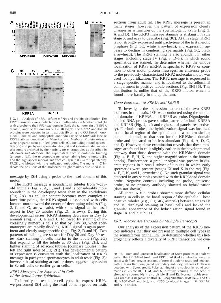
FIG. 5. Analysis of KRP3 isoform mRNA and protein distribution. TheKRP3 transcripts were detected on a multiple-tissue Northern blot (A)with a probe to the KRP3head domain (left), the tail domain of KRP3A(center), and the tail domain of KRP3B (right). The KRP3A and KRP3Bproteins were detected in testis extracts (B) using the KRP3head mono-clonal (lane H) and antipeptide antibodies (lane A: KRP3tail; lane B:KRP3Btail) as described in Materials and Methods. Protein extractswere prepared from purified germ cells (C), including round sperma-tids (RS) and pachytene spermatocytes (PS) and kinesin-related molec-ular motors enriched by their affinity for microtubules as described inMaterials and Methods. The supernatant from microtubule binding(unbound [U]), the microtubule pellet containing bound motors (B),and the high-speed supernatant from cell lysate (L) were separated byPAGE and blotted with the indicated antibodies. The marks in C in-dicate the positions of the molecular weight markers also used in B.
c
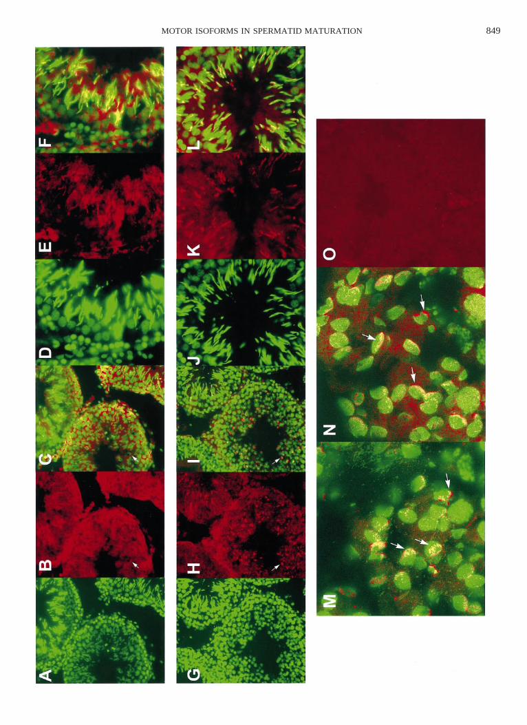
FIG. 6. Immunofluorescent localization of KRP3 proteins in adult rattestis. The KRP3Atail (A–F) and KRP3Btail (G–L ) antibodies were re-acted with fixed, frozen sections of normal adult rat testis and detectedwith a Texas Red-conjugated secondary antibody, whereas DNA wasdetected with Sytox green). Polar staining of the nucleus in round sper-matids is visible (B, H, M, and N, arrows); staining of the head ofelongating spermatids is also visible (E and K). Normal rabbit serumwas used as a negative control (O). Magnification 350 (A–C and G–H), 3100 (D–F and J–L), and 3250 (confocal images in M [KRP3A]and N [KRP3B]).
message by ISH using a probe to the head domain of thismotor.
The KRP3 message is abundant in tubules from 7-day-old animals (Fig. 2, A, E, and I) and is considerably moreabundant in the developing testis than in the epididymis(Fig. 2A). As cellular content becomes more complex atlater time points, the KRP3 signal is associated with cellslocated more toward the center of developing tubules (Fig.2, C and G, arrowheads), with some signal at the basalaspect in Day 20 tubules (Fig. 2C, arrows). During thisdevelopmental series, KRP3 staining decreases in Day 15animals (Fig. 2, B, F, and J), followed by staining of in-creasingly numerous cells so that by Day 30, when sper-matocytes are rapidly dividing, KRP3 signal is again prom-inent and clearly stage specific (e.g., Fig. 2, D and H). Twopatterns of staining are shown for Day 20 and Day 30 tes-tes: dark staining of cells interior to the tubule (Fig. 2G)that expand to fill the tubule at 30 days (Fig. 2H), andlighter staining of adjacent tubules (compare tubules in theleft and right sides of Fig. 2H). This developmental patternof expression is consistent with the localization of KRP3message in pachytene spermatocytes in adult testis (Fig. 3);however, basal staining at earlier times suggests expressionin Sertoli cells or earlier germ cells.
KRP3 Messages Are Expressed in Cellsof the Seminiferous Epithelium
To identify the testicular cell types that express KRP3,we performed ISH using the head domain probe on testis
sections from adult rat. The KRP3 message is present inmany stages; however, the pattern of expression clearlychanges as a function of the spermatogenic cycle (Fig. 3,A and B). The KRP3 message staining is striking in cyclestage X and easy to describe (Fig. 3C). At this stage, KRP3is not strongly expressed until pachytene of the first meioticprophase (Fig. 3C, white arrowhead), and expression ap-pears to decline in condensing spermatids (Fig. 3C, blackarrowhead). The KRP3 staining is also abundant in otherstages, including stage IV (Fig. 3, D–F), in which roundspermatids are stained. To determine whether the uniquelocalization of KRP3 mRNA is specific to KRP3 or com-mon to other motor protein messages, an antisense probeto the previously characterized KRP2 molecular motor wasused for hybridization. The KRP2 message is expressed ina stage-specific manner and is localized to the adluminalcompartment in positive tubule sections (Fig. 3H) [6]. Thisdistribution is unlike that of the KRP3 motor, which isfound more basally in the epithelium.
Gene Expression of KRP3A and KRP3B
To investigate the expression pattern of the two KRP3isoforms in the testis, ISH was conducted using the uniquetail domains of KRP3A and KRP3B as probe. Digoxigenin-labeled RNA probes gave similar patterns for both KRP3Aand KRP3B (Fig. 4, left and right set of panels, respective-ly). For both probes, the hybridization signal was localizedto the basal region of the epithelium in a pattern similar,but not identical, to that seen for the KRP3head domainprobe and appeared to be less abundant (Fig. 4, A, D, G,and J). However, close examination reveals that these mes-sages are found in cells slightly earlier in the developmentalpathway than those detected with the KRP3head domain(Fig. 4, B, E, H, K, and higher magnification in the bottompanels). Furthermore, a granular signal was present in dis-crete regions in a small subset of tubules in which earlyspermatids were present (stages IX and X in the rat) (Fig.4, E, F, K, and L, arrowheads). No such granular signal wasdetected in any samples stained with the KRP3head domainprobe. Negative controls including no probe, antisenseprobe, or no primary antibody showed no hybridization(data not shown).
All three KRP3 probes showed more diffuse cellularstaining in cells in the basal region of the epithelia. Thesepositive tubules (e.g., Fig. 4G, asterisk) between stages IVand VI displayed staining of basal cells and lacked thegranular appearance of the hybridization signal found instage IX and X tubules.
KRP3 Motors Are Encoded by Multiple Transcripts
Our analysis of the expression pattern of the KRP3 mo-tors indicates that they are present in multiple cell types inthe seminiferous epithelium. To determine whether this het-erogeneity reflects a diversity of KRP3 transcripts, we con-
849MOTOR ISOFORMS IN SPERMATID MATURATION

850 ZOU ET AL.
FIG. 7. KRP3B staining of early spermatids. The KRP3B tail antibodywas used to stain fixed, frozen sections of rat testis and was detectedwith a Texas Red-conjugated secondary antibody and stained for DNAwith DAPI. Sections were viewed using a triple-pass filter to visualizeKRP3B with respect to the nucleus. Arrows indicate staining outsidethe spermatid nucleus in a polar fashion. Two views are shown (A andB).
FIG. 8. RCC1 is localized to the caudal end of elongating spermatids. A polyclonal anti-RCC1 antibody raised against the Xenopus protein wasused to localize this protein in the mammalian testis. In the leftmost panel, DNA is visualized by staining with Sytox green. The middle panelshows detection of RCC1 using a Texas Red-conjugated secondary antibody, whereas the rightmost panel is the merged green and red images.The arrowheads indicate colocalization of the RCC1 protein with the caudal region of the spermatid head. Magnification 3250.
ducted Northern blot analysis of mRNA obtained from mul-tiple tissues using the probes previously used for ISH. Asexpected, the KRP3head probe, which detects all isoforms,recognizes at least three species in testicular mRNA: a rel-atively abundant transcript of 3.2 kilobases (kb), and twoless abundant transcripts of 2.7 and 1.7 kb (Fig. 5A, dotsbeside lane). This finding is consistent with our previouslypublished results concerning the sizes of KRP3 messages[6]. The 3.2-kb band is most intense in the testis samplebut is also visible in lung and brain (Fig. 5A; data notshown). Strikingly, a small transcript was detected in skel-etal muscle at approximately 750 nucleotides and was alsovisible in other experiments. The size of this transcript isquite unusual given that a typical motor domain alonewould be encoded by an approximately 1-kb message.
The KRP3Atail probe detected messages that were muchless abundant than those detected by the KRP3head probe(Fig. 5A, center panel); a 3.2-kb transcript was detected intestis. This finding suggests that the 3.2-kb transcript de-tected with the KRP3head probe might contain theKRP3Atail sequence. However, the low abundance of the
KRP3Atail transcript compared to that of the KRP3head-reactive species is consistent with KRP3Atail being a sub-set of KRP3 species of this size in the testis. A smallerKRP3Atail transcript of 2.7 kb was also visible in the testis(Fig. 5A, center panel). The KRP3Btail-positive transcriptswere also not abundant (Fig. 5A, right panel). Transcriptsof approximately 2.7 kb were detected in testis, spleen, andliver (Fig. 5C). In addition, an approximately 750-nucleo-tide species was found in the liver sample. Clearly, theKRP3 probes detect multiple isoforms, some of which maybe tissue specific.
KRP3 Motors Are Comprised of Multiple,Related Isoforms
To determine the precise cell types and intracellularstructures to which these motors associate, we developedimmunological reagents to the KRP3 isoforms. A mono-clonal antibody raised to a peptide found in the KRP3 headdomain and common to the isoforms was used to identifyKRP3-related proteins by immunoblot. The KRP3head an-tibody specifically recognizes a bacterially expressed KRP3head domain fragment (S.T. Brady, personal communica-tion). This antibody detects at least three polypeptides intestis lysates (Fig. 5B, lane H): proteins of approximately100 kDa, and a doublet at 70 kDa. Interestingly, theKRP3Atail antibody recognizes multiple polypeptides in to-tal testis lysate of 100 kDa, 70 kDa, and a small protein ofapproximately 22 kDa (Fig. 5B, lane A). The KRP3Btailantibody recognizes a protein of approximately 68 kDa(Fig. 5B, lane B). The proteins detected with each antibodyoverlap with those recognized by the KRP3head antibody.The KRP3Atail antibody detects three proteins identical insize to those found by the KRP3head antibody, and theKRP3Btail antibody recognizes an additional protein thatis also apparently detected by the KRP3head antibody.
This complex pattern indicates significant heterogeneityin the KRP3 polypeptides and reflects the multiple tran-scripts seen by Northern blot analysis (Fig. 5A). Althoughthe smallest transcript detected by Northern blot analysiswas not visible in the testis sample, a 22-kDa protein con-sistent with translation from this transcript was detected in
851MOTOR ISOFORMS IN SPERMATID MATURATION
total testis lysate and purified germ cells (Fig. 5C). In ad-dition, the 2.7- and 1.7-kb transcripts found in testicularmRNA agree with the 100- and 70-kDa doublet detectedby Western blot analysis of testis extract. Although a pro-tein of approximately 116 kDa, predicted from the 3.2-kbtranscript, was not strongly represented on immunoblot, incertain preparations we were able to detect higher-molec-ular-weight species with these antibodies (Fig. 5B, lane H).
KRP3A- and KRP3B-Related Polypeptides Are Expressedin Round Spermatids and Pachytene Spermatocytes
Molecular motor proteins were crudely purified fromgerm cell lysates by their affinity for microtubules in thepresence of the nonhydrolyzable ATP analogue AMP-PNPand the fractions immunoblotted with the KRP3Atail andKRP3Btail antibodies (Fig. 5C). Both the 70- and the 22-kDa KRP3A-reactive polypeptides are found in spermatidsand spermatocytes. The 68-kDa protein detected in whole-testis lysate with the KRP3B antibody is also found in sper-matids and spermatocytes. In addition, a 48-kDa proteinwas detected in these cells; this size is consistent with trans-lation from the 1.8-kb KRP3B clone. The enrichment ofthe 22-, 48-, and 68-kDa polypeptides in the microtubule-bound fraction (Fig. 5C, compare lanes labeled B withthose labeled U) indicates the presence of a kinesin-likemotor domain in these polypeptides. No polypeptides ofapproximately 70 kDa were detected in the total lysates orin the microtubule-bound fraction from adult Sertoli cells;however, the 22-kDa protein was detected in the boundfraction from these cells (data not shown).
KRP3A and KRP3B Are Associatedwith Developing Spermatids
Immunological reagents were developed to determinemore accurately whether the KRP3 motors associate withspecific intracellular structures in testicular cells by indirectimmunofluorescence. The KRP3head monoclonal antibodyproved to be unsuitable for indirect immunofluorescenceand was not used in these studies. Isoform-specific anti-bodies were raised in rabbits against peptides unique to thetail domain of each isoform (see Materials and Methods).Immunolocalization using the KRP3A (Fig. 6, A–F) andKRP3B (Fig. 6, G–L) antipeptide antibodies showed a sim-ilar, but not an identical, pattern. Both antibodies stain thenucleus of round (Fig. 6, B and H, arrows) and elongatespermatids (Fig. 6, E and K), although staining with theanti-KRPA antibody displayed a generalized background.Higher magnification revealed that both KRP3A- andKRP3B-isoform antibodies stain a roughly C-shaped regionat one pole of the nucleus in round spermatids, presumablythe acrosome (Fig. 6, M and N, arrows).
In addition, KRP3B was detected in very early sper-matids, being found on the surface of the nuclei in thesecells (Fig. 7, A and B, arrows). In some cases, a fibularnetwork was found to cover a small patch on the nuclearmembrane (data not shown). Both antibodies also showeda granular basal staining in stage IX tubules similar to thatseen by ISH (Fig. 4). Negative controls for these experi-ments included no primary antibody, incubation with nor-mal rabbit sera (Fig. 6O), or preabsorption with the anti-genic peptide (data not shown); all showed negligible signalcompared to the experimental sample.
Ran and Its Guanine Exchange Factor, RCC1, AreAssociated with the Nucleus of Developing Spermatids
The localization of KRP3 isoforms to the elongatingsperm head suggested a possible role in sperm head restruc-turing and/or chromatin condensation. Based on the ho-mology of KRP3B with RCC1, an antibody to RCC1 wasused to localize this protein required for chromatin conden-sation to cells of the testis. In addition to generalized cel-lular staining, particularly in the cytoplasm of spermato-gonia and spermatocytes, conspicuous staining was seen atthe caudal end of the nucleus of elongating spermatids (Fig.8, arrowheads). This staining was distinct from that of theKRP3-isoform antibodies and was located in a region ofthe approximately step-13 spermatid head occupied by themanchette structure rather than the homogeneous stainingof the nucleus displayed by the KRP3 antibodies.
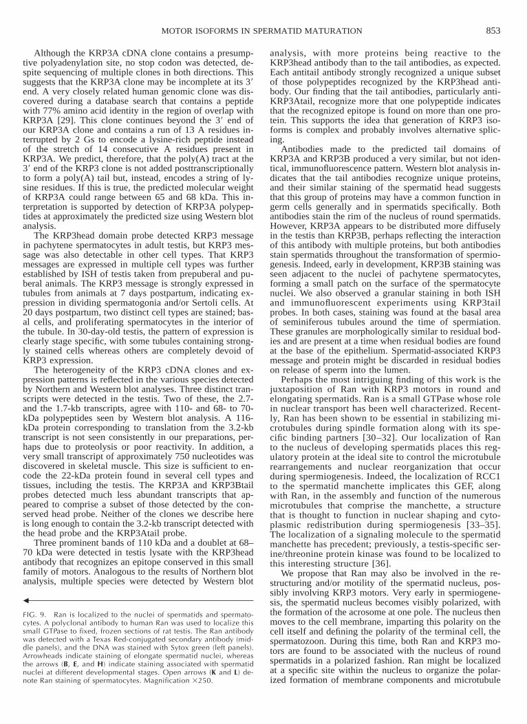
To determine whether the small GTPase Ran is associ-ated with spermatids along with RCC1, its guanine ex-change factor (GEF), and/or KRP3 motors, we localizedRan in adult testis sections. Ran is present in numerous celltypes in the epithelium, being concentrated in the nuclei ofpachytene spermatocytes (Fig. 9K, open arrows) and elon-gate spermatids (Fig. 9, B and C, arrowheads). The distri-bution of Ran in approximately step-13 spermatids is dis-tinct from its GEF RCC1 (Fig. 8), being found throughoutthe spermatid head. In some individuals, a line of Ran stain-ing is seen to outline the spermatid head (Fig. 9, B and C,arrows). In addition, Ran is localized to one pole of roundspermatids (Fig. 9, E and F, arrows). Ran staining associ-ated with spermatid nuclei is seen throughout spermiogen-esis, including spermatids just before spermiation (Fig. 9,E and F, arrows). In many views, Ran staining appears tospread over one pole of the spermatid nucleus (Fig. 9, Hand I, arrows) in a manner similar to that seen for KRP3isoforms (Fig. 6H). In addition, Ran staining takes the formof a disc-shaped structure within the nucleus of a subset ofspermatocytes (Fig. 9L, open arrows) in approximatelystage II tubules before Ran becomes strongly apparent onround spermatids.
DISCUSSION
The process of spermatogenesis involves profound re-arrangement of the microtubule cytoskeleton of germ cellswith the formation and dissolution of several microtubulecomplexes, including the mitotic and meiotic spindles andthe manchette. In addition to striking changes in the shapeof male gametes during spermatogenesis, the shape andcontent of the spermatid nucleus changes dramatically aswell. We have identified new kinesin-related molecular mo-tors, KRP3 isoforms, that are associated with developingmale germ cells and that may play roles in transformationof the spermatid nucleus.
Previously, KRP3 was shown to be expressed in cells ofthe rat seminiferous epithelium [6]. Initial library screeningusing the KRP3head domain identified earlier as probe re-vealed heterogeneity in KRP3 clones. At least two newkinesin-related cDNAs containing the KRP3head domainwere identified in the rat testis. These clones, termedKRP3A and KRP3B, are identical to one another for mostof their lengths, but they diverge 1287 nucleotides from theATG start codon in KRP3A. Genetic mapping experimentssuggest that a common gene encodes these two cDNAs.Probes specific for each tail domain cosegregate with oneanother and also with a head domain probe (N.A. Jenkinsand N.G. Copeland, personal communication).

852 ZOU ET AL.
853MOTOR ISOFORMS IN SPERMATID MATURATION
b
FIG. 9. Ran is localized to the nuclei of spermatids and spermato-cytes. A polyclonal antibody to human Ran was used to localize thissmall GTPase to fixed, frozen sections of rat testis. The Ran antibodywas detected with a Texas Red-conjugated secondary antibody (mid-dle panels), and the DNA was stained with Sytox green (left panels).Arrowheads indicate staining of elongate spermatid nuclei, whereasthe arrows (B, E, and H) indicate staining associated with spermatidnuclei at different developmental stages. Open arrows (K and L) de-note Ran staining of spermatocytes. Magnification 3250.
Although the KRP3A cDNA clone contains a presump-tive polyadenylation site, no stop codon was detected, de-spite sequencing of multiple clones in both directions. Thissuggests that the KRP3A clone may be incomplete at its 39end. A very closely related human genomic clone was dis-covered during a database search that contains a peptidewith 77% amino acid identity in the region of overlap withKRP3A [29]. This clone continues beyond the 39 end ofour KRP3A clone and contains a run of 13 A residues in-terrupted by 2 Gs to encode a lysine-rich peptide insteadof the stretch of 14 consecutive A residues present inKRP3A. We predict, therefore, that the poly(A) tract at the39 end of the KRP3 clone is not added posttranscriptionallyto form a poly(A) tail but, instead, encodes a string of ly-sine residues. If this is true, the predicted molecular weightof KRP3A could range between 65 and 68 kDa. This in-terpretation is supported by detection of KRP3A polypep-tides at approximately the predicted size using Western blotanalysis.
The KRP3head domain probe detected KRP3 messagein pachytene spermatocytes in adult testis, but KRP3 mes-sage was also detectable in other cell types. That KRP3messages are expressed in multiple cell types was furtherestablished by ISH of testis taken from prepuberal and pu-beral animals. The KRP3 message is strongly expressed intubules from animals at 7 days postpartum, indicating ex-pression in dividing spermatogonia and/or Sertoli cells. At20 days postpartum, two distinct cell types are stained; bas-al cells, and proliferating spermatocytes in the interior ofthe tubule. In 30-day-old testis, the pattern of expression isclearly stage specific, with some tubules containing strong-ly stained cells whereas others are completely devoid ofKRP3 expression.
The heterogeneity of the KRP3 cDNA clones and ex-pression patterns is reflected in the various species detectedby Northern and Western blot analyses. Three distinct tran-scripts were detected in the testis. Two of these, the 2.7-and the 1.7-kb transcripts, agree with 110- and 68- to 70-kDa polypeptides seen by Western blot analysis. A 116-kDa protein corresponding to translation from the 3.2-kbtranscript is not seen consistently in our preparations, per-haps due to proteolysis or poor reactivity. In addition, avery small transcript of approximately 750 nucleotides wasdiscovered in skeletal muscle. This size is sufficient to en-code the 22-kDa protein found in several cell types andtissues, including the testis. The KRP3A and KRP3Btailprobes detected much less abundant transcripts that ap-peared to comprise a subset of those detected by the con-served head probe. Neither of the clones we describe hereis long enough to contain the 3.2-kb transcript detected withthe head probe and the KRP3Atail probe.
Three prominent bands of 110 kDa and a doublet at 68–70 kDa were detected in testis lysate with the KRP3headantibody that recognizes an epitope conserved in this smallfamily of motors. Analogous to the results of Northern blotanalysis, multiple species were detected by Western blot
analysis, with more proteins being reactive to theKRP3head antibody than to the tail antibodies, as expected.Each antitail antibody strongly recognized a unique subsetof those polypeptides recognized by the KRP3head anti-body. Our finding that the tail antibodies, particularly anti-KRP3Atail, recognize more that one polypeptide indicatesthat the recognized epitope is found on more than one pro-tein. This supports the idea that generation of KRP3 iso-forms is complex and probably involves alternative splic-ing.
Antibodies made to the predicted tail domains ofKRP3A and KRP3B produced a very similar, but not iden-tical, immunofluorescence pattern. Western blot analysis in-dicates that the tail antibodies recognize unique proteins,and their similar staining of the spermatid head suggeststhat this group of proteins may have a common function ingerm cells generally and in spermatids specifically. Bothantibodies stain the rim of the nucleus of round spermatids.However, KRP3A appears to be distributed more diffuselyin the testis than KRP3B, perhaps reflecting the interactionof this antibody with multiple proteins, but both antibodiesstain spermatids throughout the transformation of spermio-genesis. Indeed, early in development, KRP3B staining wasseen adjacent to the nuclei of pachytene spermatocytes,forming a small patch on the surface of the spermatocytenuclei. We also observed a granular staining in both ISHand immunofluorescent experiments using KRP3tailprobes. In both cases, staining was found at the basal areaof seminiferous tubules around the time of spermiation.These granules are morphologically similar to residual bod-ies and are present at a time when residual bodies are foundat the base of the epithelium. Spermatid-associated KRP3message and protein might be discarded in residual bodieson release of sperm into the lumen.
Perhaps the most intriguing finding of this work is thejuxtaposition of Ran with KRP3 motors in round andelongating spermatids. Ran is a small GTPase whose rolein nuclear transport has been well characterized. Recent-ly, Ran has been shown to be essential in stabilizing mi-crotubules during spindle formation along with its spe-cific binding partners [30–32]. Our localization of Ranto the nucleus of developing spermatids places this reg-ulatory protein at the ideal site to control the microtubulerearrangements and nuclear reorganization that occurduring spermiogenesis. Indeed, the localization of RCC1to the spermatid manchette implicates this GEF, alongwith Ran, in the assembly and function of the numerousmicrotubules that comprise the manchette, a structurethat is thought to function in nuclear shaping and cyto-plasmic redistribution during spermiogenesis [33–35].The localization of a signaling molecule to the spermatidmanchette has precedent; previously, a testis-specific ser-ine/threonine protein kinase was found to be localized tothis interesting structure [36].
We propose that Ran may also be involved in the re-structuring and/or motility of the spermatid nucleus, pos-sibly involving KRP3 motors. Very early in spermiogene-sis, the spermatid nucleus becomes visibly polarized, withthe formation of the acrosome at one pole. The nucleus thenmoves to the cell membrane, imparting this polarity on thecell itself and defining the polarity of the terminal cell, thespermatozoon. During this time, both Ran and KRP3 mo-tors are found to be associated with the nucleus of roundspermatids in a polarized fashion. Ran might be localizedat a specific site within the nucleus to organize the polar-ized formation of membrane components and microtubule

854 ZOU ET AL.
structures for spermatid development. This idea is support-ed by the detection of Ran in the nucleus of spermatocytesto form a disc-shaped structure at one pole of the nucleus(Fig. 9L).
Certain views of spermatids reactive for Ran and KRP3isoforms show staining in a shape similar to that of thedeveloping acrosome, including an acrosomal granule. Nu-clear reshaping and formation of the acrosome occur incoordination during spermiogenesis, and these events likelyare regulated by a common mechanism. For example, aKRP3 isoform could be involved in transport of vesicles toform the acrosome at a site predetermined by the locationof signaling molecules. Similarly, nucleation and stabili-zation of manchette microtubules in a directed manner onthe spermatid nucleus could be accomplished through acommon signaling mechanism. Acrosome formation is de-pendent on microtubules and shares features with vesicletransport in other systems [37, 38]. The KRP3B staining inearly spermatids before formation of the acrosome is verysimilar to the placement and structure of the Golgi appa-ratus in these cells. Indeed, the staining we observe forKRP3 motors in round spermatids is almost identical to thelocalization of Golgi and perinuclear theca markers to thedeveloping bull acrosome [39].
Another possible function of KRP3 motors, consistentwith their immunolocalization to spermatid heads and ex-pression by Sertoli cells, is in microtubule-based movementof elongating spermatids within the epithelium [10, 40].Recently, an isoform of cytoplasmic dynein was localizedto the apical crypts concentrated in areas juxtaposed to thesperm head and postulated to be one motor responsible fortranslocation toward the apical aspect of the cell [5, 41].However, a candidate plus-end-directed motor responsiblefor movement of spermatids toward the base of the epithe-lium has not been identified. The KRP3 isoforms are pu-tative plus-end-directed motors associated with the sper-matid head that might be involved in the basal translocationof spermatids. Individual isoforms might have other rolesin spermatid maturation, including formation of the acro-some and/or spermatid head remodeling. The motor iso-forms described here are excellent candidates for motorsinvolved in the complex cellular transformations that occurduring spermiogenesis and are likely targets for regulationduring the dramatic changes in the spermatid nucleus.
ACKNOWLEDGMENTS
We thank Virginia Best, Mark Albertino, Colleen Nichols, Robin Wray,and Purnima Jani for providing expert technical assistance and Dr. ScottBrady for critical reading of the manuscript.
REFERENCES
1. Endow SA. Microtubule motors in spindle and chromosome motility.Eur J Biochem 1999; 262:12–18.
2. Hirokawa N. Kinesin and dynein superfamily proteins and the mech-anism of organelle transport. Science 1998; 279:519–526.
3. Goodson HV, Valetti C, Kreis TE. Motors and membrane traffic. CurrOpin Cell Biol 1997; 9:18–28.
4. Navolanic PM, Sperry AO. Identification of isoforms of a mitoticmotor in mammalian spermatogenesis. Biol Reprod 2000; 62:1360–1369.
5. Miller MG, Mulholland DJ, Vogl AW. Rat testis motor proteins as-sociated with spermatid translocation (dynein) and spermatid flagella(kinesin-II). Biol Reprod 1999; 60:1047–1056.
6. Sperry AO, Zhao LP. Kinesin-related proteins in the mammalian tes-tes: candidate motors for meiosis and morphogenesis. Mol Biol Cell1996; 7:289–305.
7. Hall ES, Eveleth J, Jiang C, Redenbach DM, Boekelheide K. Distri-
bution of the microtubule-dependent motors cytoplasmic dynein andkinesin in rat testis. Biol Reprod 1992; 46:817–828.
8. Molina I, Baars S, Brill JA, Hales KG, Fuller MT, Ripoll P. A chro-matin-associated kinesin-related protein required for normal mitoticchromosome segregation in Drosophila. J Cell Biol 1997; 139:1361–1371.
9. Wang W, Chi T, Xue Y, Zhou S, Kuo A, Crabtree GR. ArchitecturalDNA binding by a high-mobility-group/kinesin-like subunit in mam-malian SWI/SNF-related complexes. Proc Natl Acad Sci U S A 1998;95:492–498.
10. Beach SF, Vogl AW. Spermatid translocation in the rat seminiferousepithelium: coupling membrane trafficking machinery to a junctionplaque. Biol Reprod 1999; 60:1036–1046.
11. Carazo-Salas RE, Guarguaglini G, Gruss OJ, Segref A, Karsenti E,Mattaj IW. Generation of GTP-bound Ran by RCC1 is required forchromatin-induced mitotic spindle formation. Nature 1999; 400:178–181.
12. Wilde A, Zheng Y. Stimulation of microtubule aster formation andspindle assembly by the small GTPase Ran. Science 1999; 284:1359–1362.
13. Hetzer M, Bilbao-Cortes D, Walther TC, Gruss OJ, Mattaj IW. GTPhydrolysis by Ran is required for nuclear envelope assembly. Mol Cell2000; 5:1013–1024.
14. Lupas A, Van Dyke M, Stock J. Predicting coiled coils from proteinsequences. Science 1991; 252:1162–1164.
15. Berger B, Wilson DB, Wolf E, Tonchev T, Milla M, Kim PS. Pre-dicting coiled coils by use of pairwise residue correlations. Proc NatlAcad Sci U S A 1995; 92:8259–8263.
16. Komminoth P. Detection of mRNA in tissue sections using DIG-la-beled RNA and oligonucleotide probs. In: Non-Radioactive In SituHybridization Application Manual. Mannheim, Germany: BoehringerMannheim GmbH; 1996: 126–135.
17. Bellve AR, Cavicchia JC, Millette CF, O’Brien DA, Bhatnagar YM,Dym M. Spermatogenic cells of the prepuberal mouse. Isolation andmorphological characterization. J Cell Biol 1977; 74:68–85.
18. Newton SC, Millette CF. Sertoli cell plasma membrane polypeptidesinvolved in spermatogenic cell-Sertoli cell adhesion. J Androl 1992;13:160–171.
19. Galdieri M, Zani B. Hormonal induced changes in Sertoli cell gly-coproteins. Cell Biol Int Rep 1981; 5:111.
20. Neely MD, Boekelheide K. Sertoli cell processes have axoplasmicfeatures: an ordered microtubule distribution and an abundant highmolecular weight microtubule-associated protein (cytoplasmic dyne-in). J Cell Biol 1988; 107:1767–1776.
21. Pfister KK, Wagner MC, Bloom GS, Brady ST. Modification of themicrotubule-binding and ATPase activities of kinesin by N-ethylmal-eimide (NEM) suggests a role for sulfhydryls in fast axonal transport.Biochemistry 1989; 28:9006–9012.
22. Towbin H, Staehelin T, Gordon J. Electrophoretic transfer of proteinsfrom polyacrylamide gels to nitrocellulose sheets: procedure and someapplications. Proc Natl Acad Sci U S A 1979; 76:4350–4354.
23. Moore MS, Blobel G. The GTP-binding protein Ran/TC4 is requiredfor protein import into the nucleus. Nature 1993; 365:661–663.
24. Kozak M. Point mutations define a sequence flanking the AUG ini-tiator codon that modulates translation by eukaryotic ribosomes. Cell1986; 44:283–292.
25. Nakagawa T, Tanaka Y, Matsuoka E, Kondo S, Okada Y, Noda Y,Kanai Y, Hirokawa N. Identification and classification of 16 new ki-nesin superfamily (KIF) proteins in mouse genome. Proc Natl AcadSci U S A 1997; 94:9654–9659 [published erratum appears in ProcNatl Acad Sci U S A 1999; 96:4214].
26. Bairoch A, Bucher P, Hofmann K. The PROSITE database: its statusin 1977. Nucleic Acids Res 1997; 25:217–221.
27. Murzin AG, Brenner SE, Hubbard T, Chothia C. SCOP: a structuralclassification of proteins database for the investigation of sequencesand structures. J Mol Biol 1995; 247:536–540.
28. Ohtsubo M, Okazaki H, Nishimoto T. The RCC1 protein, a regulatorfor the onset of chromosome condensation locates in the nucleus andbinds to DNA. J Cell Biol 1989; 109:1389–1397.
29. Patel R. Human DNA sequence from clone RP5-1043E3 on chro-mosome 6p21.1–21.2 contains part of a novel gene, a transcriptionfactor E2F4 pseudogene, ESTs, STSs and GSSs. Bethesda, MD:GenBank National Center for Biotechnology Information, NationalLibrary of Medicine; 2000: accession no. AL36102.
30. Azuma Y, Dasso M. The role of Ran in nuclear function. Curr OpinCell Biol 2000; 12:302–307.

855MOTOR ISOFORMS IN SPERMATID MATURATION
31. Fleig U, Salus SS, Karig I, Sazer S. The fission yeast Ran GTPase isrequired for microtubule integrity. J Cell Biol 2000; 151:1101–1112.
32. Guarguaglini G, Renzi L, D’Ottavio F, Di Fiore B, Casenghi M, Cun-dari E, Lavia P. Regulated Ran-binding protein 1 activity is requiredfor organization and function of the mitotic spindle in mammaliancells in vivo. Cell Growth Differ 2000; 11:455–465.
33. Cole A, Meistrich ML, Cherry LM, Trostle-Weige PK. Nuclear andmanchette development in spermatids of normal and azh/azh mutantmice. Biol Reprod 1988; 38:385–401.
34. Meistrich ML, Trostle-Weige PK, Russell LD. Abnormal manchettedevelopment in spermatids of azh/azh mutant mice. Am J Anat 1990;188:74–86.
35. Russell LD, Russell JA, MacGregor GR, Meistrich ML. Linkage ofmanchette microtubules to the nuclear envelope and observations ofthe role of the manchette in nuclear shaping during spermiogenesis inrodents. Am J Anat 1991; 192:97–120.
36. Walden PD, Cowan NJ. A novel 205-kilodalton testis-specific serine/threonine protein kinase associated with microtubules of the spermatidmanchette. Mol Cell Biol 1993; 13:7625–7635.
37. Nakai M, Hess RA, Matsuo F, Gotoh Y, Nasu T. Further observationson carbendazim-induced abnormalities of spermatid morphology inrats. Tissue Cell 1997; 29:477–485.
38. Tanii I, Yoshinaga K, Toshimori K. The effects of brefeldin A onacrosome formation and protein transport to the acrosome in organcultures of rat seminiferous tubules. J Electron Microsc (Tokyo) 1998;47:161–167.
39. Moreno RD, Ramalho-Santos J, Sutovsky P, Chan EKL, Schatten G.Vesicular traffic and Golgi apparatus dynamics during mammalianspermatogenesis: implications for acrosome architecture. Biol Reprod2000; 63:89–98.
40. Vogl AW, Pfeiffer DC, Mulholland D, Kimel G, Guttman J. Uniqueand multifunctional adhesion junctions in the testis: ectoplasmic spe-cializations. Arch Histol Cytol 2000; 63:1–15.
41. Guttman JA, Kimel GH, Vogl AW. Dynein and plus-end microtubule-dependent motors are associated with specialized Sertoli cell junctionplaques (ectoplasmic specializations). J Cell Sci 2000; 113:2167–2176.