ksc 2014 pulmonary venous anomaly - embryology and anatomy · pulmonary venous anomaly - embryology...
TRANSCRIPT

Pulmonary Venous Anomaly - Embryology and Anatomy
Lucy Youngmin Eun, MD, PhD
Pediatric Cardiology
Yonsei University Severance Hospital
KSC 2014
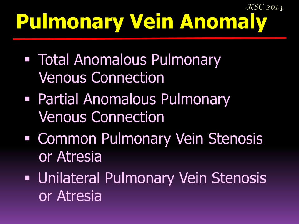
Pulmonary Vein Anomaly
Total Anomalous Pulmonary Venous Connection
Partial Anomalous Pulmonary Venous Connection
Common Pulmonary Vein Stenosis or Atresia
Unilateral Pulmonary Vein Stenosis or Atresia
KSC 2014

The development of pulmonary vein begins at 27~29 days of gestation.
The vein arise from the lung buds that are part of the vascular plexus of the forgut, the splanchnic plexus.
There are multiple connections to the umbilicovitelline and cardinal venous system.
Coalesce to form four vessels that join with common pulmonary vein that emerges from the back wall of atrium.
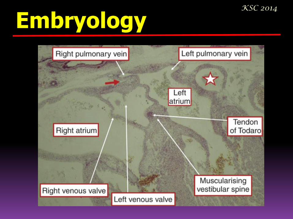
Embryology KSC 2014
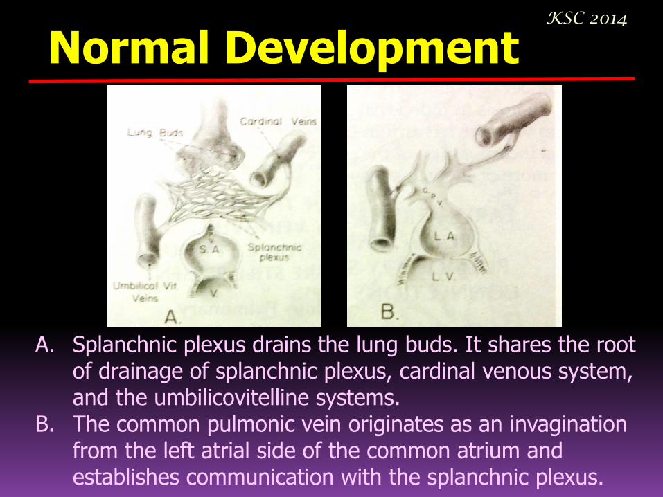
A. Splanchnic plexus drains the lung buds. It shares the root of drainage of splanchnic plexus, cardinal venous system, and the umbilicovitelline systems.
B. The common pulmonic vein originates as an invagination from the left atrial side of the common atrium and establishes communication with the splanchnic plexus.
Normal Development KSC 2014

C. No longer necessary, the primitive pulmonary venous connections disappear. D. Finally, the individual pulmonary veins are incorporated into the left atrium, and the common pulmonary vein no longer exists.
Normal Development KSC 2014
The Systemic venous tributaries identified in molecular terms expression of the transcription factor Tbx18. The Pulmonary veins do not contain this protein.
Embryology KSC 2014

Embryology - 4 wks KSC 2014
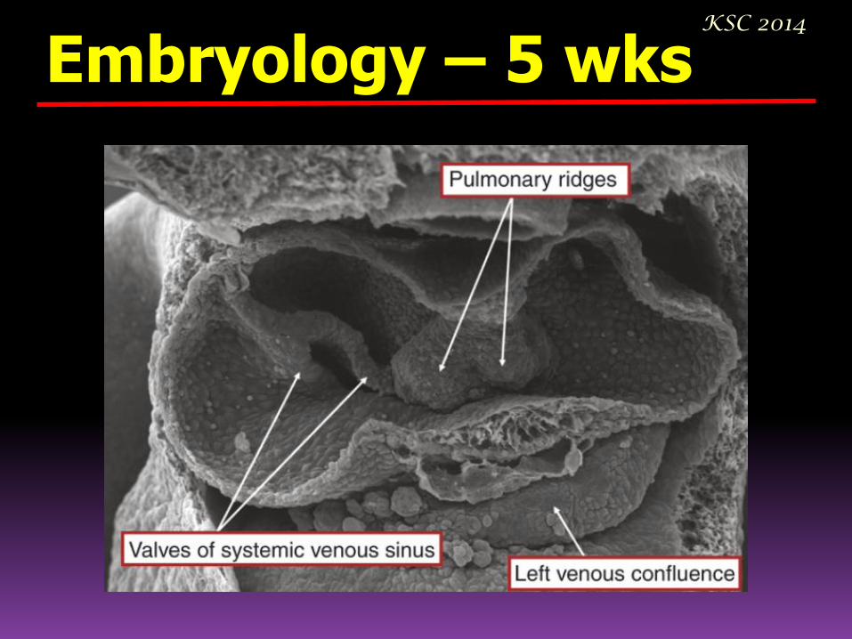
Embryology – 5 wks KSC 2014

Embryology – 6 wks KSC 2014

Embryology – 51 wks KSC 2014

Embryology KSC 2014
Embryology KSC 2014

Embryology KSC 2014

Embryologic Classification I. Atresia of common pulmonary vein while pulmonary systemic venous connections are still present
A. Partial anomalous pulmonary venous connection
B. Total anomalous pulmonary venous connection
II. Atresia of the common pulmonary vein after pulmonary systemic venous connections are obliterated
- Atresia of the common pulmonary vein
III. Stenosis of the common pulmonary vein
- Cor triatriatum
IV. Abnormal absorption of the common pulmonary vein into the left atrium
A. Stenosis of the individual pulmonary vein
B. Abnormal number of pulmonary vein
KSC 2014

Anomalous Connection KSC 2014
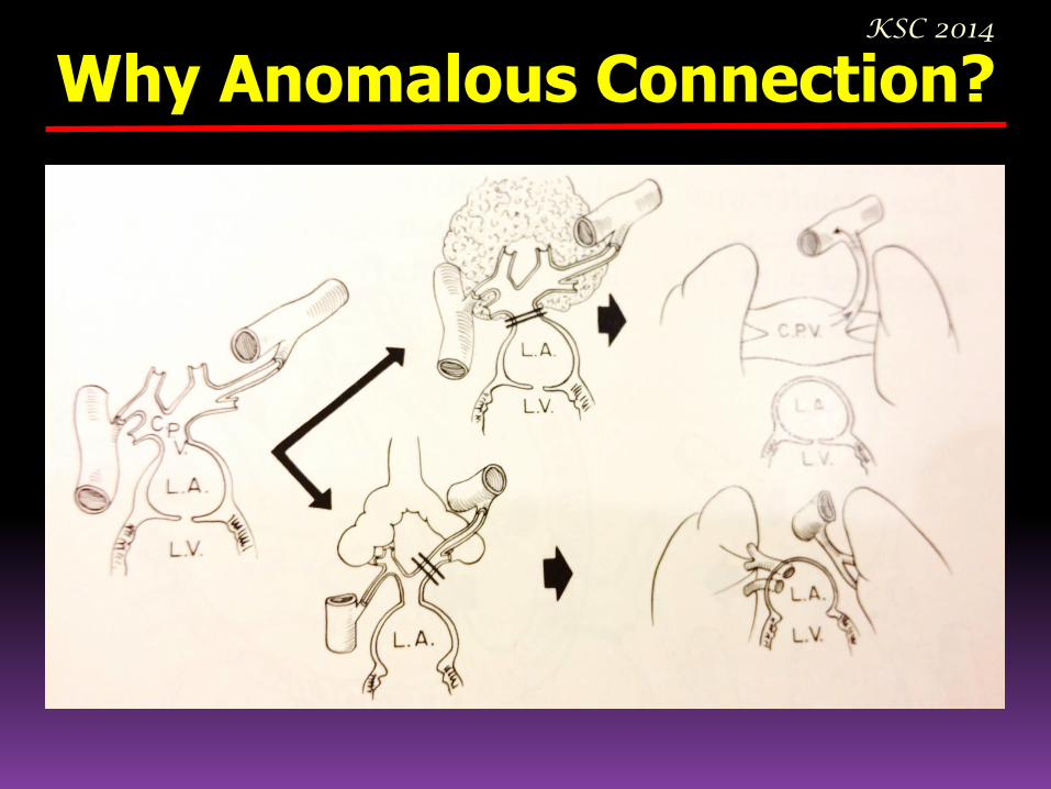
Why Anomalous Connection? KSC 2014

Why Anomalous Connection? KSC 2014

Pulmonary Vein Anomaly
Total Anomalous Pulmonary Venous Connection
Partial Anomalous Pulmonary Venous Connection
Common Pulmonary Vein Stenosis or Atresia
Unilateral Pulmonary Vein Stenosis or Atresia
KSC 2014

Total Anomalous Pulmonary Venous Connection
A pulmonary vein is connected anomalously only when it is attached to a site other than the morphologically left atrium
- 1 % of all congenital heart disease
- Boy : Girl = 4 : 1
KSC 2014

KSC 2014
Total Anomalous Pulmonary Venous Connection

Different Sites of Anomalous Connection KSC 2014

Different Sites of Anomalous Connection KSC 2014

Anatomy of TAPVC
Anomalous connections
Stenotic connections
Abnormal numbers of pulmonary veins
KSC 2014
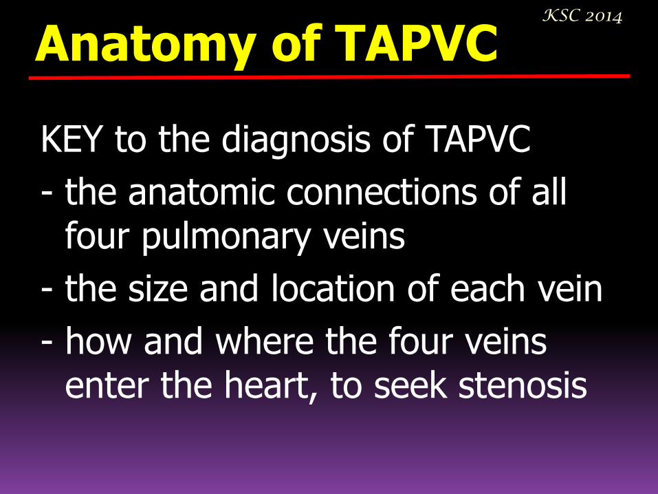
Anatomy of TAPVC
KEY to the diagnosis of TAPVC
- the anatomic connections of all four pulmonary veins
- the size and location of each vein
- how and where the four veins enter the heart, to seek stenosis
KSC 2014
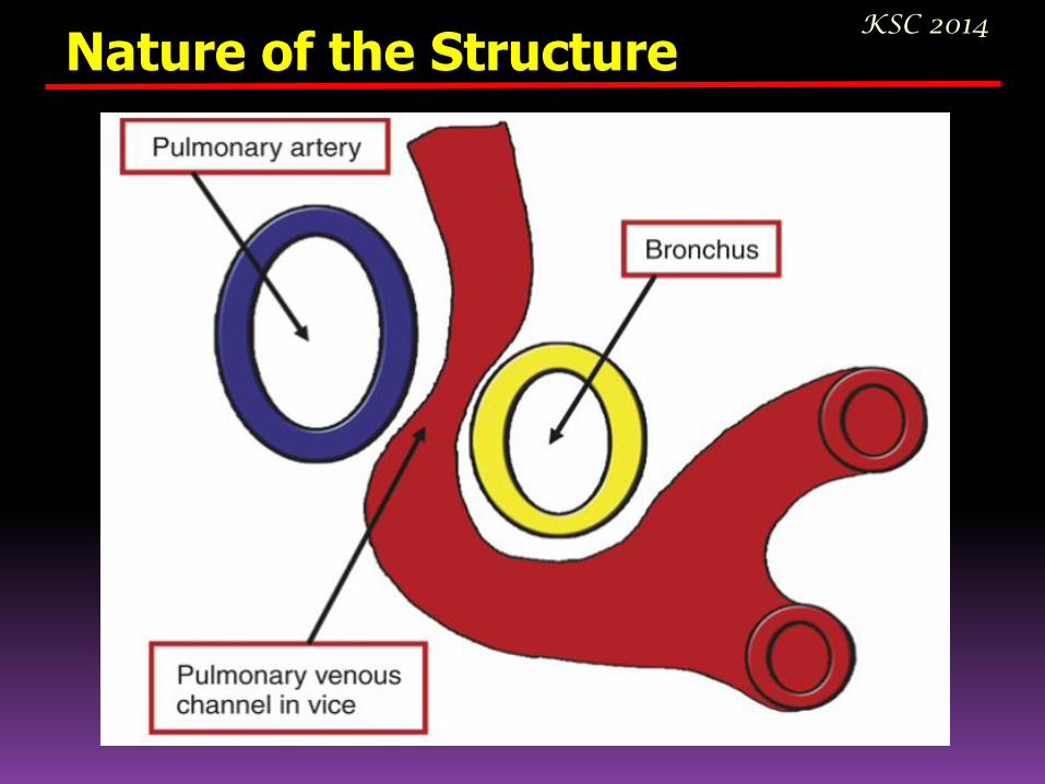
Nature of the Structure KSC 2014
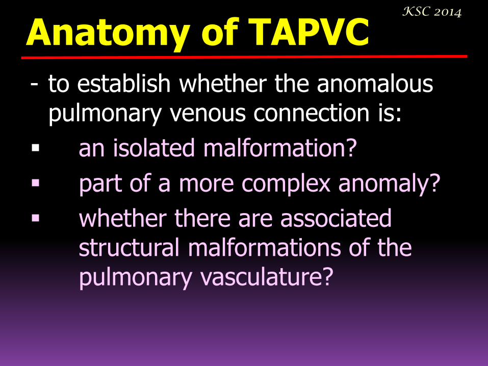
Anatomy of TAPVC
- to establish whether the anomalous pulmonary venous connection is:
an isolated malformation?
part of a more complex anomaly?
whether there are associated structural malformations of the pulmonary vasculature?
KSC 2014

2/3 of patients :
TAPVC is an isolated anomaly, associated only with the required interatrial communication.
1/3 of patients :
significant other cardiac defects occur, truncus arteriosus, pulmonary atresia, AVSD, TGA, single ventricle physiology, or heterotaxy syndrome (asplenia or polysplenia).
KSC 2014
Total Anomalous Pulmonary Venous Connection

TAPVC /c Pulmonary Venous Obstruction KSC 2014
Without severe pulmonary venous obstruction: - Present in heart failure at 2~3 months of age. - History of difficulty of feeding, pneumonia. - Cyanosis is rare. With severe pulmonary venous obstruction: - Obvious severe Cyanosis - Skin mottling reflecting poor peripheral perfusion and metabolic acidosis - Tachypnea

Inheritance of TAPVC
• Cat eye syndrome –
trisomy of the centromeric portion of chromosome 22q
• Association with a deletion of chromosome 2q31-q33
• Holt-Oram syndrome
• Asplenia syndrome
KSC 2014

TAPVC- Supracardiac KSC 2014
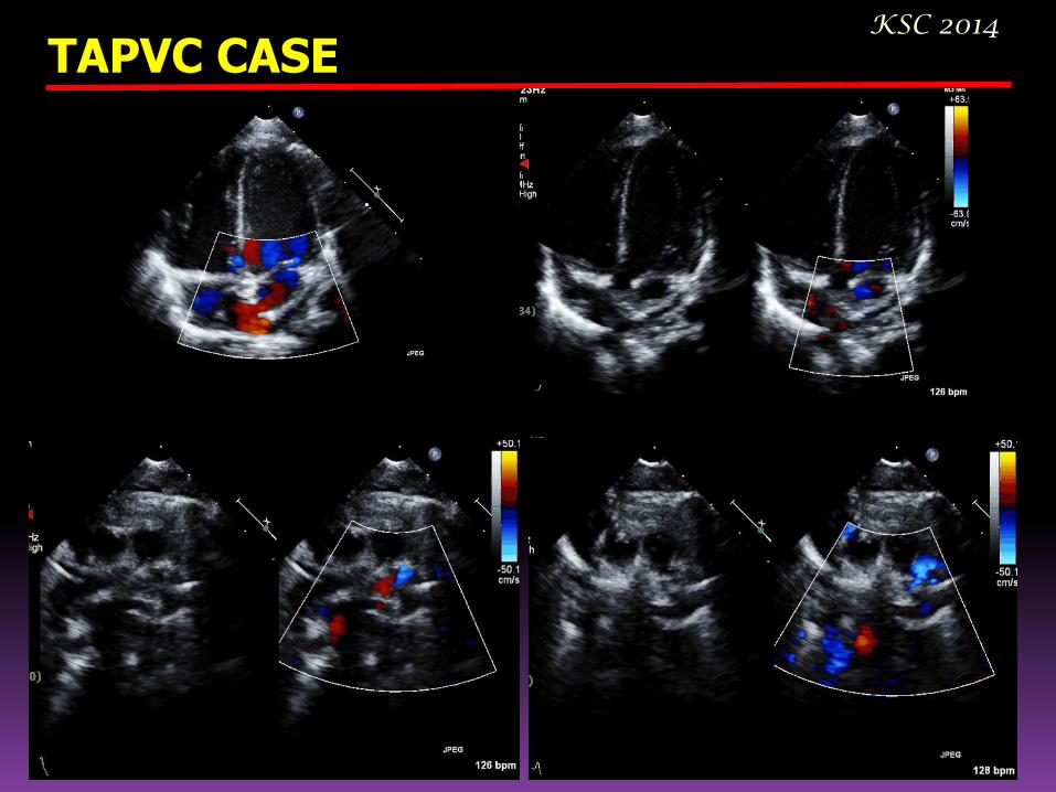
TAPVC CASE KSC 2014

TAPVC- Supracardiac and Cardiac KSC 2014

TAPVC- Cardiac KSC 2014

TAPVC- Cardiac KSC 2014

TAPVC- Right Isomerism KSC 2014

TAPVC- Infracardiac KSC 2014
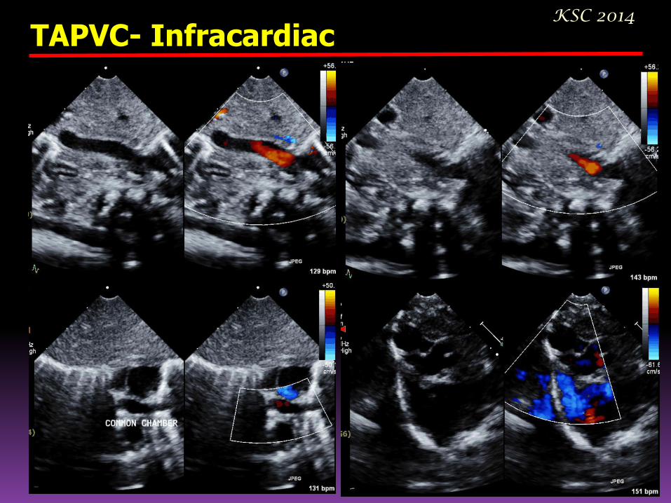
TAPVC- Infracardiac KSC 2014

TAPVC or not ? KSC 2014
Differential Diagnosis Complete TGA with large VSD AVSD with common atrium Levoatrial cardinal vein associated
with mitral atresia and intact atrial septum
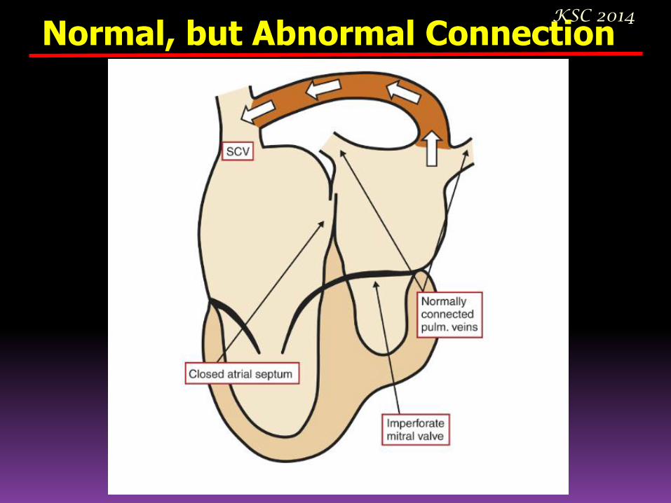
Normal, but Abnormal Connection KSC 2014

KSC 2014 Partial Anomalous Pulmonary Venous Connection
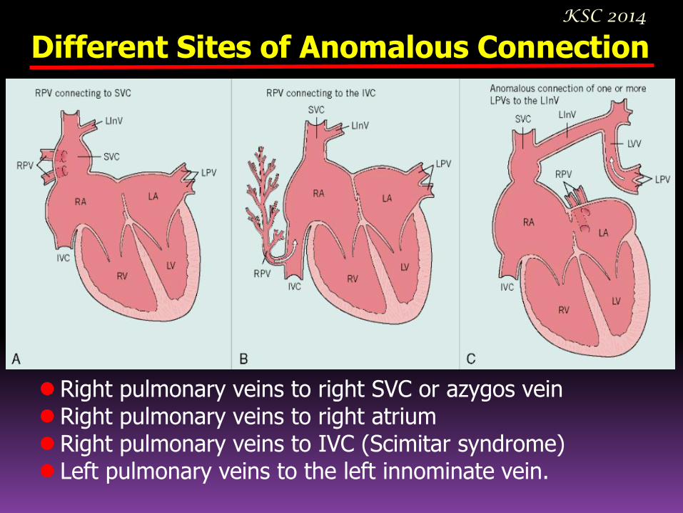
Different Sites of Anomalous Connection KSC 2014
Right pulmonary veins to right SVC or azygos vein Right pulmonary veins to right atrium Right pulmonary veins to IVC (Scimitar syndrome) Left pulmonary veins to the left innominate vein.

CASE KSC 2014
- M/ 19 yr, Palpitation, DOE - RLL, RML, part of RUL drained by SCMV connected to the intrahepatic IVC - Scimitar variant with non-restrictive connection
of meandering vein to LA

Common Pulmonary Vein Stenosis - Cor Triatriatum
KSC 2014
I. Accessory atrial chamber receives all pulmonary veins and communicates with left atrium
A. no other connection – classic cor triatriatum
B. other anomalous connection – RA or TAPVR
II. Accessory atrial chamber receives all pulmonary veins and does not communicate with left atrium
A. Anomalous connection to right atrium directly
B. With total anomalous pulmonary connection
III. Subtotal Cor triatriatum
A. Accessory atrial chamber receives part of pulmonary veins and connect to left atrium
B. Accessory atrial chamber receives part of the pulmonary veins and connects to right atrium
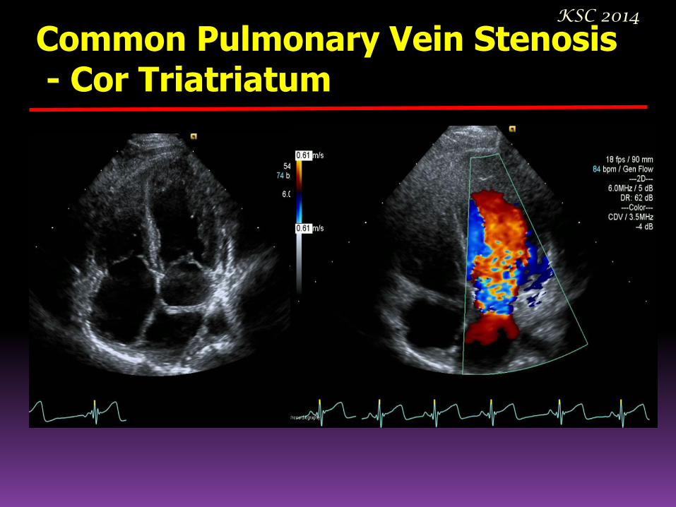
Common Pulmonary Vein Stenosis - Cor Triatriatum
KSC 2014

Common Pulmonary Vein Atresia KSC 2014

Anomalous Pulmonary to Systemic Collateral Vein & Levoatrial Cardinal Vein
KSC 2014

Stenosis or Atresia of Individual Pulmonary Vein
KSC 2014

Stenosis or Atresia of Individual Pulmonary Vein
KSC 2014
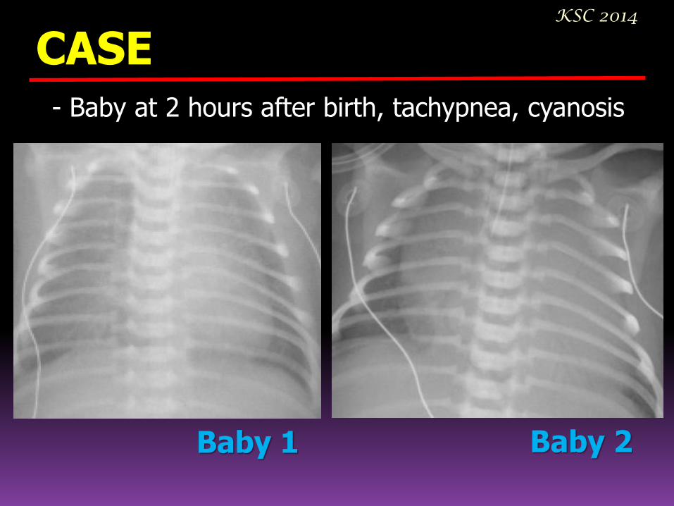
CASE KSC 2014
- Baby at 2 hours after birth, tachypnea, cyanosis
Baby 1 Baby 2

CASE KSC 2014
- Baby at 2 hours after birth, tachypnea, cyanosis
Baby 1 Baby 2

KSC 2014
Thank You ! 감사합니다.