l c a l derma journal of clinical & experimental t i n o i ... · evaluation of invasive...
TRANSCRIPT

Evaluation of Invasive Squamous Cell Carcinoma, Seborrheic Keratosis andVerruca Vulgaris in Superficial Shave Biopsies Using p16, p53, p63, andPHLDA1 ImmunohistochemistryRyanne A Brown1 and Jinah Kim1,2*
1Department of Pathology, Stanford Medicine, Stanford, CA, USA2Department of Dermatology, Stanford Medicine, Stanford, CA, USA
Corresponding author: Jinah Kim, Department of Pathology and Dermatology, Stanford University School of Medicine, 300 Pasteur Drive, L235, Stanford, CA 94305,USA, Tel: 650-736-1068; Fax: 650-725-7409; E-mail: [email protected]
Received date: February 23, 2016; Accepted date: April 19, 2016; Published date: April 25, 2016
Copyright: © 2016 Brown RA, et al. This is an open-access article distributed under the terms of the Creative Commons Attribution License, which permits unrestricteduse, distribution, and reproduction in any medium, provided the original author and source are credited.
Abstract
Occasionally, the distinction between malignant and benign is challenging in superficial shave biopsies ofsquamoproliferative lesions. This phenomenon is compounded by the increasing prevalence of conditionsencountered that weaken the immune system, such as chemotherapy, immune deficiency diseases, and anti-rejection medications for organ transplantation that have all been shown to increase the risk of the development ofsquamous cell carcinoma. We collected 30 cases (10 invasive SCC, 10 SK and 10 VV) and performedimmunohistochemical staining using a panel approach composed of markers important for proliferation and the cellcycle, including Ki-67, p16, p53, and PHLDA1. The results demonstrate that the invasive SCC group was enrichedfor high PHLDA1 (80% with PHLDA1 score=3, 100% with PHLDA1 score ≥ 2) and high p53 (50% of SCC with p53score ≥ 2 vs. 60% of SK and 90% of VV with p53 score=1). The SK group was enriched for low p16 (100% with p16score ≤ 1) and high p63 scores (100% with p63 score=3). A panel approach may be utilized to help in the distinctionbetween benign keratoses and carcinoma and may be increasingly critical to promote quality care.
Keywords: Squamous cell carcinoma; Verruca vulgaris; Seborrheickeratosis; Benign keratosis; p16; p53; PHLDA1
IntroductionExcision specimens of squamous cell carcinoma (SCC), verruca
vulgaris (VV), and seborrheic keratosis (SK) demonstrate classichistopathologic features especially along the base of the lesion,precluding the necessity for ancillary diagnostic techniques such asimmunohistochemistry (IHC). However, pathologists are oftenpresented with superficial shave biopsies, some lacking basalepithelium, rendering distinction of these squamoproliferative lesionswith overlapping histologic features challenging. An establishedimmunohistochemical panel does not yet exist for this differentialdiagnosis. Therefore, our goal was to evaluate the diagnostic utility ofimmunohistochemical stains p16, p53, p63, PHLDA1, and theproliferation marker Ki-67 in differentiating SCC, VV, and SK.Although p16 has been used as a diagnostic adjunct for diagnosinghuman papillomavirus (HPV)-related intraepithelial neoplasia of thegenital and oropharyngeal mucosa, its applications in diagnosingcutaneous SCC have not been extensively studied [1,2]. Mutations inthe tumor suppressor p53 are noted to occur early and often in skincarcinogenesis [3,4]. p63 mutations have not frequently beenimplicated in cancers, but one of its isoforms is often upregulated in
neoplasia and is expressed in SCC [3]. PHLDA1 is a hair follicle bulgemarker that has demonstrated utility in differentiating basal cellcarcinoma (BCC) from trichoepithelioma and trichoblastomas [5-10].Given the increased incidence of squamoproliferative lesions inpatients treated with BRAF inhibitors [11-14], it is increasinglyimportant to distinguish benign keratoses (SK, VV) from SCC. Weevaluate the utility of IHC to assist in the distinction of these lesionsand provide pathologists with a tool in approaching this potentiallydifficult diagnostic differential in shave biopsy specimens.
Materials and MethodsWe searched the Department of Pathology database for shave biopsy
cases received for evaluation between 2012 and 2014. 30 cases (10invasive SCC, 10 SK and 10 VV) were randomly selected.Immunohistochemical staining was performed on formalin-fixed,paraffin-embedded tissue sections sliced at 5 μm using Ki-67 (Dakomonoclonal mouse anti-human antibody, 1:200 dilution, Leicainstrument), p16 (Ventana E6H4, 1:2 dilution, Leica instrument), p53(Ventana DO-7, 1:400 dilution, Ventana instrument), p63 (BiocareMedical BC4A4, 1:100 dilution, Leica instrument), and PHLDA1(Santa Cruz Biotech RN-6E2, 1:400 dilution, Leica instrument) IHC.Positive controls were performed for each antibody withdemonstration of the appropriate staining pattern.
Brown and Kim, J Clin Exp Dermatol Res 2016, 7:3 DOI: 10.4172/2155-9554.1000352
Research Article Open Access
J Clin Exp Dermatol ResISSN:2155-9554 JCEDR an open access journal
Volume 7 • Issue 3 • 1000352
Journal of Clinical & ExperimentalDermatology ResearchJourna
l of C
linic
al &
Experimental Dermatology Research
ISSN: 2155-9554
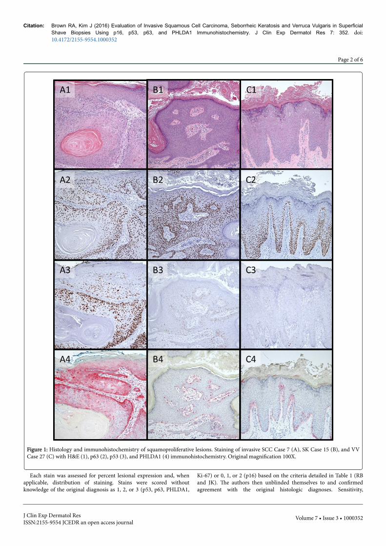
Figure 1: Histology and immunohistochemistry of squamoproliferative lesions. Staining of invasive SCC Case 7 (A), SK Case 15 (B), and VVCase 27 (C) with H&E (1), p63 (2), p53 (3), and PHLDA1 (4) immunohistochemistry. Original magnification 100X.
Each stain was assessed for percent lesional expression and, whenapplicable, distribution of staining. Stains were scored withoutknowledge of the original diagnosis as 1, 2, or 3 (p53, p63, PHLDA1,
Ki-67) or 0, 1, or 2 (p16) based on the criteria detailed in Table 1 (RBand JK). The authors then unblinded themselves to and confirmedagreement with the original histologic diagnoses. Sensitivity,
Citation: Brown RA, Kim J (2016) Evaluation of Invasive Squamous Cell Carcinoma, Seborrheic Keratosis and Verruca Vulgaris in SuperficialShave Biopsies Using p16, p53, p63, and PHLDA1 Immunohistochemistry. J Clin Exp Dermatol Res 7: 352. doi:10.4172/2155-9554.1000352
Page 2 of 6
J Clin Exp Dermatol ResISSN:2155-9554 JCEDR an open access journal
Volume 7 • Issue 3 • 1000352

specificity, positive predictive value, and negative predictive value foreach diagnosis were calculated with various combinations of stainingresults.
Stain p16 p53 p63* PHLDA1 Ki-67
Score=0 <1% of cells N/A N/A N/A N/A
1 1-30% of cells <5% of cells <50% of thickness <10% of cells <5% of cells or staining restricted to basal layer
2 >30% of cells 5-30% of cells 50-90% of thickness 10-40% of cells5-30% of cells or extending above basal layer but less than 30%of cells
3 N/A >30% of cells >90% thickness >40% of cells >30% of cells
*As measured beneath the granular layer. N/A: Not Applicable.
Table 1: Immunohistochemical scoring criteria.
ResultsThe results of p16, p53, p63, PHLDA1, and Ki-67 staining are shown
in Tables 2-4 with histology and IHC of representative SCC, VV, andSK cases shown in Figure 1.
The invasive SCC group was enriched for high PHLDA1 (80% withPHLDA1 score=3, 100% with PHLDA1 score ≥ 2).
Stain p16 p53 p63 PHLDA1 Ki-67
Intensity Weak to strong Strong Strong Weak to strong Weak to strong
DistributionNuclear and cytoplasmiclesional staining
Nuclear lesionalstaining
Nuclear lesionalstaining
Cytoplasmic lesional staining with backgroundstromal staining
Nuclear lesionalstaining
Table 2: Overall immunohistochemical stain expression patterns.
Invasive SCC p16 p53 p63 PHLDA1 Ki-67
Case 1 1 3 2 3 2
Case 2 1 1 2 3 1
Case 3 2 1 2 3 1
Case 4 1 1 2 2 1
Case 5 2 1 1 3 1
Case 6 2 2 1 3 1
Case 7 1 3 2 3 2
Case 8 2 3 3 3 2
Case 9 2 3 3 3 3
Case 10 1 1 1 2 1
SK p16 p53 p63 PHLDA1 Ki-67
Case 11 1 2 3 2 1
Case 12 0 1 3 2 1
Case 13 1 2 3 2 2
Case 14 1 2 3 1 1
Case 15 1 1 3 1 1
Case 16 0 1 3 1 1
Case 17 0 2 3 1 1
Case 18 0 1 3 1 1
Case 19 1 1 3 1 1
Case 20 0 1 3 2 1
VV p16 p53 p63 PHLDA1 Ki-67
Case 21 0 2 3 1 1
Case 22 0 1 2 1 1
Case 23 2 1 1 1 1
Case 24 1 1 2 1 1
Case 25 0 1 3 1 1
Case 26 0 1 3 1 2
Case 27 1 1 2 1 1
Case 28 0 1 2 1 1
Case 29 1 1 2 2 1
Case 30 1 1 3 1 1
SSC: Squamous Cell Carcinoma; SK: Seborrheic Keratosis; VV: VerrucaVulgaris
Table 3: Immunohistochemical scores by lesion type.
Citation: Brown RA, Kim J (2016) Evaluation of Invasive Squamous Cell Carcinoma, Seborrheic Keratosis and Verruca Vulgaris in SuperficialShave Biopsies Using p16, p53, p63, and PHLDA1 Immunohistochemistry. J Clin Exp Dermatol Res 7: 352. doi:10.4172/2155-9554.1000352
Page 3 of 6
J Clin Exp Dermatol ResISSN:2155-9554 JCEDR an open access journal
Volume 7 • Issue 3 • 1000352

Compared to VV and SK, the SCC group also showed high p53(50% of SCC with p53 score ≥ 2 vs. 60% of SK and 90% of VV with p53
score=1). The SK group was enriched for low p16 (100% with p16 score≤ 1) and high p63 scores (100% with p63 score=3).
Invasive SCC p16 p53 p63 PHLDA1 Ki-67
Score=0 0/10 N/A N/A N/A N/A
1 5/10 5/10 3/10 0/10 6/10
2 5/10 1/10 5/10 2/10 3/10
3 N/A 4/10 2/10 8/10 1/10
SK p16 p53 p63 PHLDA1 Ki-67
Score=0 5/10 N/A N/A N/A N/A
1 5/10 6/10 0/10 6/10 9/10
2 0/10 4/10 0/10 4/10 1/10
3 N/A 0/10 10/10 0/10 0/10
VV p16 p53 p63 PHLDA1 Ki-67
Score=0 5/10 N/A N/A N/A N/A
1 4/10 9/10 1/10 9/10 9/10
2 1/10 1/10 5/10 1/10 1/10
3 N/A 0/10 4/10 0/10 0/10
SCC: Squamous Cell Carcinoma; SK: Seborrheic Keratosis; VV: Verruca Vulgaris; N/A: Not Applicable.
Table 4: Immunohistochemical score frequency by lesion type.
Sensitivity, specificity, positive predictive value, and negativepredictive value for selected staining results are displayed in Table 5.
Lesions included inanalysis SK, VV , Invasive SCC SK, Invasive SCC SK, VV
Criteria for diagnosing SK p16<2 and p63=3 p63=3 p16<2 p16<2 and p63=3 p63=3 p16<2 p16<2 and p63=3 p63=3 p16<2
Sensitivity 1 1 1 1 1 1 1 1 1
Specificity 0.8 0.7 0.3 1 0.8 0.5 0.6 0.6 0.1
PPV 0.71 0.63 0.42 1 0.83 0.67 0.71 0.71 0.53
NPV 1 1 1 1 1 1 1 1 1
Lesions included in analysis SK, VV, Invasive SCC SK, Invasive SCC Invasive SCC, VV
Criteria for diagnosingInvasive SCC
PHLDA1=3 andp53=3
PHLDA1=3 p53=3
PHLDA1=3 andp53=3 PHLDA1=3 p53=3
PHLDA1=3 andp53=3 PHLDA1=3 p53=3
Sensitivity 0.4 0.8 0.4 0.67 0.8 0.4 0.4 0.8 0.4
Specificity 1 1 1 1 1 1 1 1 1
PPV 1 1 1 1 1 1 1 1 1
NPV 0.77 0.91 0.77 0.83 0.83 0.63 0.63 0.83 0.63
Lesions included in analysis SK, VV, Invasive SCC SK, VV Invasive SCC, VV
Criteria for diagnosing VVPHLDA1=1 andp53=1
PHLDA1=1 p53=1
PHLDA1=1 andp53=1 PHLDA1=1 p53=1
PHLDA1=1 andp53=1 PHLDA1=1 p53=1
Citation: Brown RA, Kim J (2016) Evaluation of Invasive Squamous Cell Carcinoma, Seborrheic Keratosis and Verruca Vulgaris in SuperficialShave Biopsies Using p16, p53, p63, and PHLDA1 Immunohistochemistry. J Clin Exp Dermatol Res 7: 352. doi:10.4172/2155-9554.1000352
Page 4 of 6
J Clin Exp Dermatol ResISSN:2155-9554 JCEDR an open access journal
Volume 7 • Issue 3 • 1000352

Sensitivity 0.8 0.9 0.9 0.8 0.9 0.9 0.8 0.9 0.9
Specificity 0.8 0.7 0.45 0.6 0.4 0.4 1 1 0.5
PPV 0.67 0.6 0.45 0.67 0.6 0.6 1 1 0.64
NPV 0.89 0.93 0.9 0.75 0.8 0.8 0.83 0.91 0.83
SK: Seborrheic Keratosis; VV: Verruca Vulgaris; SCC: Squamous Cell Carcinoma; PPV: Positive Predictive Value; NPV: Negative Predictive Value.
Table 5: Diagnostic statistics by stain criteria and differential diagnosis.
Discussionp53 is a tumor suppressor that promotes cell cycle exit, senescence
or apoptosis in response to DNA damage [3,15]. Soini and colleaguesfound an association between increased p53 expression and Ki-67expression in benign skin lesions, including seborrheic keratosis [16].They also noted clustering of p53 positive keratinocytes in areas ofdamage and inflammation. [16] Ko et al. found p53 expression ininverted follicular keratosis as well as SK [17]. Hussein et al. notedsignificantly increased p53 expression in invasive and in situ SCCcompared to normal skin [18]. p53 expression in SK was also increasedbut to a lesser degree [18]. Another study found that p53 expressionwas highest in SK with less expression in malignant and pre-malignantsquamous epithelial lesions, including SCC [19]. Bito et al. found theincidence of p53 expression to be greater in SCCs arising in sun-damaged skin [20], and p53 expression has frequently been noted inactinic keratosis (AK). A study by Gouvêa found strong p53 expressionin oral SCC arising in patients with a history of proliferative verrucousleukoplakia [21]. Other studies have replicated the finding of morefrequent p53 expression in SCC as compared to SK, with someevidence for increasing expression of p53 with higher grade histology.[22,23] Our results further support the finding of enriched p53expression in invasive squamous cell carcinoma compared to SK andVV.
p63, a p53 homologue, is normally expressed in the nuclei of thebasal epidermis, cells of the germinative hair matrix, and hair follicleexternal root sheath, and plays a role in regulating keratinocyte-specific gene expression [24-27]. Chang et al. were able to differentiatepagetoid SCC in situ from primary extramammary Paget’s disease bythe strong p63 positivity present in the former [28]. Takeuchi et al.demonstrated strong p63 expression in both SK and poorlydifferentiated SCC [29]. Other studies have noted similar findings ofp63 expression in SK and SCC [30]. A trend towards increased p63expression in less differentiated cells of invasive SCC has been noted[24]. Increased p63 expression in SK has been supported by microarrayanalysis [31]. Although we demonstrated p63 expression in VV andinvasive SCC, only SK showed diffuse and strong positivity for p63.
PHLDA1 is a marker of matrical differentiation with demonstratedutility in identifying a subset of tumors of the hair follicle [5-10]. Wefound that a high level of PHLDA1 expression was moderatelysensitive and highly specific for invasive SCC compared to SK and VV.
p16 expression has been described as a diagnostic tool fordifferentiating SCC in situ from actinic keratosis and benign squamouscutaneous lesions [32]. Hodges et al. found increasing p16 expressionin the progression from AK to SCC in situ to invasive SCC [33].Beyond its association with HPV-related oropharyngeal SCC, p16expression in other cutaneous head and neck SCC with lymph node
metastasis can be frequent [34]. Bai and colleagues noted that a smallnumber of vulvar and non-vulvar SKs demonstrated p16 expressionwith poor correlation with HPV-DNA status [35]. A study byNakamura et al. found a subset of SK with p16 staining in all lesionalcells [36]. Our findings demonstrated low p16 expression in all SK andmost VV. Recently, diffuse cyclin D1 and p16 expression wasdemonstrated more frequently in SCCIS and SCC than in AK.Although the invasive SCC group was enriched for high p16expression, this marker did not improve diagnostic yield above thatoffered by PHLDA1 and p53. Several studies have documented higherKi-67 expression in SCC compared to non-malignant lesions includinginflammatory dermatoses and SK [19,37]. Although increased Ki-67staining was noted in a subset of invasive SCC, this marker failed toadd additional diagnostic information in our cohort of cases [38,39].
This limited collection of cases provides support for the utilizationof diffuse and strong p63 staining as a sensitive marker for SK withpotential use of low to no p16 staining for its specificity for SK wheninvasive SCC is the primary differential diagnostic consideration. Thecombination of high PHLDA1 and p53 staining appeared highlyspecific for invasive SCC compared to SK and VV. Low PHLDA1 andp53 staining demonstrated moderate sensitivity for VV in this smallcollection of cases, but a specific marker for VV was not apparent fromthe markers tested [40]. Although our sample size is limited and doesnot include in situ SCC cases, our findings provide some support forutilization of IHC when a superficial shave biopsy specimen promptsthe differential diagnosis of invasive SCC, SK, and VV. Additionalstudies with more cases are merited to validate these findings.
References1. Santos M, Montagut C, Mellado B, García A, Ramón y Cajal S, et al.
(2004) Immunohistochemical staining for p16 and p53 in premalignantand malignant epithelial lesions of the vulva. Int J Gynecol Pathol 23:206-214.
2. Chaux A, Pfannl R, Rodríguez IM, Barreto JE, Velazquez EF, et al. (2011)Distinctive immunohistochemical profile of penile intraepithelial lesions:A study of 74 cases. Am J Surg Pathol 35: 553-562.
3. Missero C, Antonini D (2014) Crosstalk among p53 family members incutaneous carcinoma. Exp Dermatol 23: 143-146.
4. Serdar ZA, Eren PA, Canbakan M, Turan K, Tellioglu G, et al. (2010)Dermatologic findings in renal transplant recipients: Possible effects ofimmunosuppression regimen and p53 mutations. Transplant Proc 42:2538-2541.
5. Sellheyer K, Krahl D (2011) Phlda1 (tdag51) is a follicular stem cellmarker and differentiates between morphoeic basal cell carcinoma anddesmoplastic trichoepitheliom. British Journal of Dermatology 164:141-147.
6. Battistella M, Carlson JA, Osio A, Langbein L, Cribier B (2014) Skintumors with matrical differentiation: lessons from hair keratins, beta-catenin and PHLDA-1 expression. J Cutan Pathol 41: 427-436.
Citation: Brown RA, Kim J (2016) Evaluation of Invasive Squamous Cell Carcinoma, Seborrheic Keratosis and Verruca Vulgaris in SuperficialShave Biopsies Using p16, p53, p63, and PHLDA1 Immunohistochemistry. J Clin Exp Dermatol Res 7: 352. doi:10.4172/2155-9554.1000352
Page 5 of 6
J Clin Exp Dermatol ResISSN:2155-9554 JCEDR an open access journal
Volume 7 • Issue 3 • 1000352

7. Battistella M, Peltre B, Cribier B (2014) Phlda1, a follicular stem cellmarker, differentiates clear-cell/granular-cell trichoblastoma and clear-cell/granular cell basal cell carcinoma: A case-control study, with firstdescription of granular-cell trichoblastoma. Am J Dermatopathol 36:643-650.
8. Sellheyer K, Nelson P (2011) Follicular stem cell marker phlda1 (tdag51)is superior to cytokeratin-20 in differentiating between trichoepitheliomaand basal cell carcinoma in small biopsy specimens. Journal of cutaneouspathology 38: 542-550.
9. Battistella M, Carlson JA, Osio A, Langbein L, Cribier B (2014) Skintumors with matrical differentiation: lessons from hair keratins, beta-catenin and PHLDA-1 expression. J Cutan Pathol 41: 427-436.
10. Ohyama M, Terunuma A, Tock CL, Radonovich MF, Pise-Masison CA, etal. (2006) Characterization and isolation of stem cell-enriched humanhair follicle bulge cells. J Clin Invest 116: 249-260.
11. Huang V, Hepper D, Anadkat M, Cornelius L (2012) Cutaneous toxiceffects associated with vemurafenib and inhibition of the BRAF pathway.Arch Dermatol 148: 628-633.
12. Mandalà M, Massi D, De Giorgi V (2013) Cutaneous toxicities of BRAFinhibitors: clinical and pathological challenges and call to action. Crit RevOncol Hematol 88: 318-337.
13. Belum VR, Fischer A, Choi JN, Lacouture ME (2013) Dermatologicaladverse events from BRAF inhibitors: a growing problem. Curr OncolRep 15: 249-259.
14. Curry JL, Torres-Cabala CA, Kim KB, Tetzlaff MT, Duvic M, et al. (2014)Dermatologic toxicities to targeted cancer therapy: Shared clinical andhistologic adverse skin reactions. Int J Dermatol 53: 376-384.
15. Lane D, Levine A (2010) p53 Research: the past thirty years and the nextthirty years. Cold Spring Harb Perspect Biol 2: a000893.
16. Soini Y, Kamel D, Pääkkö P, Lehto VP, Oikarinen A, et al. (1994) Aberrantaccumulation of p53 associates with Ki67 and mitotic count in benignskin lesions. Br J Dermatol 131: 514-520.
17. Ko CJ, Shintaku P, Binder SW (2005) Comparison of benign keratosesusing p53, bcl-1, and bcl-2. J Cutan Pathol 32: 356-359.
18. Hussein MR, Al-Badaiwy ZH, Guirguis MN (2014) Analysis of p53 andbcl-2 protein expression in the non-tumorigenic, pretumorigenic, andtumorigenic keratinocytic hyperproliferative lesions. Journal of cutaneouspathology 31: 643-651.
19. Onodera H, Nakamura S, Sugai T (1996) Cell proliferation and p53protein expressions in cutaneous epithelial neoplasms. Am JDermatopathol 18: 580-588.
20. Bito T, Ueda M, Ahmed NU, Nagano T, Ichihashi M (1995) Cyclin D andretinoblastoma gene product expression in actinic keratosis andcutaneous squamous cell carcinoma in relation to p53 expression. J CutanPathol 22: 427-434.
21. Gouvêa AF, Vargas PA, Coletta RD, Jorge J, Lopes MA, et al. (2010)Clinicopathological features and immunohistochemical expression ofp53, ki-67, mcm-2 and mcm-5 in proliferative verrucous leukoplakia.Journal of Oral Pathology & Medicine 239: 447-452.
22. Urano Y, Oura H, Sakaki A, Nagae H, Matsumoto K, et al. (1992)Immunohistological analysis of P53 expression in human skin tumors. JDermatol Sci 4: 69-75.
23. Chen H, Takahara M, Xie L, Takeuchi S, Tu Y, et al. (2013) Levels of theEMT-related protein Snail/Slug are not correlated with p53/p63 incutaneous squamous cell carcinoma. J Cutan Pathol 40: 651-656.
24. Reis-Filho JS, Torio B, Albergaria A, Schmitt FC (2002) p63 expression innormal skin and usual cutaneous carcinomas. J Cutan Pathol 29: 517-523.
25. Yang A, Schweitzer R, Sun D, Kaghad M, Walker N, et al. (1999) p63 isessential for regenerative proliferation in limb, craniofacial and epithelialdevelopment. Nature 398: 714-718.
26. Mills AA, Zheng B, Wang XJ, Vogel H, Roop DR, et al. (1999) p63 is a p53homologue required for limb and epidermal morphogenesis. Nature 398:708-713.
27. Della Gatta G, Bansal M, Ambesi-Impiombato A, Antonini D, Missero C,et al. (2008) Direct targets of the trp63 transcription factor revealed by acombination of gene expression profiling and reverse engineering.Genome Res 18: 939-948.
28. Chang J, Prieto VG, Sangueza M, Plaza JA (2014) Diagnostic utility of p63expression in the differential diagnosis of pagetoid squamous cellcarcinoma in situ and extramammary paget disease: A histopathologicstudy of 70 cases. Am J Dermatopathol 36: 49-53.
29. Takeuchi Y, Tamura A, Kamiya M, Fukuda T, Ishikawa O (2005)Immunohistochemical analyses of p63 expression in cutaneous tumours.Br J Dermatol 153: 1230-1232.
30. Abbas O, Richards JE, Yaar R, Mahalingam M (2011) Stem cell markers(cytokeratin 15, cytokeratin 19 and p63) in in situ and invasive cutaneousepithelial lesions. Mod Pathol 24: 90-97.
31. Seo EY, Lee DH, Lee Y, Cho KH, Eun HC, et al. (2012) Microarrayanalysis reveals increased expression of ΔNp63α in seborrhoeickeratosis. Br J Dermatol 166: 337-342.
32. Salama ME, Mahmood MN, Qureshi HS, Ma C, Zarbo RJ, et al. (2003)p16INK4a expression in actinic keratosis and Bowen's disease. Br JDermatol 149: 1006-1012.
33. Hodges A, Smoller BR (2002) Immunohistochemical comparison of p16expression in actinic keratoses and squamous cell carcinomas of the skin.Mod Pathol 15: 1121-1125.
34. Beadle BM, William WN Jr, McLemore MS, Sturgis EM, Williams MD(2013) P16 expression in cutaneous squamous carcinomas with neckmetastases: A potential pitfall in identifying unknown primaries of thehead and neck. Head & Neck 35: 1527-1533.
35. Bai H, Cviko A, Granter S, Yuan L, Betensky RA, et al. (2003)Immunophenotypic and viral (human papillomavirus) correlates ofvulvar seborrheic keratosis. Hum Pathol 34: 559-564.
36. Nakamura S, Nishioka K (2003) Enhanced expression of p16 inseborrhoeic keratosis; a lesion of accumulated senescent epidermal cellsin G1 arrest. Br J Dermatol 149: 560-565.
37. Kawahira K (1999) Immunohistochemical staining of proliferating cellnuclear antigen (PCNA) in malignant and nonmalignant skin diseases.Arch Dermatol Res 291: 413-418.
38. Fabbrocini G, Russo N, Pagliuca MC, Delfino M, Staibano S, et al. (2000)P53, cyclin-d1, pcna, agnor expression in squamous cell cancer of the lip:A multicenter study. Photodermatol Photoimmunol Photomed 16:172-177.
39. Neto PD, Alchorne M, Michalany N, Abreu M, Borra R (2013) ReducedP53 Staining in Actinic Keratosis is Associated with Squamous CellCarcinoma: A Preliminary Study. Indian J Dermatol 58: 325.
40. Brasanac D, Stojkovic-Filipovic J, Bosic M, Tomanovic N, Manojlovic-Gacic E (2015) Expression of G1/S-cyclins and cyclin-dependent kinaseinhibitors in actinic keratosis and squamous cell carcinoma. J CutanePathol 43: 200-210.
Citation: Brown RA, Kim J (2016) Evaluation of Invasive Squamous Cell Carcinoma, Seborrheic Keratosis and Verruca Vulgaris in SuperficialShave Biopsies Using p16, p53, p63, and PHLDA1 Immunohistochemistry. J Clin Exp Dermatol Res 7: 352. doi:10.4172/2155-9554.1000352
Page 6 of 6
J Clin Exp Dermatol ResISSN:2155-9554 JCEDR an open access journal
Volume 7 • Issue 3 • 1000352