la dermatite atopica vista dal pediatra - sipps.it · •atopic dermatitis is an important...
TRANSCRIPT

La Dermatite atopicavista dal pediatra
Introduction
Pathogenesis
Triggers & Allergens
Treatments
Conclusions
Universita’ di Ferrara
Diego Peroni


•Atopic dermatitis is an important condition that affects up to 20% of children with a possible rising prevalence in westernized societies.•Williams HC. Is the prevalence of atopic dermatitis increasing? Clin Exp Dermatol 1992; 17:385–91.
•It is associated with significant morbidity, including: -sleepless nights and -poor self-esteem for the child, and -financial strain and -family stress for parents and other siblings.•Verboom P, et al. The cost of atopic dermatitis in the Netherlands: an international comparison. Br J Dermatol 2002; 147:716–24.•Kemp AS. Cost of illness of atopic dermatitis in children. Pharmacoeconomics 2003; 21:105–13.
Atopic Dermatitis: Prevalence and Morbidity

•There are at least two theories proposed to explain the development of this common disorder.
1) For years, the major theory was that patients had an aberrant and robust Th2 adaptive immune response to largely innocuous environmental antigens.
2) Recent research highlights the importance of skin barrier abnormalities and an inadequate host response to common cutaneous microbes as other highly plausible mechanisms thatmight predispose individuals to develop atopic dermatitis.
Atopic Dermatitis: Development

La Dermatite atopicavista dal pediatra
Introduction
Pathogenesis
Triggers & Allergens
Treatments
Conclusions
Universita’ di Ferrara
Diego Peroni

Immunologic pathway involved in the progression of AD.Akdis CA, JACI 2006;118:152
LC,Langerhans cell;
MC, mast cell;
TSLP, humanthymic stromallymphopoietin;
Ag, antigen;
SAg, superantigen;
AICD, activation-induced cell
death;
CLA, cutaneouslymphocyte
antigen;
MO, monocyte.
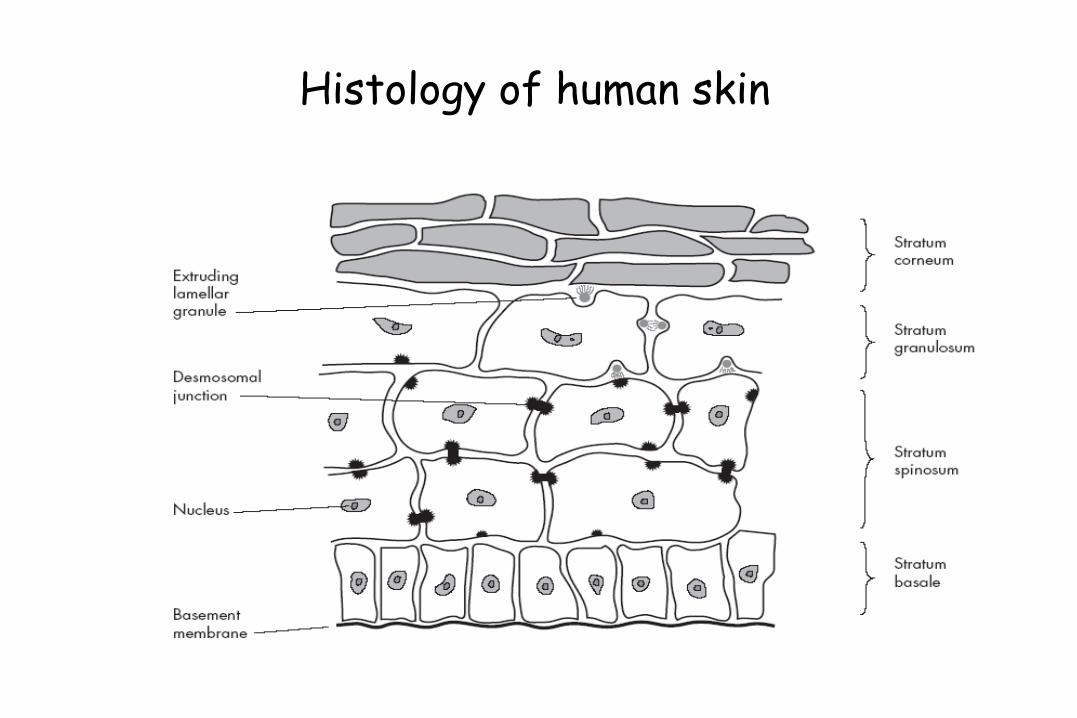
Histology of human skin
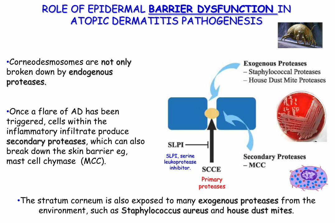
•Corneodesmosomes are not only broken down by endogenous proteases.
•Once a flare of AD has been triggered, cells within the inflammatory infiltrate produce secondary proteases, which can also break down the skin barrier eg,mast cell chymase (MCC).
SLPI, serine leukoprotease
inhibitor.
•The stratum corneum is also exposed to many exogenous proteases from the environment, such as Staphylococcus aureus and house dust mites.
Primaryproteases
ROLE OF EPIDERMAL BARRIER DYSFUNCTION INATOPIC DERMATITIS PATHOGENESIS

The brick wall analogy of the stratum corneumof the epidermal barrier.
In healthy skin thecorneodesmosomes(iron rods) are intact throughout the stratum corneum. At the surface, the corneodesmosomesstart to break down as part of the normal desquamation process, analogous to iron rods rusting (A).
ROLE OF EPIDERMAL BARRIER DYSFUNCTION INATOPIC DERMATITIS PATHOGENESIS
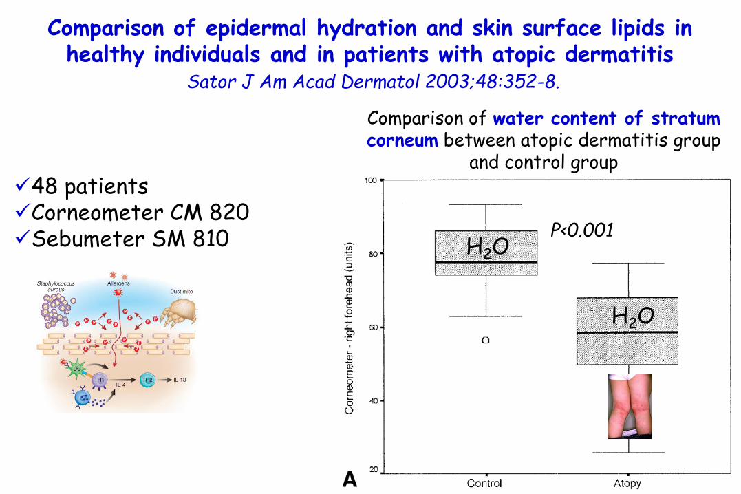
Comparison of epidermal hydration and skin surface lipids in healthy individuals and in patients with atopic dermatitis
Sator J Am Acad Dermatol 2003;48:352-8.
Comparison of water content of stratumcorneum between atopic dermatitis group
and control group
P<0.001H2O
H2O
48 patients Corneometer CM 820 Sebumeter SM 810

Comparison of epidermal hydration and skin surface lipids in healthy individuals and in patients with atopic dermatitis
Sator J Am Acad Dermatol 2003;48:352-8.
Comparison of skin surface lipidsbetween atopic dermatitis group and
control group
P<0.001
ceramides
ceramides
48 patients Corneometer CM 820 Sebumeter SM 810

Peanut allergy: Effect of environmental peanut exposure in children with filaggrin loss-of-function mutations. Brough H, JACI 2014; 134:867
Exposure to peanut antigen in dust within the first year of life
Peanut sensitization and peanut allergy at 8 and 11 years.
Genotyping was performed for 6 FLG mutations

Skin barrier impairment correlates with the risk of aeroallergen sensitization.
61 ch with AD
SCORAD index
TEWL
Corneometer
SPT aeroallergens
Allergy score
TEWL on healthy skin of
AD patients
controls

Skin barrier impairment correlates with the risk of aeroallergen sensitization
• 61 ch with AD
• 6 months and 17 years
• SCORAD index
• TEWL
• Corneometer
• SPT aeroallergens
• Allergy score

THE ROLE OF INNATE IMMUNITY IN ATOPIC DERMATITISPATHOGENESIS
•Patients with AD have a greatly increased risk of certain types of bacterial (Staphylococcus aureus), viral (herpes simplex virus and pox viruses), and fungal (Malassezia sympodialis) infections.
•Epithelial cells are equipped with a variety ofpattern-recognition receptors (eg, Toll-like receptors [TLRs]) sensing different microbial structures.
•Thus activated, epithelial cells start to produce antimicrobial peptides, including defensins, cathelicidins, dermcidin, and psoriasin.

Correlation between serum 25-hydroxyvitamin D levels and severity of atopic dermatitis in children.
Peroni DG, Br J Dermatol. 2011;164:1078-82.
37 children (8 months and 12 years) with AD,
SCORAD index,
Serum levels of 25-hydroxyvitamin D
sIgE to S.aureus and to M. furfur
Serum vitamin D levels in relation to different threshold values of AD severity.
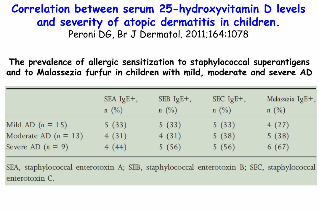
The prevalence of allergic sensitization to staphylococcal superantigensand to Malassezia furfur in children with mild, moderate and severe AD
Correlation between serum 25-hydroxyvitamin D levels and severity of atopic dermatitis in children.
Peroni DG, Br J Dermatol. 2011;164:1078

The prevalence of allergic sensitization to staphylococcal superantigensand to Malassezia furfur in children with mild, moderate and severe AD
Correlation between serum 25-hydroxyvitamin D levels and severity of atopic dermatitis in children.
Peroni DG, Br J Dermatol. 2011;164:1078
Microorganism-inducedexacerbations in atopicdermatitis: a possible
preventive role for Vitamin D?Peroni et al.
Allergy Asthma Proc 2014

La Dermatite atopicavista dal pediatra
Introduction
Pathogenesis
Triggers & Allergens
Treatments
Conclusions
Universita’ di Ferrara
Diego Peroni

THE ROLE OF PRURITUS IN ATOPIC DERMATITIS PATHOGENESIS
Jung T, JACI 2008;122:1074
•neuropeptides,
•proteases,
•IL-31,
•kallikrein 7,
•……..?
pruritus
Pruritus is an unpleasant sensation provoking the desire to scratch
and constitutes an essential feature of atopic dermatitis

Buddenkotte J, Allergy. 2010;65:805-21.
Trigger factors aggravating pruritus perception in AD
Epidermal barrier
Xerosis, a common problem of the skin of patients sufferingfrom AD, results in an increased transepidermal water loss and a decreased ability of the stratum corneum to bind water
a disturbed epidermal barrier constitutes an activator of pruritus.
scratching behaviour and induction of pruritus are triggered by water content below 10%
Buddenkotte J, Allergy. 2010;65:805-21.
Trigger factors aggravating pruritus perception in AD
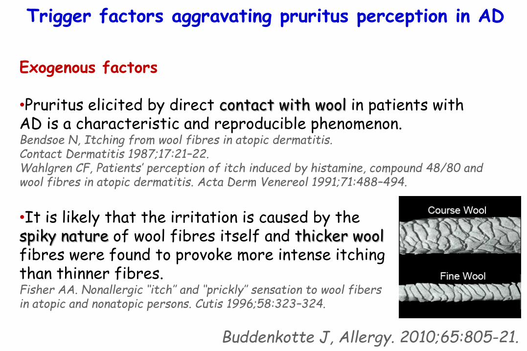
Exogenous factors
•Pruritus elicited by direct contact with wool in patients withAD is a characteristic and reproducible phenomenon.Bendsoe N, Itching from wool fibres in atopic dermatitis. Contact Dermatitis 1987;17:21–22.Wahlgren CF, Patients’ perception of itch induced by histamine, compound 48/80 and wool fibres in atopic dermatitis. Acta Derm Venereol 1991;71:488–494.
•It is likely that the irritation is caused by the spiky nature of wool fibres itself and thicker wool fibres were found to provoke more intense itching than thinner fibres.Fisher AA. Nonallergic ‘‘itch’’ and ‘‘prickly’’ sensation to wool fibers in atopic and nonatopic persons. Cutis 1996;58:323–324.

Buddenkotte J, Allergy. 2010;65:805-21.
Trigger factors aggravating pruritus perception in AD
Exogenous factors
•Pruritus elicited by direct contact with wool in patients withAD is a characteristic and reproducible phenomenon.Bendsoe N, Itching from wool fibres in atopic dermatitis. Contact Dermatitis 1987;17:21–22.Wahlgren CF, Patients’ perception of itch induced by histamine, compound 48/80 and wool fibres in atopic dermatitis. Acta Derm Venereol 1991;71:488–494.
•It is likely that the irritation is caused by the spiky nature of wool fibres itself and thicker wool fibres were found to provoke more intense itching than thinner fibres.Fisher AA. Nonallergic ‘‘itch’’ and ‘‘prickly’’ sensation to wool fibers in atopic and nonatopic persons. Cutis 1996;58:323–324.
Cotton fibres
Silk fibres

Management of pruritus in AD
Buddenkotte J, Allergy. 2010;65:805-21.
•Scratch-induced skin damage caused by nocturnal scratch movements may be improved by using cotton gloves.
•Also controlled physical exercise like gymnastics or ball games were demonstrated in a controlled study to teach patients to cope better with itch attacks

Bacterial Colonization And Infection In Ad: To Treat Or Not To Treat (With Antibiotics) Boguniewicz JACI 2010;125:4
Other approaches include silver-impregnated clothing, which has been shown:
1) to reduce staphylococcal colonization, 2) improve clinical parameters, and 3) reduce topical steroid use in patients with AD.
Gauger A, Dermatology 2003;207:15-21.Gauger A, J Eur Acad Dermatol Venereol 2006;20:534-41.
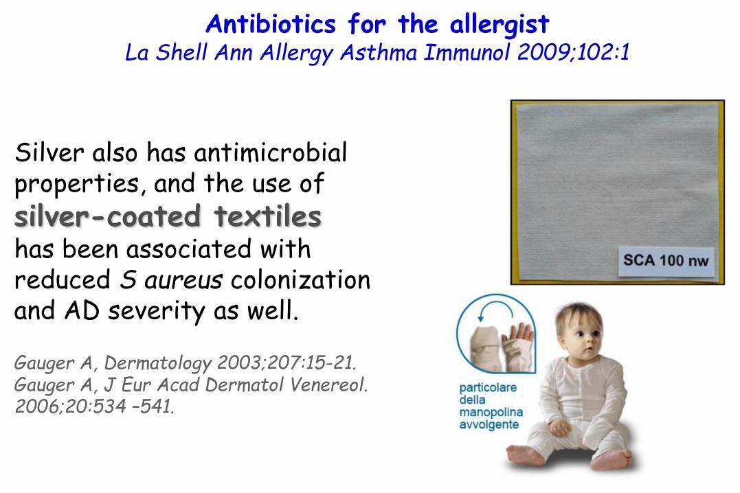
Antibiotics for the allergistLa Shell Ann Allergy Asthma Immunol 2009;102:1
Silver also has antimicrobialproperties, and the use ofsilver-coated textileshas been associated withreduced S aureus colonizationand AD severity as well.
Gauger A, Dermatology 2003;207:15-21.Gauger A, J Eur Acad Dermatol Venereol. 2006;20:534 –541.

La Dermatite atopicavista dal pediatra
Introduction
Pathogenesis
Triggers & Allergens
Treatments
Conclusions
Universita’ di Ferrara
Diego Peroni

What causes worsening of eczema? A systemic review. Langan SM. Williams HC. Br J Dermatol. 2006;155:505-514.
Medline from 1966 until 20 April 2005 The roles of: •foodstuffs (13 studies), •house dust mite (three), •other aeroallergens (two), •seasonality (two), •bacterial infections (one), •textiles (three), •detergents (one), •sunlight (one) and •stress (two)
Collectively, these studies provide some evidence that:•certain foods, •house dust mite, •stress and •seasonal factors are relevant causes of disease worsening in certain subgroups with eczema.
No good evidence could be found to support the role of detergents, textiles and irritants in causing worsening of eczema.

THE ROLE OF ADAPTIVE IMMUNITY IN ATOPIC DERMATITIS PATHOGENESIS and TREATMENT STRATEGIES
Jung T, JACI 2008;122:1074
TH2-dominated cytokine milieu downregulates
The antimicrobial peptidicresponse in AD skin
Howell MD, Immunity 2006;24:341-8.
FLG expression in keratinocytesHowell MD, JACI 2007;120:150-5.
allergen avoidance

Prevalence of IgE-mediated food allergy among Children with atopic dermatitis
Eigenmann Pediatrics 1998; 101: E8
%
C
H
I
L
D
R
E
N
40 -
30 -
20 -
10 -
37%
Approximately one third of children with refractory,moderate-severe AD have IgE-mediated clinical reactivityto food proteins

IgE food sensitization to cow’s milk, egg or peanut is a major
risk factor for the presence of atopic dermatitis in infancy
Hill J Pediatr 2000; 137: 475
%
C
H
I
L
D
R
E
N
W
I
T
H
(+)
S
P
T
80 -
70 -
60 -
50 -
40 -
30 -
20 -
10 -
AGE 6 months 12 months
83%
22%
65%
5%
36%
19%
severe atopic dermatitis
atopic dermatitis in general
controls

Guidelines for treatment of atopic eczema (atopic dermatitis) Part IJ. Ring, JEADV 2012, 26, 1045–1060
Dietary intervention: Food allergens
•Among food allergens, cow‟s milk, hen„s egg, wheat, soy, tree nutsand peanuts are most frequently responsible for eczema orexacerbation in infancy. Werfel T, Curr Opin Allergy Clin Immunol 2004; 4: 379–385.
•In older children, adolescents and adults pollen related food allergy should be taken into account.Breuer K, Allergy 2004; 59:988–994.Reekers R, J Allergy Clin Immunol 1999; 104: 466–472.
•Patients with moderate to severe AE should observe a diet eliminating those foods that elicitated clinical early or late reactions upon controlled oral provocation tests.
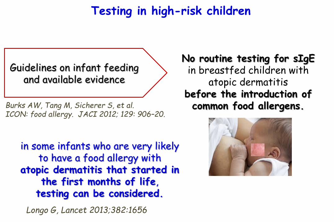
Testing in high-risk children
Guidelines on infant feeding and available evidence
No routine testing for sIgEin breastfed children with
atopic dermatitis before the introduction of common food allergens.Burks AW, Tang M, Sicherer S, et al.
ICON: food allergy. JACI 2012; 129: 906–20.
in some infants who are very likely to have a food allergy with
atopic dermatitis that started in the first months of life,
testing can be considered.
Longo G, Lancet 2013;382:1656
Guidelines for treatment of atopic eczema (atopic dermatitis) Part IJ. Ring, JEADV 2012, 26, 1045–1060
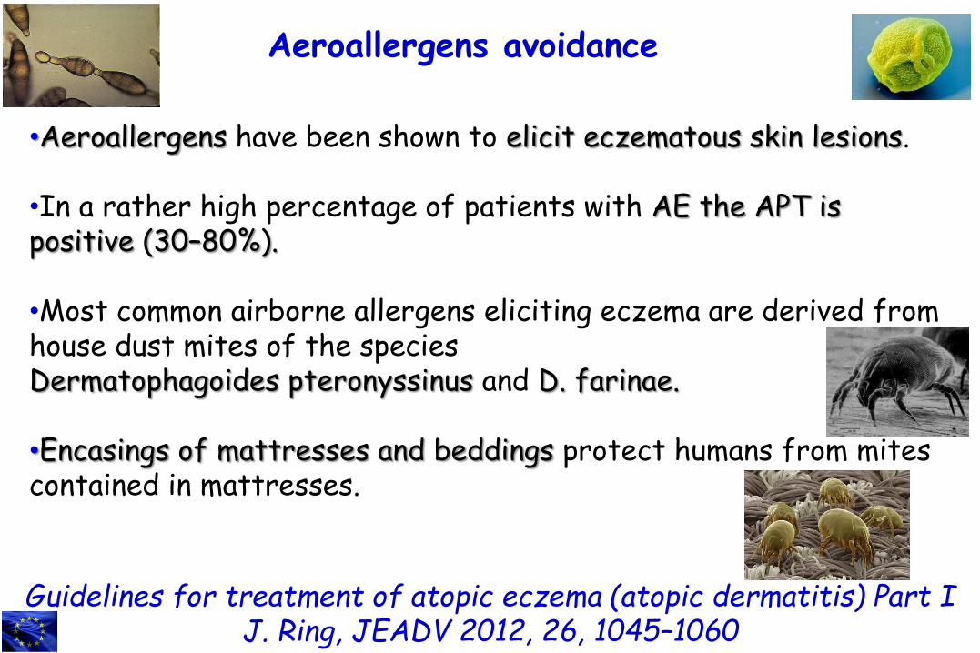
•Aeroallergens have been shown to elicit eczematous skin lesions.
•In a rather high percentage of patients with AE the APT is positive (30–80%).
•Most common airborne allergens eliciting eczema are derived from house dust mites of the species Dermatophagoides pteronyssinus and D. farinae.
•Encasings of mattresses and beddings protect humans from mitescontained in mattresses.
Aeroallergens avoidance
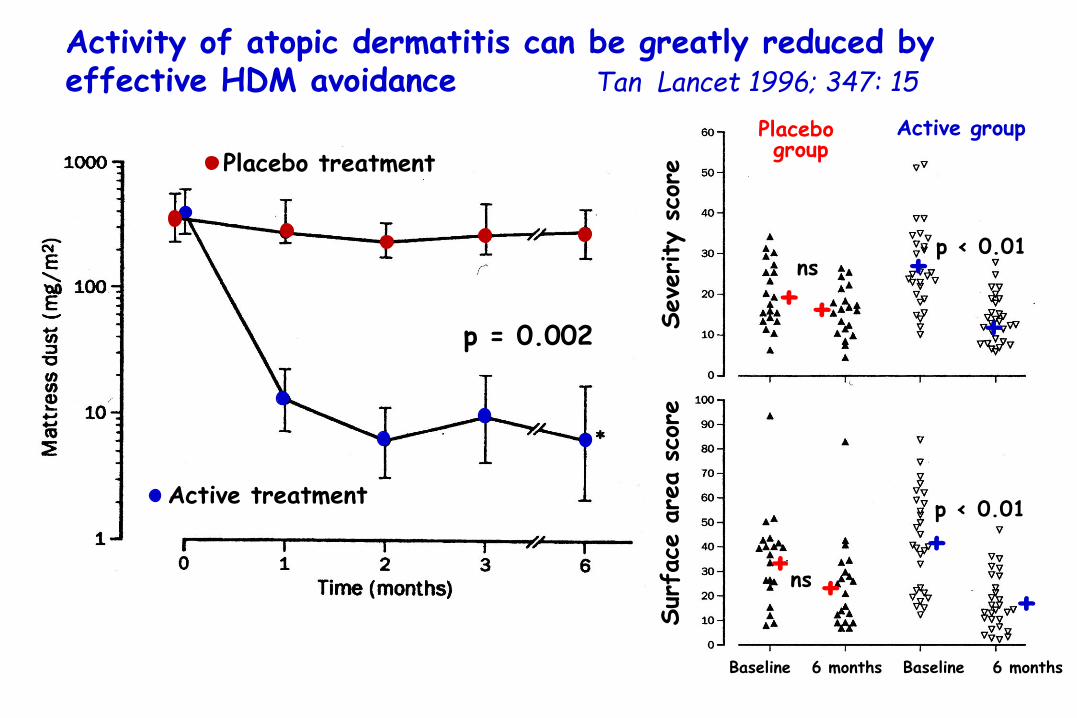
Activity of atopic dermatitis can be greatly reduced by effective HDM avoidance Tan Lancet 1996; 347: 15
Active treatment
Placebo treatment
p = 0.002
Placebo group
Active group
+
+
+
+
p < 0.01
p < 0.01
+ns
+ +ns
+
Baseline 6 months Baseline 6 monthsSur
face
are
a s
core
Seve
rity
sco
re

Eczema severity decreased significantly in both HDM sensitive and non sensitive patients (p < 0.001)
sCD30 levels were significantly reduced in both groups (p < 0.001)
Patients not sensitized to HDM allergens benefited from the bedcovers as much as sensitized patients
A result which could be due to a reduction in beds of:
• other important allergens
• supertantigens
• irritants and enzymes
Effectiveness of occlusive bedding in the treatment of atopic dermatitis a placebo-controlled trial of 12 months’ duration
Holm Allergy 2001; 56: 152
“bedcovers should be part of routine treatment for AD”

30 –
20 –
10 –
04.5ng
20ng30ng
AcarbusterEnvicon
Microair Nature Alpretec
Microair Pristine Alpretec
Pristine Basic USA
20ng
Fel d 1 permeability throughout different anti-mites encaising
Efficacy in allergen control and air permeability of different materials used for bed encasement.
Peroni DG, Allergy. 2004 Sep;59(9):969-72.
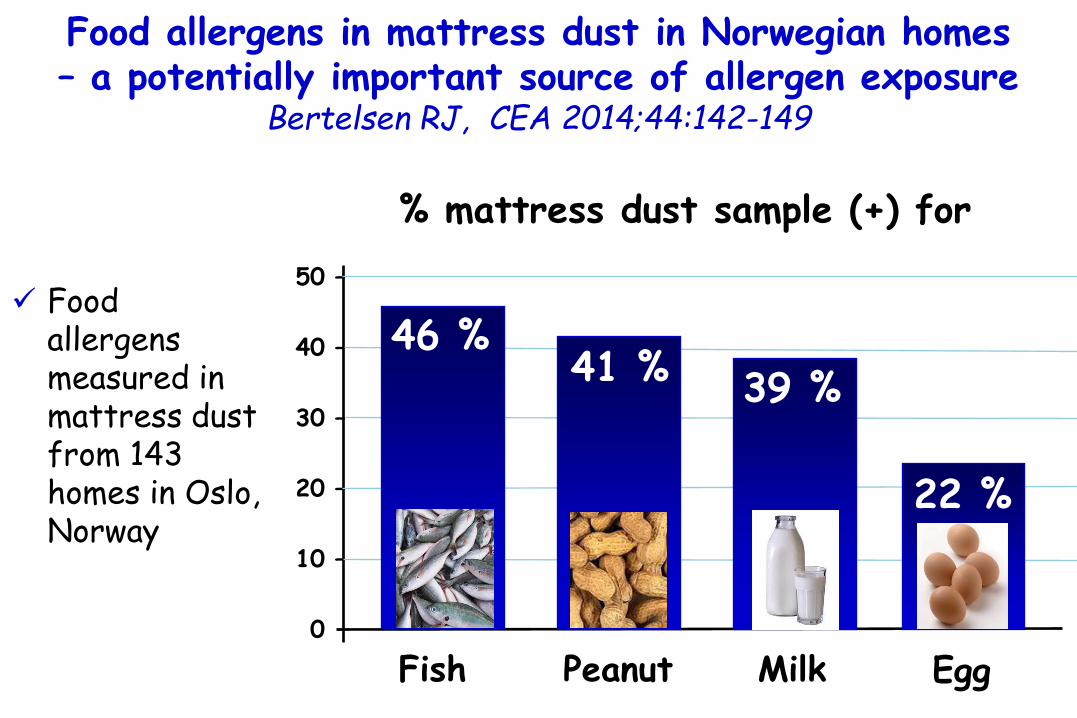
Food allergens measured in mattress dust from 143 homes in Oslo, Norway
Food allergens in mattress dust in Norwegian homes – a potentially important source of allergen exposure
Bertelsen RJ, CEA 2014;44:142-149
% mattress dust sample (+) for
50 –
40 –
30 –
20 –
10 –
00
Fish
46 %41 % 39 %
22 %
Peanut Milk Egg

La Dermatite atopicavista dal pediatra
Introduction
Pathogenesis
Triggers & Allergens
Emollient Treatments
Conclusions
Universita’ di Ferrara
Diego Peroni

Cleansing and bathing
Guidelines for treatment of atopic eczema (atopic dermatitis) Part IJ. Ring, JEADV 2012, 26, 1045–1060
•The skin must be cleansed thoroughly, but gently and carefully to get rid of crusts and mechanically eliminate bacterial contaminantsin the case of bacterial super-infection.
•Cleansers with or without antiseptics
•Salt baths may be beneficial because of removing the dead keratolytic material. In heavily impetiginized or ichthyotic skin salt baths are very useful.

22 neonates at high risk for developingAD emollient therapyfrom birth. followed up meantime of 547 days
% children whodeveloped AD
50 –
40 –
30 –
20 –
10 –
0
15%
30%
50%
Chance of developingAD in similar
high-riskinfants
Hoare C, Health Technol Assess
2000;4:1-191.
A pilot study of emollient therapy for the primaryprevention of atopic dermatitis.
Simpson EL, J Am Acad Dermatol. 2010;63:587-93.

52 ch with AD (2-12 yrs).
26 ch received a steroid cream for 2 weeks (+4 weeks follow-up with no treatment) (Group A).
26 ch received steroid cream for 2 weeks + emolients for 6 weeks (Group B).
Emollients Improve Treatment Results with Topical Corticosteroids in Childhood Atopic Dermatitis: a
Randomized Comparative StudySzczepanowska Ped All Immunol 2008;19:614
STEROID
* p=0.004 ** p=0.01 ***p<0.001
No emolient
plus emolient

52 ch with AD (2-12 yrs).
26 ch received a steroid cream for 2 weeks (+4 weeks follow-up with no treatment) (Group A).
26 ch received steroid cream for 2 weeks + emolients for 6 weeks (Group B).
* p=0.004 ** p=0.01 ***p<0.001
No emolient
STEROID
plus emolient
Emollients Improve Treatment Results with Topical Corticosteroids in Childhood Atopic Dermatitis: a
Randomized Comparative StudySzczepanowska Ped All Immunol 2008;19:614
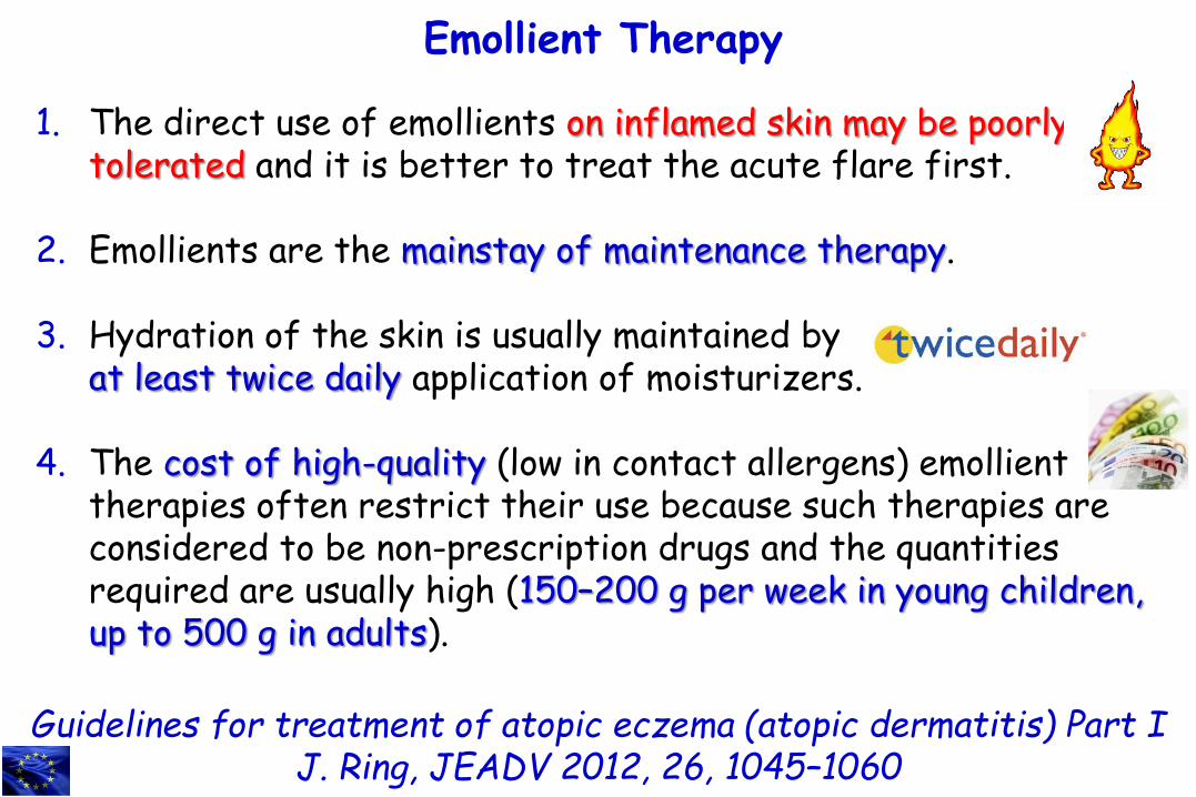
Guidelines for treatment of atopic eczema (atopic dermatitis) Part IJ. Ring, JEADV 2012, 26, 1045–1060
1. The direct use of emollients on inflamed skin may be poorly tolerated and it is better to treat the acute flare first.
2. Emollients are the mainstay of maintenance therapy.
3. Hydration of the skin is usually maintained by at least twice daily application of moisturizers.
4. The cost of high-quality (low in contact allergens) emollient therapies often restrict their use because such therapies are considered to be non-prescription drugs and the quantities required are usually high (150–200 g per week in young children, up to 500 g in adults).
Emollient Therapy

Local rhamnosoft, ceramides and L-isoleucine in atopiceczema: a randomized, placebo controlled trial
Marseglia A. PAI,2014; 25:271-275
A non-steroidal, anti-inflammatory moisturizing cream containing rhamnosoft, ceramides, and L-isoleucine (ILE) (pro-AMP cream)
107 children (72 allocated to pro-AMP cream and 35 allocated tocontrol group) with mild-to-moderate chronic AE of the face
Treatments were applied twice daily for a 6-week period.
Evolution of Eczema severity Score from baseline to week 3 and week 6
in the two study groups.
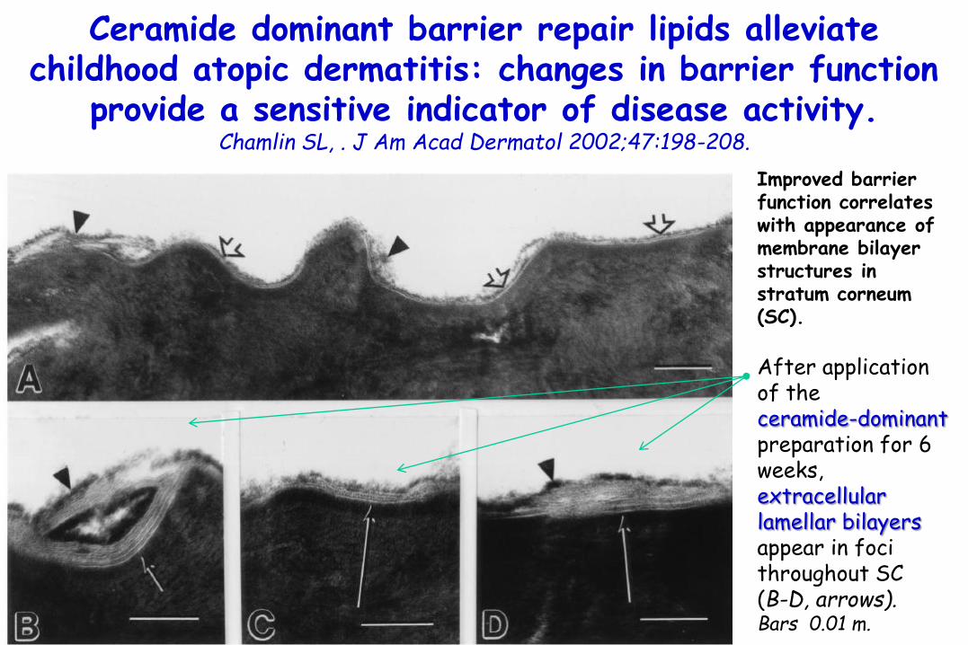
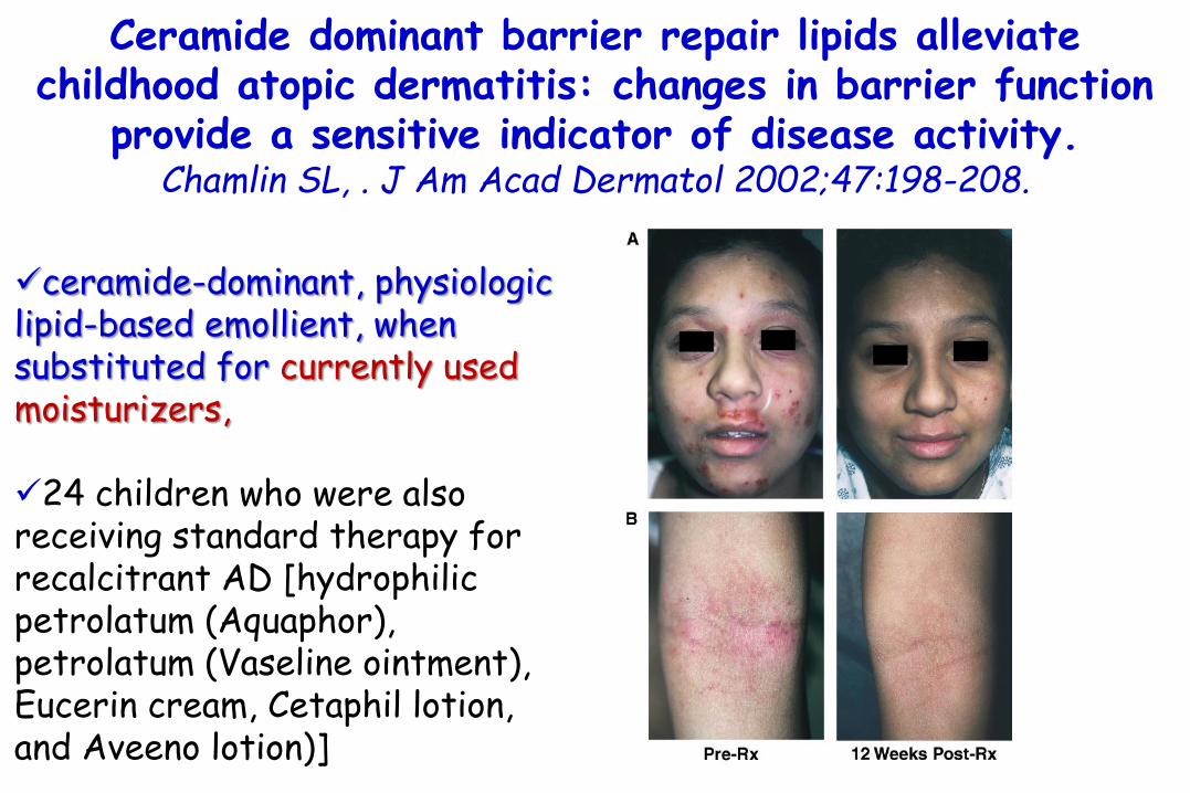
Ceramide dominant barrier repair lipids alleviate childhood atopic dermatitis: changes in barrier function
provide a sensitive indicator of disease activity. Chamlin SL, . J Am Acad Dermatol 2002;47:198-208.
Improved barrier function correlates with appearance of membrane bilayerstructures instratum corneum(SC).
After applicationof the ceramide-dominantpreparation for 6 weeks, extracellular lamellar bilayersappear in focithroughout SC (B-D, arrows). Bars 0.01 m.
Ceramide dominant barrier repair lipids alleviate childhood atopic dermatitis: changes in barrier function
provide a sensitive indicator of disease activity. Chamlin SL, . J Am Acad Dermatol 2002;47:198-208.
ceramide-dominant, physiologic lipid-based emollient, when substituted for currently used moisturizers,
24 children who were also receiving standard therapy for recalcitrant AD [hydrophilicpetrolatum (Aquaphor),petrolatum (Vaseline ointment), Eucerin cream, Cetaphil lotion, and Aveeno lotion)]

0 –
-10 –
-20 –
-30 –
-40 -
-50 –
Use of an emolient as a steroid-sparing agent in the treatment of mild to moderate atopic dermatitis in
children Lucky Pediatr Dermatol 1997;4:321
•25 ch with AD (3-15 years) •Application of hydrocortisone 2.5% cream alone or in association with a water-in-oil cream•3 weeks treatment
% REDUCTION IN CORTICOSTEROID WITH THE USE OF THE
EMOLLIENT
-50%

Bathing in Atopic Dermatitis
Anthony J. Mancini, Semin Cutan Med Surg 2012;31(suppl 3):S23-S28
The Diaper Area Analogy: Explaining Why the Wet Method Works
“Atopic dermatitis likes dry skin, but not moist and hydrated skin. Look at the area covered by your baby‟s diaper. Feel how soft and supple it is? And notice that eczema does not occur in this area.
By bathing your baby every day and immediately applying the medications and then the moisturizer, we are trying to create the same sort of environment (a moist one) as inthe diaper area on the rest of your baby‟s skin.”

MgCl salt
Skin barrierSkin hydrationInflammation
Allantoin
Hydration Skin barrierRepair
Urea ASkin hydration
NaCl salt
urea‟s effects
Loden,ActaDermVen.2002;82:45Thornfeldt,DermSurg.2005;31:873
Proksch,IntJDerm.2005;44:151
Hagstromer,SkinPhaApSkinPhy.2001;14:27
Bathing in a complementary salt solution
S.aureus attachment
Akiyama J Dermat Sci 1998;16:216

+++++
++++
INFECTION
+
VITIS VINIFERAGLYCIRRETIC
ACIDPANTHENOL
Long Term Treatment for Atopic Dermatitis: www.envicon.it
HYDRATATION
DETERGENT pH = 5.5
with anti-microbialsand C18
monogliceride
INFLAMMATION
Bath with a saline solution (MgCl, NaCl, urea, allantoina)
CERAMIDES + VIT. E (α-tocopherol) Hyaluronic acid

La Dermatite atopicavista dal pediatra
Introduction
Pathogenesis
Triggers & Allergens
Antiinflammatory
Treatments
Conclusions
Universita’ di Ferrara
Diego Peroni
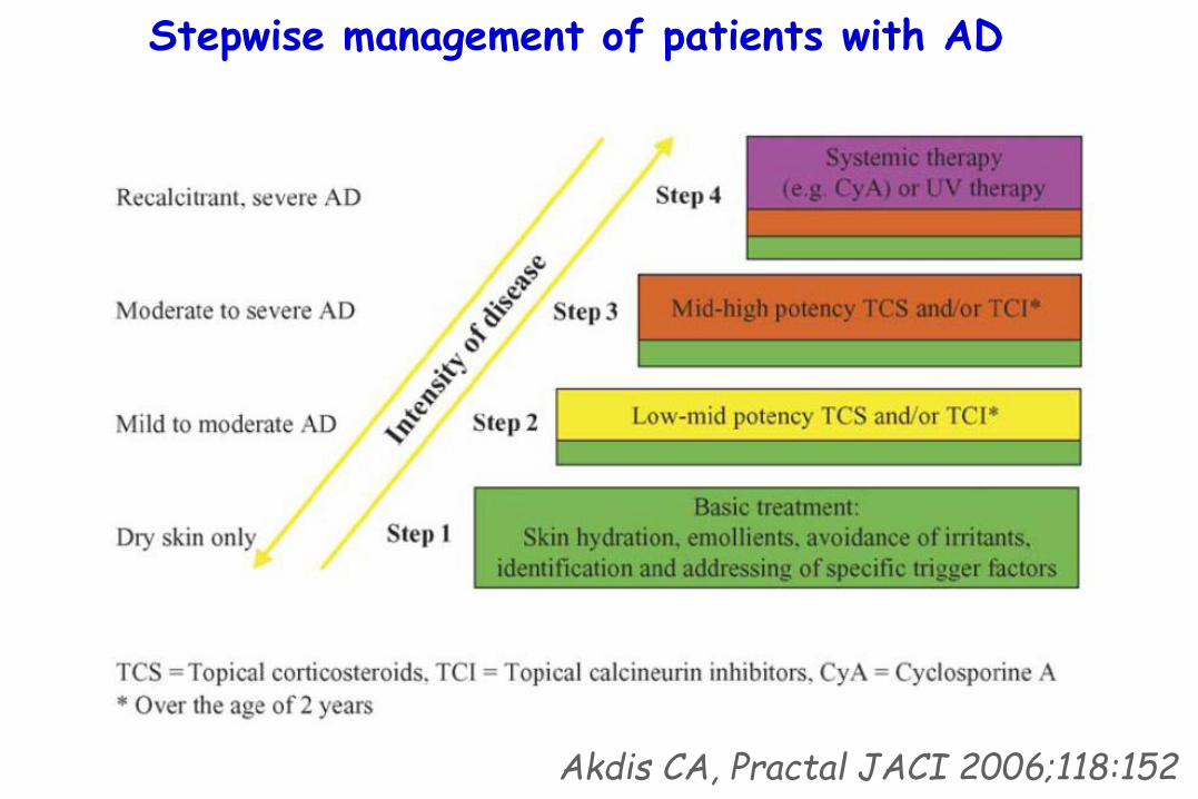
Stepwise management of patients with AD
Akdis CA, Practal JACI 2006;118:152

STEROIDI TOPICI MOLTO POTENTI (GRADO II)
Alcinonide 0,1% c. Halciderm
Amcinonide 0,1% p. Amcinil
Betametasone dipropionato 0,05% u c Diprosone; Betamesol; Betametasone dipropionato
Diflucortolone valerato 0,3% c. p. u. Nerisona forte, Temetex forte, Cortical, Dervin
Fluocinonide 0,05% p. g. l. Flu 21, Topsyn
STEROIDI TOPICI SUPERPOTENTI (GRADO I)
Clobetasolo propionato 0,05% p. u. s. sch. Clobesol; Olux sch
Potenza degli steroidi topiciAbbreviazioni: c:crema, p=pomata, u=unguento, lp= lipocrema, l= lozione, e= emulsione, s=soluzione, sch= schiuma, g= gel

STEROIDI TOPICI POTENTI B (GRADO IV)
Alclometasone dipropionato 0,1% c. u. l. Legederm
Beclometasone dipropionato 0,025% c. Menaderm simplex; Beclometasone Doc
Betametasone benzoato o,1% c. l. g. Beben
Budesonide 0,025 c. u. Bidien; Preferid
STEROIDI TOPICI POTENTI A (GRADO III)
Betametasone dipropionato 0,05% c. u. s. Diprosone, Betamesol, Betanesone dipropionato Sandoz
Betametasone valerato 0,1% c. u. e. s. Ecoval 70, Bettamousse, Betesil cerotti
Desossimetasone 0,025% e. Flubason
Diflucortolone valerato 0,1% c. u. s. Nerisona, Temetex, Dermaval, Cortical 0,2, Flu-cortanest
Fluticasone propionato 0,05% c.; 0,005% u. Flixoderm crema e unguento
Metilprednisolone aceponato 0,1% c. u .s. Advantan, Avancort
Mometasone furoato 0,1% c. u .s. Altosone, Elocon
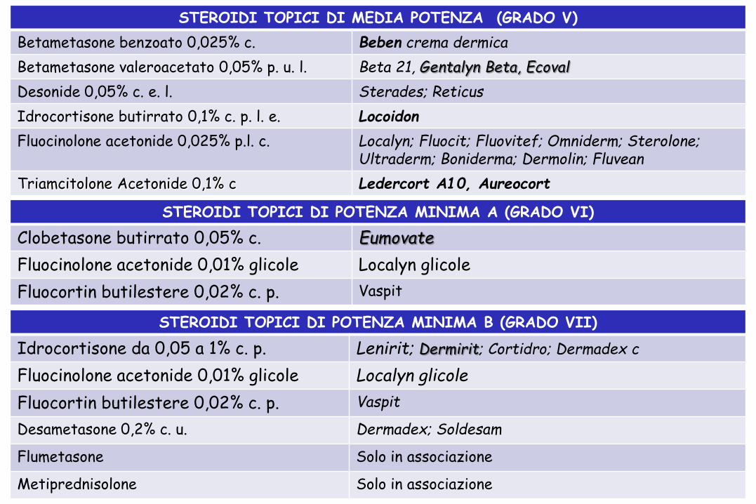
STEROIDI TOPICI DI POTENZA MINIMA A (GRADO VI)
Clobetasone butirrato 0,05% c. Eumovate
Fluocinolone acetonide 0,01% glicole Localyn glicole
Fluocortin butilestere 0,02% c. p. Vaspit
STEROIDI TOPICI DI POTENZA MINIMA B (GRADO VII)
Idrocortisone da 0,05 a 1% c. p. Lenirit; Dermirit; Cortidro; Dermadex c
Fluocinolone acetonide 0,01% glicole Localyn glicole
Fluocortin butilestere 0,02% c. p. Vaspit
Desametasone 0,2% c. u. Dermadex; Soldesam
Flumetasone Solo in associazione
Metiprednisolone Solo in associazione
STEROIDI TOPICI DI MEDIA POTENZA (GRADO V)
Betametasone benzoato 0,025% c. Beben crema dermica
Betametasone valeroacetato 0,05% p. u. l. Beta 21, Gentalyn Beta, Ecoval
Desonide 0,05% c. e. l. Sterades; Reticus
Idrocortisone butirrato 0,1% c. p. l. e. Locoidon
Fluocinolone acetonide 0,025% p.l. c. Localyn; Fluocit; Fluovitef; Omniderm; Sterolone; Ultraderm; Boniderma; Dermolin; Fluvean
Triamcitolone Acetonide 0,1% c Ledercort A10, Aureocort

Prescribing Topical Corticosteroids in Atopic Dermatitis
Charman C. Clin Dermatol. 2003:21:193-200.
Type of Preparation
•Ointment bases are more occlusive than creams and result in better penetration and an increased hydrating effect on the skin.Because preservatives are not required in ointments, they are associated with a lower incidence of hypersensitivity reactions.
•Creams, however, can be more cosmetically acceptable on the faceand are preferable in moist, hairy areas.
•Lotions, gels, and mousses are useful on the scalp but often contain alcohol, which may cause a stinging or burning sensation on inflamed skin.

Guidelines for treatment of atopic eczema (atopic dermatitis) Part IJ. Ring, JEADV 2012, 26, 1045–1060
Topical anti-inflammatory therapy
Topical Calcineurin Inhibitors
•The anti-inflammatory potency of 0.1% tacrolimus ointment issimilar to a corticosteroid with intermediate activity, while thelatter is clearly more active than 1.0% pimecrolimus cream.
•TCI do not induce skin atrophy. This favours their use overtopical corticosteroids in delicate body areas such as the eyelidregion, the perioral skin, the genital area, the axilla region or the inguinal fold and for topical long-term management.
Severe granuloma gluteale infantum

Guidelines for treatment of atopic eczema (atopic dermatitis) Part IJ. Ring, JEADV 2012, 26, 1045–1060
Topical anti-inflammatory therapy and wet wraps
•Patients with acute, oozing and erosive lesions, and children in particular, sometimes do not tolerate standard topical application, and may first be treated with ‘wet wraps’ until the oozing stops.
•They are highly effective in acute eczema and improve tolerance.
•The use of wetwrap dressings with diluted corticosteroids for up to 14 days (usual is up to 3 days) is a safe crisis intervention treatment of severe and/or refractory AE

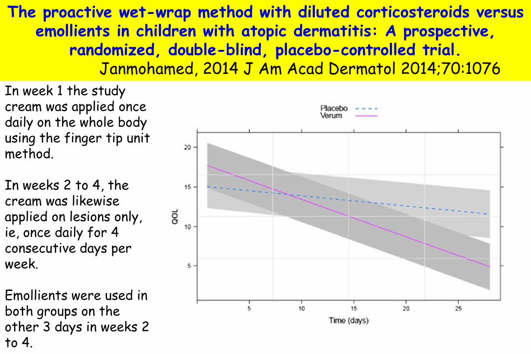
The proactive wet-wrap method with diluted corticosteroids versus emollients in children with atopic dermatitis: A prospective,
randomized, double-blind, placebo-controlled trial. Janmohamed, 2014 J Am Acad Dermatol 2014;70:1076
children aged 6 monthsto 10 years with severe AD (SCORAD > 40)
4 wks comparing dilutedcorticosteroids (1:3 mometasone furoate0.1% ointment and for the face 1:19 mometasone furoate0.1% ointment under a mask)
with emollient (petrolatum 20% in cetomacrogol cream).
The proactive wet-wrap method with diluted corticosteroids versus emollients in children with atopic dermatitis: A prospective,
randomized, double-blind, placebo-controlled trial. Janmohamed, 2014 J Am Acad Dermatol 2014;70:1076
In week 1 the study cream was applied once daily on the whole body using the finger tip unit method.
In weeks 2 to 4, the cream was likewise applied on lesions only, ie, once daily for 4 consecutive days perweek.
Emollients were used in both groups on the other 3 days in weeks 2 to 4.

Prevention of exacerbations with topical treatment
An important concept with therapeutic implications is the recognition that normal-appearing skin in patients with AD is not immunologically normal.Hamid Q, J Clin Invest 1994;94:870-6.
One approach to patients whose eczema tends to relapse in the same location is that of
proactive therapy.
After a period of stabilization, topical steroids (1,2) or calcineurin inhibitors (3-5) are applied to areas of previously involved but normal-appearing skin rather than waiting for eczema to flare. 1) Berth-Jones J, BMJ 2003;326:1367. 2) Peserico A, Br J Dermatol 2008;158:801. 3) Wollenberg A, Allergy 2008;63:742. 4) Breneman D, J Am
Acad Dermatol 2008;58:990-9. 5) Paller AS, Pediatrics 2008;122:e1210-8.

Guidelines of care for the management of atopicdermatitis. Sidbury, 2014; J Am Acad Dermatol.
Continued use of either topical corticosteroids (1-2 times/wk) or topical calcineurin inhibitors (2-3 times/wk) after disease stabilization, to previously involved skin, is recommended to reduce subsequent flares or relapses.
What are the most effective approaches to preventing flares in patients with
atopic dermatitis?
La Dermatite atopicavista dal pediatra
Introduction
Pathogenesis
Triggers & Allergens
Treatments
Conclusions
Universita’ di Ferrara
Diego Peroni

Conclusions
1. Although there has been some controversy with regard to the role of allergy in atopic dermatitis (eczema), the bulk of the data indicate that allergy plays a role in selected patients with AD.
2. In patients with AD, the rate of sensitization to foods (positive skin or in vitro test) ranges from 30 to 80 percent, depending upon the population. The rate of confirmed food allergy is much lower.
3. Food allergies play a role in exacerbating AD in up to 33% of patients with severe AD, 10% to 20% with moderate AD, and 5% percent with mild AD.
4. Elimination of food allergens in patients with AD and confirmed food allergy can lead to significant clinical improvement.

Conclusions
5. The data on the role of aeroallergens in exacerbating AD are less extensive.
6. Dust mites are consistently the most common positive aeroallergen, and also appear to be the most clinically relevant.
7. Immune reactions, both IgE and T cell-mediated, to Malasseziaspecies to S. aureus and can also worsen AD.
8. Proper early treatment of the skin is probably the simplest and most effective preventive strategy also for related diseases.
9. Allergen avoidance can be tried in selected patients.
10. Always consider comorbidities.