lab. #3 - int 256 - school of earth and climate...
TRANSCRIPT

SFR 458 – Tree Pests and Disease Lab : page 1
SFR 458 – Tree Pests and Disease Lab
I. Disease ConceptsA. Disease: Persistent, detrimental functioning
1. Which tissue is affected ?
2. Is overall growth and survival affected?
B. Cause of disease – disease triangle – disease complex
1. Stress agenta) Pestb) Pathogenc) Signs
2. Diseased treea) Symptomsb) Functions
(i) Tissue
(ii) Tree
(iii) Stand
3. Environmenta) Conditions where disease is possible.b) Depends on:
(i) Climate
(ii) Site conditions
(iii) Other species present
(iv) Management
(a) Natural regeneration
(b) Disturbance regime altered (old fields, fire)
(c) Plantations/nurseries
(d) Urban

SFR 458 – Tree Pests and Disease Lab : page 2
II. Laboratory Disease Descriptions.You are expected to complete descriptions of specific disease complexes. Many of the descriptions for the complexes can be viewed at
http://apollo.umenfa.maine.edu/SFR457_458_557/SFR457CourseInfo.html
For each sample that you needed to describe, complete the following components:
A. Disease complex name:
B. Disease complex components - disease triangle.
1. Stress Agent [pathogen(s)].a) Factor(s) or name(s):b) Signs:c) Mechanism of how disease is caused:
2. Diseased tree (plant) (s).a) Susceptible species:b) Symptom(s):c) Diseased function(s):
(i) Tissue function:
(ii) Tree/stand impact:
3. Environment (where to monitor):
Sketches should be made of disease symptoms and pathogen signs. Use proper terminology in labeling various parts of the sketches. These drawings need not be works of art, but they should serve as study aids in reviewing for quizzes and exams. Your description grade will be mostly based on your sketches.

SFR 458 – Tree Pests and Disease Lab : page 3
III. Orders of Insects(Edmonds et al. 2011: 435-473; 2000: p. 413-435)
A. Purpose:
To recognize the major taxonomic orders of insects.
B. Materials:
Dissecting microscope or hand lens, compound microscope, insect specimens.
C. Introduction.a) Phylogenetic classification.
(i) Kingdom: Animalia
(ii) Phylum: Arthropoda (spiders, mites, crabs, centipedes, etc.)
(a) Arthropoda = "jointed legs"
(iii) Class: Insecta
(iv) Order: 26 (depending on the classification used)
(v) Family: name ends in -idae (for animals)
(vi) Genus: name begins with a capital letter
(vii) Species: name begins with a lower case letter
2. Characteristics of the phylum Arthropoda.
(i) Segmented: usually 2 or 3 distinct body regions
(ii) Paired jointed appendages
(iii) External skeleton (exoskeleton)
(iv) Ventral nerve chord
3. Characteristics of the class Insectaa) 3 body regions
(i) Head
(a) Compound eyes
(b) Antennae
(c) Mouth parts
(1) Chewing
(2) Sucking
(ii) Thorax: 3 segments, 1 pair of legs/segment
(a) Prothorax
(b) Mesothorax - 1 pair of wings
(c) Metathorax - 1 pair of wings
SFR 458 – Tree Pests and Disease Lab : page 4
(d) Function: locomotion
(iii) Abdomen
(a) 11 of fewer segments
(b) Digestion, reproductionb) 3 pair of legsc) 1 or 2 pair of wingsd) 1 pair of antennaee) Compound eyes

SFR 458 – Tree Pests and Disease Lab : page 5
Name: __________________________________________ Date: ___________________
D. Insect Orders
Examine samples from each of the insects orders listed below. Make a sketch of each insect and CLEARLY label the parts that can distinguish this insect order from others. BE SURE TO DRAW AND INDICATE ALL THREE BODY REGIONS.
1. Odonata: Dragonflies, damselfliesHow ID: Front wings have many veins and cross veins; hind wings similar to front wings; wings don’t fold onto body; chewing mouthparts:
Dragonfly sketch - Draw and Label: Top and bottom views; head (eyes, antennae, mouthparts), thorax (6 legs; wings with veins), abdomen
2. Dictyoptera: roaches, mantidsHow ID: Front wings elongate, often thickened, usually with many veins; hind wings wider than front; wings fold along body; chewing mouthparts
Cockroach sketch - Draw and Label: Top and bottom views; head (eyes, antennae, mouthparts), thorax (6 legs; wings), abdomen

SFR 458 – Tree Pests and Disease Lab : page 6
Praying Mantis sketch - Draw and Label: Top and bottom views; head (eyes, antennae, mouthparts), thorax (prothorax, mesothorax, metathorax; 6 legs; wings), abdomen
3. Orthoptera: Crickets, grasshoppers, walking sticksHow ID: Front wings long and slender, thickened, with many veins; hind wings wider than front, membranous; folded on body; chewing mouthparts.
Cricket sketch - Draw and Label: Top and bottom views; head (eyes, antennae, mouthparts), thorax (prothorax, mesothorax, metathorax; 6 legs; wings), abdomen:
SFR 458 – Tree Pests and Disease Lab : page 7
Walking stick sketch - Draw and Label: Top and bottom views; head (eyes, antennae, mouthparts), thorax (prothorax, mesothorax, metathorax; 6 legs; wings), abdomen; NOTE – flightless when mature, helps explain how wings formed:
4. Coleoptera: BeetlesHow ID: Front wings hardened, protective, called “elytra”, meet in strait line on back; hind wings fold and are hidden under the elytra; chewing mouthparts.
Chafer beetle sketch - Draw and Label: Top and bottom views; head (eyes, antennae, mouthparts), thorax (prothorax, mesothorax, metathorax; 6 legs; wings), abdomen:

SFR 458 – Tree Pests and Disease Lab : page 8
Long-horned beetle sketch - Draw and Label: Top and bottom views; head (eyes, antennae, mouthparts), thorax (prothorax, mesothorax, metathorax; 6 legs; wings), abdomen:
5. Lepidoptera: Moths, butterliesHow ID: Wings triangular in form and with scales, few veins; front wings slender to rather broad; hind wings usually shorter, broader, and more rounded; wings to not fold; mouthparts are coiled tubes.
Monarch Butterfly sketch - Draw and Label: Top view only; head (antennae), thorax (wings):

SFR 458 – Tree Pests and Disease Lab : page 9
Silkworm Moth adult sketch - Draw and Label: Top and bottom views; head (eyes, antennae, NO mouthparts), thorax (6 legs; wings), abdomen:
6. Hemiptera - Heteroptera: “Bugs”How ID: Front wings upper part hardened and pigmented, few veins; hind wings membranous, shorter and wider; wings fold along body; sucking mouthparts arise from front side of head.
Shield Bug sketch - Draw and Label: Top and bottom views; head (eyes, antennae, mouthparts), thorax (prothorax, mesothorax, metathorax; 6 legs; wings), abdomen:

SFR 458 – Tree Pests and Disease Lab : page 10
7. Hemiptera – Homoptera: Cicadas, aphids, leafhoppers, scalesHow ID: Wings (many times absent) slope over side of body (tentlike); front wings uniform; sucking mouthparts arise underside of head
Cicada sketch - Draw and Label: Top and bottom views; head (eyes, antennae, mouthparts), thorax (prothorax, mesothorax, metathorax, 6 legs; wings), abdomen:
Aphid sketch from microscope slide - Draw and Label: Top and bottom views; head (eyes, antennae, mouthparts), thorax (6 legs; wings), abdomen (include structures that exude honeydew); NOTE: Wings aren’t used when mature:

SFR 458 – Tree Pests and Disease Lab : page 11
8. Hymenoptera: Ants, wasps, bees, hornetsHow ID: Font wings membranous with few veins; hind wings smaller than front; chewing mouthparts.
Yellow Jacket Wasp sketch - Draw and Label: Top and bottom views; head (eyes, antennae, mouthparts), thorax (prothorax, mesothorax, metathorax; 6 legs; wings), abdomen (note constriction where thorax joins abdomen):
Ant sketch - Draw and Label: Top and bottom views; head (eyes, antennae, mouthparts), thorax (prothorax, mesothorax, metathorax; 6 legs), abdomen (note constriction where thorax joins abdomen):

SFR 458 – Tree Pests and Disease Lab : page 12
9. Diptera: FliesHow ID: Front wings membranous; hind wings no longer functional – now halteres; mouthparts piercing to sponging.
Onion Fly sketch - Draw and Label: Top and bottom views; head (eyes, antennae, mouthparts), thorax (6 legs; wings), abdomen:

SFR 458 – Tree Pests and Disease Lab : page 13
Insect Orders
Coleoptera
Dictyoptera
DipteraHemiptera
Heteroptera
Homoptera
Hymenoptera
Lepidoptera
Odonata
Orthoptera

SFR 458 – Tree Pests and Disease Lab : page 14
Introduction to FungiI. Introduction
A. Primary attributes.
1. Eukaryotic.
2. Multicellular - hyphae
3. Mycelial growth form.
4. Cell walls with chitin.
5. Heterotrophic.
6. Reproduction by spores.a) Definition.
b) Function.
c) Asexual (imperfect).d) Sexual (perfect).
(i) Plasmogamy (N+N).
(ii) Karyogamy
(iii) Meiosis.
(iv) Haploid (1N) nuclei form spores
B. Growth.
1. Requirements.
2. Thallus.a) Hypha (hyphae)
b) Mycelium (mycelia).

SFR 458 – Tree Pests and Disease Lab : page 15
SFR 458 – Tree Pests and Disease Lab : page 16
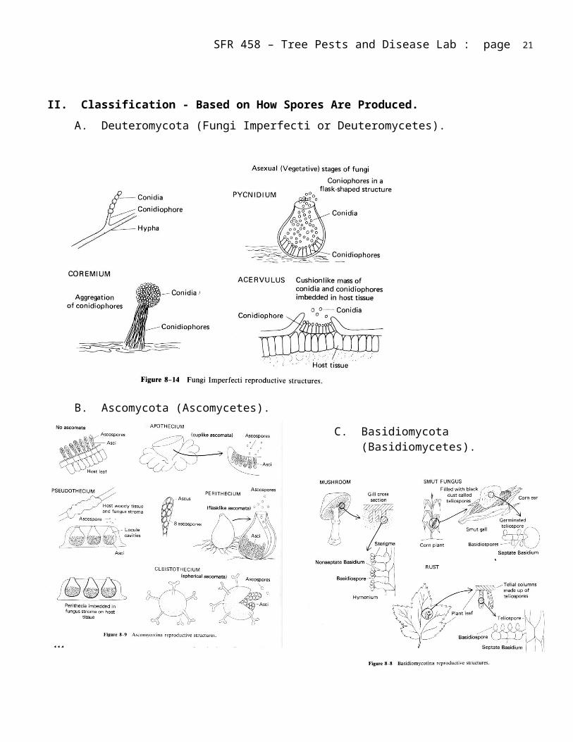
II. Classification - Based on How Spores Are Produced.A. Deuteromycota (Fungi Imperfecti or Deuteromycetes).
B. Ascomycota (Ascomycetes).
C. Basidiomycota (Basidiomycetes).

III. Introduction to Fungal Incited Diseases - ExamplesA. Tar spot of maple (Acer) (class web site)
1. Observe the signs and symptoms of tar spot caused by Rhytisma. Identify the stroma formed by the pathogen.
2. Find an apothecium of Rhytisma on a prepared slide. Label stroma, apothecium, asci, leaf tissue.
B. Needle cast (Necrophytes). Symptoms are dead tissue or needles
1. Obtain pine (Pinus spp.) needles infected with Lophodermium. a) Using a dissecting microscope, learn to recognize the distinctive fruiting structure
of this fungus (elongated apothecia or hysterothecia).
2. Obtain balsam fir (Abies balsamea) needles infected with Isthmiella (= Bifusella) and learn to recognize the pycnidia (asexual) and hysterothecia (sexual) of this species. (http://www.na.fs.fed.us/spfo/pubs/howtos/ht_bfir/ht_bfir.htm) (class web site)
C. Red belt fungus. (class web site)
1. Fomitopsis pinicola (Fomes pinicola) is the most common decay of slash and logs in North America (conifers and deciduous trees). It causes a brown, cubical, crumbly rot.
2. Examine the sporophore and note the following: a) Perennial, shelving conks, 2-10 in (2 ft) flat to hoof shape.b) Upper surface smooth to crusty, gray to black in older parts, usually with white and
red bands at margin.c) Lower surface white with round pores.d) Context buff colored, corky.e) Mycelial felts in decayed wood.
3. Fungal spores normally infect only dead stems. Brown cubicle rot can occur up to 30 ft from conk.
D. Phellinus trunk rot of aspen. (class web site)
1. Phellinus tremulae (Fomes igniarius var. populinus) is the most serious decay of aspen (Populus spp.). It causes a white trunk rot of living trees.
2. Examine the sporophore and note the following: a) Perennial conks hoof shape (hymenium convex), ca. 4 in across.b) Upper surface grayish black, cracked, cinder-like.c) Hymenium gray with round pores.d) Interior dark brown with white (silver) flecks (tubes filled with mycelium).
3. Spores enter through wounds and branch stubs.
4. Decay extends 8 or more feet from conk.
E. Tinder fungus. (class web site)
1. Fomes fomentarius. commonly causes white rot in dead stems of hardwoods, such as birches (Betula), beeches (Fagus), maples (Acer), and poplars (Populus).
2. Examine the sporophore and note the following:a) Perennial conks hoof shape (hymenium flat), ca. 3 in across.b) Upper surface gray to tan, smooth, concentric ridges.c) Hymenium gray with round pores.d) Interior dark brown.
3. Spores infect wood of dead, standing trees and sometimes decays heartwood of living trees.
4. Presence of conk indicates total cull.
F. Birch Polypore. (class web site)
1. Piptoporus betulinus (Polyporus betulinus) is the most common decay causing a brown rot in dead birch.
2. Examine the sporophore and note the following:a) Annual fruiting body growing a tree stem; has a short stalk; 4-6” dia round cap;
corky in textureb) Beige in color on top; bottom has pores with unequal lengths; can appear “toothy”;
poor layer can separate from the upper layer.
G. Shoe string root rot, Armillaria root disease. (incited by Armillaria spp., Agaricaceae). http://www.na.fs.fed.us/spfo/pubs/fidls/armillaria/armillaria.htm (and class web site)
1. Examine sporophores and identify: cap, stem, gills (attached), collar, lack of a cup.
2. Examine rhizomorphs.
3. Note the resin production and identify mycelial fans underneath the bark of a spruce tree infected with the disease.
H. Chestnut blight incited by Cryphonectria parasitica (= Endothia parasitica), a diffuse canker on American chestnut (Castanea dentata). (http://www.apsnet.org/online/feature/chestnut/top.html) (class web site)
1. Examine specimens and be able to identify and locate:a) Mycelial fans.b) Perithecia embedded in stroma.
2. Examine two prepared slides with a compound microscope for:a) Pycnidia in host tissue.b) Stroma, perithecia, and ascospores.

I. Beech bark disease, a diffuse canker incited by Cryptococcus fagisuga followed by Neonectria faginata = Nectria coccinea var. faginata..(http://www.na.fs.fed.us/spfo/pubs/fidls/beechbark/fidl-beech.htm) (class web site)
1. Examine specimens of American beech (Fagus grandifolia) infected with Cryptococcus fagisuga (a scale insect) and Neonectria faginata and be able to identify and locate:a) The beech scale.b) Clusters of perithecia.
2. Examine prepared slides for:a) Stroma.b) Perithecia.
J. Black knot (incited by Dibotryon morbosum = Apiosporina morbosa) of cherry (Prunus), a perennial gall/canker.
1. Note the conspicuous black stroma which is characteristic of this common disease of cherry. The stroma development kills a gall (symptom) that had formed after initial fungal infection.
2. The ascocarps (perithecia) are covering the surface of the stroma.
Latin NamesRhytismaLophodermiumFomitopsis pinicolaPhellinus tremulaeFomes fomentariusPiptoporus betulinusArmillariaCryphonectria parasiticaNeonectria faginataCryptococcus fagisugaApiosporina morbosa

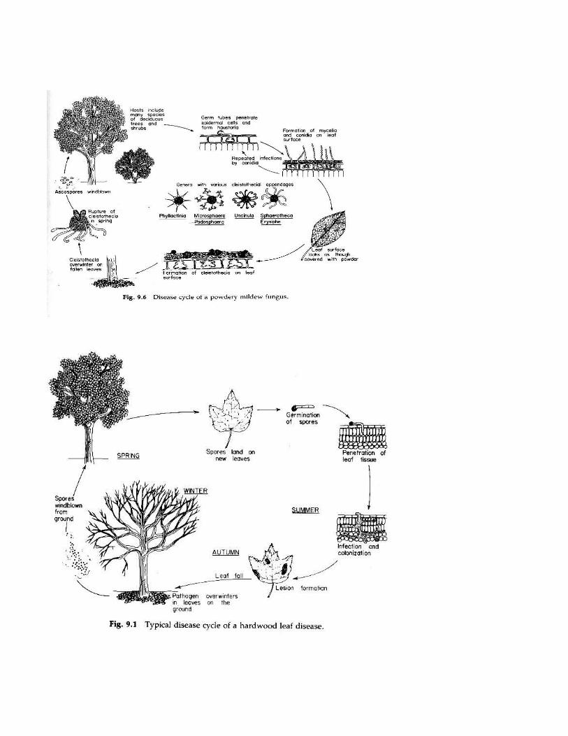
IV. Ascomycota Foliar Disease: HardwoodsA. Powdery mildews (class web site)
1. Identify the genus.a) Using the dissecting microscope, examine leaves of red oak (Quercus rubra) OR
red maple (Acer rubrum) infected by the powdery mildew fungus. Notice the signs of disease, the white mycelium on the leaf surface and the presence of black cleistothecia.
b) Identify the fungus to genus using the diagrams on the pathogen cycle and the key (see below).
(i) Pick off cleistothecia with a needle and razor blade and place them in a drop of water on a slide.
(a) Verify the presence of cleistothecia in the drop of water by using the dissecting microscope or hand lens.
(b) Place a cover slip over the water drop, but do not press.
(ii) Observe the appendages on the cleistothecia.
(iii) Split open the cleistothecia and count the number of asci.c) Key:
(i) One ascus per cleistothecia.
(a) Appendages mycelioid: Sphaerotheca.
(b) Appendages dichotomously branched: Podosphaera.
(ii) Several asci per cleistothecium.
(a) Appendages mycelioid: Erysiphe.
(b) Appendages dichotomously branched: Microsphaera.
(c) Appendages with a bulbous base: Phyllactinia.
(d) Appendages with coiled tips: Uncinula.
2. Complete a disease descriptiona) Disease tree/symptoms; inciting stress/signs, environment.b) Be sure to diagram and label the following:
(i) Overall appearance of fungus on leaf.
(ii) Cleistothecia with appendages.
(iii) Asci and ascospores.

B. Oak leaf blister. (class web site)
1. Examine oak leaf blister incited by the fungus Taphrina. Your sketch should indicate:a) Areas of discoloration.b) Any abnormal growth of the leaf tissue ("blisters") known as hypertrophy.
2. Examine a prepared slide of Taphrina and identify the ascus and ascospores.
C. Anthracnose. (http://www.na.fs.fed.us/spfo/pubs/howtos/ht_dogwd/ht_dog.htm)(http://na.fs.fed.us/spfo/pubs/fidls/anthracnose_east/fidl-ae.htm) (class web site)
On maple (Acer) incited by Kabetiella sp.
1. Examine leaves of maple for symptoms of the disease anthracnose.
2. Note location of necrotic tissue in relation to the leaf margin and veins. This will help distinguish this disease from other problems (e.g. drought, nutrient deficiencies) commonly found on trees.
3. There are no apparent signs on these samples.
D. Elm leaf spot (class web site)
1. Examine elm (Ulmus) leaves affected by leaf spot and note the differences in symptoms with those of anthracnose.
2. Examine prepared slides of elm leaf spot and locate perithecia of the pathogen (Gnomonia is the Latin name for the sexual stage, Gloeosporium is the Latin name for the asexual stage (not seen)).
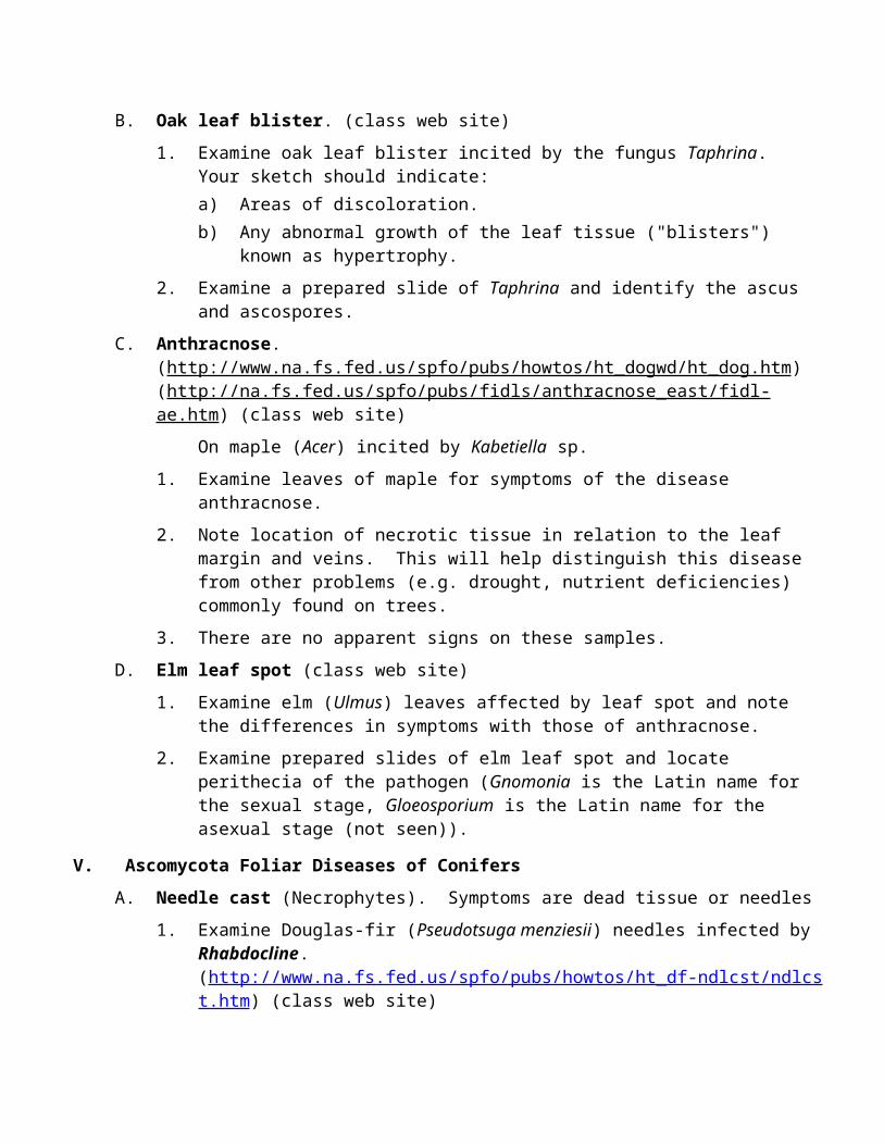
V. Ascomycota Foliar Diseases of ConifersA. Needle cast (Necrophytes). Symptoms are dead tissue or needles
1. Examine Douglas-fir (Pseudotsuga menziesii) needles infected by Rhabdocline.(http://www.na.fs.fed.us/spfo/pubs/howtos/ht_df-ndlcst/ndlcst.htm) (class web site)a) Compare apothecia of this fungus with the hysterothecia of the previous two
Ascomycetes and note the differences in shape, size, and location on the needle.b) Note the pattern and color of necrotic tissue associated with this fungus.c) Examine a prepared slide of infected needles with a compound microscope.
Identify asci and ascospores.
2. Examine Douglas-fir needles affected by Swiss needle cast, incited by Phaeocryptopus (= Adelopus). (http://www.na.fs.fed.us/spfo/pubs/howtos/ht_df-ndlcst/ndlcst.htm)a) Identify the minute perithecia emerging from stomata. b) Note how the necrotic tissue forms no distinctive pattern on the needles.c) Examine a prepared slide. Label needle tissue, stomata, and perithecia.
Latin NamesBifusellaErysiphaGloeosporiumGnomoniaIsthmiellaKabetiellaLophodermiumMicrosphaeraPhaeocryptopusPhyllactiniaPodosphaeraRhabdoclineRhytismaSphaerothecaTaphrinaUncinula
VI. Stem Decay Fungi.A. Introduction.
1. Symptomsa) Wood components.
(i) Lignin.
(ii) Cellulose.b) Brown rot.c) White rot.d) White-pocket rot.
2. Signsa) Conk.b) Mycelia.
B. Pathogen Cycle.
1. Basidiospores disseminated by wind.
2. Enter through wounds in stem.
3. Colonizes dead xylem tissue.
C. CODIT: Diagram as many walls as seen in the samples. Label the walls
CODIT: http://www.na.fs.fed.us/spfo/pubs/misc/treedecay/cover.htm
1. Wall #1 - plugging of vessels and tracheids.
2. Wall #2 - last cells in growth ring.
3. Wall #3 - ray cells.
4. Wall #4 - cambium response to wound.


D. Control
1. Prevent wounds.
2. Prune properly (handout).
How to Prune Trees: http://www.na.fs.fed.us/spfo/pubs/howtos/ht_prune/prun001.htm
3. Remove decayed trees.
Hazard Trees Web Site: http://www.na.fs.fed.us/spfo/hazard/index.htm
E. Make sketches of the three decay types (symptoms).
1. Brown rot.
2. White rot with zone lines.
3. White pocket rot.
F. Complete Decay Descriptions:
1. Disease Complex Name = Decay name.
2. Stress Agent = Decay fungus.a) Nameb) Sign: How identifyc) Mechanism: Saprophytic feeding on wood components
3. Susceptible tree species.a) Nameb) Symptom
(i) Decay type
(ii) How much decayc) Diseased function:
(i) Tissue function: (wood location - branches, stem heartwood, stem sapwood, roots)
(ii) Tree /stand impact: (living or dead tree)
4. Where to monitor: Follow the wounds!
G. Red ring rot. (class web site)
1. Phellinus pini (Fomes pini) can cause a white pocket rot in the heartwood of most living conifers in the north temperate zone.
2. Examine samples of the early decay which appears as rings of red to purple discoloration in a stem cross section.
3. Punk knots can form on the stems of living trees (samples not available). Yellow brown mycilium fill the punk knots.
4. Decay extends 2-4 ft each side of conk or punk knots.
5. Examine the sporophore and note the following:
a) On living trees - branch stubs.b) Ca. 3 in (1-12 in) across, shelving.c) Upper surface dark gray to black, concentrically furrowed.d) Hymenium (hymenial surface) with pores (round to daedaloid), yellow-brown.e) Context yellow brown when growing, darker when inactive.
6. The fungus enters through branch stubs and causes up to 50% volume loss in stands.
H. Phellinus trunk rot of hardwoods
1. Phellinus igniarius (Fomes igniarius) is a common decay of living and dead hardwoods including birch and maple.
2. It look identical to P. tremulae except for a curved margin; the margin on tremulae meats in a sharp edge.
I. Cinder conk.
1. Inonotus obliquus (Poria obliqua) commonly causes white rot and a canker in living birch, sometimes beech.
2. A sterile, cinder-like conk developing from branch stub or wound.
3. Spores enter through branch stub or wound.
4. Fungus decays heartwood and pushes through sapwood and phloem (canker develops) to form a sterile conk.
5. Fertile sporophore develops after tree dies.
6. Sterile conk indicates 15 ft or more of decay in stem.
J. Artist's Conk. (class web site)
1. Ganoderma applanatum (Fomes applanatus) can cause a white rot of dead hardwood stems, occasionally attacking heartwood of live trees.
2. Examine the sporophore and note the following:a) Large, shelving conk, up to 2 ft or more across.b) Upper surface light gray to light brown, smooth to crusty.c) Hymenium white, turns brown when touched.d) Interior: Upper layer white, lower layer brown.
3. Spores enter through branch stubs or wound.
4. Conk indicates 15 ft or more decay on live tree, 30 ft or more on dead tree.
VII. Root Rot Diseases.(Manion p. 293-306, 125-129) (Edmonds p. 275-291, 297-307)
A. Annosus Root Rot (incited by Heterobasidion annosum (Fomes annosus), Polyporaceae) http://www.na.fs.fed.us/spfo/pubs/fidls/annosus/fidl-ann.htm (and class web site)
1. Examine the perennial sporophores and decay of this basidiomycete.
Latin Names:
Armillaria sppFomes annosusFomes applanatusFomes fomentariusFomes igniariusFomes igniarius var. populinusFomes piniFomes pinicolaFomitopsis pinicola Ganoderma applanatum Heterobasidion annosum Inonotus obliquus Phellinus igniariusPhellinus piniPhellinus tremulaePiptoporus betulinusPolyporus betulinusPoria obliqua
VIII. Rust Fungi IntroductionSigns: Rust fungus – requires two hosts, 5 spore stages
A. First Host.
1. Spermagonia a) Used to bring 2 nucleae together
2. Aecia - blistersa) Aecial spores produced mitoticallyb) Infect alternate host.
B. Second Host.
1. Uredia a) Spores produced using mitosisb) Infect more alternate host; repeating stage.
2. Telia in columnsa) Grow out of urediab) Germinate to produce basidia
3. Basidia a) Used meiosis to produce basidiosporesb) Infect original host.

IX. Foliar Rust FungiA. Pine needle rust.
1. Examine pine needles infected with Coleosporium, a macrocyclic rust.a) Spermagonia (not usually visible) and aecia are produced on the pine needles.
Note the characteristic aecial blisters or aecial membranes on the needle surface.b) Examine prepared slides for aecia and aeciospores.
2. Uredia, urediospores (the repeating stage of the fungus), telia, and teliospores are found on asters (Aster spp.) and golden rod (Solidago canadensis). Examine leaves of golden rod for uredia of the fungus.
B. Ash leaf rust incited by Puccinia peridermiospora, a macrocyclic rust. (class web site)
1. Spermagonia and aecia occur on leaflets of ash species (Fraxinus).a) The fungus causes a leaf blotch.b) Spermagonia are on the upper surface.c) Aecia are formed on the lower surface with aecial membranes.
2. Uredia are in elongated lesions on marsh grass (Spartina spp.).
X. Stem Rust Fungi.A. Cedar-Apple Rust incited by Gymnosporangium juniperi-virginianae, a Demicyclic Rust.
1. Spermagonia (= pycnia) and aecia are found on leaves of apple and crabapple trees (Malus spp.). a) Examine infected leaves for spermagonia (upper surface) and aecia (on lower
surface directly below the spermagonia). The aecia are surrounded by long, curled aecial membranes.

b) Observe the spermatia (= pycniospores) and spermagonia (= pycnia) on a prepared slide (label says: "Pycnia on Pyrus leaf".
c) Observe the aecia and aeciospores on a prepared slide.
2. Galls and telia form on juniper (Juniperus) shoots.a) Examine dried specimens of cedar-apple galls on eastern red cedar.
(i) Note the size and shape.
(ii) The telial horns (= spikes) are bright orange and gelatinous when fresh.b) With forceps, break-off a small piece (<5 mm) of a telial horn and place it into a
drop of water on a microscope slide.
(i) With a razor blade, cut the telial horn into small portions (<1 mm). Let it absorb water for a minute or two.
(ii) Squash the fungal tissue between the slide and a cover slip and examine the specimen with a compound microscope.
(iii) Identify teliospores, their shape, and the number of cells per spore.
B. Fir broom rust incited by Melampsorella caryophyllacearum, a macrocyclic rust.
1. Spermagonia and aecia occur on balsam fir needles.a) Infection results in "broom" formation.b) Infected needles are shorter, thicker, and more yellow.c) Make a sketch of the needles with aecia.d) Needles fall off after one growing season.
2. Uredia, telia, and basidia are found on chickweed (Cerastium spp.). Sketch the chickweed, but the spore stages are not present on these samples.
C. White pine blister rust incited by Cronartium ribicola, a macrocyclic rust.(http://www.na.fs.fed.us/spfo/pubs/howtos/ht_white/white.htm) (class web site)
1. Spermagonia and aecia are produced on stems of white pine (Pinus strobus).a) Examine stems of white pine infected with blister rust and identify blackened areas
of spermagonial (= pycnial) scars and areas of aecial blisters scars. Note the discoloration of host tissue at the edge of the canker.
b) Examine prepared slides of aecia on white pine stems. Be able to distinguish between tree and fungal tissue.
2. Uredia and telia are produced on leaves of Ribes (currants and gooseberries).a) Examine Ribes leaves under the dissecting microscope and note the characteristics
of the necrotic areas, the uredia, and the telia (column-like structures).b) Examine prepared slides of Ribes infected with C. ribicola for uredia and telia.
D. Pine-pine gall rust incited by Endocronartium harknessii (= Peridermium harknessii) on pines. (class web site)
1. Only one host needed – 2-3 needle pines, e.g., jack pine.
2. Produces a gall with aecial-like blisters on live samples, scars on dead.

3. Aecial-like spores germinate and infect pine needles.
4. Spermagonia are rare.
E. Fusiform rust incited by Cronartium quercuum form species fusiforme on southern pines.(http://www.na.fs.fed.us/spfo/pubs/fidls/fusiform/fidl-fusi.htm)
1. Southern pines (Pinus. sp.).a) Produces a spindle shape, “fusiform” gall.b) Spermagonia on gall (not seen on samples)c) Aecial blisters on gall (can see scars)
2. Oak leaves (Quercus sp. - sorry, no samples to draw)a) Leaf spotb) Uredia, telia, basidia – similar to what is found on Ribes.
Latin Names for Rust FungiColeosporium Cronartium quercuum form species fusiformeCronartium ribicola Endocronartium harknessii Gymnosporangium juniperi-virginianae Melampsorella caryophyllacearum Peridermium harknessii Puccinia peridermiospora
XI. Cankers Diseases.A. Sirococcus shoot blight (Necrophyte) of red pine incited by Sirococcus conigenus.
(http://www.na.fs.fed.us/spfo/pubs/fidls/sirococcus/sirococcus.htm)
1. Symptoms of this disease are drooping, gray needles on branch tips.
2. Signs of the fungus are black pycnidia.
3. This is a problem of red pine plantations in the Maritimes and is a potential problem with red pine in Maine.
B. European larch canker incited by Lachnellula. (class web site)
1. Examine larch (Larix) branches for cankers infected with Lachnellula willkommii.
2. Locate the apothecia of this fungus.
C. Scleroderris canker incited by Gremmeniella abietina = Scleroderris lagerbergii.(http://www.na.fs.fed.us/spfo/pubs/fidls/scleroderris/scleroderris.htm) (class web site)
1. Examine samples of pine which show brown apothecia in the needle scars.

D. Nectria "target" canker (incited by Neonectria ditissima = Nectria galligena), a perennial canker of most deciduous trees. (class web site)
1. Examine different stages of canker development. Note the concentric rings of callus that resemble a target.
2. Sometimes in the field, you can find perithecia around the edge of the canker (not required for notebook).
E. Hypoxylon canker (Hypoxylon mammatum = Entoleuca mammata) on quaking aspen (Populus tremuloides), a diffuse canker.(http://www.na.fs.fed.us/spfo/pubs/fidls/hypoxylon/hypoxylon.htm)
1. Examine specimens of quaking aspen infected with Hypoxylon and be able to identify and locate:a) Clusters of black perithecial heads in gray stromata.b) Slightly sunken, yellow-orange areas around the edge of the canker.

Latin NamesApiosporina morbosa Cryphonectria parasiticaCryptococcus fagisugaDibotryon morbosum Endothia parasitica Entoleuca mammataNeonectria ditissima Gremmeniella abietinaHypoxylon mammatum Nectria coccinea var. faginata..Nectria galligena Neonectria faginataScleroderris lagerbergii Sirococcus conigenus
XII. Vascular Wilt Diseases and Stains.(Manion p. 209-222, 285-292) (Edmonds p. 358-362)
A. Dutch elm disease incited by Ophiostoma ulmi (Ceratocystis ulmi) and O. novo-ulmi.http://www.na.fs.fed.us/spfo/pubs/howtos/ht_ded/ht_ded.htm (class web site)
1. Examine branches showing egg and larval galleries of the European elm bark beetle (Scolytus multistriatus) (egg gallery with the grain of wood) and native elm bark beetle (Hylurgopinus rufipes) (egg gallery against the grain of wood).
2. Examine twigs of elm for feeding wounds of the European elm bark beetle. Note (1) location and (2) size.
3. Observe pattern of staining in elm twigs infected with the disease.
XIII. Parasitic Flowering Plants (The Mistletoes).(Manion p. 314-327) (Edmonds p. 333-339)
A. True Mistletoe (incited by Phoradendron spp.). http://www.fs.fed.us/r6/nr/fid/fidls/147.htm
Hosts: Primarily hardwoods in south half of U.S.
1. Signs:a) Stems, leaves, and berries.b) Sinkers (infected rays) - view cross section of infected stem.
B. Eastern dwarf mistletoe (incited by Arceuthobium pusillum). http://www.na.fs.fed.us/spfo/pubs/fidls/dwarf_mistletoe/fidl-dm.htm (class web site)
Hosts: Black, white, and red spruce (Picea mariana, P. glauca, P. rubens) rarely on larch (Larix laricina).
1. Symptoms: Witches' broom.
2. Signs:a) Male shoots – rounded tip, more than 1 slit.b) Female shoots – pointed tip, one slit.c) Seed capsules and seed.d) Sinkers (infected rays) - view prepared slides.e) Basal cups.
XIV. Forest Insect Pests.(Manion p. 314-327) (Edmonds p. 333-339)
A. White Pine Weevil – Pissodes strobi http://www.fs.fed.us/r6/nr/fid/fidls/147.htm
Hosts: White pine and Norway spruce
1. Symptoms: Shephards crook
a) Tree tip killed, curlsb) Resinc) Red needles
2. Signs:a) Larval feeding: May to Julyb) Pupal chamber with frassc) Adult (not seen)
3. Environment / where to monitora) Needs duff layer.b) Open grown trees.
B. Spruce gall adelgids (Eastern: Adelgis abietis (exotic, northeast), Cooley: Adelgis cooleyi (native))http://www.uri.edu/ce/factsheets/sheets/sprucegalladelgid.htmlhttp://www.entomology.wisc.edu/diaglab/pdfs/landscape/spruce%20gallpdf.pdfHosts: Eastern: Norway and white, sometimes others; Cooley: white and western spruces
1. Symptoms: Gall on twigsa) Cooley: 1-3 inchesb) Eastern: ½ to 1 ½ inchesc) “Pineapple” shape
2. Signs: Not seen a) Aphid like: 1/8 – 1/4 inch
3. Environment / where to monitora) Ornamental nuisance
C. Asian Longhorned Beetle (Anoplophora glabripennis)http://www.aphis.usda.gov/newsroom/hot_issues/alb/alb_general_info.shtml http://www.na.fs.fed.us/pubs/palerts/alb/alb_pa.pdfHosts: Maples (Acer) and other hardwoods
1. Symptoms: a) Oozing sap, cankersb) Declinec) Sometimes death
2. Signs:a) Adult: 0.75-1.25 inch long, black and white antennae, glossy black body with
irregular white spots, May to September.b) Larvae in wood; 12-18 monthsc) Feeding generates frassd) 3/8 inch round exit holes
3. Environment / where to monitora) Spreads on shipping crates and campfire wood
b) Monitor urban sites near import docks, infested areas.
D. Emerald Ash Borer (Agrilus planipennis)http://www.emeraldashborer.infohttp://www.emeraldashborer.info/files/E-2938.pdfHosts: All ashes.
1. Symptoms: a) Cankers, bark splitting, sproutsb) Decline and dieback – branches to entire crownc) Tree death
2. Signs:a) Adult: ½ inch long, metallic green, May to Augustb) Larvae: Year round, 1-2 yearsc) Feeding generates frass, serpentine galleries at cambiumd) D shaped exit holes.
3. Environment / where to monitora) Spreads on shipping crates and campfire woodb) Monitor urban sites near import docks, infested areas.c) Areas near camps
Latin NamesAdelgis abietisAdelgis cooleyiAgrilus planipennisAnoplophora glabripennisArceuthobium pusillumCeratocystis ulmiHylurgopinus rufipesOphiostoma novo-ulmiOphiostoma ulmiPhoradendronPissodes strobiScolytus multistriatus