lab manual accelerated biology - escience labs | 1: diffusion and osmosis lab 2: enzyme catalysis...
TRANSCRIPT

Lab ManualAccelerated Biology
© 2011 eScience Labs, LLCAll rights reserved
www.esciencelabs.com • 888.375.5487


Lab 1: Diffusion and Osmosis
Lab 2: Enzyme Catalysis
Lab 3: Mitosis and Meiosis
Lab 4: Plant Pigments and Photosynthesis
Lab 5: Respiration
Lab 6: Molecular Biology
Lab 7: Genetics of Organisms
Lab 8: Population Genetics and Evolution
Lab 9: Transpiration
Lab 10: Physiology of the Circulatory System
Lab 11: Animal Behavior
Lab 12: Dissolved Oxygen and Aquatic Primary Productivity
Table of Contents


Molecular Biology
AP BIOLOGY
Lab 6: Molecular Biology
Concepts covered
restriction endonucleases
Figure 6.1 Restriction endonucleases can snipDNA in two ways, leaving either blunt ends or
sticky ends.

Electrophoresis
recombinantDNA
Molecular Biology
Figure 6.2 Filling the wells of an electrophoresis gel
Figure 6.3 A fluorescent dye that binds DNA, can beused to visualize bands after the gel has been run.

Conjugation
Transduction
transformation
Figure 6.4 Scientists are able to engineer plasmids sothey contain a gene of interest from another organism.
Molecular Biology
Figure 6.5 Incorporation of a foreign gene can lead to cells with new characteristics.

ori
antibioticresistance
engineer plasmids
Pre-Laboratory Questions
Use additional paper as necessary
Molecular Biology

Molecular Biology
Figure 6.6
Experiment 6.1: Restriction Endonucleases and Electrophoresis
Materials
ProcedureTable 6.1

(Note: Enzyme 2 and 3 means that first, enzyme 2fragments the DNA, then enzyme 3 cuts the fragments made by enzyme 2).
Questions
Molecular Biology
Lab 6.2: Electrophoresis
Materials
Note: Gels must be analyzed immediately after electrophoresis.Color bands will diffuse and bleed over time.
1.Note: If the 10X solution has precipitated, place the bottle in a hot
water bath and swirl until precipitant dissolves.
Molecular Biology

2.
Note: The agarose will become clear whenmelted, about 40 45 seconds.
5.
Hint: The grooves are noticeable from above and from the side, about 1/4 inch fromthe top and bottom of the chamber. If you prefer, it may be helpful to be eye level with thechamber when pouring the agarose. When looking straight on, from a lateral view, you can seethe height of the grooves and where to stop when pouring the agarose.
Hint: Use a pipette tip to “poke” the bubbles or move them to theside.
Hint: You may have to softly jiggle the comb out of the gel.
Note: The dyes have been mixed with a sucrose solution to increase theirdensity and insure your sample will fall gently into the well. However, it is still important to runthe gel relatively quickly so the dyes do not diffuse out of the wells.
Molecular Biology

16. Remember to use aclean pipette for each sample!
Molecular Biology
Figure 6.7 Connecting the leads to themini gel chamber and power supply
Molecular Biology
Remember to use aclean pipette for each sample!
Note: DO NOT let the dyes move off the gel!
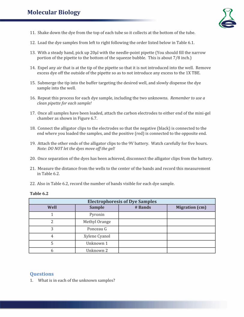
Table 6.2
Questions
Electrophoresis of Dye SamplesWell Sample # Bands Migration (cm)

Molecular Biology


1500 West Hampden AvenueSuite 5 H
Sheridan, CO 80110303 741 0674 888 ESL KITSwww.eScienceLabs.com