laboratory 1 safety and security measures microscope parts identification cell ...€¦ · ·...
TRANSCRIPT

Laboratory 1
Safety and Security measures
Microscope parts identification
Cell fractionation technique

Lab Safety Rules
1. Conduct yourself in a responsible manner at all times in the laboratory.
2. Follow all written and verbal instructions carefully. If you do not understand a
direction or part of a procedure, ASK YOUR TEACHER BEFORE PROCEEDING WITH
THE ACTIVITY.
3. Never work alone in the laboratory. No student may work in the science
classroom without the presence of the teacher.
4. When first entering a science room, do not touch any equipment, chemicals, or
other materials in the laboratory area until you are instructed to do so.
5. Perform only those experiments authorized by your teacher. Carefully follow all
instructions, both written and oral. Unauthorized experiments are not allowed.
6. Do not eat food, drink beverages, or chew gum in the laboratory. Do not use
laboratory glassware as containers for food or beverages.
7. Be prepared for your work in the laboratory.
Read all procedures thoroughly before entering the laboratory.
8. Always work in a well-ventilated area.
9. Keep hands away from face, eyes, mouth, and body while using chemicals or lab
equipment. Wash your hands with soap and water after performing all experiments.
10. Know the locations and operating procedures of all safety equipment including:
first aid kit(s), and fire extinguisher. Know where the fire alarm and the exits are
located.
11. Know what to do if there is a fire drill during a laboratory period; containers
must be closed, and any electrical equipment turned off.
12. Any time chemicals, heat, or glassware are used, students will wear safety
goggles.
13. Contact lenses may be not be worn in the laboratory.
14. Dress properly during a laboratory activity. Long hair must be tied back, and
dangling jewelry and baggy clothing must be secured. Shoes must completely cover
the foot. No sandals allowed on lab days.
15. A lab coat or smock should be worn during laboratory experiments.

To study cells
The cell is the basic structural and functional unit of all known living
organisms.
To study cells, biologists use microscopes and the tools of biochemistry
It can be difficult to understand how a cell, usually too small to be seen by
the unaided eye, can be so complex.
Cells are studied by using two Techniques:
- Microscopy
- cell fractionation
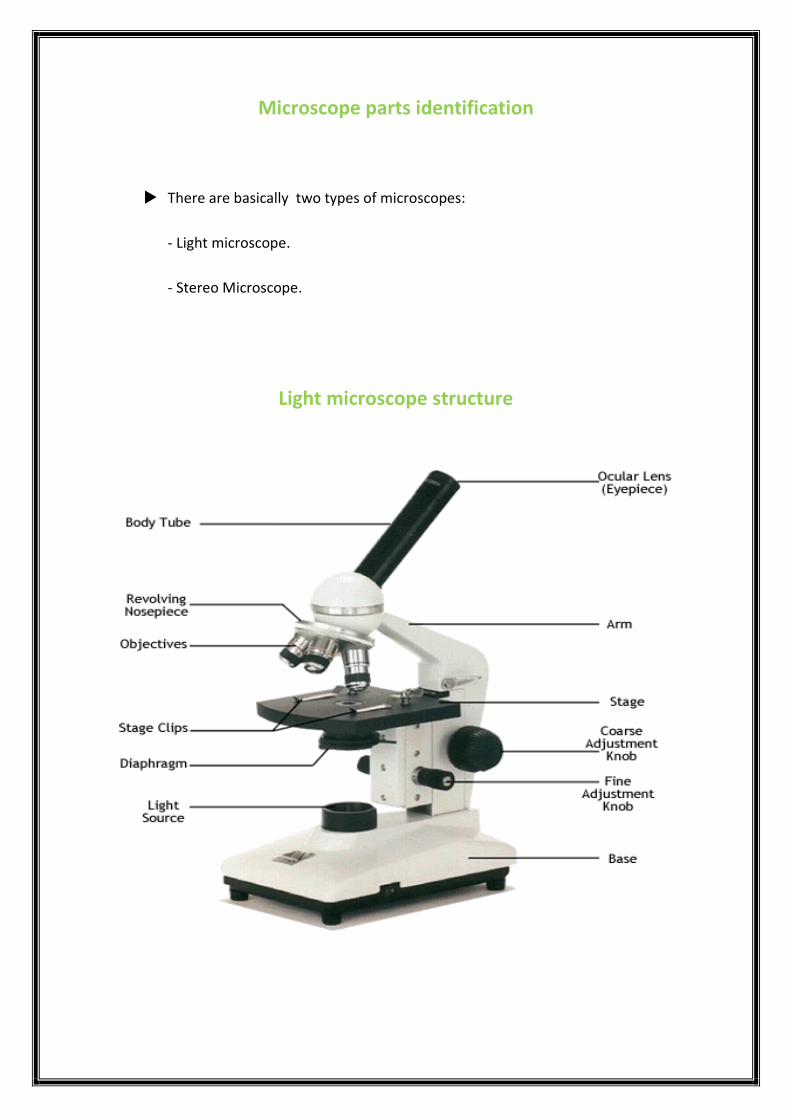
Microscope parts identification
There are basically two types of microscopes:
- Light microscope.
- Stereo Microscope.
Light microscope structure

The part � Function
Eyepiece � Magnifies the specimen image.
Body Tube The body tube holds the eye piece at the proper
distance.
Nose Piece The objective lenses can be turned to increase the
magnification.
Objective Lenses � The objective lenses increase magnification
(usually from 10x to 40x).
Stage Clips � These 2 clips hold the slide in place on the
stage.
Diaphragm � Used to vary the amount of light passing
through the slide. Usually it is better if the
amount of light is low.
Light Source � Some have lights, others have mirrors where
you must move the mirror to reflect light.
Arm � Used to support the microscope when carried,
and holds the body tube, nose piece and
objective lenses.
Stage � Supports the slide.
Coarse Adjustment Knob � Moves the stage up and down (quickly) for
focusing your image.
Fine Adjustment Knob � This knob moves the stage slightly to sharpen
the image.
Base � Supports the microscope.

Stereo Microscope Structure

Main differences between Light and Stereo Microscope
Light microscope Stereo microscope
40x – 1000 x 7x – 40x Magnification
4 objectives lenses 2 objectives lenses Structure
For higher magnification
(slides examination)
For lower magnification
(samples examination) Use
One source of lighting
Two
sources of lighting
(top/bottom)
Illumination
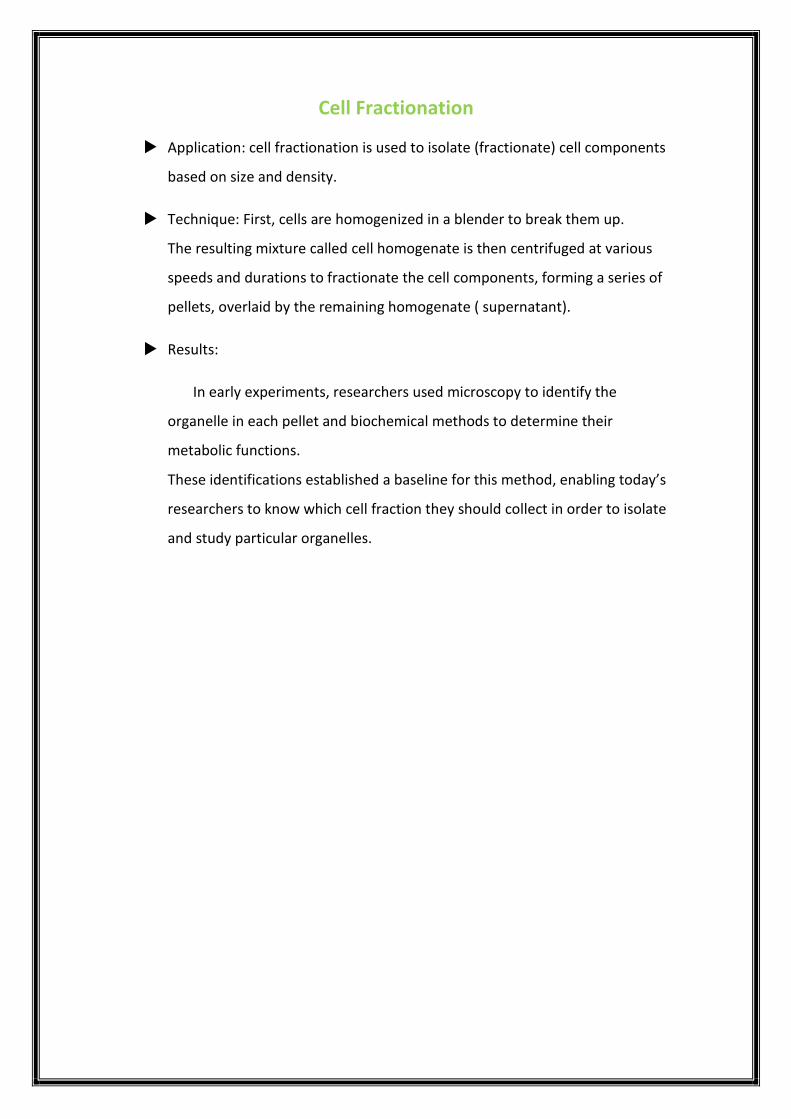
Cell Fractionation
Application: cell fractionation is used to isolate (fractionate) cell components
based on size and density.
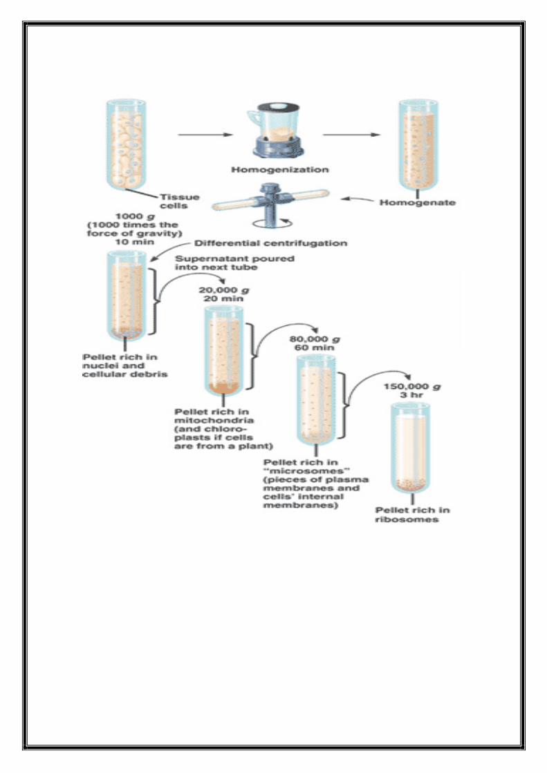
Technique: First, cells are homogenized in a blender to break them up.
The resulting mixture called cell homogenate is then centrifuged at various
speeds and durations to fractionate the cell components, forming a series of
pellets, overlaid by the remaining homogenate ( supernatant).
Results:
In early experiments, researchers used microscopy to identify the
organelle in each pellet and biochemical methods to determine their
metabolic functions.
These identifications established a baseline for this method, enabling today’s
researchers to know which cell fraction they should collect in order to isolate
and study particular organelles.

Laboratory 2
Animal cell and Plant cell
Cell organelles
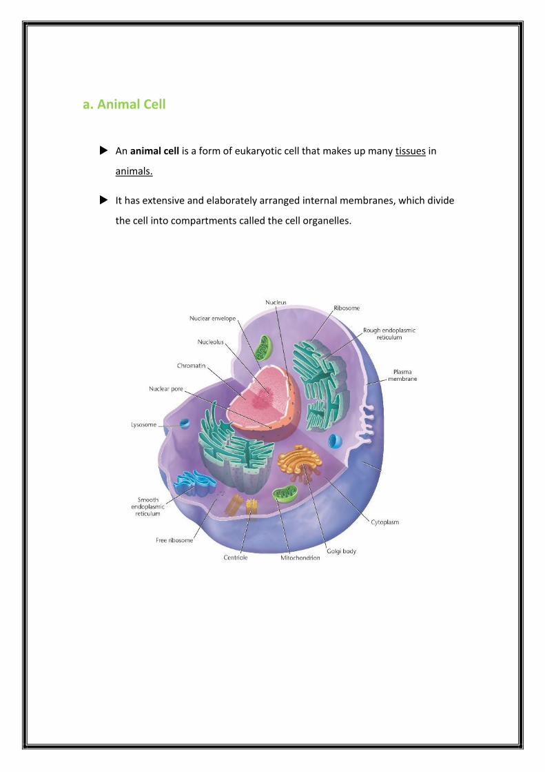
a. Animal Cell
An animal cell is a form of eukaryotic cell that makes up many tissues in
animals.
It has extensive and elaborately arranged internal membranes, which divide
the cell into compartments called the cell organelles.

Animal cell structures and their function
Function
Structure
Organelles
Boundary of the cell Phospholipid bilayer Cell membrane
Control all activity of the
cell, DNA maintenance
Double membrane
compartment Nucleus
Storage container Single membrane
compartment Vacuole
Cellular respiration Double membrane
compartment Mitochondria
Breakdown of large
molecules
Single membrane
compartment Lysosomes
Expression of lipids Single membrane
compartment
Smooth endoplasmic
reticulum
Translation and folding of
new proteins
Single membrane
compartment
Rough endoplasmic
reticulum
Translation of RNA into
proteins RNA-protein Ribosome
sorting, packaging,
processing and modification
of proteins
Single membrane
compartment Golgi apparatus

Function
Structure
Organelles
breakdown of metabolic
hydrogen peroxide
Single membrane
compartment Peroxisome
ribosome production protein-DNA-RNA Nucleolus
helps in cell division by
forming spindle fibers Microtubule protein
Centrioles

b. Plant Cell
Plant cells are eukaryotic cells found in plants and differ in several key
aspects from the cells of other eukaryotic organisms by:
A Large central vacuole .
A Cell wall.
Presence of plastids the most notable is the chloroplast.

Plant cell structures and their function
Organelles
Structure
Function
Cell wall Made of cellulose,
hemicellulose, and pectin.
Rigid, protective barrier
Chloroplast Double membrane
compartment Photosynthesis
Main differences between animal and plant cell
Animal cell
Plant cell
Don’t have chloroplast Have chloroplast
Don’t have a cell wall Have a cell wall
small vacuoles Large central vacuole

Prepare animal and plant cells experiment
Purpose :
In this lab, you will prepare and observe cells from a plant (onion) and an animal
(your own cheek).
Materials :
Water-methylene blue stain-iodine stain-glass slides-covers-cotton buds-dropper-
filter papers-onion.
Procedure :
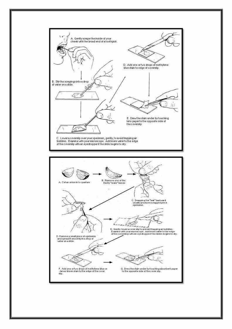
a. animal cell preparation
Take clean cotton buds and gently scrape the inside of your cheek.
Place one drop of water on the slide.
Swirl the cotton bud with cheek cells, in the water drop on the slide.
Put one drop of Methylene blue solution , using dropper and add this to the
mixture on the slide.
Remove any excess water and stain from the slide using a filter paper.
Take a clean cover slip and lower it carefully on the slide.
Observe animal cells under the microscope , making sure the microscope is
on low power.
Go through the three powers (4X,10X and 40X) and label any parts you see.
After you draw your diagrams, rotate the nosepiece back to low power.
Remove the slide and turn off the microscope.
b. plant cell preparation
Peel off a small section of onion skin.
Place the onion skin in the center of the slide.
Add a small drop of iodine stain directly to the tissue.
Place a cover slip over the stained tissue.
Gently tap the slide with a pencil to remove any air bubbles and draw excess
stain from the slide using a filter paper.
Observe plant cells under the microscope like you did above.

Results
a. Animal cell
b. Plant cell
Plant cell
Animal cell
Cell shape
Nucleus position

Laboratory 3
Cell membrane Structure and Function
Movement Across membranes:
Diffusion Experiment
Osmosis Experiment
Cell membrane
The cell membrane or also called plasma membrane surrounds the living cell
and control the passage of substances in and out.
The ability of the cell to discriminate in its chemical exchanges with its
environment is fundamental to life, and it is the plasma membrane and its
components molecules that make this selectively possible.
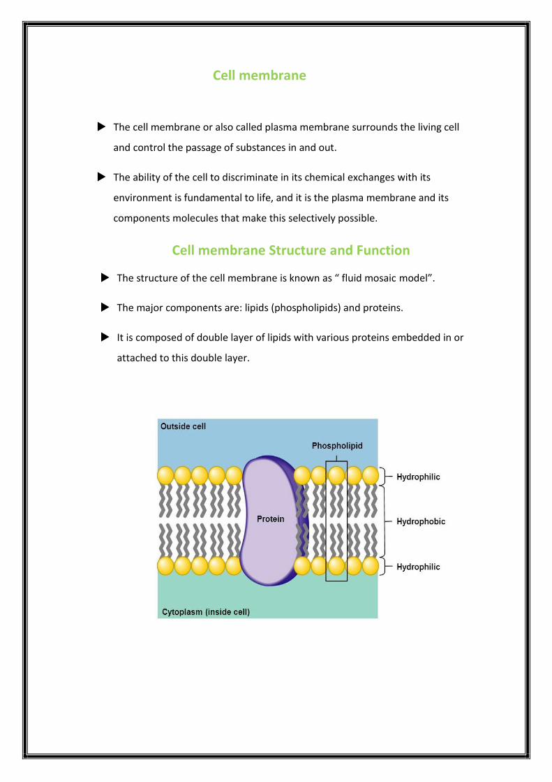
Cell membrane Structure and Function
The structure of the cell membrane is known as “ fluid mosaic model”.
The major components are: lipids (phospholipids) and proteins.
It is composed of double layer of lipids with various proteins embedded in or
attached to this double layer.

The fluid mosaic model of the plasma membrane helps regulate the cell’s
molecular traffic.
The cell membrane exhibits a “ selective permeability”.

Movement across membranes
There are different ways of transporting molecules across the membrane.
The simplest and spontaneous ways are the ones which occurs passively.
These ways are called: Passive transport and include:
Diffusion
Osmosis
Diffusion
The rule of diffusion is that :
Substance will diffuse from where it is more concentrated to where it is less
concentrated.
We would like to demonstrate that diffusion occurs through the selective
permeable membrane.
Materials :
A large beaker that is half filled with water.
Tea spoon of corn flour.
Some cold water to mix and make a starch solution.
Sandwich plastic bag.
Iodine solution.

Procedure:
Pour the starch solution into the plastic bag.
Make sure that the bag is completely closed, you can use an elastic band.
Close the bag and place it down.
Then, place about 20 drops of iodine solution into the water in the beaker.
Insert the plastic bag filled with starch into the beaker that contains dilute
iodine solution.
Results
After a while, you will see that the starch solution inside the plastic bag has
changed to a blue black color.
The only way that could have occurred is if the iodine molecules have moved
from a region of higher concentration (the solution in the beaker) to a
region of lower concentration inside the plastic bag.
Conclusion
We can prove that the iodine molecules have indeed moved through the
plastic bag considered as a selective permeable barrier.
The longer you leave the bag in the iodine solution The greater the
results.
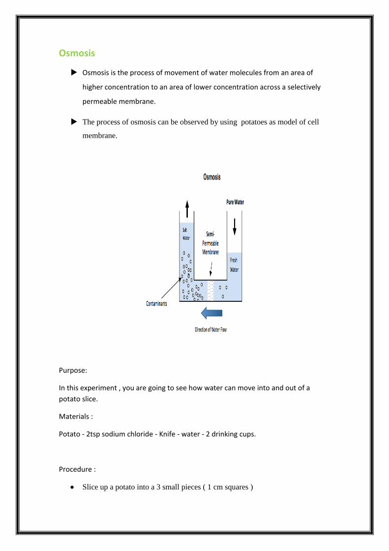
Osmosis
Osmosis is the process of movement of water molecules from an area of
higher concentration to an area of lower concentration across a selectively
permeable membrane.
The process of osmosis can be observed by using potatoes as model of cell
membrane.
Purpose:
In this experiment , you are going to see how water can move into and out of a
potato slice.
Materials :
Potato - 2tsp sodium chloride - Knife - water - 2 drinking cups.
Procedure :
Slice up a potato into a 3 small pieces ( 1 cm squares )
Fill two cups with water.
In one of the cups add 2-3 table spoons of salt, and stir it in.
Make sure you label which cup has the salt, and which one doesn't.
Place one potato slice into plain water, and one into salty water.
Leave it for about 30 – 60 minutes.
Remove the pieces onto a plate.
Write your observation on these pieces by pay attention to size, how flexible it
is, etc.
Explain your result.

Laboratory 4
Cell Division
Mitosis
Meiosis
Technique of Preparing a Karyotype
Technique of testing a fetus for genetic disorders
Cell Division :
Cell division is the process by which a parent cell divides into two or more
daughter cells.
This process enable organisms to grow and to reproduce, it play a key role in
repairing tissues and in formation of new organisms.
There are 2 types of Cell division: Mitosis and Meiosis.
Somatic cells divide by mitosis.
Sex cells or gametes divide by meiosis.
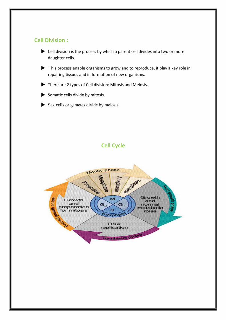
Cell Cycle

The cell cycle consists of four distinct phases
Gap 1 (G1) Cells increase in size
Synthesize (S) DNA Replication
Gap 2 (G2) Cell continue to grow
Mitosis (M) Division into 2 daughter cells
The Cell cycle consist of two phases:
Interphase: is the phase of the cell cycle in which the cell spends the majority of its
time and performs the majority of its purposes including preparation for cell division.
(G1, S, G2).
Mitosis: Results in the production of two daughter cells from a single parent cell.
The daughter cells and the original parent cell have the same number of
chromosomes (M).

Phases of Mitosis :
Prophase:
The chromatin condenses into double rod-shaped structures called
chromosomes in which the chromatin becomes visible and they attached to
each other by a central part called centromere.
Metaphase:
Characterized by positioning and alignment of chromosomes in the
equatorial level of the cell.
Anaphase:
The paired chromosomes (sister chromatids) separate and begin moving to
opposite ends (poles) of the cell.
Telophase:
The final stage of mitosis or meiosis during which the chromosomes of
daughter cells are grouped in new nuclei.
Chromosomes duplication Interphase
Condensation of DNA Prophase
Formation of the equatorial plate Metaphase
Migration of chromatids Anaphase
Decondensation of chromosome Telophase

Phases of mitosis under the microscope
Cytokinesis :
Mitosis is usually followed by cytokinesis, which is the division of the original
cell's cytoplasm.
At the end of cytokinesis, there are two distinct daughter cells.
Animal cells carry out cytokinesis by cleavage furrow.
Plant cells carry out cytokinesis by formation of cell plate.

Meiosis :
Meiosis includes two cycles of cell division result in production of four cells.
Each daughter cell has half number of chromosomes of the parent cell.
The four daughter cells are not identical between each other.

Comparison between Mitosis & Meiosis
Meiosis Mitosis
Definition A type of cellular
reproduction in which the
number of chromosomes
are reduced by half through
the separation of
homologous chromosomes
in a haploid cell.
A process of asexual
reproduction in which the
cell divides in two, with
an equal number of
chromosomes in diploid
cell.
Function sexual reproduction Cellular Reproduction &
general growth and repair
of the body
Type of reproduction Sexual Asexual
Occurs in Humans, animals, plants,
fungi
all organisms
Genetically Different Identical
Number of division 2 1
Number of daughter cells 4 2
Chromosome number Reduced by half Remains the same

This a figure that shows the difference between Mitosis &
Meiosis

Set of chromosomes in human cells :
Each human somatic cell has 46 chromosomes.
Chromosomes differ in size, and in the positions of their centromeres so they
can be distinguished from one another by microscopic examination when
sufficiently condensed.
Karyotype is the image of the arrangement of chromosome in pairs, starting
with the longest ones.
Preparing a Karyotype :
Application:
A karyotype is a display of condensed chromosomes arranged in pairs.
Karyotyping can be used to screen for abnormal numbers of chromosomes or
defective chromosomes associated with certain congenital disorders, such as Down
syndrome.
Preparing a Karyotype :
Technique:
Karyotypes are prepared from isolated somatic cells, which are treated with a drug
to stimulate mitosis and then grown in culture for several days.
Cells arrested in metaphase are stained and then viewed with a microscope
equipped with a digital camera.
A photograph of the chromosomes is displayed on a computer monitor, and the
images of the chromosomes are arranged into pairs according to size & shape.

Results:
This karyotype shows the chromosomes from a normal human male. The size of the
chromosome, position of the centromere, and pattern of stained bands help identify
specific chromosomes. Although difficult to discern in the karyotype, each
metaphase chromosome consists of two closely attached sister chromatids.

Technique of testing a fetus for genetic disorders
What is Amniocentesis?
Amniocentesis is a diagnostic procedure performed on pregnant women that looks
for genetic and chromosomal abnormalities (birth defects) in the fetus.
Some common conditions that may be detected include Down syndrome (trisomy
21), cystic fibrosis, and others.
This test is generally performed between weeks 14 and 20 of pregnancy.
Karyotyping shows whether the chromosomes of the fetus are normal in number
and appearance or not.
Technique of Amniocentesis
1. A sample of amniotic fluid can be taken starting at the 14th to 16th week of
pregnancy.
2. Centrifugation of the sample to separate the fluid from the fetal cells.
3. Biochemical tests can be performed immediately on the amniotic fluid or
later on the cultured fetal cells.
4. Fetal cells must be cultured for several weeks to obtain sufficient numbers
for Karyotyping.

Laboratory 5
DNA structure
PCR technique
DNA extraction experiment

DNA Structure :
Experiments with bacteria and with phages provided the first strong evidence
that the genetic material is DNA.
DNA is a double helix molecule.
Two antiparallel sugar-phosphate chains wind around the outside of the
molecule.
The Nitrogenous bases project into the interior.
The nitrogenous bases hydrogen-bond in specific pairs:
A with T
G with C
A chromosome consists of a DNA molecule packed together with proteins.
During Cell division, the DNA molecule is duplicated in the interphase and
passed to daughter cells in mitosis.
The DNA inherited by an organism leads to specific traits by dictating the synthesis of proteins and of RNA molecules involved in the process of Gene Expression.
Techniques of molecular genetics:
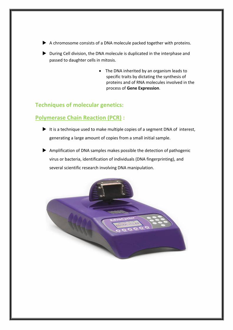
Polymerase Chain Reaction (PCR) :
It is a technique used to make multiple copies of a segment DNA of interest,
generating a large amount of copies from a small initial sample.
Amplification of DNA samples makes possible the detection of pathogenic
virus or bacteria, identification of individuals (DNA fingerprinting), and
several scientific research involving DNA manipulation.
Basic Tools:
What you need for the reaction is:
1. The DNA sequence of interest
2. Enzyme: DNA polymerase (heat resistant enzyme)
3. DNA primers
4. Free nucleotides (A,C,T,G).
Technique:
You combine all the tools together in the PCR tube and put it in the machine.
The PCR cycle begins.
Each cycle consists of three steps:
1. Denaturation (95ºC)
2. Annealing (55ºC)
3. Extension
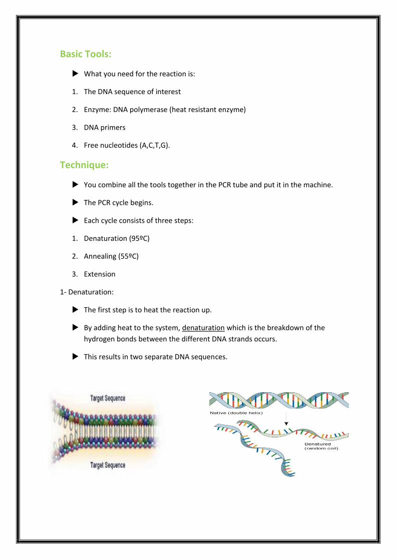
1- Denaturation:
The first step is to heat the reaction up.
By adding heat to the system, denaturation which is the breakdown of the
hydrogen bonds between the different DNA strands occurs.
This results in two separate DNA sequences.

2- Annealing:
The second step is to allow the system to cool.
As it cools, the DNA primers are able to hybridize to the separate strands.
DNA synthesis starts from the 5’ to 3’ direction.
This means that the primers have to match the 3’ end of the sequences of
interest.
3- Extension:
The third step is that the DNA polymerase enzyme is going to bind to the DNA
sequence of interest with the primer.
Once the DNA polymerase is bound, it takes some of the free nucleotides and
slowly add them to the primer to finish up the new DNA strand.
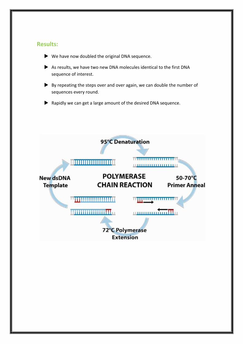
Results:
We have now doubled the original DNA sequence.
As results, we have two new DNA molecules identical to the first DNA
sequence of interest.
By repeating the steps over and over again, we can double the number of
sequences every round.
Rapidly we can get a large amount of the desired DNA sequence.

DNA Extraction Experiment
First we need to do Extraction Solution
Materials (100 ml):
1. 1 tape spoon of shampoo.
2. ½ tea spoon of salt.
3. 1 cup of Water.
4. Beaker.
Procedure:
1. Mix the water and salt together in a beaker..
2. Add shampoo and Stir slowly to avoid foaming of the shampoo.
Seconded DNA Extraction
Materials :
1. 3 freshly Strawberry fruit.
2. Zipper bag.
3. Extraction Solution.
4. strainer.
5. beaker .
6. Ice cold 95% ethanol.
7. Tweezers.
Procedure:
1. Put the strawberry in zipper bag .
2. Add 3 spoons of extraction solution inside the zipper bag and close it.
3. Mash the strawberry with your hand for about 5 minutes.
4. Strain the mixture into a beaker using a strainer .
5. Gently Pour the ice cold ethanol down the side of the beaker to form a layer
on top .

6. Let the mixture sit for 2-3 minutes.
7. finally use the tweezers to remove the cloudy DNA from the beaker .

Laboratory 6
Plant Tissues
Animal Tissues
Plant Tissues
Definition of tissue:
A tissue is composed of group of cells from the same origin that share the same
structure and the same specific function.
Plant tissue:
Plants do have a higher level of structure called plant tissue systems.
A plant tissue system can be defined as a functional unit, which connects all organs
of a plant.
Like animal tissue system, plant tissue system is also grouped into various tissues
based on their functions.

Meristematic tissues:
Consist of actively dividing cells, these tissues in a plant consist of small,
densely packed cells that can keep dividing to form new cells and lead to
increase the length and the thickness of the plant.
They have the following characteristics:
o the cells are small.
o the cells walls are thin.
o cells have large nuclei.
o vacuoles are absent or very small.

Permanent tissues:
Are those tissues which have lost the power of cell division.
Cells of permanent tissues are matured, have a definite shape, size and
function.
On basis of the constituents cells, permanent tissues are classified into two
types.
I. Simple
II. Complex
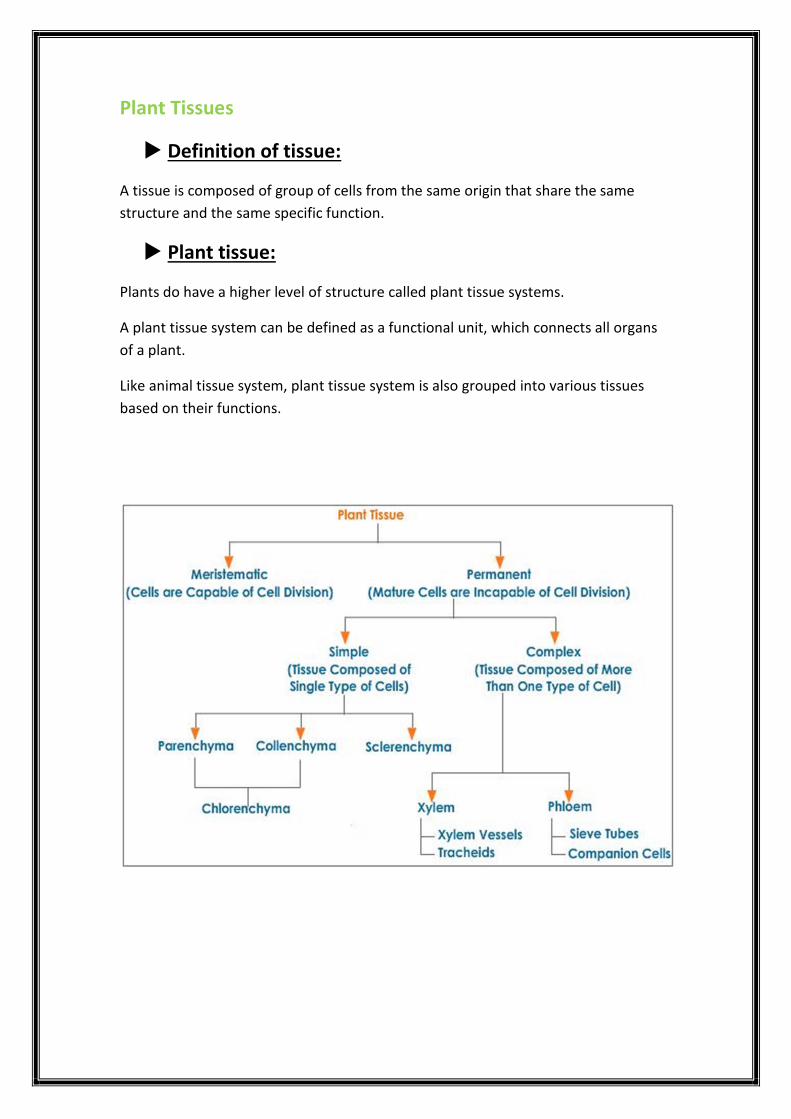
Simple Permanent tissues:
Composed of single type of cells.
There are three types:
1. PARENCHYMA CELLS: Thin walled, they function in storage & photosynthesis.
2. COLLENCHYMA CELLS: Unevenly thick walls. Give flexible support to plant.
3. SCLERENCHYMA CELLS: Evenly thick walls. Provide rigid support to the plant.
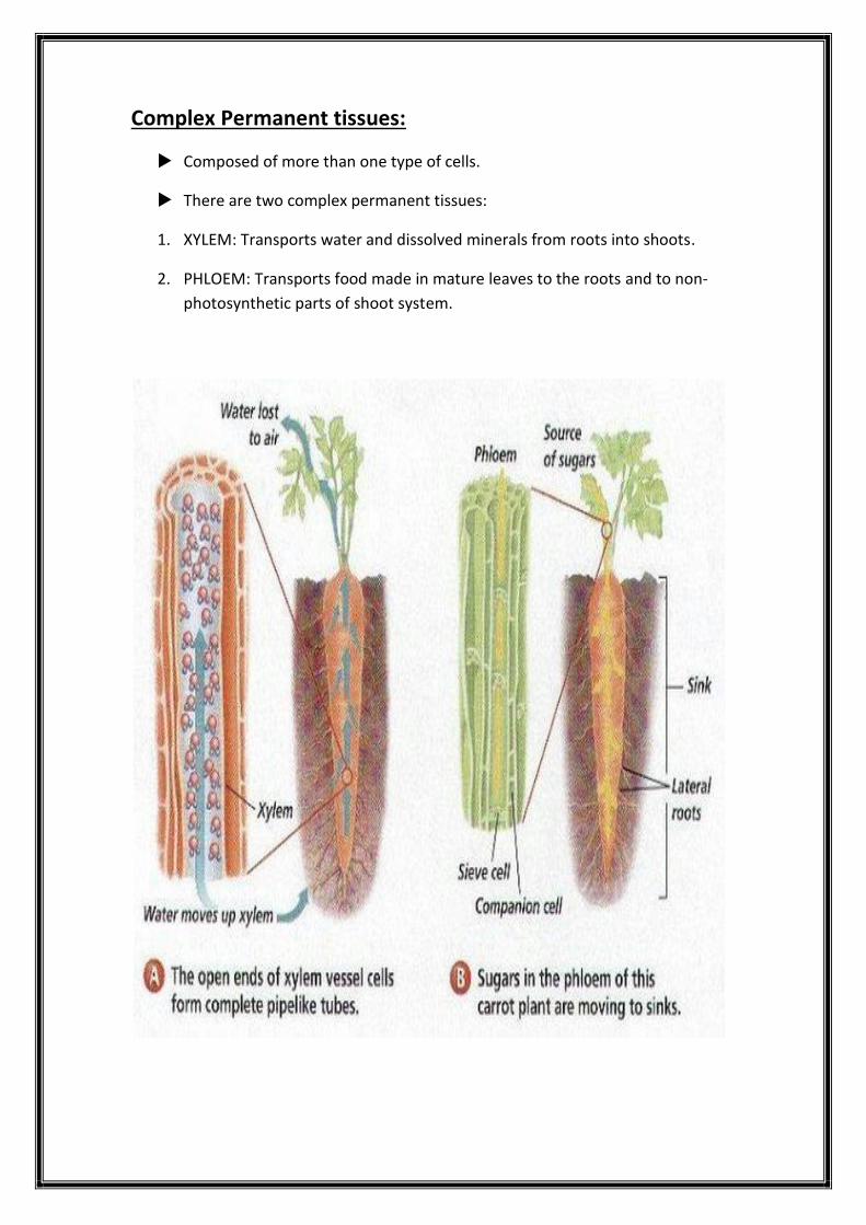
Complex Permanent tissues:
Composed of more than one type of cells.
There are two complex permanent tissues:
1. XYLEM: Transports water and dissolved minerals from roots into shoots.
2. PHLOEM: Transports food made in mature leaves to the roots and to non-
photosynthetic parts of shoot system.

Plant Organs :
Tissues are organized together to form organs.
Organs are composed of two or more tissues performing specific function.
Each plant has three specific organs:
i. Root.
ii. Leaf.
iii. Stem.
Each plant organ (root –leaf-stem) has dermal, vascular and ground tissues.
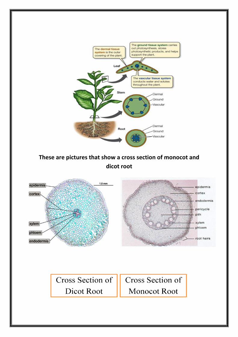
These are pictures that show a cross section of monocot and
dicot root

Animal Tissues
There are four basic types of animal tissues.
1. Epithelial tissues
2. Connective tissues
3. Muscle tissues
4. Nervous tissues
Each organ of the animal organism is made up of 2 or more types of animal
tissues.

Epithelial tissue
Covers the outside of the body
Lines organs and cavities inside the body.
Epithelial tissues can be classified on the basis of two criteria:
1- Shape.
2- Arrangement.
:TYPES ON BASIS OF SHAPE
Cuboidal epithelium
Squamous epithelium
Columnar epithelium
TYPES ON BASIS OF ARRANGEMENT:
Simple epithelium
Stratified epithelium
Pseudo stratified epithelium

Simple epithelial tissue
Stratified epithelial tissue

This is some microscope pictures of epithelial tissues
Simple cuboidal
Simple squamous
Pseudostratified
columnar
Simple columnar

Connective tissue
Connective tissue binds and supports other tissues in the body.
There are many types of connective tissues:
1. Loose connective tissue.
2. Cartilage.
3. Fibrous connective tissue.
4. Adipose tissue.
5. Blood.
6. Bone.
1- Loose connective tissue:
Is characterized by the presence of many gaps between the cells. It is composed of a
network of fibres which bind the epithelia to the underlying tissues.
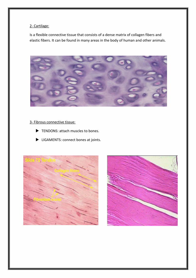
2- Cartilage:
Is a flexible connective tissue that consists of a dense matrix of collagen fibers and
elastic fibers. It can be found in many areas in the body of human and other animals.
3- Fibrous connective tissue:
TENDONS: attach muscles to bones.
LIGAMENTS: connect bones at joints.
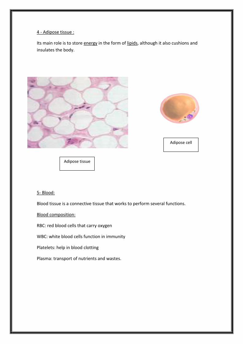
4 - Adipose tissue :
Its main role is to store energy in the form of lipids, although it also cushions and
insulates the body.
5- Blood:
Blood tissue is a connective tissue that works to perform several functions.
Blood composition:
RBC: red blood cells that carry oxygen
WBC: white blood cells function in immunity
Platelets: help in blood clotting
Plasma: transport of nutrients and wastes.
Adipose cell
Adipose tissue

6- Bone:
Bone is a mineralized connective tissue. It forms the skeleton in most vertebrates.
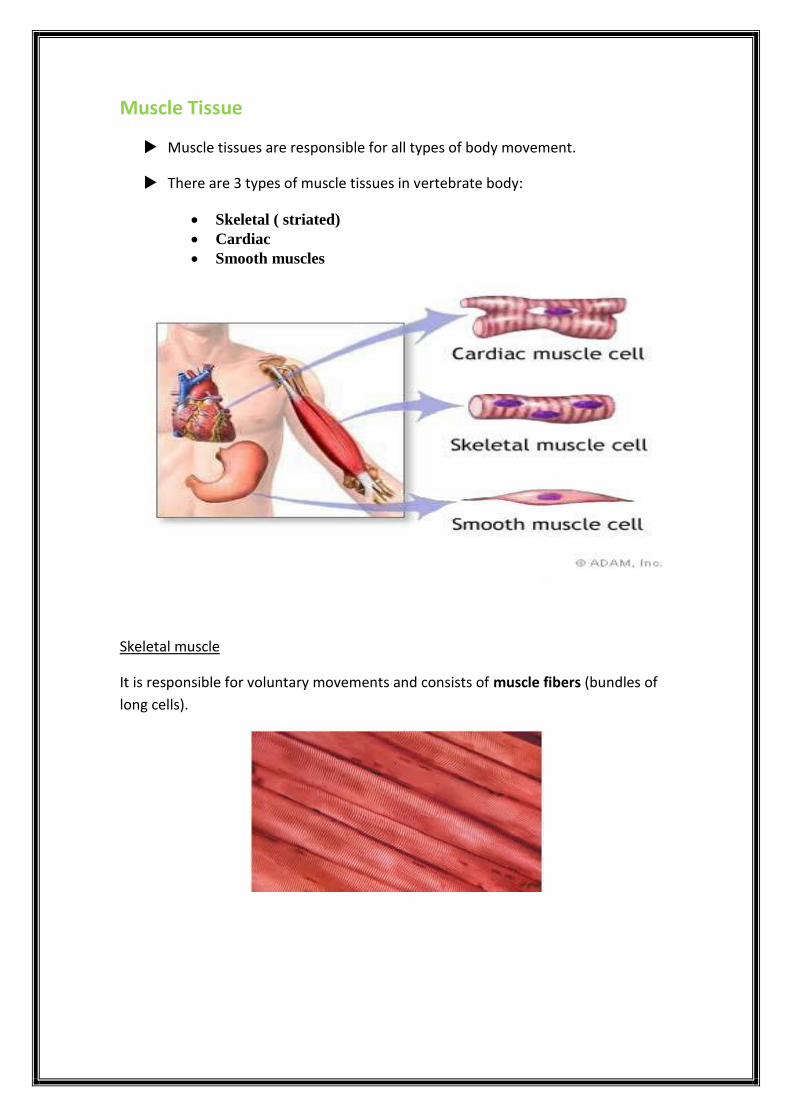
Muscle Tissue
Muscle tissues are responsible for all types of body movement.
There are 3 types of muscle tissues in vertebrate body:
Skeletal ( striated)
Cardiac
Smooth muscles
Skeletal muscle
It is responsible for voluntary movements and consists of muscle fibers (bundles of
long cells).
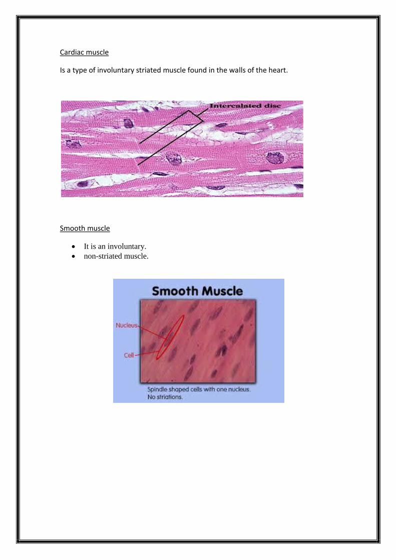
Cardiac muscle
Is a type of involuntary striated muscle found in the walls of the heart.
Smooth muscle
It is an involuntary.
non-striated muscle.

Nervous tissue
Nervous tissue is made of nerve cells.
It is specialized to react to stimuli and to conduct impulses to various organs
in the body which bring about a response to the stimulus.
It’s found in the brain and the spinal cord.

Laboratory 7
The Five kingdoms of life:
1. Monera Kingdom.
2. Protista Kingdom.
3. Fungi Kingdom.

Every living creature on Earth belongs to a kingdom.
These kingdoms are based on how living things are the same and how they
are different.
Scientists divide all living things into 5 kingdoms*:
1-Monera. 2. Protista. 3. Fungi. 4.Plantae. 5.Animalia.
Kingdoms are divided into categories called phyla, each phylum is divided
into classes, each class into orders, each order into families, each family into
genera, and each genus into species.

To which kingdom do viruses belong?
Viruses are not classified in any kingdom yet ,because they are not really
alive. They only show signs of life after they infect a host cell.
Virus: lifeless particle that does not carry out any METABOLIC functions on its
own and CANNOT REPRODUCE on its own until it invades a living HOST cell.
Viruses are much smaller than bacteria, about 20nm in diameter.
Most viruses are too small to be seen directly with a light microscope.
Viral genomes may consist of double-stranded DNA, single-stranded DNA,
double-stranded RNA or single-stranded RNA depending on the kind of virus.

Capsids and Envelopes
The protein shell enclosing the viral genome is called a capsid.
Depending on the type of virus, the capsid can be rod-shaped, polyhedral, or
more complex in shape.
Capsids are built from a large number of protein subunits called capsomeres.
Some viruses have a membranous envelope surrounding the capsids and
help in infecting hosts.

Viral structure
Viruses can reproduce only in host cells
Viruses lack metabolic enzymes and equipment for making proteins, such as
ribosomes.
They are obligate intracellular parasites, they can reproduce only within a
host cell.
Each type of virus can infect cells of only a limited variety of hosts, called the
host range of the virus.

Human cold viruses infect only the cells lining the upper respiratory tract, and
the AIDS virus binds to receptors present only on certain types of white blood
cells.
A simplified viral reproductive cycle
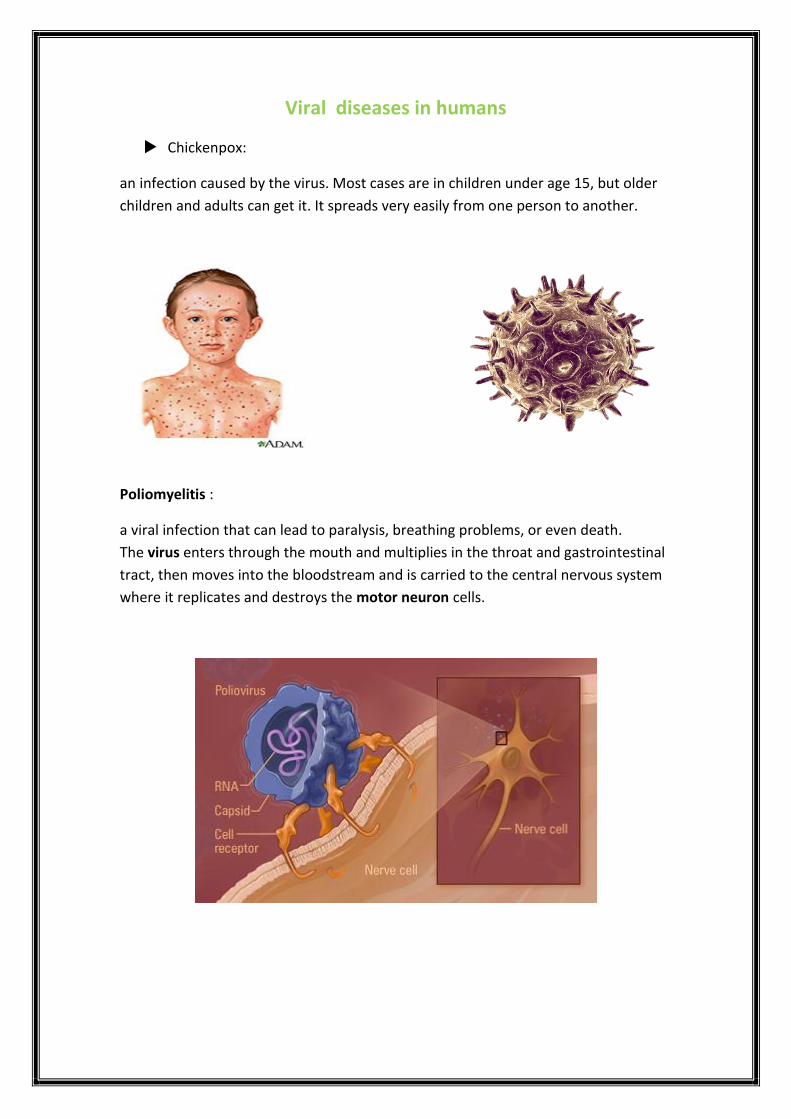
Viral diseases in humans
Chickenpox:
an infection caused by the virus. Most cases are in children under age 15, but older
children and adults can get it. It spreads very easily from one person to another.
Poliomyelitis :
a viral infection that can lead to paralysis, breathing problems, or even death.
The virus enters through the mouth and multiplies in the throat and gastrointestinal
tract, then moves into the bloodstream and is carried to the central nervous system
where it replicates and destroys the motor neuron cells.
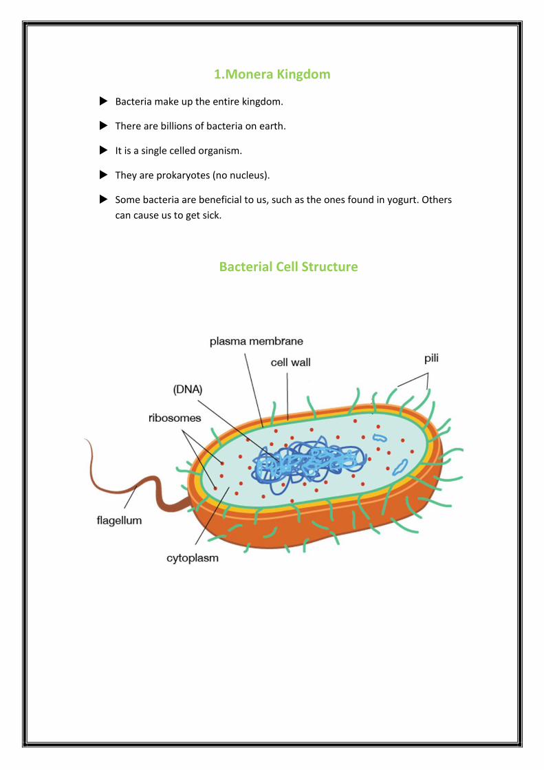
1.Monera Kingdom
Bacteria make up the entire kingdom.
There are billions of bacteria on earth.
It is a single celled organism.
They are prokaryotes (no nucleus).
Some bacteria are beneficial to us, such as the ones found in yogurt. Others
can cause us to get sick.
Bacterial Cell Structure
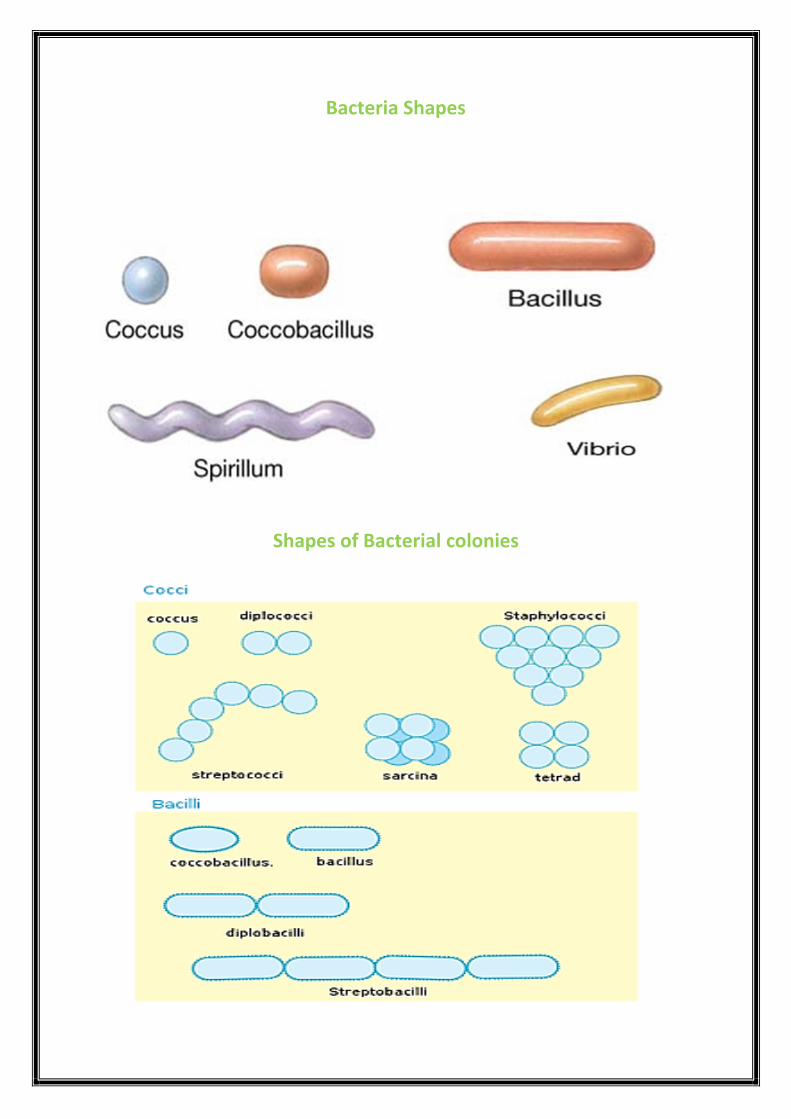
Bacteria Shapes
Shapes of Bacterial colonies

Bacteria Shapes under microscope
Pathogenic bacteria cause many human diseases
Streptococcus pneumoniae : lives in the throats of most healthy people, but it can multiply and cause pneumonia when the host’s defenses are down.�
(d) vibrio

Vibrio cholerae: it’s causes cholera , a dangerous disease characterized by
severe diarrhea. Resulting from the consumption of water contaminated with
human feces.
How cholera affects the body

2. Protista kingdom:
Protozoa are unicellular eukaryotic organisms. They are the smallest of all
animals.
There are three main types of protozoa:
1. Amoeba 2. Paramecium 3. Euglena
Pathogenic Protists cause many human diseases
Plasmodium parasites:
Responsible for most malarial infection, that spread by mosquitoes.
Plasmodium Protozoa
Plasmodium protozoa cause malaria. The parasites are spread by a mosquito vector.
Parasites enter a host’s blood through the bite of an infected mosquito. The
parasites infect the host’s red blood cells, causing symptoms such as fever, joint
pain, anemia, and fatigue. In fact, malaria is one of the most common infectious

diseases on the planet. Malaria is also a very serious disease. It kills several million
people each year, most of them children.
Entamoeba histolytica: Responsible for Amoebic dysentery (amoebiasis) an infection of the intestine, cause
sever diarrhea with blood.

Amoeba
shape No definite shape
Movement Pseudopods (fake feet)
Ingestion Engulfs (swallows) food with pseudopods.
Digestion Food is broken down in food vacuole
Excretion
Wastes are excreted through the cell
membrane. Excess water is pumped out by
the contractile vacuole
Reproduction Binary fission, asexual
Structure Of Amoeba

Paramecium
shape Slipper shaped
Movement Cilia like hair
Ingestion Food is swept into oral groove by cilia then
into the gullet.
Digestion Food is broken down in food vacuole
Excretion
Wastes are excreted through the anal pore.
Excess water is pumped out by the contractile
vacuole
Reproduction Binary fission and conjugation
Structure Of Paramecium

Euglena
shape Oval shaped
Movement Flagella like tail
Ingestion Makes food in chloroplast or food enters
through the cell membrane.
Digestion Food is broken down by enzymes in the
cytoplasm
Excretion
Contractile vacuole uses to excrete water
and waste from inside and discharges it to
outside through a hole in the cell
membrane.
Reproduction Binary fission
Structure Of Euglena

3. Fungi Kingdom:
Fungi are Eukaryotic organisms that include microorganisms such as yeast,
mold, mushroom.
Grow best in warm, moist environments.
They are heterotrophs (can’t make their own food)
Most fungi grow as tubular filaments called hyphae.
Fungi Produce both sexual and asexual spores.
Pathogenic Fungi cause many human diseases
Ringworm is a common skin infection that is caused by a fungus. It’s called
“ringworm” because it can cause a circular rash (shaped like a ring) that is
usually red and itchy.
Nail fungus is a common condition that begins as a white or yellow spot
under the tip of your fingernail or toenail. As the fungal infection goes
deeper, nail fungus may cause your nail to discolor, thicken and crumble at
the edge.�


Laboratory 8
Animal Kingdom

Animal Kingdom
There are many different types of animals in the world. Many animals are
quite similar to each other, others are quite different. Animals can be
classified based on their similarities.
The animal kingdom is divided into about 35 phyla encompassing 1.3 million
known species.
We divided animal kingdom into two major groups:
Vertebrates
Invertebrates

A. Vertebrates
Vertebrates are often larger and have more complex bodies than
invertebrates .
There are approximately 52,000 species of vertebrates.
It have only one phylum (Chordata).
Kingdom :Animalia, or "animal“.
Phylum : Chordata.
Sub phylum: Vertebrate.
Class :
1. Amphibia.
2. Fish.
3. Reptiles.
4. Birds.
5.Mammals.
1. class: Amphibia
includes semi-aquatic animals with moist skin.
They must return to the water to breed.
Ex: Frog, Salamander and newt.

2.Class: fish
Breathe through gills.
Have a moist skin covered in scales.
Ex : Goldfish, shark and Cod.
3. class: Reptilia
They have dry, scaly skin.
Ex : Crocodile, Snake and Turtle.
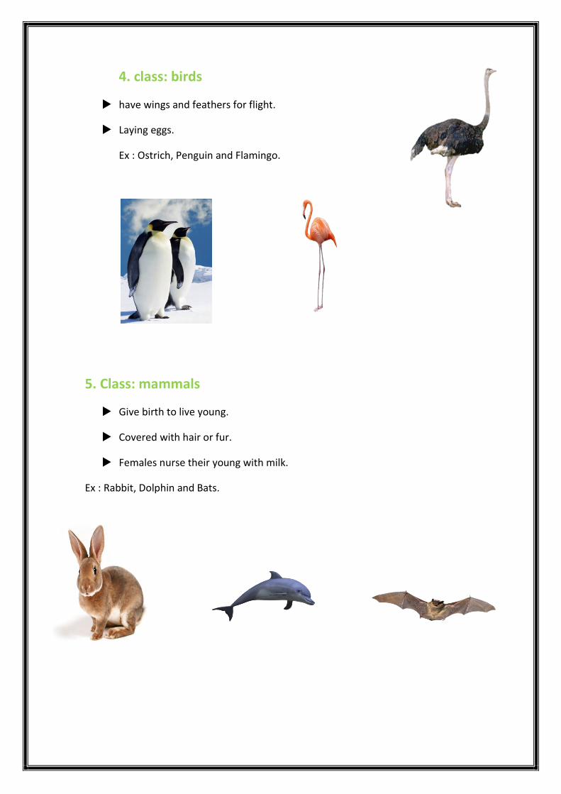
4. class: birds
have wings and feathers for flight.
Laying eggs.
Ex : Ostrich, Penguin and Flamingo.
5. Class: mammals
Give birth to live young.
Covered with hair or fur.
Females nurse their young with milk.
Ex : Rabbit, Dolphin and Bats.

1. Vertebrates
Examples Main features Class
Frog, salamander , newt. Moist permeable, skin. Amphibia
Co
ld
blo
od
ed
Goldfish, shark, cod. Gills, Wet scales. Fish
Crocodile, lizard, snake, turtles. Dry scales, lay eggs . Reptiles
Ostrich , penguins , budgerigar. Feathers, Beaks. Birds
War
m
blo
od
ed
Rabbit, dolphin, Bats. Fur, produce milk. Mammals

2. Invertebrates
Animals without a Backbone or Spinal Column.
Invertebrates account for 95% of known animal species.
32 phylum – 735,000 species.
more than 90% of the invertebrates are insects.
We’ll examine 8 of those phyla with more detail.
1. Phylum: Porifera
Sponges are sessile animals
(they spend their lives attached to rocks).
Water enters the small pores of a sponge, travels through canals, and exits through a
large hole at the top of the sponge.
Ex : Sponges.
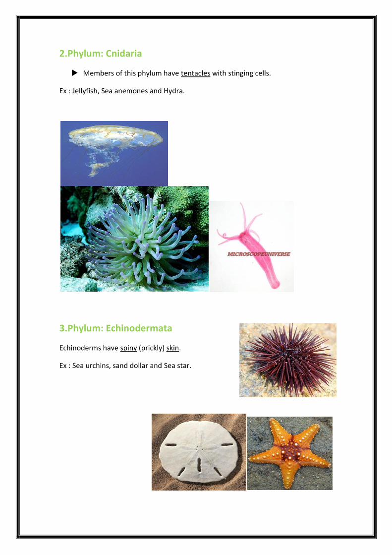
2.Phylum: Cnidaria
Members of this phylum have tentacles with stinging cells.
Ex : Jellyfish, Sea anemones and Hydra.
3.Phylum: Echinodermata
Echinoderms have spiny (prickly) skin.
Ex : Sea urchins, sand dollar and Sea star.

4.Phylum: Mollusca
Most mollusks have a hard shell covering their soft bodies.
Ex : Snails, Clams and Octopus.
5.Phylum: Annelida (Segmented worms)
Annelids have bodies that are segmented (divided into sections).
Ex : Earthworm, Sandworm and Leeches.

6.Phylum: Platyhelminthes (Flatworms)
The worms in this phylum are all very thin and flat, and parasitic .
Ex : Tapeworm, Planarians and Flukes.

7. Phylum: Nematoda (Roundworms)
Nematodes are not segmented; their body surfaces appear smooth.
Many nematodes are parasites
Ex : Hookworm.
8.Phylum: Arthropoda
largest phylum.
All arthropods, including this tick, have jointed legs.
Arthropods have a crunchy shell called an exoskeleton.
Divided into 3 classes:
1. Crustaceans.
2. Insects.
3. Arachnids.

CRUSTACEANS
have claws
Ex: Shrimps ,lobster.
INSECTS
have 6 legs, 3 body
segments and wings.
Ex: Bees ,mosquitoes, grasshopper.
ARACHNIDS
have 8 legs. Ex: Scorpion ,
spider

ssppeecciieess EExxaammpplleess MMaaiinn ffeeaattuurreess PPhhyylluumm
5,500 Sponges. No tissues or organs, pores.
11..PPoorriiffeerraa
7,000 Sea Urchins, Sand Dollars,
Sea Stars. Spiny skin 22..EEcchhiinnooddeerrmmaattaa
10,000 Jellyfish, sea anemones,
hydra. Gastrovascular
cavity 33..CCnniiddaarriiaa
16,500 Earthworms, sandworms,
leeches. Segmented
worms 44..AAnnnneelliiddaa
20,000 Tapeworms, planarians,
and flukes. Flatworms 55..PPllaattyyhheellmmiinntthheess
25,000 Hookworm. Roundworms 66..NNeemmaattooddaa
93,000 Snails, clams, and
octopuses. Soft body 77..MMoolllluussccaa
1,000,000 Crustaceans, Arachnids,
and Insects. Segmented
exoskeleton 88..AArrtthhrrooppooddaa

Laboratory 9
Plant Kingdom

Plant Kingdom:
This kingdom has organisms that are multi-cellular.
have cell walls and chlorophyll.
produce their own food.
and don’t physically move from one place to another.
Scientists divided The plant kingdom is into two groups:

Non-vascular plants
plants that do not have tubes to carry water up the plant or tubes to carry
food made in the leaves down the plant.
Spore: the reproductive cell of a nonvascular plant
Examples:


Vascular Plant
Plants that have tubes to carry water up and food down the plant.
Examples:
Tree
Roses
Grass
Photosynthesis
Plant cells produce their own food through a process called photosynthesis.
Photosynthesis allows plants to convert light energy into food energy.

Morphology
1.Roots
A true root system consists of a primary root and secondary roots (or lateral
roots).
Generally, two categories are recognized:
1.Adventitious roots.
2.Taproots.
1.Roots 2.Stems
3.Fruits 4.leaves
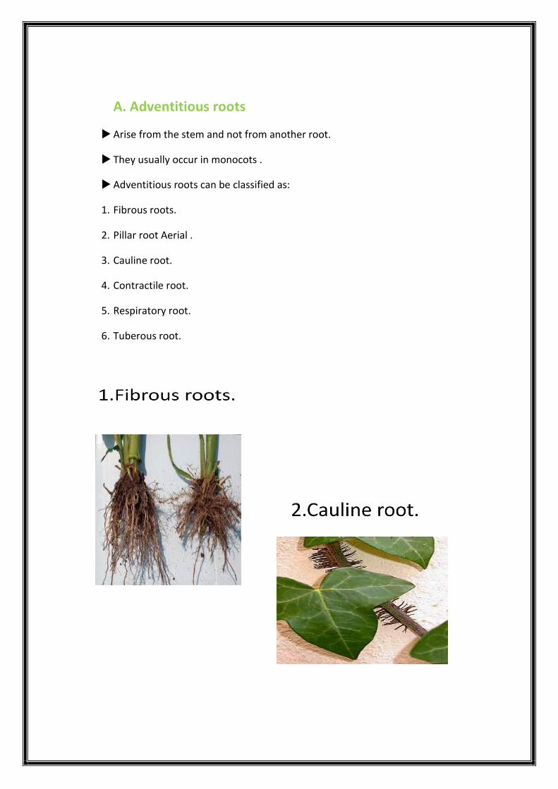
A. Adventitious roots
Arise from the stem and not from another root.
They usually occur in monocots .
Adventitious roots can be classified as:
1. Fibrous roots.
2. Pillar root Aerial .
3. Cauline root.
4. Contractile root.
5. Respiratory root.
6. Tuberous root.


B. Tap Root
The primary root is prominent which tapers at the end and there are fibrous
secondary roots running outward.
Most common in dicots.
tap root divided into:
A stem is the part of a plant
from which shoots and buds arise.
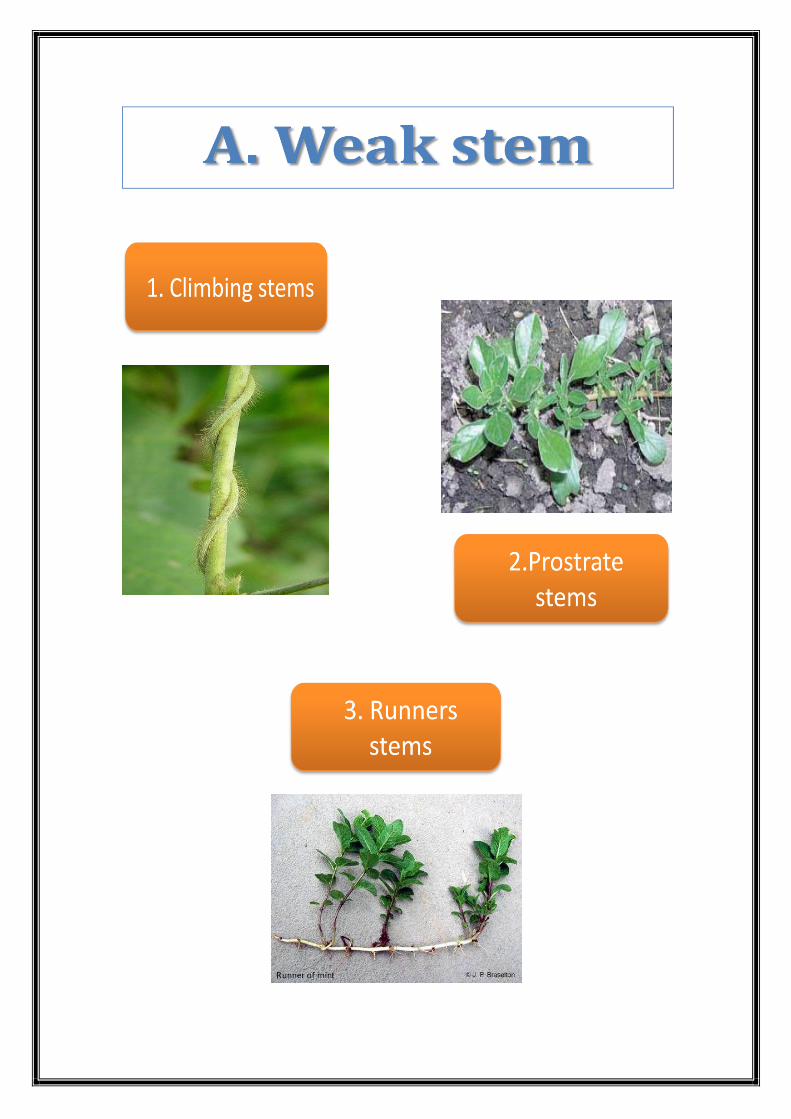
Stems are classified as:
1. Weak
2.Metamorphosis stem
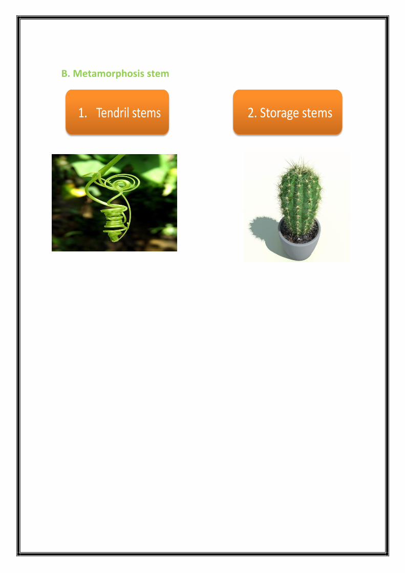
B. Metamorphosis stem

A fruit is a part of a flowering plant that derives from specific tissues of the
flower one or more ovaries.
It can be divided into 3 main categories :
A. Compound Fruits
A compound fruit is one that develops from Several
ovaries in a multiple flowers.
Examples : Pineapple, Mulberries, Figs.
Compound fruits
Aggregate fruits
simple fruits

B. Aggregate Fruits
Aggregate fruits form from single flowers
that have multiple carpels which are not
joined together.
Examples : Raspberry , Strawberry .
C. Simple Fruits
A simple fruit develops from
one ovary in a one flower.
Examples : Cherry, tomato.

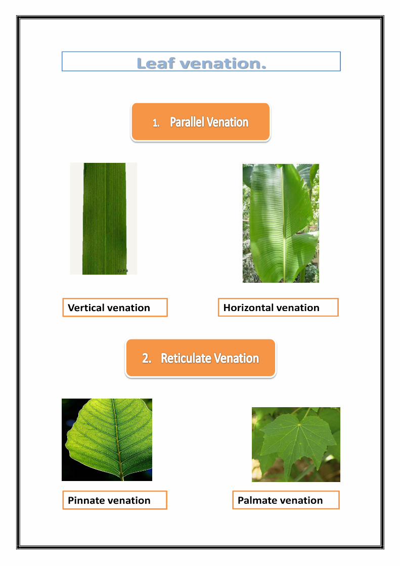
The leaf can be classified based on:
1. Leaf shape.
2. Leaf margin.
3. Leaf venation.
4.Leaf arrangement.
1.Simple leaf
2.Compound Leaf
Compound pinnate
Cordate
Sagittate
Compound palmate

3.Lobed Leaf
Palmate lobed
Pinnate lobed


Leaf arrangement
