laboratory procedure manual...laboratory procedure manual analytes: cadmium, lead, manganese,...
TRANSCRIPT
Laboratory Procedure Manual
Analytes: Cadmium, Lead, Manganese, Mercury,
and Selenium
Matrix: Whole Blood
Method: blood multi-element analysis by ICP-DRC-MS
Method No: DLS 3016.8-05 As performed by: Inorganic and Radiation Analytical Toxicology Branch
Division of Laboratory Sciences National Center for Environmental Health
Contact: Jeffery M. Jarrett, MS
Phone: 770-488-7906 Fax: 770-488-4097 Email: [email protected]
James L. Pirkle, M.D., Ph.D. Director, Division of Laboratory Sciences
Important Information for Users The Centers for Disease Control and Prevention (CDC) periodically refines these laboratory methods. It is the responsibility of the user to contact the person listed on the title page of each write-up before using the analytical method to find out whether any changes have been made and what revisions, if any, have been incorporated.

Public Release Data Set Information
This document details the Lab Protocol for testing items in the following table:
Data File Name Variable Name SAS Label
PBCD_H
LBXBCD Cadmium (µg/L)
LBDBCDSI Cadmium (µmol/L)
LBXBPB Lead (µg/dL)
LBDBPBSI Lead (µmol/L)
LBXTHG Mercury, total (μg/L)
LBDTHGSI Mercury, total (μmol/L)
LBXBMN Manganese (μg/L)
LBDBMNSI Manganese (μmol/L)
LBXBSE Selenium (ug/L)
LBDBSESI Selenium (μmol/L)
Blood Metals Panel in Whole Blood NHANES 2015-2016 2

1. Clinical relevance & summary of test principle
a. Clinical relevance:
Metals ions affect human health in various ways. Some metals (i.e. lead, cadmium, and mercury) show only deleterious effects on human health. Some (i.e. selenium and manganese) play an essential role in the human biological system if within certain concentration ranges, while negative health implications are observed when concentrations in biological systems are in deficit or excess. Determination of a person’s level of environmental exposure to chemicals through direct measurement of the substances or their metabolites in human specimens such as blood is called biomonitoring. Biomonitoring reduces the uncertainty of determining levels of exposure over making these determinations through calculations of estimated dose based on analysis of environmental samples and assumptions about exposure pathways [1]. Biomonitoring measurements are the most health-relevant assessments of exposure because they indicate the amount of the chemical that actually gets into people from all environmental sources (e.g., air, soil, water, dust, or food) combined, rather than the amount that gets into them. The laboratory method described here is a multi-element technique for monitoring the concentrations of cadmium (Cd), lead (Pb), manganese (Mn), mercury (Hg), and selenium (Se) in whole human blood for the purpose of biomonitoring.
There is no known biological role of mercury in the human body. The main sources of mercury intake in humans are fish, dental amalgams, and occupational exposures [2]. The main organs affected by mercury are the brain and the kidneys. Exposure of childbearing-aged women is of particular concern because of the potential adverse neurologic effects of Hg in fetuses. The health effects of mercury are diverse and depend on the form of mercury encountered and the severity and length of exposure. The general population is be exposed to three forms of mercury: elemental, inorganic, and organic (predominantly methyl). However, this method tests only for the total amount of mercury in the blood without regard to chemical form. In the general population, total blood mercury is due mostly to the dietary intake of organic forms which are formed through microbial action from inorganic mercury that has deposited in aquatic environments and bioaccumulated through the food chain (especially into large predatory fish)[3]. Exposure to inorganic or elemental mercury (e.g. dental amalgams or occupational exposures) is particularly reflected in urine excretion rather than blood. Psychic and emotional disturbances are the initial signs of chronic intoxication by elemental mercury vapors or salts. Those exposed are at increased risk for paresthesia, neuralgias, renal disease, digestive disturbances, and ocular lesions [4]. Massive exposure over a longer period of time results in violent muscular spasms, hallucinations, delirium, and death [5]. Except for methylmercury exposures, blood is considered useful if samples are taken within a few days of exposure. This is because most forms of mercury in the blood decrease by one-half every three days if exposure has been stopped. Thus, mercury levels in the blood provide more useful information after recent exposures than after long-term exposures. Several months after an exposure, mercury levels in the blood and urine are much lower. Blood mercury reference ranges for the U.S. population are listed in Table 10 in Appendix B.
Blood Metals Panel in Whole Blood NHANES 2015-2016 3

There is no known biological role of lead in the human body. Lead, a naturally occurring metal, has had many different commercial uses from which a person can be exposed either in the occupational / manufacturing process or by the manufactured products such as paint (paint chips, or dust and soil contaminated from deteriorating paint), solder or pipes (only now in older homes), gasoline (now outlawed for all but specialized applications), glazes on pottery, hobby uses (e.g. stained glass), commercial products (e.g. batteries, lead-containing jewelry), home remedy medicines containing lead compounds and non-Western cosmetics. Soil contains lead naturally, or from man-made uses of lead such as paint (near older homes), gasoline (near roadways), mining, manufacturing, and disposal. The main target for lead toxicity is the nervous system, both in adults and children. The developing biological systems of children are most sensitive to the effects of Pb, where effects are being recognized even at blood lead levels <5 µg/dL [6-10]. Acute, elevated lead exposure is associated with anorexia, dyspepsia, and constipation followed by diffuse paroxysmal abdominal pain. When lead exposure is high, particularly in children, the person is at increased risk for encephalopathy [11]. The alkyl lead species are highly toxic to the central nervous system [12]. The primary screening method for lead exposure is blood lead, which primarily reflects recent exposures (excretory half-life in blood is approximately 30 days) [13]. Lead in blood is primarily (99%) in the red blood cells. Blood lead reference ranges for the U.S. population are listed in Table 10 in Appendix B. The CDC now uses a reference level of 5 µg/dL to identify children with blood lead levels that are much higher than most children’s levels. This new level is based on the U.S. population of children ages 1-5 years who are in the highest 2.5% of children when tested for lead in their blood. This reference value is based on the 97.5th percentile of the National Health and Nutrition Examination Survey (NHANES)’s blood lead distribution in children. CDC will update the reference value every four years using the two most recent NHANES surveys [14]. There is no known biological role of cadmium in the human body. The predominant commercial use of cadmium is in battery manufacturing. Other uses include pigment production, coatings and plating, plastic stabilizers, and nonferrous alloys. Since 2001, U.S. cadmium use has declined in response to environmental concerns. In the United States, for nonsmokers the primary source of cadmium exposure is from the food supply. People who regularly consume shellfish and organ meats will have higher exposures. In general, leafy vegetables such as lettuce and spinach, potatoes and grains, peanuts, soybeans, and sunflower seeds contain high levels of cadmium due to bioaccumulation from the soil. Tobacco leaves accumulate high levels of cadmium from the soil, and smoking is the primary non-occupational source of cadmium exposure for smokers. Generally, the critical organ for Cd is the kidney. Kidney dysfunction is one of the most characteristic signs of exposure to Cd. Workers in an environment with high exposure levels have developed proteinuria, renal glucosuria, aminoaciduria, hypercalciuria, phosphaturia, and polyuria. Chronic obstructive lung disease of varying degrees of severities is frequently seen in Cd workers. Concentration of cadmium in blood of healthy unexposed adults are in the range 0.1 – 4 µg/L [15]. Newborn babies are practically free of Cd [16]. Exposure to high concentration of fumes appearing from heated cadmium metal or compounds has led to acute poisoning and in some cases to
Blood Metals Panel in Whole Blood NHANES 2015-2016 4

the death of workers [11]. Principal symptoms reported were respiratory distress due to chemical pneumonitis and edema. It has been estimated that 8 hrs. exposure to 5 g Cd/m3 will be lethal [11]. Ingestion of high amounts of Cd puts a person at increased risk to a rapid onset with severe nausea, vomiting, and abdominal pain. Cadmium levels in blood, urine, feces, liver, kidney, hair, and other tissues have been used as biological indicators of exposure to cadmium. Blood cadmium levels are principally indicative of recent exposure(s) to cadmium rather than whole-body burdens [17-20]. Urine cadmium levels primarily reflect total body burden of cadmium, although urine levels do respond somewhat to recent exposure [21]. Blood cadmium reference ranges for the U.S. population are listed in Table 10 in Appendix B. Manganese (Mn) is a trace element essential to humans and is associated with the formation of connective and bony tissue, growth and reproductive functions and with carbohydrate and lipid metabolism [22]. Manganese is also a known neurotoxin but little information exists about levels of manganese that cause toxicity. Symptoms of manganese toxicity are similar to Parkinson’s Disease and can also include disorientation, memory impairment, anxiety and compulsive behavior [23]. There is much concern for the levels of manganese in humans whom are occupationally exposed to it [24-30]. Recently, there are growing concerns over exposure due to contamination of drinking water with manganese [31-33] and as a result of methylcyclopentadienyl mangangese tricarbonyl (MMT) used as an anti-knocking additive in gasoline [34-40]. Populations suffering from iron deficiencies are at an increased risk to manganese toxicity because iron deficiency can result in an accumulation of manganese in the central nervous system [37]. To fully understand the essentiality and toxicity of manganese, further investigations are needed regarding the levels of manganese in biological matrices. Group average levels in blood appear to be related to manganese body burden, while average urinary excretion levels appear to be most indicative of recent exposures [41]. On an individual basis the correlation between the level of workplace exposure and the levels in blood or urine has always been found to be a reliable predictor of exposure [25, 41-43]. Manganese in blood or urine are useful in detecting groups with above-average current exposure, but measurements of manganese in these body fluids in individuals are sometimes be related to exposure dose after the exposure has ceased. In addition to individual variability, another factor that limits the usefulness of measuring manganese in blood, urine, or feces as a measure of excess manganese exposure is the relatively rapid rate of manganese clearance from the body. Excess manganese in blood is rapidly removed by the liver and excreted into the bile, with very little excretion in urine [44, 45]. Thus, levels of manganese in blood or urine are not expected to be the most sensitive indicators of exposure [46]. Typical blood manganese concentrations in humans, which have been reported in the literature, are listed in Table 11 of Appendix B. Selenium is an essential element that is required to maintain good health but both selenium deficiency and excessive levels of selenium are associated with several disorders [47, 48]. Selenium is a naturally occurring mineral element that is distributed widely in nature in most rocks and soils. Most processed selenium is used in the electronics industry, but it is also used: as a nutritional supplement; in the glass industry; as a component of pigments in plastics, paints, enamels, inks, and rubber; in the preparation of pharmaceuticals; as a nutritional feed additive for poultry and
Blood Metals Panel in Whole Blood NHANES 2015-2016 5

livestock; in pesticide formulations; in rubber production; as an ingredient in antidandruff shampoos; and as a constituent of fungicides. Radioactive selenium is used in diagnostic medicine. In the body, selenium is incorporated into proteins to make selenoproteins, which are important antioxidant enzymes. The antioxidant properties of selenoproteins help prevent cellular damage from free radicals. Free radicals are natural by-products of oxygen metabolism that increase risk of chronic diseases such as cancer and heart disease [48, 49]. Other selenoproteins help regulate thyroid function and play a role in the immune system [50-53]. Human selenium deficiency is rare in the U.S. but is seen in other countries where soil concentration of selenium is low[54]. There is evidence that selenium deficiency increases the risk of a form of heart disease, hypothyroidism, and a weakened immune system [55, 56]. There is also evidence that selenium deficiency does not usually cause illness by itself. Rather, it can make the body more susceptible to illnesses caused by other nutritional, biochemical or infectious stresses [57]. Symptoms of very high exposure to selenium, a condition called selenosis, include gastrointestinal upsets, hair loss, white blotchy nails, garlic breath odor, fatigue, irritability, and mild nerve damage [47]. Selenium can be detected in the blood, feces, urine, hair, and nails of exposed individuals, however, field studies have used primarily blood or urine levels to indicate the degree of selenium exposure [47]. Typical blood selenium concentrations in humans which have been reported in the literature are listed in Table 11 of Appendix B. The laboratory method presented here can be used to achieve rapid and accurate quantification of five elements of toxicological and nutritional interest including cadmium (Cd), lead (Pb), mercury (Hg), manganese (Mn) and selenium (Se) in whole human blood. Use this method to screen blood when people are suspected to be acutely exposed to these elements or to evaluate chronic environmental or other non-occupational exposure.
b. Test principle: This method directly measures the Cd, Mn, Hg, Pb, and Se content of whole blood specimens using mass spectrometry after a simple dilution sample preparation step. During the sample dilution step, a small volume of whole blood is extracted from a larger whole blood patient specimen after the entire specimen is mixed (vortexed) to create a uniform distribution of cellular components. This mixing step is important because some metals (e.g. Pb) are known to be associated mostly with the red blood cells in the specimen and a uniform distribution of this cellular material must be produced before a small volume extracted from the larger specimen will accurately reflect the average metal concentration of all fractions of the larger specimen. Coagulation is the process in which blood forms solid clots from its cellular components. If steps are not taken to prevent this process from occurring, i.e. addition of anti-coagulant reagents such as EDTA in the blood collection tube prior to blood collection, blood will immediately begin to form clots once leaving the body and entering the tube. These clots prevent the uniform distribution of cellular material in the blood specimen even after rigorous mixing, making a representative sub-sample of the larger specimen unattainable. It is important that prior to or during sample preparation the analyst identify any sample having clots or micro-clots (small clots). Clotted samples are not
Blood Metals Panel in Whole Blood NHANES 2015-2016 6

analyzed by this method due to the inhomogeneity concerns (i.e. all results for the sample are processed as “not reportable”). Dilution of the blood in the sample preparation step prior to analysis is a simple dilution of 1 part sample + 1 part water + 48 parts diluent. The effects of the chemicals in the diluent are to release metals bound to red blood cells making them available for ionization, reduce ionization suppression by the biological matrix, prevent clogging of the sample introduction system pathways by undissolved biological solids, and allow introduction of internal standards to be utilized in the analysis step. Tetramethylammonium hydroxide (TMAH, 0.4% v/v) and Triton X-100 (0.05%) in the sample diluent solubilizes blood components. Triton X-100 also helps prevent biological deposits on internal surfaces of the instrument’s sample introduction system and reduce collection of air bubbles in sample transport tubing. Ammonium pyrrolidine dithiocarbamate (APDC) in the sample diluent (0.01%) aids in solubilizing metals released from the biological matrix. Ethyl alcohol in the sample diluent (1%) aids solubility of blood components and aids in aerosol generation by reduction of the surface tension of the solution. The internal standards, rhodium, iridium, and tellurium, are at a constant concentration in all blanks, calibrators, QC, and samples. Monitoring the instrument signal ratio of a metal to its internal standard allows correction for instrument noise and drift, and sample-to-sample matrix differences. Liquid samples are introduced into the mass spectrometer through the inductively coupled plasma (ICP) ionization source. The liquid diluted blood sample is forced through a nebulizer, which converts the bulk liquid into small droplets in an argon aerosol. The smaller droplets from the aerosol are selectively passed through the spray chamber by a flowing argon stream into the ICP. By coupling radio-frequency power into flowing argon, plasma is created in which the predominant species are positive argon ions and electrons and has a temperature of 6000-8000 K. The small aerosol droplets pass through a region of the plasma and the thermal energy vaporizes the liquid droplets, atomizes the molecules of the sample and then ionizes the atoms. The ions, along with the argon, enter the mass spectrometer through an interface that separates the ICP (at atmospheric pressure, ~760 torr) from the mass spectrometer (operating at a pressure of 10-5 torr). The ions first pass through a focusing region, then the dynamic reaction cell (DRC), the quadrupole mass filter, and finally are selectively counted in rapid sequence at the detector allowing individual isotopes of an element to be determined.
Generally, the DRC operates in one of two modes. In ‘vented’ (or ‘standard’) mode the cell is not pressurized and ions pass through the cell to the quadrupole mass filter unaffected. In ‘DRC’ mode, the cell is pressurized with a gas for the purpose of causing collisions and/or reactions between the fill gas and the incoming ions. In general, collisions or reactions with the incoming ions selectively occur to either eliminate an interfering ion, change the ion of interest to a new mass, which is free from interference, or collisions between ions in the beam and the DRC gas can focus the ion beam to the middle of the cell and increase the ion signal. In this method, the instrument is operated in DRC mode when analyzing for manganese, mercury, and selenium. For selenium, the DRC is pressurized with methane gas (CH4, 99.999%) which reduces the signal
Blood Metals Panel in Whole Blood NHANES 2015-2016 7

from 40Ar2+ while allowing the 80Se+ ions to pass relatively unaffected through the DRC on toward the analytical quadrupole and detector. Manganese and mercury are both measured when the DRC is pressurized with oxygen gas (O2, 99.999%). They are analyzed at the same flow rate of oxygen to the DRC cell to avoid lengthening analysis time due to pause delays that would be necessary if different gas flows were used for the two analytes. The oxygen reduces the ion signal from several interfering ions (37Cl18O+, 40Ar15N+, 38Ar16O1H+, 54Fe1H+) while allowing the Mn+ ion stream to pass relatively unaffected through the DRC on toward the analytical quadrupole and detector. In the case of mercury, collisional focusing of the mercury ions occurs, increasing the observed mercury signal at the detector by approximately a factor of two (2x). Once ions pass through the DRC cell and electrically selected for passage through the analytical quadrupole, electrical signals resulting from the ions striking the discrete dynode detector are processed into digital information that is used to indicate the intensity of the ions. The intensity of ions detected while aspirating an unknown sample is correlated to an elemental concentration through comparison of the analyte: internal standard signal ratio with that obtained when aspirating calibration standards. This method was originally based on the method by Lutz et al [58]. The DRC portions of the method are based on work published by Tanner et al. [59, 60].
2) Limitations of Method; Interfering Substances and Conditions
a. Interferences addressed by this method
i. Reduction of argon dimer (40Ar2+) interference on selenium (80Se+) using ICP-DRC-MS: 40Ar2+ is a polyatomic ion formed in the plasma as a result of a reaction between the plasma gas (Ar) and itself. The dynamic reaction cell of the ICP-MS is used to reduce ion signals from polyatomic ions via ion-molecule reaction chemistry [60, 61]. In the reaction cell, methane (CH4) molecules react with 40Ar2+ ions through a charge transfer reaction. The products of the reaction are 40Ar+ (ion at a different mass) and 40Ar (neutral). The background ion signal at m/z 80 is reduced by six orders of magnitude because of this reaction.
ii. Reduction of argon nitride (40Ar15N+), argon hydroxide (38Ar16O1H+) interference on manganese (55Mn) using ICP-DRC-MS: 40Ar15N+ and 38Ar16O1H+ are polyatomic ions formed in the plasma as a result of reactions between the plasma gas (Ar) and atmospheric gases (N2, O2) or the solvent (H2O). The dynamic reaction cell of the ICP-MS is used to reduce ion signals from polyatomic ions via ion-molecule reaction chemistry [60, 61]. In the reaction cell, oxygen molecules react with 40Ar15N+ and 38Ar16O1H+ ions through either charge transfer reactions or oxygen transfer reactions. The products of the reactions are either neutral molecules and are not detected (charge transfer), or a new ion with higher mass (oxygen transfer). In either case, attenuation of the background ion signal at m/z 55 occurs.
iii. Reduction of 37Cl18O+, 39K16O+, 54Fe1H+ interferences on manganese (55Mn) using
ICP-DRC-MS: 37Cl18O+, 39K16O+, 54Fe1H+ are polyatomic ions created in the plasma as a result of reactions between elements present in the blood matrix (Cl, K, and Fe)
Blood Metals Panel in Whole Blood NHANES 2015-2016 8

and the solvent (H2O). Due to the high concentrations of Cl, K, and Fe in the blood matrix the resulting ion signals of 37Cl18O+, 39K16O+, and 54Fe1H+ interfere with the measurement of 55Mn+ at m/z 55. The dynamic reaction cell of the ICP-MS is used to reduce ion signals from polyatomic ions via ion-molecule reaction chemistry [60, 61]. In the reaction cell, oxygen molecules react with 37Cl18O+, 39K16O+, 54Fe1H+ ions through either charge transfer reactions or oxygen transfer reactions. The products of the reactions are either neutral molecules and are not detected (charge transfer), or a new ions with higher mass (oxygen transfer). In either case, attenuation of the background ion signal at m/z 55 occurs.
b. Limitations of method (interferences remaining in method)
i. MoO2 interference on 130Te: Molybdenum will combine with oxygen in the DRC
conditions used in this method for Hg analysis to form a polyatomic ion, 98Mo16O2+, which interferes with the measurement of the internal standard 130Te+. Increased signal at m/z 130 (due to measuring both 130Te+ and 98Mo16O2+) results in an erroneously low net intensity for Hg (net intensity = measured intensity for analyte isotope / measured intensity for internal standard isotope). If this interference occurs during the measurement of the calibration standards, (i.e. a multi-element calibration stock standard includes high levels of Mo) it can result in a positive bias for observed mercury concentrations as a consequence of a nonlinear calibration curve having an artificially low slope. If this interference occurs during the measurement of an unknown sample, the reduced net intensity observed can result in reporting an erroneously low Hg result. This interference has been verified to be of concern (>5% effect negative bias) at blood molybdenum concentrations greater than 15 ug/L. However, typical levels of molybdenum in whole blood (0.2 – 4.6 ug/L [62, 63]) are below this. Also, levels of molybdenum in whole blood after acute exposures have been observed to be ≤15 µg/L [62]. Molybdenum concentrations below 5 µg/mL in stock calibration standard solutions do not produce an observable interference.
3) Procedures for collecting, storing, and handling specimens; criteria for specimen
rejection; specimen accountability and tracking a. Procedures for collecting, storing, and handling specimens: Specimen handling
conditions, special requirements, and procedures for collection and transport are discussed in the Division of Laboratory Science’s (DLS) Policies and Procedures Manual [64]. In general,
i. No fasting or special diets are required before collection of blood
ii. Specimen type – whole blood
iii. Optimal amount of specimen is 1+ mL. Request a minimum volume of 0.25 mL. Volume for one analytical measurement is 0.05 mL.
iv. Verify sample collection devices and containers are free of significant contamination
(“pre-screened”) before use.
Blood Metals Panel in Whole Blood NHANES 2015-2016 9

v. Draw the blood through a stainless steel needle into a pre-screened vacutainer.
vi. Do not freeze blood in blood collection tubes due to risk the tubes cracking. Transfer
to plastic, pre-screened cryovials before freezing.
vii. Once received, store blood collection tubes at refrigerated temperatures (2–8 oC). Transfer to plastic, pre-screened cryovials before freezing. Specimen stability has been demonstrated for over 1 year at ≤ -20 °C.
b. Criteria for specimen rejection: The criteria for an unacceptable specimen include:
i. Contamination: Improper collection procedures, collection devices, or sample
handling can contaminate the blood through contact with dust, dirt, etc. Manganese is present in the general environment, found often in combination with iron, and is present in many alloys (especially stainless steel).
ii. Low Volume: Request a minimum volume of 0.25 mL. Volume for one analytical
measurement is 0.05 mL.
In all cases, request a second blood specimen.
c. Transfer or referral of specimens, procedures for specimen accountability and tracking: Location, status, and final disposition of the specimens will be tracked at least by paper document in the “Study Folder” (created before analysts receive the samples). Apart from this specimen tracking form, this folder will also contain the paper print outs of results from analysis of the specimens. Maintain records for a minimum of 3 years. Use only numerical identifiers for samples within the laboratory (e.g., case ID numbers) in order to safeguard confidentiality. Access to personal identifiers for samples will be limited to the medical supervisor or project coordinator (e.g. non-CDC personnel).
4) Safety precautions
a. General safety
i. Observe all safety regulations as detailed in the Laboratory Safety Manual and the Chemical Hygiene Plan. Participate in training regarding blood-borne pathogens prior to performing this method.
ii. Observe Universal Precautions when working with blood.
iii. Wear appropriate gloves, lab coat, and safety glasses while handling all solutions.
iv. Take special care when handling and dispensing bases and concentrated acids. Use
additional personal protective equipment, which protects face, neck, and front of body. If TMAH or concentrated hydrochloric acid comes in contact with any
Blood Metals Panel in Whole Blood NHANES 2015-2016 10

part of the body, quickly wash with copious quantities of water for at least 15 minutes.
v. Use secondary containment for containers holding biological or corrosive liquids.
vi. The use of the foot pedal on the benchtop automatic pipette is recommended
because it reduces analyst contact with work surfaces that have been in contact with blood and also keeps the analyst’s hands free to hold the specimen cups and autosampler tubes and to wipe off the tip of benchtop automatic pipette.
vii. There are many potential hazards on an operating ICP-MS instrument including
ultraviolet radiation, high voltages, radio-frequency radiation, and high temperatures. This information is detailed in the ICP-MS System Safety Manual.
viii. Transport and store compressed gas cylinders with proper securing harnesses. For
compressed oxygen gas, use regulators, which are oil-free and are equipped with a flash arrestor.
ix. Wipe down all work surfaces at the end of the day with disinfectant. Disinfectant may
be either daily remake of diluted bleach (1 part household bleach containing 5.25% sodium hypochlorite + 9 parts water) or an equivalent disinfectant
b. Waste disposal:
i. Autoclaving: All diluted biological specimens, original biological specimens being disposed, or consumables, which come into contact with biological specimens (even diluted or aerosolized). Use sharps containers or special autoclave pans for broken glass / quartz or items, which puncture autoclave bags (e.g. pipette tips).
ii. Other liquid waste
1. Waste discarded down sink: Only non-corrosive liquid waste (EPA defines as pH >2 and pH<12.5, 40CFR §261.22) from the ICP-MS instrument can be discarded at the sink. Flush the sink with copious amounts of water.
2. Waste to be picked up by CDC hazardous waste program: Submit request for hazardous waste removal of all other liquid waste generated in the CDC laboratory for this method.
5) Instrument & material sources
a. Sources for ICP-MS instrumentation
i. ICP-MS: Inductively Coupled Plasma Mass Spectrometer with Dynamic Reaction
Cell Technology (ELAN® DRC II) (PerkinElmer Norwalk, CT, www.perkinelmer.com).
Blood Metals Panel in Whole Blood NHANES 2015-2016 11

ii. Recirculating chiller / heat exchanger for ICP-MS: Refrigerated chiller (PolyScience 6105PE) or heat exchanger (PolyScience 3370) (PerkinElmer Norwalk, CT, www.perkinelmer.com).
iii. Autosampler: ESI SC4-DX autosampler (Elemental Scientific Inc., Omaha, NE) or
equivalent. iv. Computer: Computer controller provided or recommended by ICP-MS manufacturer
is recommended to ensure proper communication between computer and ICP-MS. Recommend 1-2 Gb RAM and secondary internal hard disk for nightly backups (if network backups are not possible).
v. FAST sample introduction system (optional): Standard peristaltic pump on ICP-MS
replaced by DXi-FAST micro-peristaltic pump / FAST actuator and valve combination unit. Like part # DXI-54-P4-F6. If DXi-FAST upgrade on ICP-MS is not used, a separate FAST actuator (built-in option on ESI SC4-DX autosampler or stand-alone FAST actuator) will be necessary to complete the FAST sample introduction system.
b. Sources for ICP-MS parts & consumables NOTE: The minimum number of spares recommended before reordering (if owning one instrument) are listed as “# Spares = X amount” in the descriptions below.
i. Adapter, PEEK: Securely connects 1.6mm O.D. PFA tubing to 0.03” I.D. peristaltic
tubing. Composed of three PEEK parts. 1. Female nut for 1.6mm O.D. (1/16”) tubing. Like part P-420 (Upchurch Scientific,
Oak Harbor, WA, www.upchurch.com). 2. PEEK ferrule. Like part P-260x (10pk SuperFlangeless ferrule, Upchurch
Scientific, Oak Harbor, WA, www.upchurch.com). 3. Conical Adapter Body. Like part P-692 (Upchurch Scientific, Oak Harbor, WA,
www.upchurch.com). ii. Bottles (for rinse solution): Four liter screw-cap polypropylene container with built-in
luer connections (2) designed for use with FAST sample introduction system (like catalog# SC-0305-1, Elemental Scientific Inc., Omaha, NE., www.elementalscientific.com).
iii. Carboy and cap assembly for waste collection: 10-15 L, polypropylene wide-mouth carboy (100 mm neck size) with handles and no spigot (Like part #7BE-25126, Lab Safety Supply, Janesville, WI, www.lss.com) with cap assembly like part # N0690271 (PerkinElmer, Norwalk, CT, www.perkinelmer.com) with tubing connections built into the cap for addition of liquid waste.
iv. Coolant, for polyscience chiller or heat exchanger: Only PerkinElmer part # WE01-6558 (PerkinElmer Norwalk, CT, www.perkinelmer.com) is approved for use by PerkinElmer. # Spares = 6.
Blood Metals Panel in Whole Blood NHANES 2015-2016 12

v. Cones: Platinum or Nickel cones have been used and tested to be comparable in performance from either PerkinElmer or Spectron. Platinum cones are more expensive, but will last longer, can be refurbished (often for free by the manufacturer), and will frequently yield higher sensitivity. 1. Sampler (nickel/platinum): PerkinElmer part # WE021140 / WE027802
(PerkinElmer Norwalk, CT, www.perkinelmer.com). # Spares = 4. 2. Skimmer (nickel / platinum): PerkinElmer part # WE021137 / WE027803
(PerkinElmer Norwalk, CT, www.perkinelmer.com). # Spares = 4. vi. Connector (for tubing): Use to connect 1/8” I.D. PVC tubing to 0.125” I.D peristaltic
pump tubing. Use part # 3140715 (PerkinElmer Norwalk, CT, www.perkinelmer.com) or equivalent. # Spares = 4.
vii. Detector, electron multiplier: Like part # N8125001 (PerkinElmer Norwalk, CT, www.perkinelmer.com). # Spares = 1.
viii. FAST accessories 1. Valve: CTFE High-flow valve head for SC-FAST (uses ¼-28 fittings). Like part #
SC-0599-1010 (Elemental Scientific Inc., Omaha, NE., www.elementalscientific.com).
2. Stator: CTFE Stator for 6 port SC-FAST high flow valve (¼-28 fittings). Like part # SC-0599-1010-01 (Elemental Scientific Inc., Omaha, NE., www.elementalscientific.com).
3. Rotor: Composite rotor for 6 port SC-FAST high flow valve (¼-28 fittings). Like part # SC-0599-1010-05 (Elemental Scientific Inc., Omaha, NE., www.elementalscientific.com).
4. Sample Loop: 1 mL Teflon, white connector-nuts for high flow valve head (¼-28 fittings). Like part # SC-0315-10 (Elemental Scientific Inc., Omaha, NE., www.elementalscientific.com).
5. Probe, Autosampler: Teflon, carbon fiber support, 0.8mm i.d., blue marker, 1/4-28 fittings. Like part number SC-5037-3751 (Elemental Scientific Inc., Omaha, NE., www.elementalscientific.com). # Spares = 2.
6. Probe, Carrier Solution: Teflon, carbon fiber support, 0.5mm i.d., orange marker, 1/4-28 fittings. Like part number SC-5037-3501 (Elemental Scientific Inc., Omaha, NE., www.elementalscientific.com). # Spares = 2.
7. Tubing, FAST vacuum: Vacuum line for SC-FAST high flow valve, connects to port #6, black nut for connection to valve head, natural brown color nut on other end for connection to SC autosampler vacuum port. Like part # SC-0321 (Elemental Scientific Inc., Omaha, NE., www.elementalscientific.com).
8. Tubing, connects nebulizer to valve: See “Nebulizer, PolyPro-ST micro flow” ix. Hose, for connection to chiller: Push on hose. I.D. = ½”, O.D. = ¾”. Use part # PB-8
(per inch, Georgia Valve and Fitting, Atlanta, GA, www.swagelok.com) or equivalent. Do not normally need spare hose (unless moving instrument into a new location).
Blood Metals Panel in Whole Blood NHANES 2015-2016 13

x. Hose, for exhaust of ICP-MS: Available as part of ICP-MS installation kit from Perkin Elmer (PerkinElmer Norwalk, CT, www.perkinelmer.com). Available direct from manufacturer as part # S-LP-10 air connector (Thermaflex, Abbeville, SC, www.thermaflex.net), or equivalent. # Spares = 10 feet of 4” diameter and 10 feet of 6” diameter hose.
xi. Injector, quartz with ball joint: I.D. = 2.0 mm. PerkinElmer part # WE023948 (PerkinElmer Norwalk, CT, www.perkinelmer.com). Available direct from manufacturer as part # 400-30 (Precision Glass Blowing, Centennial, CO, www.precisionglassblowing.com) or from various distributors. # Spares = 2.
xii. Ion lens: PerkinElmer part # WE018034 (PerkinElmer Norwalk, CT, www.perkinelmer.com). # Spares = 3.
xiii. Nebulizer: PolyPro-ST micro flow polypropylene nebulizer with external 1/4-28 threaded connector for liquid delivery, low-pressure version or equivalent. Like part # ES-4040-7010 (Elemental Scientific Inc., Omaha, NE., www.elementalscientific.com). # Spares = 1. Different nebulizers are acceptable; however, the nebulizer gas flow rate, sample flush time, read delay time, loop fill time, loop size, blood sample dilution preparation volume, and sample-to-sample carry-over must be evaluated and optimized.
1. Gas connection: a. Teflon tubing: 4mm o.d., 2.4mm i.d. Teflon tubing (like part # ES-2502,
Elemental Scientific Inc., Omaha, NE., www.elementalscientific.com). # Spares = 1.
b. Adapter kit: Plastic adapters to connect Teflon tubing (2.4 mm i.d) to ¼” male Swagelok (compression) port on ICP-DRC-MS. Parts can be obtained as components in a “gas fittings kit for microflow nebulizer”, kit like part # ES-2501-1000 (Elemental Scientific Inc., Omaha, NE., www.elementalscientific.com). # Spares = 1.
2. Liquid connection: Connects nebulizer to port #3 of high flow FAST valve head with green, 1/4- 28 fitting. Like part # SC-0317-0250 (Elemental Scientific Inc., Omaha, NE., www.elementalscientific.com). # Spares = 2.
xiv. Nut: (for flanged connections of 1.59mm (1/16”) o.d. PFA tubing) Flanged, for 1/16” o.d. tubing, 1/4-28 threads. Use part # P-406x (pkg. of 10, Upchurch Scientific, Oak Harbor, WA, www.upchurch.com) or equivalent. Use a Teflon-coated Viton o-ring with this nut instead of the stainless steel washer that comes with part # P-406x). # Spares = 10.
xv. Nut and ferrule set, 1/8” Swagelok: Such as part # SS-200-NFSET (stainless steel) or part # B-200-NFSET (brass) (Georgia Valve and Fitting, Atlanta, GA, www.swagelok.com) or equivalent. For part numbers listed here a quantity of 1 means 1 nut, 1 front ferrule, and 1 back ferrule. Spares = 20.
xvi. Nut and ferrule set, 1/4” Swagelok: Such as part # SS-400-NFSET (stainless steel) or part # B-400-NFSET (brass) (Georgia Valve and Fitting, Atlanta, GA, www.swagelok.com) or equivalent. For part numbers listed here a quantity of 1 means 1 nut, 1 front ferrule, and 1 back ferrule. Spares = 20.
Blood Metals Panel in Whole Blood NHANES 2015-2016 14

xvii. Oil for roughing pumps: 1. Welch Directorr Gold: For roughing pumps. Available direct from manufacturer
as part # 8995G-15 (1 gallon, Welch Rietschle Thomas, Skokie, IL, www.welchvacuum.com), or equivalent. # Spares = 4.
2. Fomblin Y14/5 fluid: PerkinElmer part # N8122265 (1 kg bottle, PerkinElmer, Shelton, CT, www.perkinelmer.com) or equivalent. # Spares =1 per instrument.
xviii. O-ring: (for sampler cone) PerkinElmer part # N8120511 (pkg. of 5, PerkinElmer, Shelton, CT, www.perkinelmer.com) or equivalent. # Spares = 20 o-rings.
xix. O-ring: (for skimmer cone) PerkinElmer part # N8120512 (pkg. of 5, PerkinElmer, Shelton, CT, www.perkinelmer.com) or equivalent. # Spares = 20 o-rings.
xx. O-ring: (for flanged connections of 1.59mm (1/16”) o.d. PFA tubing) Teflon-coated Viton o-ring, i.d. = 1/16", thickness = 1/16”, o.d. = 3/16”. Such as part # V75-003 (O-rings West, Seattle, WA, www.oringswest.com) or equivalent. # Spares = 20.
xxi. O-ring: (for injector support). 1. Internal o-rings: ID = ¼”, OD = 3/8”, thickness = 1/16”. Need 2 o-rings per
injector support setup. PerkinElmer part # N8122008 (PerkinElmer, Shelton, CT, www.perkinelmer.com) or equivalent (such as part # V75-010, O-rings West, Seattle, WA, www.oringswest.com). # Spares = 20.
2. External o-rings: ID = 3/8”, OD = 1/2”, thickness = 1/16”. Need 2 o-rings for each injector support setup. PerkinElmer part # N8122009 (PerkinElmer, Shelton, CT, www.perkinelmer.com) or equivalent (such as part # V75-012, O-rings West, Seattle, WA, www.oringswest.com). # Spares = 20.
xxii. O-ring (for inside nebulizer port on standard PerkinElmer cyclonic quartz spray chamber for the ELAN): Such as part # 120-56 (Precision Glass Blowing, Centennial, CO, www.precisionglassblowing.com). Additional o-rings can sometimes be obtained free of charge or at reduced price when acquired while purchasing spray chambers. # Spares = 20.
xxiii. O-ring: (for inside of bayonet torch mount): Part # WE017284 (PerkinElmer, Shelton, CT, www.perkinelmer.com). Do not substitute. The PerkinElmer o-ring is specially metal impregnated to minimize RF leakage though the torch mount. # Spares = 2.
xxiv. Photon stop: PerkinElmer part # WE018278 (PerkinElmer, Shelton, CT, www.perkinelmer.com). # Spares = 1.
xxv. Plugs, quick change for roughing pump oil: These plugs will only work on the Varian roughing pumps, which come standard on ELAN DRC II ICPMS instruments. These plugs will not fit the Leybold pumps which come standard on the ELAN DRC Plus instruments. Part # W1011013 (PerkinElmer, Shelton, CT, www.perkinelmer.com). No spares typically needed.
xxvi. Probes 1. for ESI autosampler: Teflon, carbon fiber support, 0.8 mm i.d., blue marker, 1/4-28
fittings. Like part number SC-5037-3751 (Elemental Scientific Inc., Omaha, NE., www.elementalscientific.com). # Spares = 2.
Blood Metals Panel in Whole Blood NHANES 2015-2016 15

2. for carrier solution of FAST sample introduction system: Teflon, carbon fiber support, 0.5mm i.d., orange marker, 1/4-28 fittings. Like part number SC-5037-3501 (Elemental Scientific Inc., Omaha, NE., www.elementalscientific.com). # Spares = 2.
xxvii. RF coil: PerkinElmer part # WE02-1816 (PerkinElmer, Shelton, CT, www.perkinelmer.com) or equivalent. # Spares = 2.
xxviii. Spray chamber, quartz concentric: PerkinElmer part # WE025221 (PerkinElmer, Shelton, CT, www.perkinelmer.com) or equivalent. Available direct from manufacturer as part # 400-20 (Precision Glass Blowing, Centennial, CO, www.precisionglassblowing.com) or from various distributors. # Spares = 2.
xxix. Torch, quartz: PerkinElmer part # N812-2006 (PerkinElmer, Shelton, CT, www.perkinelmer.com) or equivalent. # New Spares = 2.
xxx. Tubing and adapter, for SC autosampler rinse station drain: Tygon tubing and adapter to attach to back of SC autosampler for draining rinse station waste (like part # SC-0303-002, Elemental Scientific Inc., Omaha, NE., www.elementalscientific.com).
xxxi. Tubing and adapters, for SC autosampler rinse station filling: Teflon tubing and adapters (to attach to back of SC autosampler for filling rinse stations and to attach to rinse containers). Like part # SC-0302-0500, Elemental Scientific Inc., Omaha, NE., www.elementalscientific.com).
xxxii. Tubing and nut, for FAST carrier solution: 0.5 mm i.d. Teflon tubing (orange marker) with red ¼-28 male nut. Connects to high flow FAST valve head, port #2. Like part # SC-0316-0500 (Elemental Scientific Inc., Omaha, NE., www.elementalscientific.com).
xxxiii. Tubing, FAST vacuum: Vacuum line for SC-FAST high flow valve, connects to port #6, black nut for connection to valve head, natural brown color nut on other end for connection to SC autosampler vacuum port. Like part # SC-0321 (Elemental Scientific Inc., Omaha, NE., www.elementalscientific.com).
xxxiv. Tubing, main argon delivery to instrument: I.D. = 1/8”, O.D. = ¼”. Like part # C-06500-02 (pkg. of 100ft, polypropylene, Fisher Scientific International, Hampton, NH, www.fishersci.com) or equivalent. # Spares = 50 ft.
xxxv. Tubing, PFA: I.D. = 0.5 mm, O.D. = 1.59 mm (1/16”). Used to transfer liquid between rinse solution jug and peristaltic pump tubing The Perfluoroalkoxy (PFA) copolymer is a form of Teflon®. Like part # 1548 (20ft length, Upchurch Scientific, Oak Harbor, WA, www.upchurch.com) or equivalent.# Spares = 20ft.
xxxvi. Tubing, peristaltic, 0.03” i.d. (carrier solution for ESI autosampler): use either 1. Standard PVC, 2-stop (black / black) peristaltic pump tubing, i.d. = 0.03”.
PerkinElmer part # 09908587 (PerkinElmer, Shelton, CT, www.perkinelmer.com) or equivalent. # Spares = 6 packs of 12 tubes.
2. Standard PVC, 3-stop (black/ black/black) peristaltic pump tubing, i.d. 0.76 mm. Spectron part # SC0056 (Spectron, Ventura, CA, www.spectronus.com) or
Blood Metals Panel in Whole Blood NHANES 2015-2016 16
equivalent. #Spares = 6 packs of 12 tubes. Use this type of tubing with ESI DXi micro-peristaltic pump.
xxxvii. Tubing, peristaltic, 0.125” i.d. (spray chamber drain): use either 1. Standard PVC, 2-stop (black / white) peristaltic pump tubing, i.d. = 0.125” or
equivalent. PerkinElmer part # N812-2012 (PerkinElmer, Shelton, CT, www.perkinelmer.com) or equivalent. # Spares = 6 packs of 12 tubes.
2. Standard Santoprene, 3-stop (grey/ grey/ grey) peristaltic pump tubing, i.d. 1.30 mm. Spectron part # SC0311 (Spectron, Ventura, CA, www.spectronus.com) or equivalent. #Spares = 6 packs of 12 tubes. Use this type of tubing with ESI DXi micro-peristaltic pump.
xxxviii. Tubing, PVC, i.d. = 1/8”, o.d. = 3/16”. Used to transfer liquid 1. between spray chamber waste port and peristaltic pump 2. between peristaltic pump and liquid waste jug
Like part # 14-169-7A (pkg. of 50 ft, Fisher Scientific International, Hampton, NH, www.fishersci.com) or equivalent. # Spares = 20ft.
xxxix. Tubing, Stainless Steel, o.d. = 1/8”, wall thickness = 0.028”: Used to connect gas cylinders to NexIONUCT gas ports. Like part # SS-T2-S-028-20 (20ft, Georgia Valve and Fitting, Atlanta, GA, www.swagelok.com) or equivalent. Spares = 20 ft.
xl. Tubing, Teflon, corrugated, ¼” o.d.: Connects to the auxiliary and plasma gas side-arms of the torch. Part # WE015903 (PerkinElmer, Shelton, CT, www.perkinelmer.com) or equivalent. # Spares = 2.
xli. Tubing, vinyl (argon delivery to nebulizer): Vinyl Tubing, 1/8" ID x 1/4" OD. Like part # EW-06405-02 (Cole Parmer, Vernon Hills, Illinois, www.coleparmer.com) or equivalent. # Spares = 10 ft.
xlii. Union elbow, PTFE ¼” Swagelok (ELAN bayonet mount): Connects argon tubing to torch auxiliary gas sidearm on bayonet mount NEXION ICP-MS instruments. Like part # T-400-9 (Georgia Valve and Fitting, Atlanta, GA, www.swagelok.com) or equivalent. Spares = 2.
xliii. Union tee, PTFE, ¼” Swagelok (ELAN bayonet mount): Connects argon tubing to torch plasma gas sidearm and holds igniter inside torch sidearm on bayonet mount NEXION ICP-MS instruments. Like part # T-400-3 (Georgia Valve and Fitting, Atlanta, GA, www.swagelok.com) or equivalent. Spares = 2.
c. \Sources for ICP-MS maintenance equipment & supplies
i. Anemometer: Like digital wind-vane anemometer (Model 840032, SPER Scientific LTD., Scottsdale, AZ, www.sperscientific.com) or equivalent. Use to verify adequate exhaust ventilation for ICP-MS (check with hoses fully disconnected).
ii. Pan, for changing roughing pump oil: Like part # 53216 (United States Plastics Corporation, Lima, OH, www.usplastic.com) or equivalent.
Blood Metals Panel in Whole Blood NHANES 2015-2016 17
iii. Container, to hold acid baths for glassware: Polypropylene or polyethylene containers with lids (must be large enough for torch, injector, or spray chamber submersion). Available from laboratory or home kitchen supply companies.
iv. Cotton swabs: Any vendor. For cleaning of cones and glassware. v. Cutter (for 1/8” o.d. metal tubing): Terry tool with 3 replacement wheels. Like part #
TT-1008 (Chrom Tech, Inc., Saint Paul, MN, www.chromtech.com) or equivalent. vi. Getter regeneration Kit: Part # WE023257 (PerkinElmer, Shelton, CT,
www.perkinelmer.com). Use this as needed (at least annually) to clean the getter in the pathway of channel A DRC gas.
vii. Magnifying glass: Any 10x + pocket loupe for inspection of cones and other ICP-MS parts. Plastic body is preferred for non-corrosion characteristics. Like part # 5BC-42813 (Lab Safety Supply, Janesville, WI, www.labsafety.com).
viii. Ultrasonic bath: Like ULTRAsonik™ Benchtop Cleaners (NEYTECH, Bloomfield, CT, www.neytech.com) or equivalent.
d. Sources for general laboratory equipment and consumables
i. Bar code scanner: Like Code Reader 2.0 (Code Corporation, Draper, UT, www.codecorp.com) or equivalent. For scanning sample IDs during analysis setup. Any bar code scanner capable of reading Code 128 encoding at a 3 mil label density can be substituted.
ii. Carboy (for preparation of blood quality control pool and waste jug for ICPMS sample introduction system): Polypropylene 10-L carboy (like catalog # 02-960-20C, Fisher Scientific, Pittsburgh, PA, www.fischersci.com) or equivalent. Carboys with spouts are not advised due to potential for leaking.
iii. Containers for diluent and rinse solution: Two liter Teflon™ containers (like catalog# 02-923-30E, Fisher Scientific, Pittsburgh, PA., www.fishersci.com, or equivalent) and 4L polypropylene jugs (like catalog# 02-960-10A, Fisher Scientific, Pittsburgh, PA, www.fishersci.com, or equivalent) have both been used. Acid rinse before use.
iv. Gloves: Powder-free, low particulate nitrile (like Best CleaN-DEX™ 100% nitrile gloves, any vendor).
v. Paper towels: For general lab use, any low-lint paper wipes such as KIMWIPES®EX-L Delicate Task Wipers or KAYDRY®EX-L Delicate Task Wipers (Kimberly-Clark Professional, Atlanta, GA, www.kcprofessional.com). For sensitive applications in cleanrooms, use a wipe designed for cleanrooms such as the Econowipe or Wetwipe (Liberty, East Berlin, CT, www.liberty-ind.com).
vi. Pipette, benchtop automatic (for preparation of blood dilutions to be analyzed): Like the Microlab 625 advanced dual syringe diluter (Hamilton, Reno, NV, http://www.hamilton.com/) equipped with a 5.0 mL left syringe, a 250 µL right syringe, a 12 gauge Concorde CT probe dispense tip, the Microlab cable management system and a foot pedal. Alternatives are acceptable, including the Micromedic Digiflex™
Blood Metals Panel in Whole Blood NHANES 2015-2016 18
(Titertek, Huntsville, AL, http://www.titertek.com/) equipped with 10.0-mL dispensing syringe, 200 µL sampling syringe, 0.75-mm tip, and foot pedal.
vii. Pipettes (for preparation of intermediate stock working standards & other reagents): Like Brinkmann Research Pro Electronic pipettes (Brinkmann Instruments, Inc., Westbury, NY, http://www.brinkmann.com/home/). 5-100 µL (catalog #4860 000.070), 20-300 µL (catalog #4860 000.089), 50-1000 µL (catalog #4860 000.097), 100-5000 µL (catalog #4860 000.100). Note: pipette catalog numbers are without individual chargers. Can purchase individual chargers (pipette catalog numbers will differ) or a charging stand that will hold four pipettes (catalog #4860 000.860). When purchasing pipette tips (epTips), purchase one or more boxes, then “reloads” for those boxes after that: 5-100 µL (box catalog # 22 49 133-4, reload catalog # 22 49 153-9), 20-300 µL (box catalog # 22 49 134-2, reload catalog # 22 49 154-7), 50-1000 µL (box catalog # 22 49 135-1, reload catalog # 22 49 155-5), 100-5000 µL (box catalog # 22 49 138-5, reload catalog # 22 49 198-9, bulk bag catalog # 22 49 208-0). Equivalent pipettes and tips can be substituted.
viii. Tubes for sample analysis (for autosampler): Like polypropylene 15-mL conical tubes, BD Falcon model #352097 (Becton Dickinson Labware, FranklinLakes, NJ, www.bd.com) or equivalent. Clear plastics tend to have lowest trace metal contamination. Blue colored caps have also been used successfully for this method.
ix. Tubes for storage of intermediate working stock standards: Like polypropylene 50-mL conical tubes, BD Falcon model #352098 (Becton Dickinson Labware, FranklinLakes, NJ, www.bd.com) or equivalent. For use in storage of intermediate working stock standards. Clear plastics tend to have lowest trace metal contamination. Blue colored caps have also been used successfully for this method.
x. Vortexer: Like MV-1 Mini Vortexer (VWR, West Chester, PA, www.vwr.com). Used for vortexing blood specimens before removing an aliquot for analysis. Equivalent item can be substituted.
e. Sources of chemicals, gases, and regulators
i. Acid, hydrochloric acid: Veritas™ double-distilled grade, 30–35% (GFS Chemicals Inc. Columbus, OH, www.gfschemicals.com) or equivalent. This is referred to as “concentrated” hydrochloric acid in this method write-up. For use in preparation of intermediate working stock standards.
ii. Acid, nitric acid: Veritas™ double-distilled grade, 68-70% (GFS Chemicals Inc. Columbus, OH, www.gfschemicals.com). For use in cleaning any bottles, vials, tubes, and flasks. This is referred to as “concentrated” nitric acid in this method write-up.
iii. Blood, whole (human or bovine): Bags of human blood can be purchased from various sources such as American Red Cross (http://www.redcross.org) or Tennessee Blood services (Memphis, TN, http://tennesseebloodservices.com/). Request that human blood be screened for infectious diseases such as Hepatitis B and HIV. Source for bovine blood includes the Wisconsin State Laboratory of Hygiene (WSLH, Madison, WI, http://www.slh.wisc.edu).
Blood Metals Panel in Whole Blood NHANES 2015-2016 19

iv. Ethanol (EtOH): USP dehydrated 200 proof (Pharmco Products, Inc.) or equivalent. v. Ammonium pyrrolidine dithiocarbamate, laboratory grade (Fisher Scientific, Fairlawn,
NJ) or equivalent. vi. Argon gas (for plasma & nebulizer) and regulator: High purity argon (>99.999%
purity, Specialty Gases Southeast, Atlanta, GA, www.sgsgas.com) for torch and nebulizer. Minimum tank source is a dewar of liquid argon (180-250 L). Bulk tank (1500+L is preferred).
1. Regulator for argon (at dewar): Stainless steel, single stage, specially cleaned regulator with 3000 psig max inlet, 0–200 outlet pressure range, CGA 580 cylinder connector, and needle valve shutoff on delivery side terminating in a ¼” Swagelok connector. Part number “KPRCGRF415A2/AG10-AR1” (Georgia Valve and Fitting, Atlanta, GA, www.swagelok.com) or equivalent. # Spares = 1.
2. Regulator for argon (between bulk tank and PerkinElmer filter regulator): Single Stage 316SS Regulator, with 0-300 psi Inlet Gauge, 0-200 psi Outlet Gauge, Outlet Spring Range, 0-250 psi, ¼” Swagelok Inlet Connection, ¼ turn Shut off Valve on Outlet with ¼” Swagelok Connection and Teflon Seals. Part number KPR1GRF412A20000-AR1 (Georgia Valve and Fitting, Atlanta, GA, www.swagelok.com) or equivalent. # Spares = 1.
3. Regulator for argon (filter regulator on back of ICP-MS): Argon regulator filter kit. Catalog number N812-0508 (PerkinElmer, Shelton, CT, www.perkinelmer.com).
vii. Disinfectant, for work surfaces: Daily remake of diluted bleach (1 part household bleach containing 5.25% sodium hypochlorite + 9 parts water), or an equivalent disinfectant.
viii. Methane: Methane (Research Grade 5.0, 99.99% purity), for DRC channel A. Typically purchased in cylinder size 200 (part # ME R200, Airgas South, Atlanta, GA, www.airgas.com).
1. Regulator for methane: Stainless steel, two stage, specially cleaned regulator with 3000 psig max inlet, 0-25 outlet pressure range, CGA 350 cylinder connector, and needle valve shutoff on delivery side terminating in a ¼” Swagelok connector. Like part number KCYADPF412A2AD10 (Georgia Valve and Fitting, Atlanta, GA, www.swagelok.com), or equivalent. # Spares = 1.
2. Flash Arrestor: Like part # 6104a (Matheson Tri Gas, Montgomeryville, PA, www.mathesontrigas.com) or equivalent.
ix. Oxygen: Oxygen (“Research Grade Research Grade 5.0”, 99.9999% purity) for DRC channel B. Like part # OX R33A (Airgas South, Atlanta, GA, www.airgas.com).
1. Regulator for oxygen: Stainless steel, two stage regulator for use with high purity oxygen (cleaned to be free of all oils). Maximum inlet pressure 3600-5000 psi. Inlet gauge pressure 0-5000 psi (no oil in gauge). Maximum delivery pressure 50–100 psi with a 0-30 psi outlet gauge (no oil in gauge). CGA 540 cylinder connector on inlet side and an angle pattern (90 degree) stainless steel needle valve on the delivery side terminating in a 1/8” stainless steel Swagelok
Blood Metals Panel in Whole Blood NHANES 2015-2016 20

connector. Like part # GEORG/KCYCFR/ORS2/540 (Georgia Valve and Fitting, Atlanta, GA, www.swagelok.com), or equivalent.
2. Flash arrestor: Like part # 6104A (Matheson Tri Gas, Montgomeryville, PA, www.mathesontrigas.com), or equivalent. # Spares = 1.
x. Standard, iridium: Like 1,000 µg/mL, item #CGIR1-1 (Inorganic Ventures, Christiansburg, VA http://www.inorganicventures.com). Used as an internal standard in diluent. Standard must be traceable to the National Institute for Standards and Technology.
xi. Standard, multi-element stock calibration standard: Item number SM-2107-042 (High Purity Standards, Charleston, SC, http://www.hps.net/). Standard must be traceable to the National Institute for Standards and Technology.
xii. Standard, rhodium: Like 1,000 mg/L, item # PLRH3-2Y. (SPEX Industries, Inc., Edison, NJ, www.spexcsp.com). Used as an internal standard in diluent. Standard must be traceable to the National Institute for Standards and Technology.
xiii. Standard, single element stock standards for preparation of calibrators and blood quality control pools: National Institute of Standards and Technology (NIST) Standard Reference Materials (SRMs): 3108 (Cd), 3132 (Mn), 3128 (Pb), 3133 (Hg), 3149 (Se). (Gaithersburg, MD, www.nist.gov). Standard must be traceable to the National Institute for Standards and Technology.
xiv. Standard, tellurium: Like 1,000 mg/L, item #CGTE1-1 (Inorganic Ventures, Christiansburg, VA http://www.inorganicventures.com).Used as an internal standard in diluent. Standard must be traceable to the National Institute for Standards and Technology.
xv. Tetramethylammonium hydroxide, 25% w/w, or equivalent (AlfaAesar, 30 Bond St., Ward Hill, MA 01835).
xvi. Triton X-100™ surfactant: Like “Baker Analyzed” TritonX-100™ (J.T. Baker Chemical Co., www.jtbaker.com).
6) Preparation of reagents and materials a. Internal standard intermediate mixture:
i. Purpose: Preparation of single intermediate solution containing all internal standards simplifies the addition of the internal standard(s) into the final diluent solution. This solution can be purchased rather than prepared.
ii. Preparation: To prepare 50 mL of 20 mg/L Rh, Ir, Te in 1% v/v HNO3: 1. If not previously dedicated to this purpose, acid wash a 50 mL volumetric flask
(PP, PMP, or Teflon™). For example, with 1% (v/v) HNO3 and >18 Mohm∙cm water (at least 3 times each) and verify cleanliness through analysis of rinsate.
2. Partially fill the 50 mL volumetric flask with 1% v/v HNO3 (approximately 25-40 mL).
3. Add 1 mL of 1,000 µg/mL Rh standard, 1 mL of 1,000 µg/mL Ir standard, and 1 mL of 1,000 µg/mL Te standard. If initial Rh, Ir, or Te standard concentration is different, adjust volume proportionally.
Blood Metals Panel in Whole Blood NHANES 2015-2016 21

4. Fill to mark (50 mL) with 1% v/v HNO3 and mix thoroughly. 5. Store at room temperature and label appropriately. Expiration is 1 year from date
of preparation.
b. Intermediate Triton X-100 solution: i. Purpose: To ease daily preparation of the diluent and rinse solutions by first preparing
an intermediate Triton X-100 solution. ii. Preparation: To prepare 1 L of 20% Triton x-100®
1. If not previously dedicated to this purpose, acid wash a 200 mL volumetric flask (PP, PMP, or Teflon™). For example, with 1% (v/v) HNO3 and >18 Mohm∙cm water (at least 3 times each) and verify cleanliness through analysis of rinsate.
2. Add 200 mL of Triton X-100 to the 1L container that is partially filled with >18 Mohm∙cm water.
3. Fill to 1 L with >18 Mohm∙cm water and mix until the Triton X-100 has completely dissolved into solution (overnight). A magnetic stirring plate can be used to assist mixing by adding an acid-washed Tefloncoated stirring bar to the bottle.
4. Store at room temperature and label appropriately. Expiration is 1 year from date of preparation.
c. Sample diluent and carrier i. Purpose: This solution will be used in the preparation of all samples and calibrators
during the dilution process prior to analysis. Make all samples, standards, blanks, QC, etc. in a run from the same diluent solution so that the concentration of the internal standards will be the same among all calibrators and samples in the run. When using a flow-injection component in the sample introduction system (i.e. the Elemental Scientific SC4-FAST autosampler), use the same solution for the ‘carrier’ and sample diluent. The diluent is an aqueous solution of 5 µg/L internal standard mixture (Rh, Ir, Te), in 0.4% v/v tetramethyl ammonia hydroxide (TMAH), 1% ethyl alcohol, 0.01% APDC, and 0.05% v/v Triton X-100. Larger volumes of these solutions can be prepared by adjusting component volumes proportionally.
ii. Preparation: To prepare 2L of 5 µg/L Rh, Ir and Te, 0.01% APDC in 0.4% v/v TMAH, 1% ethanol, and 0.05% v/v Triton X-100: 1. If not previously dedicated to this purpose, acid wash a 2L container (PP, PMP, or
Teflon™). For example, with 1% (v/v) HNO3 and >18 Mohm∙cm water (at least 3 times each) and verify cleanliness through analysis of rinsate.
2. Partially fill the 2L container with >18 Mohm∙cm water. 3. Add 0.2 g of APDC , 8 mL of 25% v/v TMAH, 20 mL of ethanol, and 5 mL of 20%
Triton X-100. 4. Dilute to volume (2L) with >18 Mohm∙cm water.
5. Spike 500 µL of 20 mg/L Rh, Ir, Te to the final diluent.
Blood Metals Panel in Whole Blood NHANES 2015-2016 22

6. Invert bottle a few times to insure thorough mixing. Allow to sit for several hours or overnight before using.
7. Store at room temperature and label appropriately. Expiration is 1 year from date of preparation.
d. ICP-MS rinse solution
i. Purpose: The rinse solution used in this method is an aqueous solution of 0.01% APDC in 0.4% v/v TMAH, 1% ethanol, and 0.05% v/v Triton X-100. This solution will be pumped through the autosampler rinse station, probe, and sample loop between sample analyses to prevent carry-over of analytes from one sample measurement to the next.
ii. Preparation: To Prepare 4 L of 0.01% APDC in 0.4% v/v TMAH, 1% ethanol, and 0.05% v/v Triton X-100: 1. If not previously dedicated to this purpose, acid wash a 4L container (PP, PMP, or
Teflon™). For example, with 1% v/v HNO3 and >18 Mohm∙cm water (at least 3 times each) and verify cleanliness through analysis of rinsate.
2. Partially fill the 4 L bottle with >18 Mohm∙cm water (approximately 2-3 L). Use of volumetric flask is not required.
3. Add 0.4 g of APDC 4. Add 16 mL of TMAH 5. Add 40 mL of ethyl alcohol,
6. Add 10mL of 20% Triton X-100, (See Section 6.b for details on preparation) 7. Fill to 4 L using >18 Mohm∙cm water. 8. Store at room temperature and prepare as needed. To prepare volumes other
than specified here, add proportionally larger or smaller volumes of the solution constituents.
9. Invert bottle a few times to ensure thorough mixing. Allow to sit for several hours or overnight before using.
10. Store at room temperature and label appropriately. Expiration is 1 year from date of preparation.
e. Standards, calibrators, base blood and QC
i. Multi-element stock calibration standards 1. Purpose: This multi-element stock standard will be used to prepare the
intermediate working calibration standards. 2. Purchase & Storage:
Blood Metals Panel in Whole Blood NHANES 2015-2016 23
a. Purchasing from vendors: Whether purchased or prepared in-house, the starting materials must be NIST-traceable. Matrix and concentrations of Pb, Cd, Hg, Mn and Se are listed in Table 3 of Appendix B.
b. Storage: Store at room temperature and label appropriately. Expiration is as defined by the manufacturer or 1 year from date of opening, whichever comes first.
ii. Diluent for intermediate calibration standard preparations: 1. Purpose: This diluent is used to dilute stock and intermediate stock calibration
standards, not to prepare working calibrators or blood samples for analysis. 2. Preparation: To prepare 2L of 3% v/v HCl:
a. If not previously dedicated to this purpose, acid wash a 2L container (PP, PMP, or Teflon™). For example, with 3% HCl and >18 Mohm∙cm water (at least 3 times each) and verify cleanliness through analysis of rinsate.
b. In the 2 L flask, add 1-1.5L >18 Mohm∙cm water. c. Add 60 mL high purity concentrated HCl. d. Fill to the mark and mix thoroughly. e. Store at room temperature and label appropriately. Expiration is 1 year from
date of preparation. iii. Multi-element intermediate stock calibration standard
1. Purpose: This multi-element intermediate stock standard will be used to prepare the intermediate working calibration standards.
2. Preparation: To prepare 3% v/v HCl solutions containing Cd, Pb, Hg, Se, and Mn with concentrations listed in Table 4 of Appendix B:
a. Acid-rinse one 100 mL, PP (or PMP) volumetric flask. For example, with 3% HCl and >18 Mohm∙cm water (at least 3 times each) and verify cleanliness through analysis of rinsate. Mark flask according to intended use. Dedicate to purpose.
b. Partially fill (50-75% full) the 100 mL flask with the 3% (v/v) HCl diluent prepared in Section 6.e.ii.
c. Using the volume listed in Table 4 of Appendix B, pipette the appropriate volume of the multi-element stock calibration standard solution into the volumetric flask. Dilute to the volumetric mark with the 3% HCl (v/v) diluent using a pipette for the final drops. Mix each solution thoroughly. Final concentrations are listed in Table 4 of Appendix B.
d. Once mixed, transfer to acid-cleaned, labeled, 50 mL containers (PP, PMP, or Teflon™) for storage.
e. Store at room temperature and label appropriately. Expiration is 1 year from date of preparation.
iv. Intermediate working calibration standards
Blood Metals Panel in Whole Blood NHANES 2015-2016 24

1. Purpose: Used each day of analysis to prepare the final working calibrators that will be placed on the autosampler.
2. Preparation: To prepare 3% v/v HCl solutions containing Cd, Pb, Hg, Se, and Mn with concentrations listed in Table 3 of Appendix B:
a. Acid-rinse eight 100 mL, PP (or PMP) volumetric flasks and one 2 L PP (or PMP) volumetric flasks. For example, with 3% HCl and >18 Mohm∙cm water (at least 3 times each) and verify cleanliness through analysis of rinsate. Mark each flask according to intended use. Dedicate to purpose.
b. Fill each 100 mL flask 50-75% with the 3% (v/v) HCl diluent prepared in Section 6.e.ii.
c. Using the volumes listed in Table 5 of Appendix B; pipette the appropriate volume of the multi-element intermediate stock calibration standard solutions into each of the volumetric flasks. Dilute each to the volumetric mark with the 3% HCl diluent using a pipette for the final drops. Mix each solution thoroughly. Final concentrations are listed in Table 5 of Appendix B.
d. Once mixed, transfer to acid-cleaned, labeled, 50 mL containers (PP, PMP, or Teflon™) for storage.
e. Store at room temperature and label appropriately. Expiration is 1 year from date of preparation.
f. Pour aliquots of each standard into clean 15mL polypropylene tubes and label for daily use.
v. Working calibrators 1. Purpose: The working calibrators will be analyzed in each run to provide a signal-
to-concentration response curve for each analyte in the method. The concentration of the analyte of interest in a patient blood sample dilution is determined by comparing the observed signal ratio (element/internal standard) from the dilution of the patient blood sample to the signal ratio response curve from the working calibrators.
2. Content: Dilutions (1:50) of the corresponding eight intermediate working calibration standards with base blood and sample diluent.
3. Preparation: Mix with base blood and diluent (Section 6.c) using a benchtop automatic pipette to make 1:50 dilutions of the corresponding eight intermediate working calibration standards immediately prior to analysis (see Table 8 of Appendix B).
vi. Base blood 1. Purpose: This blood pool material will be mixed with the intermediate working
calibrators just prior to analysis to matrix-match the calibration curve to the blood matrix of the unknown samples.
2. Preparation: To prepare a mixture of multiple blood sources collected from anonymous donors to approximate an average blood matrix:
Blood Metals Panel in Whole Blood NHANES 2015-2016 25
a. Purchase several bags of whole blood. b. Screen each individual bag of blood for concentration of analytes of interest.
See Table 2 in Appendix B for minimum acceptable values c. Once screened, mix the acceptable blood together in a larger container (i.e.
acid washed polypropylene (PP), polymethylpentene (PMP), or Teflon™) and stir for 30+ minutes on a large stir plate (acid wash large Teflon™ stir bar before use).
d. Store long-term as smaller portions for daily use (e.g. 2 mL cryovials) according the same storing and handling criteria described in Section 3.
vii. Internal quality control materials (“bench” QC) 1. Purpose: Internal (or “bench”) quality control (QC) materials are used to evaluate
the accuracy and precision of the analysis process, and to determine if the analytical system is “in control” (is producing results that are acceptably accurate and precise). They are included in the beginning and at the end of each analytical run.
2. Preparation: To prepare pooled animal or human blood at low-normal and high-normal concentrations: Both purchased or in-house prepared quality control materials are suitable for this purpose if volumes, concentrations meet method requirements and any spikes of elemental levels are traceable to the National Institute for Standards and Technology (NIST).
3. Screening blood: Screen bags of blood for analyte of interest concentration before mixing together to make 2 separate base blood pools (for preparing the low and high bench QC materials). Samples can be screened individually
a. Keep blood refrigerated whenever possible to minimize microbial growth. b. Because this is only a quick screen of the analyte of interest concentration,
the number of replicates in the blood method can be reduced to one in order to reduce analysis time.
c. Select blood for the low bench QC pool which has analyte concentrations in the low-normal population range. Select blood for the high and elevated bench QC pools which has analyte concentrations less than some pre-selected target concentration values in the high normal population range. See Table 2 in Appendix B for recommended concentration ranges.
4. Combining collected blood: The goal is for combining samples is to approach an ‘average’ matrix for each pool.
a. Graduate four acid-washed 10 L carboys (PP or PMP) in 0.5 L increments (two will be used for decanting into).
b. Combine collected blood samples into two separate acid-washed 10 L carboys (PP or PMP), according to their concentrations, for the low bench and high bench QC pools.
Blood Metals Panel in Whole Blood NHANES 2015-2016 26

c. Mix each blood pool using carboy stirrers and large stir plates. Keep blood refrigerated whenever possible.
5. Spiking of blood a. Analyze three samples of each blood pool. Record these results for future
recovery calculations. b. Use these results to determine target analyte concentrations possible for the
pools c. Calculate the volume of single element standards needed to spike each pool
to the desired concentrations. See Table 2 in Appendix B for recommended concentration ranges.
d. While stirring the pools on large stir plates, spike each pool with calculated volumes of single element standards (all spiking standards used must be traceable to NIST).
e. Continue to stir pools overnight after spiking, then reanalyze. f. Repeat steps 4 and 5 until all analytes reach target concentrations keeping
track of the total volume of spiking solution added to each blood pool. 6. Dispensing and storage of blood
a. Container types: Dispense blood into lot screened containers (i.e. – 2 mL polypropylene tubes). If possible, prepare tubes of QC, which have only enough volume for one typical run + 1 repeat analysis. This allows for one vial of QC to be used per day of analysis, reducing chances of contamination of QC materials due to multi-day use.
b. Labels: Place labels on vials after dispensing and capping if the vials are originally bagged separately from the caps. This minimizes the chance for contamination during the process. Include at least the name of QC pool (text and bar code), date of preparation, and a vial number on the labels.
c. Dispensing: Dispensing can be accomplished most easily using a benchtop automatic pipette in continuous cycling dispense mode. Dispense the pools in a clean environment (i.e. a class 100 cleanroom area or hood).
1. Allow blood to reach room temperature before dispensing (to prevent temperature gradients possibly causing concentration gradients across the large number of vials being dispensed and to prevent condensation problems during labeling of vials).
2. Replace the tubing attached to the dispensing syringe (left when looking at front of the benchtop automatic pipette) with a length of clean Teflon™ tubing long enough to reach into the bottom of the 10 L carboy while it is sitting on the stir plate.
3. Check cleanliness of the benchtop automatic pipette before use by analyzing 1-2% (v/v) HNO3 which has been flushed through the benchtop automatic pipette with a portion of the same solution which has not been through the benchtop automatic pipette.
Blood Metals Panel in Whole Blood NHANES 2015-2016 27

4. Approximately one hour before dispensing begins, a. With the large stir plate close to the left side of the benchtop
automatic pipette, begin stirring the blood pool to be dispensed. b. Also during this time, flush the benchtop automatic pipette with
blood from the pool to be dispensed. Place the ends of the tubing attached to both the sample and dispensing syringes into the carboy of blood so that blood won’t be used up during this process. Be sure to secure both ends of tubing in the carboy with Parafilm so they will not come out during the flushing process.
5. After dispensing the blood into the vials, cap the vials and label them. Placing labels on vials after capping minimizes the chance for contamination during the process.
d. Homogeneity test: Check homogeneity of analyte concentrations in pool aliquots.
e. Storage: Store long-term as smaller portions for daily use (e.g. 2 mL cryovials) according the same storing and handling criteria described in Section 3.
f. Optimization solutions i. DRC optimization:
1. Purpose: For periodic testing of the DRC cell parameters. Procedure requires at a minimum a blank (i), an analyte solution (ii), a blank with interference (iii), and an analyte and interference containing solution (iv). For Se, only the blank (i), an analyte solution (ii) are needed because the interference on Se is plasma based.
2. Content:
Diluent in this section refers to sample diluent (5 µg/L internal standard mixture (Rh, Ir, Te), 0.4% v/v tetramethyl ammonia hydroxide (TMAH), 1% ethyl alcohol, 0.01% APDC, and 0.05% v/v Triton X-100 as described in Section 6c. a. Solutions for testing elimination of 54Fe1H interference on 55Mn:
i. Base blood in diluent (1 + 49) ii. Base blood in diluent (1 + 49) + 4.5 µg/L Mn iii. Base blood in diluent (1 + 49) + 500 µg/L Fe iv. Base blood in diluent (1 + 49) + 4.5 µg/L Mn + 500 µg/L Fe
b. Solutions for testing elimination of 40Ar2 interference on 80Se: i. Base blood in diluent (1 + 49) ii. Base blood in diluent (1 + 49) + 90 µg/L Se
3. Preparation & storage: Prepare different volumes, if needed, by adding
proportionally larger or smaller volumes of solution constituents. Interference concentrations can be prepared higher as needed by adjusting the volume of this
Blood Metals Panel in Whole Blood NHANES 2015-2016 28

spike. Keep interference spike volume small (<0.3 mL) using a high concentration stock solution (i.e. 1000 mg/mL). Analyte concentrations can be made higher if needed for sensitivity reasons by preparing a higher concentration calibrator.
a. Solutions for testing elimination of 54Fe1H interference on 55Mn: i. Base blood in diluent (1 + 49)
1. In a 50 mL lot screened or acid-washed polypropylene tube, prepare a 50 mL portion of working calibrator 0 as described in Table 6 (multiply volumes by 5).
ii. Base blood in diluent (1 + 49) + 4.5 µg/L Mn 1. In a 50 mL lot screened or acid-washed polypropylene tube, prepare
a 50 mL portion of working calibrator 2 as described in Table 6 (multiply volumes by 5).
iii. Base blood in diluent (1 + 49) + 500 µg/L Fe 1. In a 50 mL lot screened or acid-washed polypropylene tube, prepare
a 50 mL portion of working calibrator 0 as described in Table 6 (multiply volumes by 5).
2. Add 0.025 mL of 1000 mg/mL Fe. iv. Base blood in diluent (1 + 49) + 4.5 µg/L Mn + 500 µg/L Fe
1. In a 50 mL lot screened or acid-washed polypropylene tube, prepare a 50 mL portion of working calibrator 2 as described in Table 6 (multiply volumes by 5).
2. Add 0.025 mL of 1000 mg/mL Fe.
b. Solutions for testing elimination of 40Ar2 interference on 80Se: i. Base blood in diluent (1 + 49)
1. In a 50 mL lot screened or acid-washed polypropylene tube, prepare a 50 mL portion of working calibrator 0 as described in Table 6 (multiply volumes by 5).
ii. Base blood in diluent (1 + 49) + 90 µg/L Se 1. In a 50 mL lot screened or acid-washed polypropylene tube, prepare
a 50 mL portion of working calibrator 2 as described in Table 6 (multiply volumes by 5).
c. Store at room temperature and prepare as needed. d. Label appropriately (see Section 6.f.i.2), “Store at room temperature”,
preparation date, expiration date one year from preparation date, and preparer’s initials.
ii. Dual detector calibration:
1. Purpose: Use as necessary to perform the dual detector calibration.
Blood Metals Panel in Whole Blood NHANES 2015-2016 29

2. Content: Aqueous dilutions of single element stock standard solutions in 2% (v/v)nitric acid. Current solution in use contains Pb with a final concentration of 200ug/L.
3. Preparation & storage: Prepare different volumes, if needed, by addingproportionally larger or smaller volumes of solution constituents.
a. To prepare a total of 50 mL: In a 50 mL lot screened polypropylene tubes,spike in 0.01 mL of 1000 mg/mL single element stock solution for eachelement desired in the final solution.
b. Dilute to the 50 mL mark with 2% (v/v) nitric acid.c. Store at room temperature and prepare as needed.d. Label appropriately, e.g. “200 ug/L Pb in 2% (v/v) HNO3”, “Store at room
temperature”, preparation date, expiration date one year from preparationdate, and preparer’s initials.
7) Analytical instrumentation setup
(See Section 5 for details on hardware used, including sources)
a. Instrumentation and equipment setup:
i. Configuration for liquid handling
1. FAST valve setup: See Appendix B, Figure 1 for diagram and Section 5.b “FAST /ESI SC4-DX autosampler accessories” for source information.
a. Port 1: sample loop (white nut).b. Port 2: 0.5 mm ID probe (red nut) for carrier solution.c. Port 3: nebulizer line (green nut) for transfer of liquid to nebulizer.d. Port 4: sample loop (white nut).e. Port 5: 0.8 mm ID probe (blue nut) for diluted samples.f. Port 6: vacuum line (black nut).
2. Carrier solution uptake: Use peristaltic pump to control uptake flow rate of carriersolution to the SC-FAST valve. Use of a ‘peristaltic to Teflon tubing adapter’ forprevents damage to small i.d. tubing when making connections (see consumablesdescriptions in Section 5.b).
3. Spray chamber waste removal
Use of a ‘peristaltic to Teflon tubing adapter’ for prevents damage to small i.d.tubing when making connections (see consumables descriptions in Section 5.b).
Blood Metals Panel in Whole Blood NHANES 2015-2016 30
a. Between spray chamber and peristaltic tubing: i. Spray chambers with threaded connection: Use vendor-supplied
threaded connector on base of chamber, connecting tubing directly to peristaltic pump tubing through a PEEK adapter or directly.
ii. Spray chambers without threaded connection: Use of specialized push-on connectors available from various vendors (like UFT-075 from Glass Expansion, Pocasset, MA) are preferred for safety reasons to direct connection of PVC tubing (e.g. 1/8” i.d. x ¼” o.d.).
b. Between peristaltic pump tubing and waste container: Connect 1/8” i.d. x ¼” o.d. PVC tubing to the white / black peristaltic pump tubing using a tubing connector (PerkinElmer item # B3140715). Place the free end of the PVC tubing through the lid of the waste jug (be sure it is secure). Place waste container in a deep secondary containment tray in case of overflow.
4. Rinse solution for autosampler:
a. Rinse solution jug: Leave one of the caps on the top of the rinse jug loose to allow air venting into the jug as liquid is removed. Otherwise, the jug will collapse on itself as the liquid is removed and a vacuum is created inside. Use secondary containment tray.
b. Rinse solution uptake to autosampler rinse station: Use tubing of different lengths and inner diameters between the rinse solution container and the autosampler rinse station to control uptake rate of rinse solution. These can be obtained from the autosampler manufacturer, their distributors, or custom built in the lab. Optimize these factors along with fill time in the software so that waste of rinse solution is minimized and rinse station does not go empty.
c. Autosampler rinse station waste removal: Gravity drain of waste to the waste container is sufficient. Use minimum drain tubing to make this connection. If this tube is too long, the rinse station will not drain properly.
ii. Gas delivery and regulation
1. ICP-MS modifications: a. Plastic tubing between mass flow controllers and dynamic reaction cell have
been replaced with stainless steel. Stainless steel tubing is preferred between the reaction gas cylinder / regulator and the back of the ICP-MS instrument.
b. A second mass flow controller will be needed (channel B) that does not send the DRC gas through a ‘getter’.
2. Argon gas: Used for various ICP-MS functions including plasma and nebulizer.
a. Regulator for argon source (if a Dewar): Set delivery pressure of this regulator
at least 10 psi higher than the delivery pressure of the step-down regulator to allow for pressure drop across tubing that stretches to the instrument.
b. Step down regulator (if source of argon is a bulk tank): Place this single stage regulator in the lab so that incoming argon pressure can be monitored and
Blood Metals Panel in Whole Blood NHANES 2015-2016 31

adjusted. Set delivery pressure to 10 psig above the delivery pressure of the filter regulator on the ICP-MS.
c. Filter regulator at ICP-MS: Single stage “argon regulator filter kit” supplied with the ICP-DRC-MS. Set the delivery pressure depending on the instrument setup: i. ELAN with a 0-60psi gauge on the filter regulator: 52±1 psi when plasma
is running (need 0-150 psi regulator if using a PolyPro or PFA nebulizer made by Elemental Scientific Inc).
ii. ELAN with a 0-150psi gauge on the filter regulator: 90-100 psi when plasma is running.
3. Methane (99.99%) gas: Used for dynamic reaction cell interference removal from
selenium isotopes. a. Connect to DRC channel A b. Set the delivery pressure of regulator to 5-7 psig when gas is flowing. See
section 5.e for part numbers and details.
4. Oxygen (99.999+%) gas: Used for dynamic reaction cell interference removal from manganese isotopes. a. Connect to DRC channel B. b. Set the delivery pressure of regulator to 5-7 psig when gas is flowing. See
Section 5.e for part numbers and details. c. Use a brass flash arrestor on outlet side of regulator. See Section 5.e for
part numbers and details. iii. Chiller / heat exchanger: If using refrigerated chiller, set temperature control to
approximately 18 °C.
b. Instrument and method parameters: See Tables and Figures in Appendix B for a complete listing of the instrument and method parameters and software screen shots.
8) The run: quality, execution, evaluation, and reporting
a. Bench QC, reference materials and calibration verification:
i. Bench “QC”: Analysis of bench QC permits assessment of methodological imprecision, determination of whether the analytical system is ‘in control’ during the run, and assessment of time-associated trends. Before QC materials can be used in the QC process, they must be characterized by at least twenty (20) analytical runs to determine appropriate QC parameters. Bench QC pool analyte concentrations in this method span the analyte concentration range of the calibrators including “low-normal” (‘Low QC’), “high-normal” (‘High QC’), and “above-normal” (‘Elevated QC’) concentrations. In each analytical run, the analyst will test each of the three bench QC samples two times, subjecting them to the complete analytical process. Bench QC pool samples are analyzed first in the run after the calibration standards but before any patient
Blood Metals Panel in Whole Blood NHANES 2015-2016 32

samples are analyzed. This permits making judgments on calibration linearity and blank levels prior to analysis of patient samples. The second analysis of the bench QC pools is done after analysis of all patient samples in the run (typically 40-50 patient samples total when analyzing for all elements in the method) to ensure analytical performance has not degraded across the time of the run. If more patient samples are analyzed on the same calibration curve after the second run of the bench QC, all bench QC must be reanalyzed before and after the additional samples. For example, the schemes shown in Table 6 in Appendix B are both acceptable ways to analyze multiple consecutive “runs”.
ii. Reference materials: Use standard reference materials (SRM) from the National Institute of Standards and Technology (NIST) (i.e. SRM 955c Levels 1-4) to verify method accuracy. Use previously characterized samples from proficiency testing program or commercially-produced reference materials when NIST SRMs are unavailable.
iii. Calibration verification: The test system is calibrated as part of each analytical run with NIST-traceable calibration standards. These calibrators, along with the QCs and blanks, are used to verify that the test system is performing properly.
b. Perform, evaluate and report a run
i. Starting the equipment for a run 1. Power on the computer, printer, and autosampler, and instrument computer
controller. 2. Peristaltic pump: Set proper tension on peristaltic pump tubing. 3. Software: Start software for the ICP-MS and autosampler control. 4. Daily pre-ignition maintenance checks: Perform and document daily maintenance
checks (e.g., Ar supply pressure, interface components cleanliness and positioning, interface pump oil condition, vacuum pressure, etc.).
5. Place probe in adequate volume of carrier or rinse solution: If using an ESI FAST, manually place carrier probe into carrier solution. If not, send the autosampler probe to a rinse solution (e.g. autosampler rinse station).
6. Start the plasma 7. Start the peristaltic pump: Start the pump running slowly, making sure that the
rotational direction is correct for the way the tubing is set up. 8. Warm-up time: Allow warm-up time suggested by the manufacturer for the ICP-
MS (e.g. RF generator) after igniting the plasma. There will be another warm-up time (or “stability time”) for the DRC later in this procedure.
9. Daily performance check: Perform and document a daily performance check and any optimizations necessary. Save new parameters to the “default.tun” and “default.dac” files.
Blood Metals Panel in Whole Blood NHANES 2015-2016 33

10. DRC stability time: Best analyte-to-internal standard ratio stability is typicallyobserved after 1-1.5 hours of analysis of diluted blood samples using the DRCmode method (~15 measurements of the 5-element panel can be made in 1hour). Prepare 50mL+ of a calibration standard (e.g. standard 2) to be analyzedrepeatedly before the beginning of the run to achieve a stable analyte-to-internalstandard ratio. Time to reach stability is instrument-specific and learned fromperformance of runs. See Table 7 in Appendix B for example of setup in theSamples / Batch window and Table 8 in Appendix B for details of making aworking standard.
11. Readying the instrument for quick-start analysis: Leave the plasma running toeliminate the need for an initial instrument warm-up period and / or a DRCstabilization period as long as appropriate planning is made for sufficient solutionsupply and waste collection. Analysis of conditioning samples (diluted bloodmatrix) can also be scheduled to occur at roughly a predetermined time.Accomplish this by setting up multiple sample analyses with extended rinse times(e.g. one 5-element analysis with a 1500s rinse time will take approximately 30minutes to complete). Initial samples would be non-matrix, while final sampleswould be diluted matrix for conditioning. If running a DRC-only method duringthese scheduled analyses, the ICP-MS will remain in DRC-mode forapproximately 45 minutes without depressurizing the cell.
12. Software setup for analysis:a. Workspace (files & folders): Verify & set up the correct files and data
directories for your analysis (See Table 1 in Appendix B for defaults).b. Samples / batch window: Update the software to reflect the current sample
set. Use a bar code scanner to input data whenever possible. See Table 1 inAppendix B for times and speeds.
1. Blood vs. aqueous method files:a. The difference: There are two method files for this one method
(see Table 1 in Appendix B). It is necessary to use both toaccomplish each run because the current PerkinElmer softwarewill not allow for more than one blank per method file. The ONLYDIFFERENCE between these two files is on the Sampling tabwhere one lists the autosampler positions of the blood blank andblood-based calibrators (the “bldblk” method file) and the otherlists the autosampler position of the aqueous blank (the “aqblk”method file).
b. Use: The ONLY TIME when it matters which of these files is usedis when the measurement action includes “Run blank” or “Runstandards”. When the measurement action is only ‘run sample’, itdoes not matter whether the “bldblk” or “aqblk” method file isused. Analysts typically follow the pattern below, however, for thesake of consistency and as a reminder of which blank must beused for which type of sample. See Table 7 in Appendix B.
Blood Metals Panel in Whole Blood NHANES 2015-2016 34

i. The “bldblk” method file: Use to analyze the initial blood blank (blank for the calibration curve), the blood calibrators, and the blood blank checks at the very beginning of the run. The blood blank method defines the autosampler location of the blood blank and the blood calibration standards.
ii. The “aqblk” method file must be used to analyze all QC materials and patient samples. The aqueous blank method defines the aqueous blank in autosampler location.
ii. Preparation of samples for analysis (See Table 6 in Appendix B) 1. Thaw blood samples; allow them to reach ambient temperature. 2. Prepare the following solutions into pre-labeled containers using the benchtop
automatic pipette or other volumetric sample transfer device. See Table 8 in Appendix B for a summary.
a. Aqueous Blank: Prepare a minimum of two aqueous blanks. One will be the actual aqueous blank and the other will be a backup (“Aqueous Blank Check”) in case the original aqueous blank is unusable.
b. Calibrators: Prepare the working calibrators (S0-S8). Prepare S0 in triplicate. One of these S0 preparations will be the zero calibrator (blood blank) for the calibrators; the other two will be analyzed twice after the last calibrator to collect run blank data that can be used in calculating method limit of detection (LOD).
c. Patient & QC Samples: Before taking an aliquot for analysis, homogenize the sample thoroughly.
After preparation, mix and cover. Place prepared dilutions on the autosampler of the ICP-MS in the order corresponding to the sequence setup in the ICP-MS software. Room temperature is acceptable for the original samples for the workday. NOTE: Samples must be analyzed within 24 hours of preparation to obtain valid results for selenium. The method has been validated to produce valid results for other Pb, Cd, Hg, and Mn even 48 hrs after sample preparation. See critical parameter test results in Appendix A for details.
iii. Start the analysis using the ICP-MS software. iv. Monitor the analysis in real-time as much as possible. If necessary, leave the run to
complete itself unattended as long as appropriate planning is made for either overnight operation or Auto Stop (see below). Monitor the analysis for the following:
1. Verify proper operation of the instrument (proper loop filling, sample-reaching nebulizer in correct timing, autosampler arm moving properly, etc.).
2. Verify that background signal from instrument and reagents are low. Helpful checks when diagnosing high background problems include:
Blood Metals Panel in Whole Blood NHANES 2015-2016 35
a. Water to be used in Aq Blank Checks and dilutions. b. Diluent before and after being flushed through the benchtop automatic
pipette. If contamination is observed from the pipette, flush the pipette with ≥500 mL of nitric acid solution (≤ 5% v/v HNO3) and retest.
c. Comparison with other instruments. 3. Verify analyte / internal standard ratio stability
The net intensity (analyte / internal standard ratio) of the measurements made while stabilizing the DRC can be evaluated to determine the readiness of the system to begin analysis. Continual trending in this ratio indicates that unwanted instrument drift will occur within the run.
4. Verify calibration curves meet R2 requirements (minimum of 0.98, typically 0.99 to 1.000).
5. Verify bench QC results are within the acceptable limits. If an analyte result for the beginning QC material(s) falls outside of the ± 3SD limits, then the following steps are recommended:
a. Evaluate the blank results. b. Evaluate the reproducibility of the 3 replicates within the measurements. c. Evaluate the consistency of the internal standard across the measurements
(esp. the calibrators). d. Evaluate calibration curves. If a particular calibration standard is obviously in
error, it can be re-analyzed as a sample (old or new dilution) and incorporated into the curve through data reprocessing as a calibrator. As a last resort, a single calibration point per analyte between or including S2 and S7 can be removed from the curve (Do not drop S0, S1 or S8). Follow up problems with calibration standards with appropriate corrective actions (e.g. re-preparation of intermediate working standards or troubleshooting instrument parameters).
e. Prepare a fresh dilution of the failing QC material (same vial) and reanalyze it to see if the QC dilution was not properly made.
f. Prepare a fresh dilution of the failing QC material (unused vial) and analyze it to see if the QC vial had become compromised.
g. Prepare and analyze new working calibrators. h. Test a different preparation of intermediate working calibration standards.
If these steps do not result in correction of the out-of-control values for QC materials, consult the supervisor for other appropriate corrective actions. 6. Verify good precision among replicates of each measurement. 7. Verify consistent measured intensities of the internal standards.
Some sample-to-sample variations are to be expected, however, intensities drifting continuously in one direction resulting in failing results for ending QC
Blood Metals Panel in Whole Blood NHANES 2015-2016 36

indicate the instrument needs additional pre-conditioning before the run or environmental conditions are changing too much around the instrument.
8. Verify elevated patient results.
Refer to Figure 4 in Appendix B for flowchart.a. Confirming an elevated concentration: Repeat for confirmation any sample
having a concentration greater than the 1UB threshold. See Table 9 inAppendix B.
b. Dilution of a sample to within the calibration range: Repeat in duplicate withextra dilution any sample having a concentration greater than the highestcalibration standard to bring the observed result within the concentrationrange of the calibrators. See Table 7 in Appendix B for validated extradilutions.
c. Confirming proper washout after an elevated sample: When monitoring theanalysis in real-time, if a sample concentration is greater than the highestconcentration validated for washout (see Table 9 of Appendix B), do thefollowing to verify that the run is still in control for low concentration samplesbefore proceeding with analysis.i. Stop run following elevated sampleii. Verify that the run is still in control for lower concentration samples before
proceeding with analysis. Analyze 2 blood blank checks followed by a lowbench QC washout check. If the low bench QC wash check is not incontrol (within ± 3SD limits), repeat these 3 check samples until washoutis verified before proceeding with analysis.
Example:3016 BldBlkChk Wash13016 BldBlkChk Wash2LBXXXXX Wash
iii. If the run is not verified in-control for low concentration samples beforethe next samples are analyzed, see Section 8.b.vii.2. for directions.
v. Overnight operation or using auto stop: The run may be left to complete itselfunattended as long as appropriate planning is made (e.g. sufficient solution supplyand waste collection). Turn on the AutoStop feature of the ICP-MS software. Delaythe shutdown at least 10 minutes (use peristaltic pump speed approximately that ofthe method wash) to rinse the sample introduction system of blood matrix beforeturning off the plasma. It will be necessary to replace the sample peristaltic pumptubing the next day since it will have been clamped shut overnight. Enable “Auto Start/ Stop” is on the “AutoStop” tab of the Instrument window.
vi. Records of results: Run results will be documented after each run in both electronicand paper form.
Blood Metals Panel in Whole Blood NHANES 2015-2016 37

1. Electronic records: Transfer data electronically to the laboratory information system. When keyboard entry must be used, proofread transcribed data after entry.
a. Export data from the ICP-MS software using “original conditions” or files and folders used during the analysis. Use descriptive report filenames (e.g. 2005-0714a_group55.txt). In the ICP-MS software under “Report Format” (METHOD window, REPORT tab) choose the “Use Separator” option, and under the “File Write” Section choose “Append.”
b. Move the generated .TXT data file to the appropriate subdirectory on the network drive where exported data are stored prior to import to the laboratory information management system.
c. Import the instrument file into the laboratory information system with appropriate documentation (e.g. instrument ID, analyst, calibration standards lot number, and run or sample specific comments).
2. Paper records: Printed run sheets must be documented with i. Analyst initials ii. Instrument ID iii. Date of analysis and run # for the day
vii. Analyst evaluation of run results: 1. Bench quality control: After completing a run, and importing the results into the
laboratory information system, evaluate the run bench QC according to laboratory QC rules. The QC limits are based on the average and standard deviation of the beginning and ending analyses of each of the bench QC pools, so it will not be possible to know if the run is in control until statistically reviewed.
a. Rules for bench quality control evaluation: The following are the CDC DLS QC rules for three QC pools per run with two or more QC results per pool. i. If all three QC run means are within 2Sm limits and individual results are
within 2Si limits, then accept the run. ii. If one of the three QC run means is outside a 2Sm limit - reject run if:
1. Extreme Outlier – Run mean is beyond the characterization mean ± 4Sm
2. 3S Rule - Run mean is outside a 3Sm limit 3. 2S Rule – Two or more of the run means are outside the same 2Sm
limit 4. 10 X-bar Rule – Current and previous 9 run means are on same side
of the characterization mean iii. If one of the QC individual results is outside a 2Si limit - reject run if:
1. Extreme Outlier – One individual result is beyond the characterization mean ± 4Sm
Blood Metals Panel in Whole Blood NHANES 2015-2016 38

2. R 4S Rule – 2 or more of the within-run ranges in the same run exceed 4Sw (i.e., 95% range limit) Note: Since runs have multiple results per pool for 3 pools, the R 4S rule is applied within runs only.
Abbreviations: Si = Standard deviation of individual results. Sm = Standard deviation of the run means. Sw = Within-run standard deviation.
b. Implications of QC failures: If the DLS SAS program declares the run “out of control” for any analyte, use the following to determine the implications on usability of the data from the run. i. For 1 or 2 analytes: ONLY the analytes which were “out of control” are
invalid for reporting from the run. ii. For 3 or more analytes: All results, regardless of analyte, are invalid for
reporting from the run. 2. Patient results:
a. Elevated concentrations: Refer to Figure 5 in Appendix B for flowchart. i. Boundaries requiring confirmatory measurement:
1. Results greater than the first (1UB) or second (2UB) upper boundaries. The concentrations assigned to 1UB and 2UB for an element is determined by study protocol but default concentrations are in Table 9 in Appendix B. a. Results greater than the first upper boundary (1UB): Confirm by
repeat analysis of a new sample preparation concentrations observed greater than the “first upper boundary” (defined in the laboratory database as the “1UB”). Report the first analytically valid result, as long as the confirmation is within 10%. Continue repeat analysis until a concentration can be confirmed.
b. Analyst reporting of elevated results: Report any patient results confirmed to be greater than the second upper boundary (2UB) as an “elevated result”.
2. Results greater than highest calibrator: Samples that exceed the high calibrator must be prepared with minimum extra dilution in duplicate to bring the observed result within the calibration range (≤ S8). Report the first analytically valid result (i.e. the first one within the calibration range), as long as the confirmation is within 10%. Continue repeat analysis until a concentration can be confirmed.
Blood Metals Panel in Whole Blood NHANES 2015-2016 39

ii. Concentrations requiring verification of washout: Following a resultgreater than the highest concentrations validated for washout (see Table9 of Appendix B) do the following:
1. If the run was determined to be in-control for low concentrationsamples before the next samples were analyzed, no further action isrequired.
2. If the run was not determined to be in-control for low concentrationsamples before the next samples were analyzed confirm by re-analysis the results for the 2 samples immediately following theelevated sample. Report the results if they confirm the initial resultswithin ±10% or ±3SD of the low bench QC, whichever is greater.
b. Unacceptable reproducibility: If the range of the three replicate readings(maximum replicate concentration value - minimum replicate concentrationvalue) for a single sample analysis is greater than the range maximum criterialisted in Table 9 in Appendix B and the range of the three replicate readingsis greater than 10% of the observed concentration, do not use themeasurement for reporting. Repeat the analysis of the sample.
viii. Submitting final work for review: All analyses must undergo quality control and qualityassurance review. After appropriately documenting the run in the laboratoryinformation system (e.g. sample and run QC, and run and sample comments), informthe first level reviewer of the completed work and submit any printed documentation.
9) Routine equipment maintenance and data backupsMaintenance activities will be documented in the instrument logbook.a. Equipment maintenance: Analysts are expected to regularly evaluate the need for, and
when necessary perform, cleaning, replacement, or re-positioning of components inICP-MS the sample introduction system, interface, ion optics region, and equipmentrequired resources (e.g. autosampler, exhaust, compressed gases, and coolant).Frequency of equipment maintenance will be dependent on instrument throughput.
b. Parameter optimizations: Analysts are expected to optimize instrument parameters.i. Dual detector calibration: Perform dual detector calibration regularly for any element
exceeding 1,000,000 cps for calibration standard 8. This is typically only Pb. Dualdetector calibration solution is described in Section 6.f.ii. Frequency of dual detectorcalibration is typically monthly when throughput requires multiple analytical runs perweek, or as needed for optimized linearity.
ii. DRC optimizations: DRC conditions (cell gas flow rate and RPq value) can be verifiedby analyzing the DRC optimization solutions (see Section 6.f.i) as needed to ensureproper reduction of potential ICP-MS interferences.
c. Data backup: Data on the instrument computer will be backed up via two backuproutines. Files used and produced by the ICP-MS in analyzing samples will be backedup and kept a minimum of two years after analysis.
Blood Metals Panel in Whole Blood NHANES 2015-2016 40

i. Daily backups to secondary hard drive: Program automatic backups of the relevantcomputer files to occur each night onto a secondary hard drive to prevent loss of datafrom failure of primary hard drive.
ii. Weekly backup: Backup relevant computer files weekly either to secondary harddrive which is remote to the laboratory or to removable media which will be placedremote to the laboratory for retrieval in the case of catastrophic data loss elsewhere.
10) Reporting thresholdsa. Reportable range: Blood elemental concentrations are reportable in the range between
the method LOD and the highest calibrator (see ‘calibrator concentrations’ in Table 1)times the maximum validated extra dilution (see Table 8). Above the highestconcentration verified, extra dilutions are made of the blood sample to bring it within thereportable range.
b. Reference ranges (normal values): In this method, the 95% reference ranges (seeAppendix B, Table 10) for these elements in blood fall within the range of thecalibrators.
c. Action levels: Report concentrations observed greater than the “second upperboundary” (defined in the laboratory database as the “2UB”) to the QC reviewer as an“elevated result”. The concentration assigned to the 2UB for an element is determinedby study protocol but default concentrations are listed in Table 9 in Appendix B. Theprotocol for supervisors reporting elevated results to medical personnel is definedaccording to the study protocol. But typically,i. Lead: Levels of lead in blood of children ages 1-5 are considered elevated above 5µg/dL and chelation treatment is recommended at blood lead levels ≥45 µg/dL [65].The Occupational Safety and Health Administration regulations use a blood lead levelof 40 µg/dL as cause for written notification and a medical exam, and a blood leadlevel of 60 µg/dL as cause for medical removal from exposure [66].
ii. Cadmium: Levels of concern for cadmium in blood is >5 µg/L [67, 68].iii. Mercury: The American Conference of Governmental Industrial Hygienists has a
biological exposure index (BEI) of 15 µg/L for inorganic mercury in blood (end of shiftat end of workweek) [68].
iv. Manganese: Insufficient data to establish an action level.
v. Selenium: >500 µg/L [69, 70]
11) Method Calculationsa. Method limit of detection (LODs): The method detection limits for elements in blood
specimens are defined as 3 times s0, where s0 is the estimate of the standard deviationat zero analyte concentration. S0 is taken as the y-intercept of a linear or 2nd orderpolynomial regression of standard deviation versus concentration (4 concentrationlevels of the analytes in blood each measured 60 times across at least a 2-monthtimeframe). Method LODs are re-evaluated periodically.
Blood Metals Panel in Whole Blood NHANES 2015-2016 41

b. Method limit of quantitation (LOQ): The Division of Laboratory Sciences does notcurrently utilize limits of quantitation in regards to reporting limits [71].
c. QC Limits: Quality control limits are calculated based on concentration results obtainedin at least 20 separate runs. It is preferable to perform separate analyses on separatedays and using multiple calibrator lot numbers, instruments, and analysts to best mimicreal-life variability. The statistical calculations are performed using the SAS programdeveloped for the Division of Laboratory Sciences (DLS_QC_compute_char_stats.sas).
12) Alternate methods for performing test and storing specimens if test system fails:If the analytical system fails, the analysis may be setup on other ICP-MS instruments in thelaboratory. If no other instrument is available, store the specimens at ~4 °C until theanalytical system can be restored to functionality. If interruption longer than 4 weeks inanticipated, then store blood specimens at ≤ -20 °C.
13) Summary Statistics and QC GraphsSee following pages
Blood Metals Panel in Whole Blood NHANES 2015-2016 42

2015-2016 Summary Statistics and QC Chart for Blood cadmium (µg/L)
Lot N Start Date
End Date Mean
Standard Deviation
Coefficient of Variation
1094 252 16JAN15 06FEB17 2.993 0.082 2.7 1156 252 16JAN15 06FEB17 0.445 0.039 8.8 1507 252 16JAN15 06FEB17 44.256 1.135 2.6
Blood Metals Panel in Whole Blood NHANES 2015-2016 43
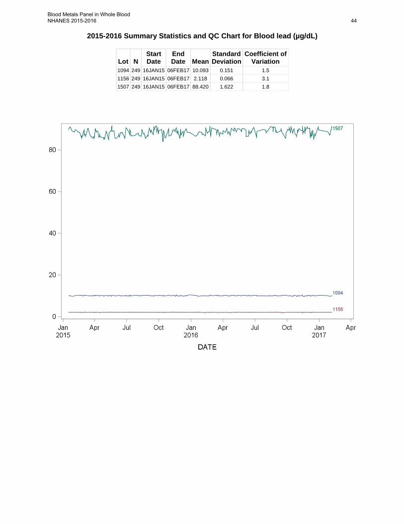
2015-2016 Summary Statistics and QC Chart for Blood lead (µg/dL)
Lot N Start Date
End Date Mean
Standard Deviation
Coefficient of Variation
1094 249 16JAN15 06FEB17 10.093 0.151 1.5 1156 249 16JAN15 06FEB17 2.118 0.066 3.1 1507 249 16JAN15 06FEB17 88.420 1.622 1.8
Blood Metals Panel in Whole Blood NHANES 2015-2016 44

2015-2016 Summary Statistics and QC Chart for Blood manganese (µg/L)
Lot N Start Date
End Date Mean
Standard Deviation
Coefficient of Variation
1094 260 20JAN15 06FEB17 14.590 0.529 3.6 1156 260 20JAN15 06FEB17 7.978 0.423 5.3 1507 260 20JAN15 06FEB17 42.725 1.465 3.4
Blood Metals Panel in Whole Blood NHANES 2015-2016 45

2015-2016 Summary Statistics and QC Chart for Blood mercury,total (µg/L)
Lot N Start Date
End Date Mean
Standard Deviation
Coefficient of Variation
1094 253 16JAN15 06FEB17 5.888 0.168 2.9 1156 253 16JAN15 06FEB17 0.616 0.064 10.4 1507 253 16JAN15 06FEB17 42.442 4.337 10.2
Blood Metals Panel in Whole Blood NHANES 2015-2016 46
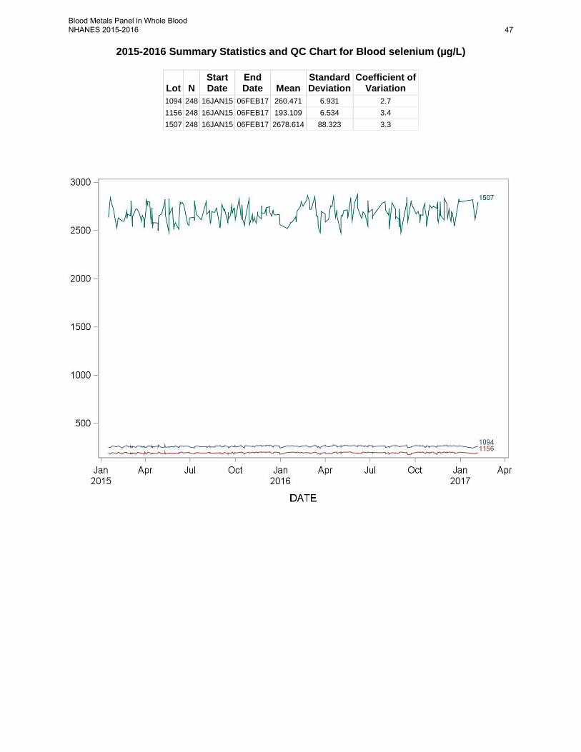
2015-2016 Summary Statistics and QC Chart for Blood selenium (µg/L)
Lot N Start Date
End Date Mean
Standard Deviation
Coefficient of Variation
1094 248 16JAN15 06FEB17 260.471 6.931 2.7 1156 248 16JAN15 06FEB17 193.109 6.534 3.4 1507 248 16JAN15 06FEB17 2678.614 88.323 3.3
Blood Metals Panel in Whole Blood NHANES 2015-2016 47

References
1. Pirkle, J.L., et al., National exposure measurements for decisions to protect public health from environmental exposures. International Journal of Hygiene and Environmental Health, 2005. 208(1-2): p. 1-5.
2. Agency for Toxic Substances and Disease Registry, Toxicological Profile for Mercury. 1999: Atlanta, GA.
3. Mahaffey, K.R. NHANES 1999 - 2002 Update on Mercury. in Northeast Regional Mercury Conference. 2005.
4. Sieler, H.G., ed. Handbook of Toxicity of Inorganic Compounds. 1988, Marcel Dekker, INC.
5. World Health Organization, Environmental Health Criteria 118: Inorganic Mercury. 1991, Geneva.
6. Centers for Disease Control and Prevention, Preventing Lead Poisoning in Young Children. 2005: Atlanta, GA.
7. Needleman, H., et al., Bone lead levels in adjudicated delinquents. A case control study. Neurotoxicology and teratology, 2002. 24(6): p. 711-7.
8. Dietrich, K., et al., Early exposure to lead and juvenile delinquency. Neurotoxicology and teratology, 2001. 23(6): p. 511-518.
9. Bellinger, D.C., Low-level lead exposure, intelligence and academic achievement: A long- term follow-up study. Pediatrics, 1992. 90(6): p. 855-861.
10. Bellinger, D.C., Intellectual Impairment and Blood Lead Levels. The New England Journal of Medicine, 2003. 349(5): p. 500-502.
11. Sigel, H. and A. Sigel, Handbook of Toxicity of Inorganic Compounds, H.G. Sieler, Editor. 1988, Marcel Dekker, INC.
12. Batley, G.E., Handbook of Trace Element Speciation: Analytical Methods. 1991, Boca Raton: CDC Press.
13. Agency for Toxic Substances and Disease Registry, Toxicological Profile for Lead. 2007: Atlanta, GA.
14. Centers for Disease Control and Prevention, CDC Response to Advisory Committee on Childhood Lead Poisoning Prevention Recommendations in “Low Level Lead Exposure Harms Children: A Renewed Call of Primary Prevention”. 2012: Atlanta, GA.
15. World Health Organization, Environmental Health Criteria 134: Cadmium. 1992. 16. Elinder, C.G., International Journal of Environmental Studies, 1982. 19(3-4): p. 187-193. 17. Ghezzi, I., et al., Behavior of biological indicators of cadmium in relation to occupational
exposure. International archives of occupational and environmental health, 1985. 55(2): p. 133-140.
18. Jarup, L., C. Elinder, and G. Spang, Cumulative blood-cadmium and tubular proteinuria: a dose-response relationship. International archives of occupational and environmental health, 1988. 60(3): p. 223-229.
19. Lauwerys, R., et al., Cadmium - Exposure Markers as Predictors of Nephrotoxic Effects. Clinical Chemistry, 1994. 40(7B): p. 1391-1394.
20. Roels, H., et al., Health significance of cadmium induced renal dysfunction: a five year follow up. British journal of industrial medicine, 1989. 46(11): p. 755-764.
21. Bernard, A. and R. Lauwerys, Cadmium in human population, in Cadmium in the Environment. 1986, Springer. p. 114-123.
Blood Metals Panel in Whole Blood NHANES 2015-2016 48

22. Milne, D.B., Trace Elements, in Tietz textbook of clinical chemistry, C.A. Burtis, Ashwood, Edward R., Editor. 1999, W. B. Saunders Company: Philadelphia. p. 1029-1055.
23. Chiswell, B. and D. Johnson, Manganese, in Handbook on Metals in Clinical and Analytical Chemistry, A.S. Hans G. Seiler, Helmut Sigel, Editor. 1994, Marcel Dekker: New York. p. 467-478.
24. Smargiassi, A., et al., Peripheral Markers of Catecholamine Metabolism among Workers Occupationally Exposed to Manganese (Mn). Toxicology Letters, 1995. 77(1-3): p. 329-333.
25. Roels, H.A., et al., Assessment of the Permissible Exposure Level to Manganese in Workers Exposed to Manganese-Dioxide Dust. British Journal of Industrial Medicine, 1992. 49(1): p. 25-34.
26. Cowan, D.M., et al., Manganese exposure among smelting workers: blood manganese-iron ratio as a novel tool for manganese exposure assessment. Biomarkers, 2009. 14(1): p. 3-16.
27. Gennart, J.P., et al., Fertility of Male Workers Exposed to Cadmium, Lead, or Manganese. American Journal of Epidemiology, 1992. 135(11): p. 1208-1219.
28. Bader, M., et al., Biomonitoring of manganese in blood, urine and axillary hair following low-dose exposure during the manufacture of dry cell batteries. International Archives of Occupational and Environmental Health, 1999. 72(8): p. 521-527.
29. Lauwerys, R., et al., Fertility of Male Workers Exposed to Mercury-Vapor or to Manganese Dust - a Questionnaire Study. American Journal of Industrial Medicine, 1985. 7(2): p. 171-176.
30. Standridge, J.S., et al., Effect of Chronic Low Level Manganese Exposure on Postural Balance: A Pilot Study of Residents in Southern Ohio. Journal of Occupational and Environmental Medicine, 2008. 50(12): p. 1421-1429.
31. Woolf, A., et al., A child with chronic manganese exposure from drinking water. Environmental Health Perspectives, 2002. 110(6): p. 613-616.
32. Wasserman, G.A., et al., Water manganese exposure and children's intellectual function in Araihazar, Bangladesh. Environmental Health Perspectives, 2006. 114: p. 124-129.
33. Ljung, K.S., et al., Maternal and Early Life Exposure to Manganese in Rural Bangladesh. Environmental Science & Technology, 2009. 43(7): p. 2595-2601.
34. Bazzi, A., J.O. Nriagu, and A.M. Linder, Determination of toxic and essential elements in children's blood with inductively coupled plasma-mass spectrometry. Journal of Environmental Monitoring, 2008. 10(10): p. 1226-1232.
35. Rollin, H.B., et al., Examining the association between blood manganese and lead levels in schoolchildren in four selected regions of South Africa (vol 103, pg 160, 2007). Environmental Research, 2008. 106(3): p. 426-426.
36. Rollin, H., et al., Blood manganese concentrations among first-grade schoolchildren in two South African cities. Environmental Research, 2005. 97(1): p. 93-99.
37. Aschner, M., Manganese: Brain transport and emerging research needs. Environmental Health Perspectives, 2000. 108: p. 429-432.
38. Yokel, R.A., Brain uptake, retention, and efflux of aluminum and manganese. Environmental Health Perspectives, 2002. 110: p. 699-704.
39. Davis, J.M., Methylcyclopentadienyl manganese tricarbonyl: Health risk uncertainties and research directions. Environmental Health Perspectives, 1998. 106: p. 191-201.
Blood Metals Panel in Whole Blood NHANES 2015-2016 49

40. Davis, J.M., et al., The EPA health risk assessment of methylcyclopentadienyl manganese tricarbonyl (MMT). Risk Analysis, 1998. 18(1): p. 57-70.
41. Roels, H., et al., Relationship Between External and Internal Parameters of Exposure to Manganese in Workers From a Manganese Oxide and Salt Producing Plant. American journal of industrial medicine, 1987. 11(3): p. 297-305.
42. Jarvisalo, J., et al., Urinary and blood manganese in occupationally nonexposed populations and in manual metal arc welders of mild-steel. International archives of occupational and environmental health, 1992. 63(7): p. 495-501.
43. Smyth, L., et al., Clinical manganism and exposure to manganese in the production and processing of ferromanganese alloy. Journal of occupational medicine, 1973. 15(2): p. 101-9.
44. Klaassen, C., Biliary-Excretion of Manganese in Rats, Rabbits, and Dogs. Toxicology and applied pharmacology, 1974. 29(3): p. 458-468.
45. Malecki, E., et al., Biliary manganese excretion in conscious rats is affected by acute and chronic manganese intake but not by dietary fat. The Journal of nutrition, 1996. 126(2): p. 489-498.
46. Agency for Toxic Substances and Disease Registry, Toxicological Profile for Manganese, ATSDR, Editor. 2000: Atlanta, GA.
47. Agency for Toxic Substances and Disease Registry, Toxicological Profile for Selenium. 2003: Atlanta, GA.
48. Goldhaber, S.B., Trace element risk assessment: essentiality vs. toxicity. Regulatory Toxicology and Pharmacology., 2003. 38: p. 232-242.
49. Combs, G.F. and W.P. Gray, Chemopreventive agents. Pharmacology and Therapeutics, 1998. 79: p. 179-192.
50. Arthur, J.R., The role of selenium in thyroid hormone metabolism. Can J Physiol Pharmacol, 1991. 69: p. 1648-1652.
51. Corvilain, B., et al., Selenium and the thyroid: How the relationship was established. Am J Clin Nutr, 1993. 57 (2 Suppl): p. 244S-248S.
52. Levander, O.A., Nutrition and newly emerging viral diseases: An overview. J Nutr, 1997. 127: p. 948S-950S.
53. McKenzie, R.C., T.S. Rafferty, and G.J. Beckett, Selenium: an essential element for immune function. Immunol Today, 1998. 19: p. 342-345.
54. Ellis, D.R. and D.E. Salt, Plants, selenium and human health. Curr Opin Plant Biol, 2003. 6: p. 273-279.
55. Combs, G.F., Food system-based approaches to improving micronutrient nutrition: the case for selenium. Biofactors, 2000. 12: p. 39-43.
56. Zimmerman, M.B. and J. Kohrle, The impact of iron and selenium deficiencies on iodine and thyroid metabolism: biochemistry and relevance to public health. Thyroid, 2002. 12: p. 867-878.
57. Beck, M.A., O. Levander, and J. Handy, Selenium deficiency and viral infection. Journal of Nutrition, 2003. 133: p. 1463S-1467S.
58. Lutz, T.M., P.M.V. Nirel, and B. Schmidt, Whole-blood analysis by ICP-MS. Applications of Plasma Source Mass Spectrometry, ed. G. Holland and A.N. Eaton. 1991, Cambridge: Royal Soc Chemistry. 96-100.
59. Tanner, S.D., Baranov, Vladimir I, Theory, Design, and Operation of a Dynamic Reaction Cell for ICP-MS. Atomic Spectroscopy, 1999. 20(2): p. 45-52.
Blood Metals Panel in Whole Blood NHANES 2015-2016 50

60. Tanner, S.D., V.I. Baranov, and D.R. Bandura, Reaction cells and collision cells for ICP-MS: a tutorial review. Spectrochimica Acta Part B-Atomic Spectroscopy, 2002. 57(9): p. 1361-1452.
61. Tanner, S.D. and V.I. Baranov, Theory, design, and operation of a dynamic reaction cell for ICP-MS. Atomic Spectroscopy, 1999. 20(2): p. 45-52.
62. Burguera, J.L., et al., Electrothermal atomic absorption spectrometry determination of molybdenum in whole blood. Spectrochimica Acta Part B-Atomic Spectroscopy, 2002. 57(3): p. 561-569.
63. Jarrett, J.M., et al., Eliminating molybdenum oxide interference in urine cadmium biomonitoring using ICP-DRC-MS. Journal of Analytical Atomic Spectrometry, 2008. 23(7): p. 962-967.
64. Division of Laboratory Sciences, Division of Laboratory Sciences Policies and Procedures Manual. 2015, Centers for Disease Control and Prevention: Atlanta, GA.
65. Centers for Disease Control and Prevention, CDC Response to Advisory Committee on Childhood Lead Poisoning Prevention Recommendations in “Low Level Lead Exposure Harms Children: A Renewed Call of Primary Prevention””, Department of Health and Human Services, Editor. 2012: Atlanta, GA.
66. Occupational Safety and Health Administration, Occupational Safety and Health Standards, in 29 CFR part 1910, Subpart Z, Standard number 1910.1025, "Lead",. 1989.
67. Occupational Safety and Health Administration, Cadmium (OSHA 3136-06R 2004). 2004.
68. American Conference of Governmental Industrial Hygienists, Tlvs and Beis 2007: Based on the Documentation for Chemical Substances and Physical Agents & Biological Exposure Indices. 2007: American Conference of Governmental Industrial Hygienists.
69. Baselt, R.C., Disposition of Toxic Drugs and Chemicals in Man,. 2011, Seal Beach, CA: Biomedical Publications.
70. Nuttall, K., Evaluating selenium poisoning. Annals of clinical & laboratory science, 2006. 36(4): p. 409-420.
71. Office of Health and Safety in the Division of Laboratory Sciences, Policies and Procedures Manual. 2002, Division of Laboratory Sciences (DLS), National Center for Environmental Health, Centers for Disease Control and Prevention, Public Health Service, Department of Health and Human ServicesCenters for Disease Control and Prevention, .
72. Centers for Disease Control and Prevention, Fourth National Report on Human Exposure to Environmental Chemicals, February 2015 Update. 2015, CDC: Atlanta, GA.
73. Carson, B.L., H.V.E. III, and J.L. McCann, Selenium, in Toxicology and biological monitoring of metals in humans., B.L. Carson, H.V.E. III, and J.L. McCann, Editors. 1986, Lewis Publishers, Inc.: Chelsea, Michigan. p. 213-218.
74. Fell, J.M.E., et al., Manganese toxicity in children receiving long-term parenteral nutrition. Lancet, 1996. 347(9010): p. 1218-1221.
75. Henn, B.C., et al., Early Postnatal Blood Manganese Levels and Children's Neurodevelopment. Epidemiology, 2010. 21(4): p. 433-439.
76. Ikeda, M., et al., Cadmium, chromium, lead, manganese and nickel concentrations in blood of women in non-polluted areas in Japan, as determined by inductively coupled
Blood Metals Panel in Whole Blood NHANES 2015-2016 51

plasma-sector field-mass spectrometry. International Archives of Occupational and Environmental Health, 2011. 84(2): p. 139-150.
Blood Metals Panel in Whole Blood NHANES 2015-2016 52

Appendix A: Critical parameter test results
Critical parameter test #1: Testing scenario of something preventing a set of prepared samples from being analyzed immediately.
Test details: Day 1: Prepared a set of dilutions (calibrators, blanks, reference material, and fake samples)
for analysis in triplicate. Analyzed set 1 immediately (normal practice). Cap sets 2 and 3 and leave at room temperature for later analysis.
Day 2: Prepared run set 4 and analyzed it sequentially with run set 2 Day 3: Prepared run set 5 and analyzed it sequentially with run set 3
Table 1. Ruggedness testing results: Evaluating the significance of time from preparation to analysis on sample stability. Test performed 12/6-8/10 by Deanna Jones. Results are the average of the beginning and ending QC results for each analytical run.
ID Time, prep to analysis Hg (µg/L) Pb (µg /dL) Cd (µg /L) Mn (µg /L) Se (µg /L)
LB08
707
_WB2
target mean and 3SD range
0.585 0.318 – 0.852
2.12 1.99 – 2.25
0.488 0.353 – 0.623
7.98 6.38 – 9.59
0 hr 0.418 2.03 0.399 6.09 24 hr (fresh) 0.504 (0.522) 1.99 (2.18) 0.419 (0.47) 7.06 (7.88) 48 hr 0.396 (0.418) 2.04 (2.03) 0.509 (0.40) 7.82 (6.09)
HB0
8708
_W
B2
target mean and 3SD range
6.19 5.74 – 6.63
10.1 9.73 – 10.4
3.14 2.84 – 3.44
14.9 12.8 – 17.1
0 hr 5.86 10.0 3.03 12.5 24 hr 5.46 (5.7) 9.5 (10.7) 2.85 (3.17) 13.6 (14.7) 48 hr 2.64 (5.9) 9.2 (10.0) 2.79 (3.03) 13.5 (12.5)
QM
EQAS
07
B-03
*
target mean and 3SD range
228 206 – 251
0 hr 192 24 hr 202 (217) 48 hr 56 (192)
QM
EQAS
10
B-06
*
target mean and 2SD range
239 215 – 253
0 hr 212 24 hr 221 (238) 48 hr 62 (212)
*samples purchase from Le centre de toxicology du Quebec (Quebec, Canada)
Conclusion: Samples which have been diluted 1+1+48 for analysis up to one (1) day previously can still be analyzed.
Blood Metals Panel in Whole Blood NHANES 2015-2016 53
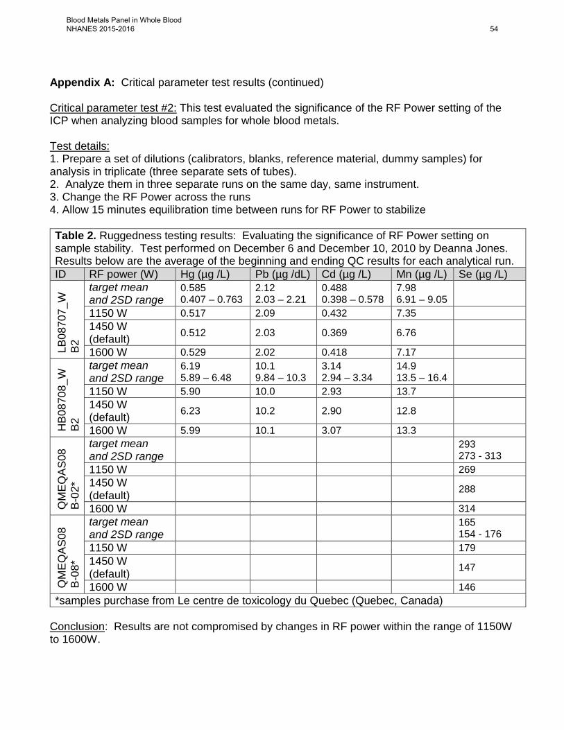
Appendix A: Critical parameter test results (continued) Critical parameter test #2: This test evaluated the significance of the RF Power setting of the ICP when analyzing blood samples for whole blood metals. Test details: 1. Prepare a set of dilutions (calibrators, blanks, reference material, dummy samples) for analysis in triplicate (three separate sets of tubes). 2. Analyze them in three separate runs on the same day, same instrument. 3. Change the RF Power across the runs 4. Allow 15 minutes equilibration time between runs for RF Power to stabilize Table 2. Ruggedness testing results: Evaluating the significance of RF Power setting on sample stability. Test performed on December 6 and December 10, 2010 by Deanna Jones. Results below are the average of the beginning and ending QC results for each analytical run. ID RF power (W) Hg (µg /L) Pb (µg /dL) Cd (µg /L) Mn (µg /L) Se (µg /L)
LB08
707_
WB2
target mean and 2SD range
0.585 0.407 – 0.763
2.12 2.03 – 2.21
0.488 0.398 – 0.578
7.98 6.91 – 9.05
1150 W 0.517 2.09 0.432 7.35 1450 W (default) 0.512 2.03 0.369 6.76
1600 W 0.529 2.02 0.418 7.17
HB0
8708
_WB2
target mean and 2SD range
6.19 5.89 – 6.48
10.1 9.84 – 10.3
3.14 2.94 – 3.34
14.9 13.5 – 16.4
1150 W 5.90 10.0 2.93 13.7 1450 W (default) 6.23 10.2 2.90 12.8
1600 W 5.99 10.1 3.07 13.3
QM
EQAS
08B-
02*
target mean and 2SD range 293
273 - 313 1150 W 269 1450 W (default) 288
1600 W 314
QM
EQAS
08B-
08*
target mean and 2SD range 165
154 - 176 1150 W 179 1450 W (default) 147
1600 W 146 *samples purchase from Le centre de toxicology du Quebec (Quebec, Canada)
Conclusion: Results are not compromised by changes in RF power within the range of 1150W to 1600W.
Blood Metals Panel in Whole Blood NHANES 2015-2016 54

Appendix A: Critical parameter test results (continued)
Critical parameter test #3: This test evaluated the significance of the dynamic reaction cell gas flow rate of the reaction gas (oxygen and methane) while analyzing blood samples for elements analyzed in DRC mode (Hg, Mn, and Se). The cell gas flow rate for Mn and Hg is oxygen (O2) and the per method setting is 1.2 mL/min. The cell gas flow rate for Se is methane (CH4) and the per method setting is 0.84 mL/min.
Test details: 1. Prepare a set of dilutions (calibrators, blanks, reference material, dummy samples) foranalysis in triplicate (three separate sets of tubes).2. Analyze them in three separate runs on the same day using the same instrument.3. Change the cell gas flow rate.
Table 3. Ruggedness testing results: Evaluating the significance of dynamic reaction cell gas flow rate on sample stability. Test performed on December 6, 2010 and January 4, 2010 by Deanna Jones. Results below are the average of the beginning and ending QC results for each analytical run.
ID cell gas flow rate Hg (µg /L) Pb (µg /dL) Cd (µg /L) Mn (µg /L) Se (µg /L)
LB08
707_
WB2
target mean and 2SD range
0.585 0.407 – 0.763
2.12 2.03 – 2.21
0.488 0.398 – 0.578
7.98 6.91 – 9.05
See Table 4
0.96 mL/min O2; 0.7 mL/min CH4
0.457 2.10 0.471 8.49
1.2 mL/min O2; 0.84 mL/min CH4 0.479 2.10 0.438 8.15
1.44 mL/min O2; 1.0 mL/min CH4 0.555 2.11 0.457 8.12
HB0
8708
_WB2
Target Mean and 2SD Range
6.19 5.89 – 6.48
10.1 9.84 – 10.3
3.14 2.94 – 3.34
14.9 13.5 – 16.4
0.96 mL/min O2; 0.7 mL/min CH4
4.71 10.0 3.19 14.4
1.2 mL/min O2; 0.84 mL/min CH4 5.45 10.1 2.92 15.2
1.44 mL/min O2; 1.0 mL/min CH4 5.34 10.3 3.04 14.6
Conclusion: Accuracy of Mn and Hg results are not compromised by changes in cell gas flow rate within the range tested (0.96 – 1.44 mL/min).
Blood Metals Panel in Whole Blood NHANES 2015-2016 55

Appendix A: Critical Parameter Test Results (Continued) Table 4. Ruggedness testing results: Evaluating the significance of dynamic reaction cell gas flow rate on sample stability. Test performed on December 6, 2010 and January 4, 2010 by Deanna Jones. Results below are the average of the beginning and ending QC results for each analytical run.
ID cell gas flow rate Hg (µg /L) Pb (µg /dL) Cd (µg /L) Mn (µg /L) Se (µg /L)
QM
EQAS
07B-
09* target mean
and 2SD range
See Table 3, Appendix A
157 146 - 168
0.96 mL/min O2; 0.7 mL/min CH4
187
1.2 mL/min O2; 0.84 mL/min CH4 186
1.44 mL/min O2; 1.0 mL/min CH4 191
QM
EQAS
08B-
02* target mean
and 2SD range 293 273 - 313
0.96 mL/min O2; 0.7 mL/min CH4
328
1.2 mL/min O2; 0.84 mL/min CH4 334
1.44 mL/min O2; 1.0 mL/min CH4 339
*samples purchase from Le centre de toxicology du Quebec (Quebec, Canada) Conclusion: Accuracy of Se results are not compromised by changes in cell gas flow rate within the range tested (0.7 – 1.0 mL/min).
Blood Metals Panel in Whole Blood NHANES 2015-2016 56

Appendix A: Critical parameter test results (continued)
Critical parameter test #4: This test evaluated the significance of the RPq value while analyzing blood samples for Se, Mn and Hg. The RPq value setting per method for Mn and Hg is 0.6, and for Se it is 0.65. The reduced and elevated RPq values for Mn and Hg are 0.48 and 0.72, respectively. The reduced and elevated RPq values for Se are 0.52 and 0.78, respectively. The results are presented in Tables 5 and 6.
Test details: 1. Prepare a set of dilutions (calibrators, blanks, reference material, fake samples) for analysisin triplicate (three separate sets of tubes).2. Analyze them in three separate runs on the same day, using the same instrument.3. Change the RPq value.
Table 5. Ruggedness testing results: Evaluating the significance of RPq value on sample stability. Test performed on December 21, 2010 by Deanna Jones. Results below are the average of the beginning and ending QC results for each analytical run.
ID RPq Hg (µg /L) Pb (µg /dL) Cd (µg /L) Mn (µg /L) Se (µg /L)
LB08
707_
WB2
Target Mean and 2SD Range
0.585 0.407 – 0.763
2.12 2.03 – 2.21
0.488 0.398 – 0.578
7.98 6.91 – 9.05
See Table 6
0.48 Mn and Hg; 0.52 Se 0.455 1.97 0.361 7.86
0.6 Mn and Hg; 0.7 Se 0.418 2.03 0.399 6.09
0.72 Mn and Hg; 0.78 Se 0.402 2.07 0.402 7.99
HB0
8708
_WB2
Target Mean and 2SD Range
6.19 5.89 – 6.48
10.1 9.84 – 10.3
3.14 2.94 – 3.34
14.9 13.5 – 16.4
0.48 Mn and Hg; 0.52 Se 5.54 9.4 2.79 14.4
0.6 Mn and Hg; 0.7 Se 5.86 10.0 3.03 12.5
0.72 Mn and Hg; 0.78 Se 5.53 9.7 2.88 14.9
Conclusion: Accuracy of Mn and Hg results are not compromised by changes in RPq settings within the range tested (0.48 – 0.72).
Blood Metals Panel in Whole Blood NHANES 2015-2016 57

Appendix A: Critical Parameter Test Results (Continued)
Table 6. Ruggedness testing results: Evaluating the significance of RPq value on sample stability. Test performed on December 21, 2010 by Deanna Jones. Results below are the average of the beginning and ending QC results for each analytical run.
ID RPq Hg (µg /L) Pb (µg /dL) Cd (µg /L) Mn (µg /L) Se (µg /L)
QM
EQAS
07B-
09* target mean
and 2SD range
See Table 5, Appendix A
293 273 – 313
0.48 Mn and Hg; 0.52 Se 262
0.6 Mn and Hg; 0.7 Se 250
0.72 Mn and Hg; 0.78 Se 277
QM
EQAS
08B-
02* target mean
and 2SD range 361 337 - 385
0.48 Mn and Hg; 0.52 Se 347
0.6 Mn and Hg; 0.7 Se 349
0.72 Mn and Hg; 0.78 Se 364
*samples purchase from Le centre de toxicology du Quebec (Quebec, Canada)
Conclusion: Accuracy of Se results are not compromised by changes in RPq settings within the range tested (0.52 – 0.78 for Se).
Blood Metals Panel in Whole Blood NHANES 2015-2016 58
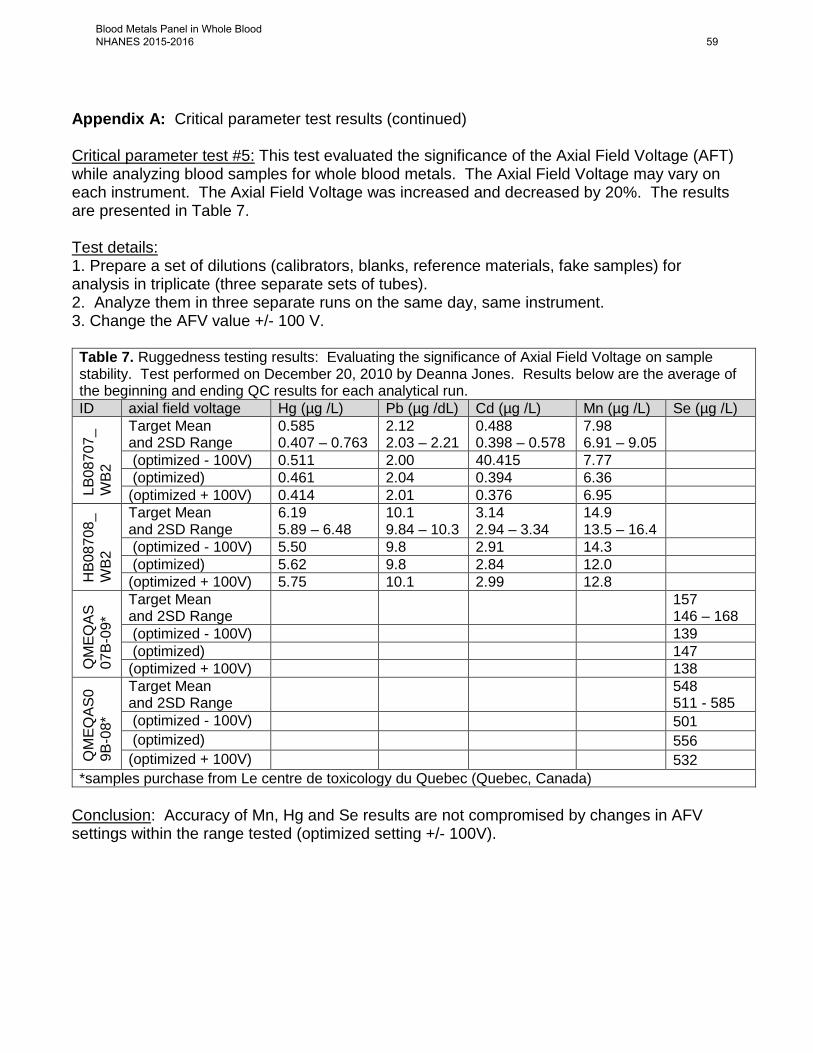
Appendix A: Critical parameter test results (continued) Critical parameter test #5: This test evaluated the significance of the Axial Field Voltage (AFT) while analyzing blood samples for whole blood metals. The Axial Field Voltage may vary on each instrument. The Axial Field Voltage was increased and decreased by 20%. The results are presented in Table 7. Test details: 1. Prepare a set of dilutions (calibrators, blanks, reference materials, fake samples) for analysis in triplicate (three separate sets of tubes). 2. Analyze them in three separate runs on the same day, same instrument. 3. Change the AFV value +/- 100 V. Table 7. Ruggedness testing results: Evaluating the significance of Axial Field Voltage on sample stability. Test performed on December 20, 2010 by Deanna Jones. Results below are the average of the beginning and ending QC results for each analytical run. ID axial field voltage Hg (µg /L) Pb (µg /dL) Cd (µg /L) Mn (µg /L) Se (µg /L)
LB08
707_
WB2
Target Mean and 2SD Range
0.585 0.407 – 0.763
2.12 2.03 – 2.21
0.488 0.398 – 0.578
7.98 6.91 – 9.05
(optimized - 100V) 0.511 2.00 40.415 7.77 (optimized) 0.461 2.04 0.394 6.36 (optimized + 100V) 0.414 2.01 0.376 6.95
HB0
8708
_W
B2
Target Mean and 2SD Range
6.19 5.89 – 6.48
10.1 9.84 – 10.3
3.14 2.94 – 3.34
14.9 13.5 – 16.4
(optimized - 100V) 5.50 9.8 2.91 14.3 (optimized) 5.62 9.8 2.84 12.0 (optimized + 100V) 5.75 10.1 2.99 12.8
QM
EQAS
07B-
09*
Target Mean and 2SD Range 157
146 – 168 (optimized - 100V) 139 (optimized) 147 (optimized + 100V) 138
QM
EQAS
09B
-08*
Target Mean and 2SD Range 548
511 - 585 (optimized - 100V) 501 (optimized) 556 (optimized + 100V) 532
*samples purchase from Le centre de toxicology du Quebec (Quebec, Canada) Conclusion: Accuracy of Mn, Hg and Se results are not compromised by changes in AFV settings within the range tested (optimized setting +/- 100V).
Blood Metals Panel in Whole Blood NHANES 2015-2016 59

Appendix A: Critical parameter test results (continued) Parameter test #6: Evaluate the impact on observed concentration if an extra dilution is performed on the sample relative to the calibration standards. Test details: 1. A large blood sample was spiked to elevated concentrations, and mixed well. The spiked
sample was then prepared for analysis at various extra dilution levels and the observed results compared to results obtained with no extra dilution performed.
normalized concentration ± 1RSD Dilution level Mn Hg Se Cd Pb No Extra (N=8) 1.00 1.00 1.00 1.00 1.00 2x dilution (N=8) 1.00 ± 0.01 1.03 ± 0.05 1.02 ± 0.03 1.00 ± 0.01 1.01 ± 0.01 5x dilution (N=6) 1.01 ± 0.01 1.06 ± 0.06 1.01 ± 0.02 1.01 ± 0.01 1.02 ± 0.01 10x dilution (N=8) 1.01 ± 0.03 1.04 ± 0.06 1.04 ± 0.06 1.00 ± 0.02 1.02 ± 0.02 20x dilution (N=8) 1.02 ± 0.04 1.09 ± 0.05 1.06 ± 0.08 1.01 ± 0.03 1.02 ± 0.02
Conclusion: Results show that all analytes of the method (Pb, Cd, Hg, Mn, and Se) can be analyzed at up to a 20x extra dilution without significant effect (> ± 10% error) to the observed concentration.
Blood Metals Panel in Whole Blood NHANES 2015-2016 60

Appendix B
Table 1. Instrument and method parameters.
Instrument: PerkinElmer ELAN DRC II ICP-MS ESI SC4 autosampler with (optional) PC3 Peltier cooled spray chamber
Optimization window parameters RF power 1450 W Plasma Gas Flow (Ar) 15 L/min Auxiliary Gas Flow (Ar) 1.2 L/min Nebulizer Gas Flow (Ar) ~0.90 – 1.0 L/min (optimized as needed for sensitivity) Ion Lens Voltage(s) AutoLens (optimized as needed for sensitivity) AFV, QRO, CRO, CPV, Discriminator Threshold
Optimized per instrument by service engineer, or advanced user.
Parameters of x-y alignment, nebulizer gas flow, AutoLens voltages, mass calibration, dual detector calibration and detector voltages are optimized regularly. Optimization file name = default.dac. Configurations window parameters cell gas changes pause times
Pressurize Delay (From Standard to DRC mode) = 60 Exhaust Delay (From DRC to Standard mode) = 30 Flow Delay (Gas changes while in DRC mode) = 30 Channel Delay (Gas channel change in DRC mode) = 30
File names & directories method file names calibration curve (programmed for blood blank)
CDC_DLS3016_bldblk.mth For QC & patient sample analysis (programmed for aqueous blank) CDC_DLS3016_aqblk.mth
dataset Create a new dataset subfolder each day. Name as “2011-0820” for all work done on August 20, 2011
sample file Create for each day’s work report file name For sample results printouts
cdc_quant comprehensive.rop
For calibration curve information CDC_Quant Comprehensive (calib curve info).rop
tuning Default.tun optimization Default.dac calibration N/A polyatomic elan.ply report options template (transferring results to the database)
CDC_Database Output.rop Report Format Options: select only “Use Separator” File Write Option: Append Report File name: make descriptive including date (e.g. 2005-0311b_DRC2A_HM-0364.txt)
Blood Metals Panel in Whole Blood NHANES 2015-2016 61

Table 1. Instrument and method parameters.
Method Parameters Method Parameters: Timing Page (see Figures 1a, 2a and 2d in Appendix B) sweeps/reading 30 readings/replicate 1 replicates 3 enable qc checking On isotopes monitored and internal standard associations (exact mass)
use 103Rh, 130Te, 193Ir as internal standards 103Rh (102.905): 55Mn (54.93805) 130Te(129.907): 202Hg (201.971), 80Se(79.9165) 193Ir(192.963):208Pb(207.977), 114Cd(113.904)
dwell times 100 ms for 55Mn,202Hg, 80Se, 208Pb, and 114Cd 50 ms for 130Te,103Rh, and 193Ir
scan mode Peak Hopping for all isotopes (1 MCA channel) DRC channel A gas flow rate
99.999% methane (5-7 psig delivery pressure) typically 0.84 L/min (0.7 – 1.0) * *optimized per instrument, and periodically verified
DRC channel B gas flow rate
99.99% oxygen (5-7 psig delivery pressure) typically 1.2 L/min (0.96 – 1.44) * *optimized per instrument, and periodically verified
RPa 0 for all isotopes
RPq
Typically* 0.6 (0.48 – 0.72) for 103Rh,55Mn,130Te, and 202Hg. 0.65 (0.52 – 0.78) for 130Te and 80Se. 0.25 for 193Ir, 208Pb, and 114Cd Use the same RPQ for each analyte and its IS. (* Optimize per instrument, and periodically verified)
Method parameters: processing page (see Figures 1b in Appendix B) detector mode Dual process spectral peak N/A autolens On isotope ratio mode Off enable short settling time
Off
blank subtraction After internal standard measurement units cps process signal profile N/A Method parameters: equations page (see Figure 1c in Appendix B) equations +Hg 200
-0.027250 * Sn118 +Pb 206 +Pb 207
Method parameters: calibration page (see Figures 1d in Appendix B)
Blood Metals Panel in Whole Blood NHANES 2015-2016 62
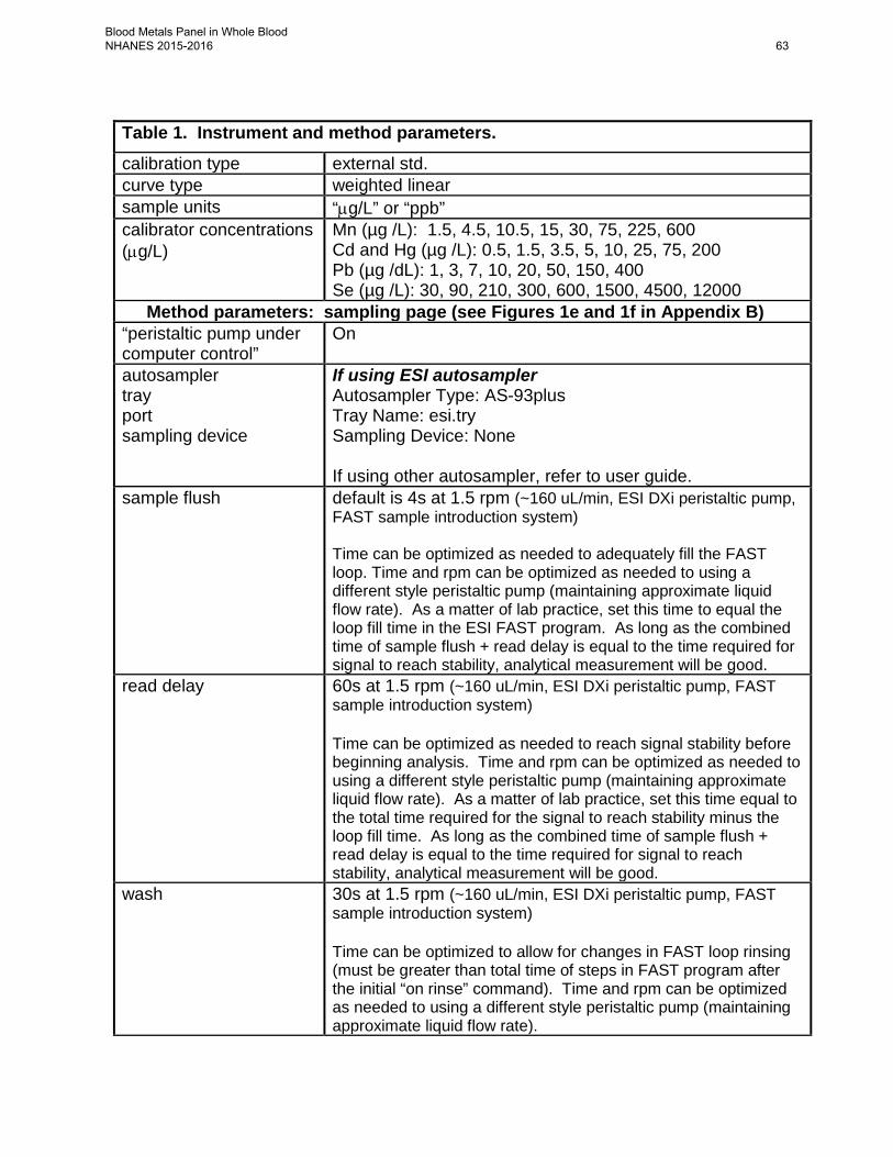
Table 1. Instrument and method parameters. calibration type external std. curve type weighted linear sample units “µg/L” or “ppb” calibrator concentrations (µg/L)
Mn (µg /L): 1.5, 4.5, 10.5, 15, 30, 75, 225, 600 Cd and Hg (µg /L): 0.5, 1.5, 3.5, 5, 10, 25, 75, 200 Pb (µg /dL): 1, 3, 7, 10, 20, 50, 150, 400 Se (µg /L): 30, 90, 210, 300, 600, 1500, 4500, 12000
Method parameters: sampling page (see Figures 1e and 1f in Appendix B) “peristaltic pump under computer control”
On
autosampler tray port sampling device
If using ESI autosampler Autosampler Type: AS-93plus Tray Name: esi.try Sampling Device: None If using other autosampler, refer to user guide.
sample flush default is 4s at 1.5 rpm (~160 uL/min, ESI DXi peristaltic pump, FAST sample introduction system) Time can be optimized as needed to adequately fill the FAST loop. Time and rpm can be optimized as needed to using a different style peristaltic pump (maintaining approximate liquid flow rate). As a matter of lab practice, set this time to equal the loop fill time in the ESI FAST program. As long as the combined time of sample flush + read delay is equal to the time required for signal to reach stability, analytical measurement will be good.
read delay 60s at 1.5 rpm (~160 uL/min, ESI DXi peristaltic pump, FAST sample introduction system) Time can be optimized as needed to reach signal stability before beginning analysis. Time and rpm can be optimized as needed to using a different style peristaltic pump (maintaining approximate liquid flow rate). As a matter of lab practice, set this time equal to the total time required for the signal to reach stability minus the loop fill time. As long as the combined time of sample flush + read delay is equal to the time required for signal to reach stability, analytical measurement will be good.
wash 30s at 1.5 rpm (~160 uL/min, ESI DXi peristaltic pump, FAST sample introduction system) Time can be optimized to allow for changes in FAST loop rinsing (must be greater than total time of steps in FAST program after the initial “on rinse” command). Time and rpm can be optimized as needed to using a different style peristaltic pump (maintaining approximate liquid flow rate).
Blood Metals Panel in Whole Blood NHANES 2015-2016 63

Table 1. Instrument and method parameters. extra wash (via ICP-MS software QC checking)
For sample concentrations greater than these, setup the ICP-MS software’s ‘QC checking’ feature to “Wash for X and continue”
Analyte Concentration Extra Rinse Time Cd 200 µg/L 200s Hg 200 µg/L 200s Mn 600 µg/L 200s Pb 400 µg/dL 200s Se 1200 µg/L 200s
autosampler locations of blanks and standards
For calibration curve (points to blood blank) CDC_DLS3016_bldblk.mth Calibration Stds 0 – 8 in autosampler positions 105 – 113 by default, but can be customized.
For QC & patient sample analysis (points to aqueous blank) CDC_DLS3016_aqblk.mth Aqueous Blank in autosampler position 117 by default, but can be customized.
FAST parameters: See Figures 4a through 4h in Appendix B for details configuration file default.sc
(saved at C:\Program Files\ESI\ESI-SC\) FAST program cdc_dls3016_5element_loop1ml_scfast.txt
Potential Emergency Response Modifications: mercury: Analyze mercury in standard mode with tellurium as the internal
standard. Set dwell time to 100ms, DRC gas flow to 0, and RPq to 0.25.
Non-FAST sample introduction system:
If the FAST sample introduction system is not available on any instruments, the method can still be implemented, but these changes will need to be made in the ELAN (and ESI software if present). Peristaltic pump speeds are for DXi pump; adjust accordingly if another pump is installed.
• Sample flush: Default is ~30s at -16 rpm. Set so that solution reaches nebulizer.
• Read delay: Default is 45s at -5 rpm. Set for best reproducibility of replicate measured intensities.
• Wash: Default is 60s at -11rpm. Set to prevent significant carry-over from one sample to the next.
• If using ESI autosampler without FAST, disable FAST in the ESI software before running analysis.
Blood Metals Panel in Whole Blood NHANES 2015-2016 64

Appendix B (continued) Table 3. Stock calibration standard concentrations
Analyte Stock calibration concentration (mg/L) High Purity Standards Item # SM-2107-042 10% v/v HCl
Cd 50 Hg 50 Mn 150 Pb 1000 Se 3000
Table 2. Suggested concentrations for base blood analyte (units) suggested concentration Cd (µg/L) <0.5 Hg (µg/L) <0.5 Mn (µg/L) < 8 Pb (µg/dL) <2 Se (µg/L) <200
Blood Metals Panel in Whole Blood NHANES 2015-2016 65

Appendix B (continued) Table 4. Preparation of intermediate stock calibration standard volume of flask (mL) 100 volume of spike of stock standard solution 2 concentrations ( mg /L) Cd 1 Hg 1 Mn 3 Pb (mg /dL) 20 Se 60
Table 5. Preparation of intermediate working standards Standard # 1 2 3 4 5 6 7 8 volume of flask (mL) 100 100 100 100 100 100 100 100
volume spike of stock std. (mL)
0.05 0.15 0.4
volume spike of int. stock Std. (mL)
0.05 0.15 0.35 0.50 1.00
concentrations ( µg /L) * Cd 0.5 1.5 3.5 5 10 25 75 200 Hg 0.5 1.5 3.5 5 10 25 75 200 Mn 1.5 4.5 10.5 15 30 75 225 600 Pb ( µg /dL) 1 3 7 10 20 50 150 400 Se 30 90 210 300 600 1500 4500 12000
* These same concentrations are entered in the ICP-MS software’s calibration page to describe the concentrations of the working calibrators (preparations analyzed during a run). This eliminates the need to multiply ICP-MS observed results by a dilution factor except for the case of extra dilutions (see Table 8).
Blood Metals Panel in Whole Blood NHANES 2015-2016 66
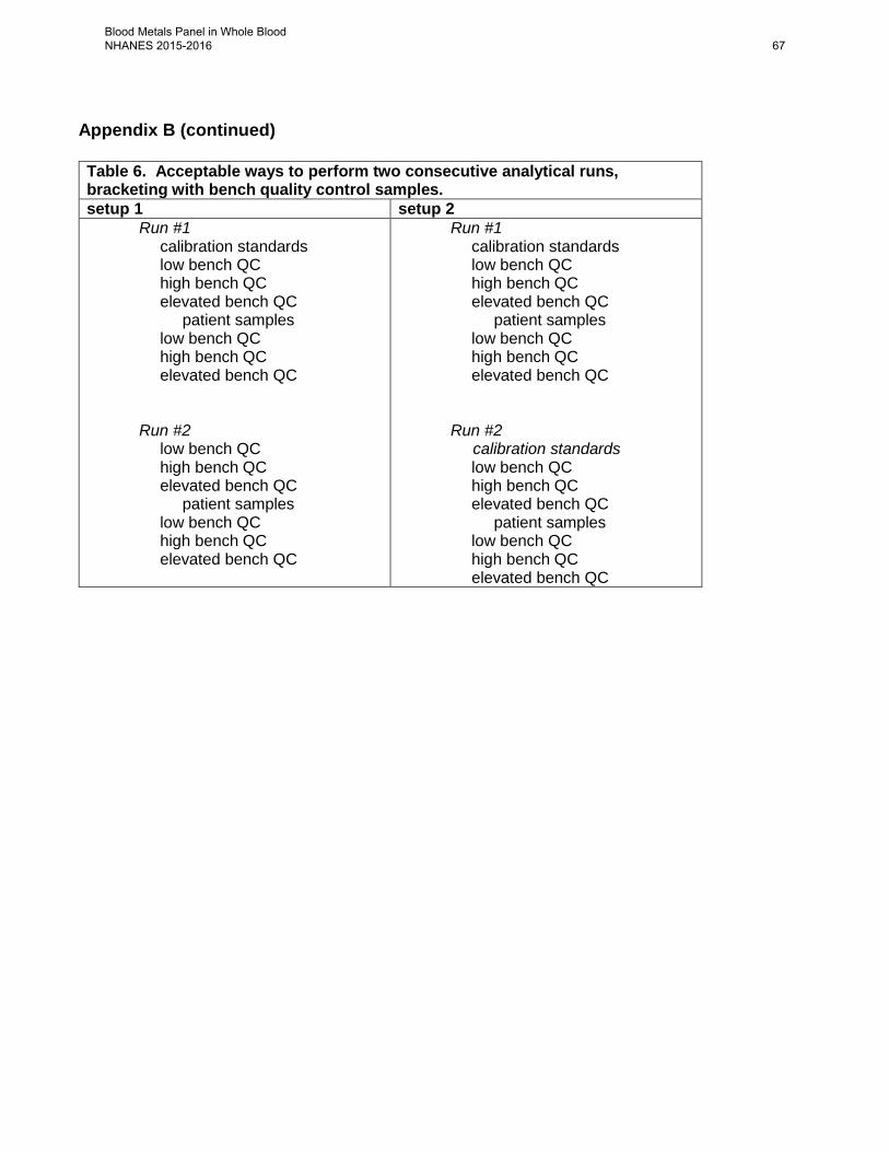
Appendix B (continued)
Table 6. Acceptable ways to perform two consecutive analytical runs, bracketing with bench quality control samples. setup 1 setup 2
Run #1 calibration standards low bench QC high bench QC elevated bench QC patient samples low bench QC high bench QC elevated bench QC
Run #2 low bench QC high bench QC elevated bench QC patient samples low bench QC high bench QC elevated bench QC
Run #1 calibration standards low bench QC high bench QC elevated bench QC patient samples low bench QC high bench QC elevated bench QC
Run #2 calibration standards
low bench QC high bench QC elevated bench QC patient samples low bench QC high bench QC elevated bench QC
Blood Metals Panel in Whole Blood NHANES 2015-2016 67
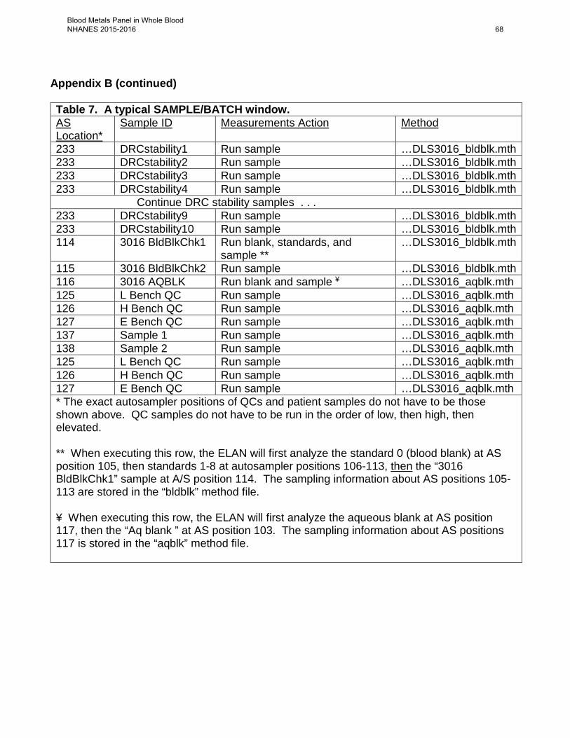
Appendix B (continued) Table 7. A typical SAMPLE/BATCH window. AS Location*
Sample ID Measurements Action Method
233 DRCstability1 Run sample …DLS3016_bldblk.mth 233 DRCstability2 Run sample …DLS3016_bldblk.mth 233 DRCstability3 Run sample …DLS3016_bldblk.mth 233 DRCstability4 Run sample …DLS3016_bldblk.mth Continue DRC stability samples . . . 233 DRCstability9 Run sample …DLS3016_bldblk.mth 233 DRCstability10 Run sample …DLS3016_bldblk.mth 114 3016 BldBlkChk1 Run blank, standards, and
sample ** …DLS3016_bldblk.mth
115 3016 BldBlkChk2 Run sample …DLS3016_bldblk.mth 116 3016 AQBLK Run blank and sample ¥ …DLS3016_aqblk.mth 125 L Bench QC Run sample …DLS3016_aqblk.mth 126 H Bench QC Run sample …DLS3016_aqblk.mth 127 E Bench QC Run sample …DLS3016_aqblk.mth 137 Sample 1 Run sample …DLS3016_aqblk.mth 138 Sample 2 Run sample …DLS3016_aqblk.mth 125 L Bench QC Run sample …DLS3016_aqblk.mth 126 H Bench QC Run sample …DLS3016_aqblk.mth 127 E Bench QC Run sample …DLS3016_aqblk.mth * The exact autosampler positions of QCs and patient samples do not have to be those shown above. QC samples do not have to be run in the order of low, then high, then elevated. ** When executing this row, the ELAN will first analyze the standard 0 (blood blank) at AS position 105, then standards 1-8 at autosampler positions 106-113, then the “3016 BldBlkChk1” sample at A/S position 114. The sampling information about AS positions 105-113 are stored in the “bldblk” method file. ¥ When executing this row, the ELAN will first analyze the aqueous blank at AS position 117, then the “Aq blank ” at AS position 103. The sampling information about AS positions 117 is stored in the “aqblk” method file.
Blood Metals Panel in Whole Blood NHANES 2015-2016 68
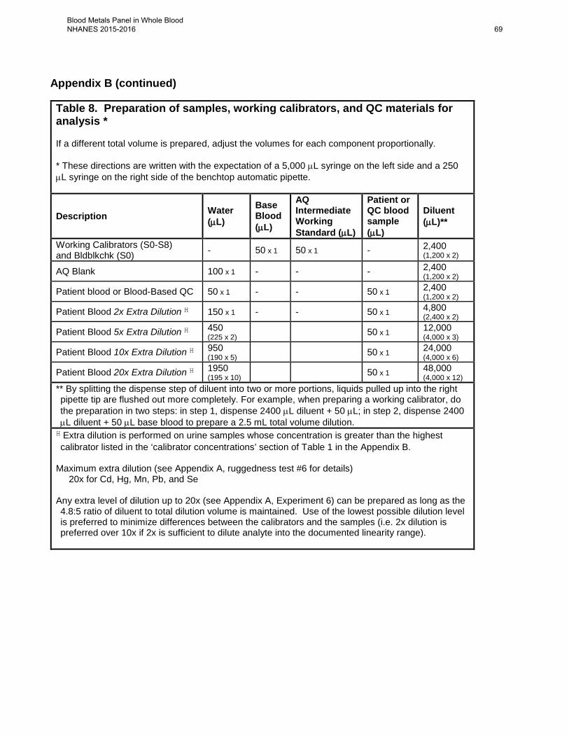
Appendix B (continued)
Table 8. Preparation of samples, working calibrators, and QC materials for analysis * If a different total volume is prepared, adjust the volumes for each component proportionally. * These directions are written with the expectation of a 5,000 µL syringe on the left side and a 250 µL syringe on the right side of the benchtop automatic pipette.
Description Water (µL)
Base Blood (µL)
AQ Intermediate Working Standard (µL)
Patient or QC blood sample (µL)
Diluent (µL)**
Working Calibrators (S0-S8) and Bldblkchk (S0) - 50 x 1 50 x 1 - 2,400
(1,200 x 2)
AQ Blank 100 x 1 - - - 2,400 (1,200 x 2)
Patient blood or Blood-Based QC 50 x 1 - - 50 x 1 2,400 (1,200 x 2)
Patient Blood 2x Extra Dilution H 150 x 1 - - 50 x 1 4,800 (2,400 x 2)
Patient Blood 5x Extra Dilution H 450 (225 x 2) 50 x 1 12,000
(4,000 x 3) Patient Blood 10x Extra Dilution H 950
(190 x 5) 50 x 1 24,000 (4,000 x 6)
Patient Blood 20x Extra Dilution H 1950 (195 x 10) 50 x 1 48,000
(4,000 x 12) ** By splitting the dispense step of diluent into two or more portions, liquids pulled up into the right pipette tip are flushed out more completely. For example, when preparing a working calibrator, do the preparation in two steps: in step 1, dispense 2400 µL diluent + 50 µL; in step 2, dispense 2400 µL diluent + 50 µL base blood to prepare a 2.5 mL total volume dilution. H Extra dilution is performed on urine samples whose concentration is greater than the highest calibrator listed in the ‘calibrator concentrations’ section of Table 1 in the Appendix B.
Maximum extra dilution (see Appendix A, ruggedness test #6 for details) 20x for Cd, Hg, Mn, Pb, and Se Any extra level of dilution up to 20x (see Appendix A, Experiment 6) can be prepared as long as the 4.8:5 ratio of diluent to total dilution volume is maintained. Use of the lowest possible dilution level is preferred to minimize differences between the calibrators and the samples (i.e. 2x dilution is preferred over 10x if 2x is sufficient to dilute analyte into the documented linearity range).
Blood Metals Panel in Whole Blood NHANES 2015-2016 69

Appendix B (continued) Table 9. Boundary concentrations for whole blood concentrations
analyte (units)
1st upper boundary (“1UB”) *
2nd upper boundary (“2UB”) **
range maximum (“Lim Rep Delta”) †
Highest Concentration Validated for Washout
Mn (µg/L) 20 35 2.0 600 Pb (µg/dL) 5.0 5.0 1.0 400 Cd (µg/L) 5.0 5.0 1.0 200 Hg (µg/L) 10.0 10.0 1.0 200 Se (µg/L) 400 400 20 12,000 * Typically, the 1st upper boundary (1UB) is the 99th percentile of non-weighted concentration results from the NHANES 1999-2000 subset groups, a concentration significant to public health, or a concentration defined by study protocol. The default 1UB concentrations are listed in this table. ** The 2nd upper boundary (2UB) may be 2x the 1UB, a concentration significant to public health, or defined by study protocol. † Range maximum (Lim Rep Delta) is the allowed limit to the range of the three replicate readings for a single sample analysis.
Blood Metals Panel in Whole Blood NHANES 2015-2016 70

Appendix B (continued)
Table 10. Reference ranges for blood concentrations [72]. analyte (units)
survey years
geometric mean 50th 75th 90th 95th N
Cd (µg/L) 07-08 0.315 0.270 0.500 1.00 1.52 8266 09-10 0.302 0.260 0.480 0.960 1.40 8793 11-12 0.279 0.250 0.460 0.960 1.50 7920
Hg (µg/L) 07-08 0.769 0.740 1.48 2.95 4.64 8266 09-10 0.863 0.790 1.68 3.43 5.13 8793 11-12 0.703 0.640 1.38 2.87 4.40 7920
Pb (µg/dL) 07-08 1.27 1.22 1.90 2.80 3.70 8266 09-10 1.12 1.07 1.70 2.58 3.34 8793 11-12 0.973 0.930 1.52 2.38 3.16 7920
Mn (µg/L) 11-12 9.35 9.22 11.5 14.4 16.7 7920 Se (µg/L) 11-12 190 190 206 223 236 7920
Table 11. Reference concentrations from published literature for blood Mn and Se. analyte (units) published concentrations
Se (µg/L) 157 – 265 µg/L [73]
Mn ( µg /L)†
Non-exposed 4 – 14 ( µg /L) [46] Exposed workers (adults) 3.2 – 101 µg /L [28] Children receiving long term parenteral nutrition 33.8 – 101 µg /L [74] Ohio adults (N=49) residing near a refinery (possible Mn emission): Mean (range) 9.4 (4.2-21.7) µg/L [30] Mexican infants Age 1, mean (SD) = 24.3 (4.5) µg/L, median = 23.7 µg/L, N=270 Age 2, mean (SD) = 21.1 (6.2) µg/L, median = 20.3 µg/L, N=430 [75] Japanese women (N = 1420) GM 13.2 µg/L overall, Range of median (max) across 8 regions 12.0-14.3 (25.0-33.4) µg/L [76] South African children, ages 8-10 years old (n = 49) Mean (SD) 8.48 (2.45) µg/L, range 4.58-18.20 µg/L. [34]
Blood Metals Panel in Whole Blood NHANES 2015-2016 71
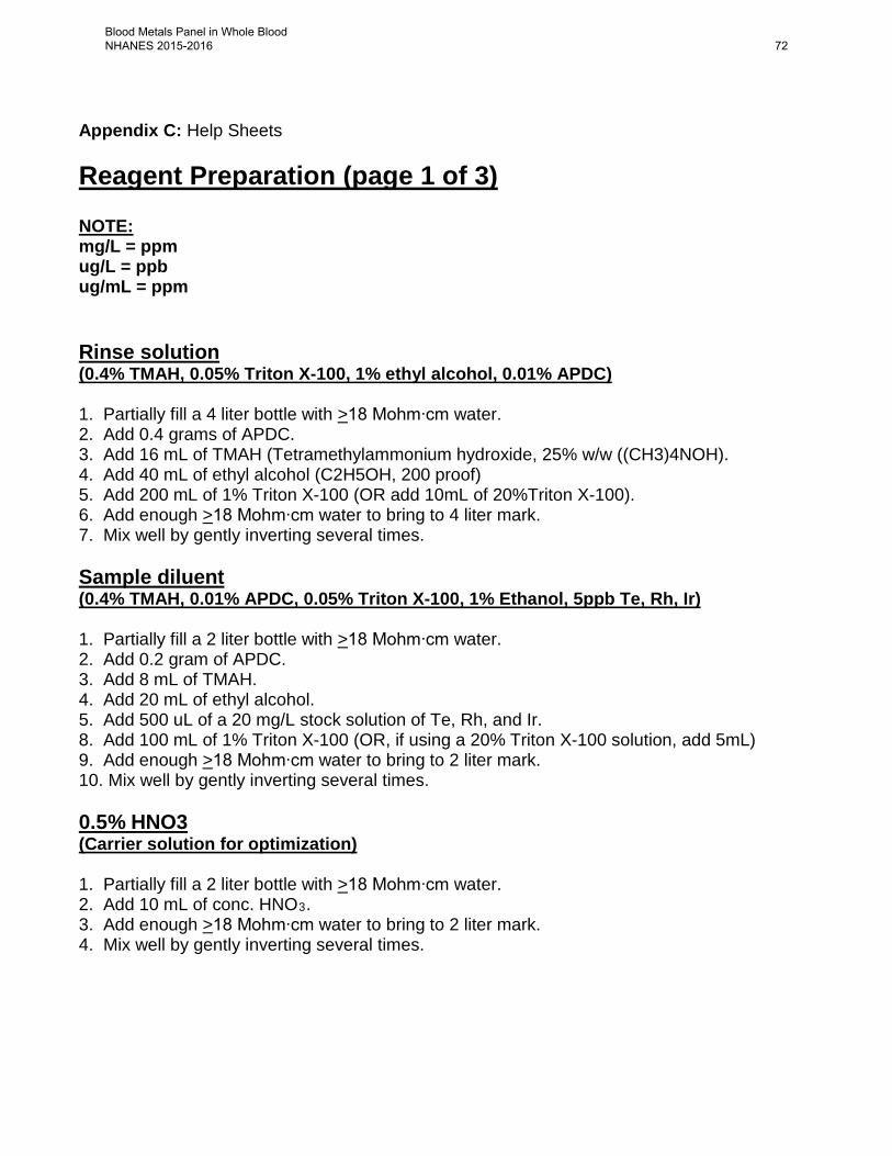
Appendix C: Help Sheets Reagent Preparation (page 1 of 3) NOTE: mg/L = ppm ug/L = ppb ug/mL = ppm Rinse solution (0.4% TMAH, 0.05% Triton X-100, 1% ethyl alcohol, 0.01% APDC) 1. Partially fill a 4 liter bottle with >18 Mohm∙cm water. 2. Add 0.4 grams of APDC. 3. Add 16 mL of TMAH (Tetramethylammonium hydroxide, 25% w/w ((CH3)4NOH). 4. Add 40 mL of ethyl alcohol (C2H5OH, 200 proof) 5. Add 200 mL of 1% Triton X-100 (OR add 10mL of 20%Triton X-100). 6. Add enough >18 Mohm∙cm water to bring to 4 liter mark. 7. Mix well by gently inverting several times. Sample diluent (0.4% TMAH, 0.01% APDC, 0.05% Triton X-100, 1% Ethanol, 5ppb Te, Rh, Ir) 1. Partially fill a 2 liter bottle with >18 Mohm∙cm water. 2. Add 0.2 gram of APDC. 3. Add 8 mL of TMAH. 4. Add 20 mL of ethyl alcohol. 5. Add 500 uL of a 20 mg/L stock solution of Te, Rh, and Ir. 8. Add 100 mL of 1% Triton X-100 (OR, if using a 20% Triton X-100 solution, add 5mL) 9. Add enough >18 Mohm∙cm water to bring to 2 liter mark. 10. Mix well by gently inverting several times. 0.5% HNO3 (Carrier solution for optimization) 1. Partially fill a 2 liter bottle with >18 Mohm∙cm water. 2. Add 10 mL of conc. HNO3. 3. Add enough >18 Mohm∙cm water to bring to 2 liter mark. 4. Mix well by gently inverting several times.
Blood Metals Panel in Whole Blood NHANES 2015-2016 72

Appendix C: Help Sheets (continued) Reagent Preparation (page 2 of 3) 1% v/v HNO3 1. Partially fill a 10 liter bottle with >18 Mohm∙cm water. 2. Add 100 mL of conc. HNO3. 3. Add enough >18 Mohm∙cm water to bring to 10 liter mark. 4. Mix well by gently swirling several times. 5% v/v HNO3 1. Partially fill a 2 liter bottle with >18 Mohm∙cm water. 2. Add 100 mL of conc. HNO3. 3. Add enough >18 Mohm∙cm water to bring to 2 liter mark. 4. Mix well by gently inverting several times. 20% Triton X-100 1. Partially fill a 1 liter bottle with >18 Mohm∙cm water. 2. Add 200 mL of Triton X-100. 3. Add enough >18 Mohm∙cm water to bring to 1 liter mark. 4. Allow to dissolve overnight (or add a Teflon magnetic stirring bar and stir on stirrer until dissolved). 5. Mix well by gently inverting several times. 1% Triton X-100 1. Partially fill a 1 liter bottle with >18 Mohm∙cm water. 2. Add 10 mL of Triton X-100. 3. Add enough >18 Mohm∙cm water to bring to 1 liter mark. 4. Allow to dissolve overnight (or add a Teflon magnetic stirring bar and stir on stirrer until dissolved). 5. Mix well by gently inverting several times. 20 ppm Rh, Te and Ir internal standard solution 1. Partially fill an acid rinsed, 50 mL flask with 1% v/v HNO3. 2. Add 1mL of Rh from 1000ppm stock standard. 3. Add 1mL of Te from 1000ppm stock standard. 4. Add 1mL of Ir from 1000ppm stock standard. 5. Add enough 1% v/v HNO3 to fill to 50mL mark. 6. Mix well by gently inverting several times. 7. Pour the standard solution over into an appropriately labeled 50mL polypropylene tube.
Blood Metals Panel in Whole Blood NHANES 2015-2016 73

Appendix C: Help Sheets (continued) Reagent Preparation (page 3 of 3) Daily solution (1ppb) in 2% v/v HNO3 1. Partially fill a 1 liter volumetric flask with >18 Mohm∙cm water. 2. Add 1mL of High Purity Standard: SM-2107-018 (or current lot #) 3. Add 20mL of concentrated HNO3 4. Add enough >18 Mohm∙cm water to bring to 1 liter mark. 5. Mix well by gently inverting several times. Stability test solution (1 liter bulk prep) 1. Use a 1 liter bottle dedicated to stability test solution preparation 2. Add 960 mL of Sample Diluent 3. Add 20 mL of “junk” whole blood 4. Add 20 mL of Intermediate Working Calibration Standard (may use S1 or S2) OR add 1.5mL of Intermediate Stock Calibration Standard. 5. Mix well by gently inverting several times. 6. Store in the refrigerator (when not using).
Blood Metals Panel in Whole Blood NHANES 2015-2016 74

Appendix C: Help Sheets (continued) Standard Preparation (page 1 of 1) (from single element stock standards) Prepare 3% HCl v/v solution:
1. Partially fill a clean 2 liter bottle with >18 Mohm∙cm water. 2. Using a clean 50 mL polypropylene tube to measure, add 60 mL of high purity
concentrated HCl. 3. Add enough >18 Mohm∙cm water to bring to 2 liter mark. 4. Gently invert to mix.
Prepare intermediate stock standard (see Table 4 in Appendix B):
1. Partially fill a 100 mL volumetric flask with 3% v/v HCl solution. 2. Label as: “HgPbCdMnSe Intermediate Stock Std” 3. Add 2 mL of HgPbCdMnSe multi-element stock solution. 4. Add enough 3% v/v HCl to bring to 100 mL mark. 5. Mix well by gently inverting several times.
Prepare intermediate working standards (see Table 5 in Appendix B):
1. Partially fill each of eight, 100 mL volumetric flasks with 3% v/v HCl solution. 2. Label as: Intermediate Working Std “S1”, “S2”, “S3” and “S4”, “S5”, “S6”, “S7” and “S8”. 3. For “S1 Intermediate Working Std”: add 50 uL of the Intermediate Stock Std. 4. For “S2 Intermediate Working Std”: add 150 uL of the Intermediate Stock Std. 5. For “S3 Intermediate Working Std”: add 350 uL of the Intermediate Stock Std. 6. For “S4 Intermediate Working Std”: add 500 uL of the Intermediate Stock Std. 7. For “S5 Intermediate Working Std”: add 1mL of the Intermediate Stock Std. 8. For “S6 Intermediate Working Std”: add 50 uL of the Multi-Element Stock Std. 9. For “S7 Intermediate Working Std”: add 150 uL of the Multi-Element Stock Std. 10. For “S8 Intermediate Working Std”: add 400 uL of the Multi-Element Stock Std. 11. Add enough 3% v/v HCl solution to bring to 100 mL mark. 12. Mix well by gently inverting several times. 13. These intermediate working standards may be poured over into clean 15 mL Falcon
tubes for daily use (NOTE: “S0 Intermediate Working Std” is 3% HCl only).
Blood Metals Panel in Whole Blood NHANES 2015-2016 75