lack of the c-terminal domain of nebulin in a...
TRANSCRIPT
CASE OF THE MONTH ABSTRACT: The most common autosomal recessive form of nemalinemyopathy is due to mutations in the nebulin gene. Among eight patientsstudied, we identified one, a 14-year-old girl, with a specific pattern of diffuserods in muscle fibers. Western blot analysis detected absence of the C-terminal domain of nebulin. Protein analysis may represent a good screen-ing method to direct molecular studies in the case of very large and complexgenes such as the large 1298 kb nebulin gene.
© 2002 Wiley Periodicals, Inc. Muscle Nerve 25: 747–752, 2002
LACK OF THE C-TERMINAL DOMAIN OFNEBULIN IN A PATIENT WITHNEMALINE MYOPATHY
J. GURGEL-GIANNETTI, PhD, 1,2 M.-L. BANG, PhD, 3 U. REED, PhD,2
S. MARIE, PhD,2 M. ZATZ, PhD,1 S. LABEIT, PhD, 3 and M. VAINZOF, PhD 1
1 Center for the Study of the Human Genome, Biosciences Institute, University ofSao Paulo, R. do Matao 106, Sao Paulo SP–CEP 05508-900, Brazil2 Department of Neurology, School of Medicine, University of Sao Paulo,Sao Paulo, Brazil3 European Molecular Biology Laboratory, Heidelberg, Germany
Accepted 18 December 2001
Nemaline myopathy is a structural congenital my-opathy associated with the presence of rod-like struc-tures (nemaline bodies) within the muscle fibers andwith an estimated incidence of 2 in every 100,000 livebirths.12 Clinically, it is characterized by the pres-ence of hypotonia as well as proximal and facialweakness and skeletal deformities. Depending onthe degree of muscle weakness and age at onset, andbased on correlations from the international data-base on nemaline myopathy, five forms were clini-cally defined by the Nemaline Consortium of theEuropean Neuromuscular Center.18
Nemaline myopathy may show both autosomaldominant and autosomal recessive inheritance pat-terns, and mutation in at least five genes has beenidentified in affected patients: the gene for slowa-tropomyosin 3 (TPM3) at 1q22–23,9,16 the nebulingene (NEB) at 2q21.1-q22,14 the actin gene (ACTA1)at 1q42,13 the b-tropomyosin (TPM2) gene at 9p13,2
and the slow skeletal troponin gene (TNNT1).7 Thetypical autosomal recessive form appears to be themost common and is caused by mutations in thenebulin gene.14
Nebulin is a large protein (700–900 kDa) foundin skeletal muscles, making up 3–4% of the totalmyofibrillar proteins.19 The C-terminal region ofnebulin is located at the Z lines, whereas its N-terminal end is at the pointed end of the thin fila-ment.4,8,11,19 It has been suggested that nebulin actsas a molecular ruler for the regulation of thin fila-ment lengths and is important for the assembly andintegration of Z-discs with the sarcomere.8,14 Thecorrelation between mutations in the nebulin geneand the expression of the protein in muscle is stillunder investigation.
We have characterized the nebulin protein fromeight patients with nemaline myopathy both by im-munofluorescence microscopy and Western blots.Using antibodies against four different domains ofnebulin, we identified one patient who showed a spe-cific loss of the C-terminal SH3 domain of nebulin,whereas three other more N-terminally located epi-topes from the N2, M176, and serine-rich domains ofnebulin were present. This patient showed the phe-notype of the typical form, and a pattern of smalland diffusely distributed rods.
PATIENTS AND METHODS
We studied eight patients (from unrelated families)with a diagnosis of nemaline myopathy. The diagno-sis was based on clinical examination, course of the
Key words: congenital myopathies; nebulin; nemaline myopathy; West-ern blotCorrespondence to: M. Vainzof; e-mail: [email protected]
© 2002 Wiley Periodicals, Inc.Published online 11 March 2002 in Wiley InterScience (www.interscience.wiley.com). DOI 10.1002/mus.10097
Nemaline Myopathy MUSCLE & NERVE May 2002 747

disease (using the protocol of the International Ne-maline Myopathy Consortium,18 family history, se-rum creatine kinase levels, electromyography, andmuscle biopsy.
Muscle samples were obtained from biceps ordeltoid biopsies, frozen in liquid nitrogen immedi-ately after removal, and stored at −70°C until use.Routine histological and histochemical procedureswere performed.3 The analysis of the proportion oftype I/II fibers, number of fibers containing rods,and the type of rods was carried out as describedpreviously.5
Immunohistochemical staining of frozen sectionswas done through single and/or double-labeling re-actions,17 using a rabbit polyclonal antibody for a-ac-tinin 2 (kindly provided by Dr. A. Beggs) diluted1:100, as a marker for rod structure. Four differentantibodies for nebulin were used (Table 1): a mousemonoclonal antibody directed to an I-band epitopenear the N2 line region, diluted 1:200 (Sigma, St.Louis, MO), and three rabbit polyclonal antibodiesraised to the expressed SH3 domain, the serine-richdomain, and to the M176–M181 domains.11–14 Thepolyclonal antibodies were diluted 1:10. As secondantibodies, anti-rabbit and anti-mouse IgG antibod-ies, both FITC and CY3 conjugated, were used. Thesections were analyzed under a Zeiss Axiophot orconfocal microscope with epifluorescence using fil-ters for fluorescein and rodamine.
Western blots were done as described previ-ously.6 The blots were incubated first with antibodiesspecific for nebulin and subsequently with an anti-body against dystrophin (Dy4/6D3, kindly providedby Dr. L.V.B. Anderson). The incubations with pri-mary antibodies were done overnight, and boundprimary antibodies were detected using alkalinephosphatase–conjugated secondary antibody.
The index patient (Case 2) is a 14-year-old girlwith a typical clinical course. Clinical manifestations
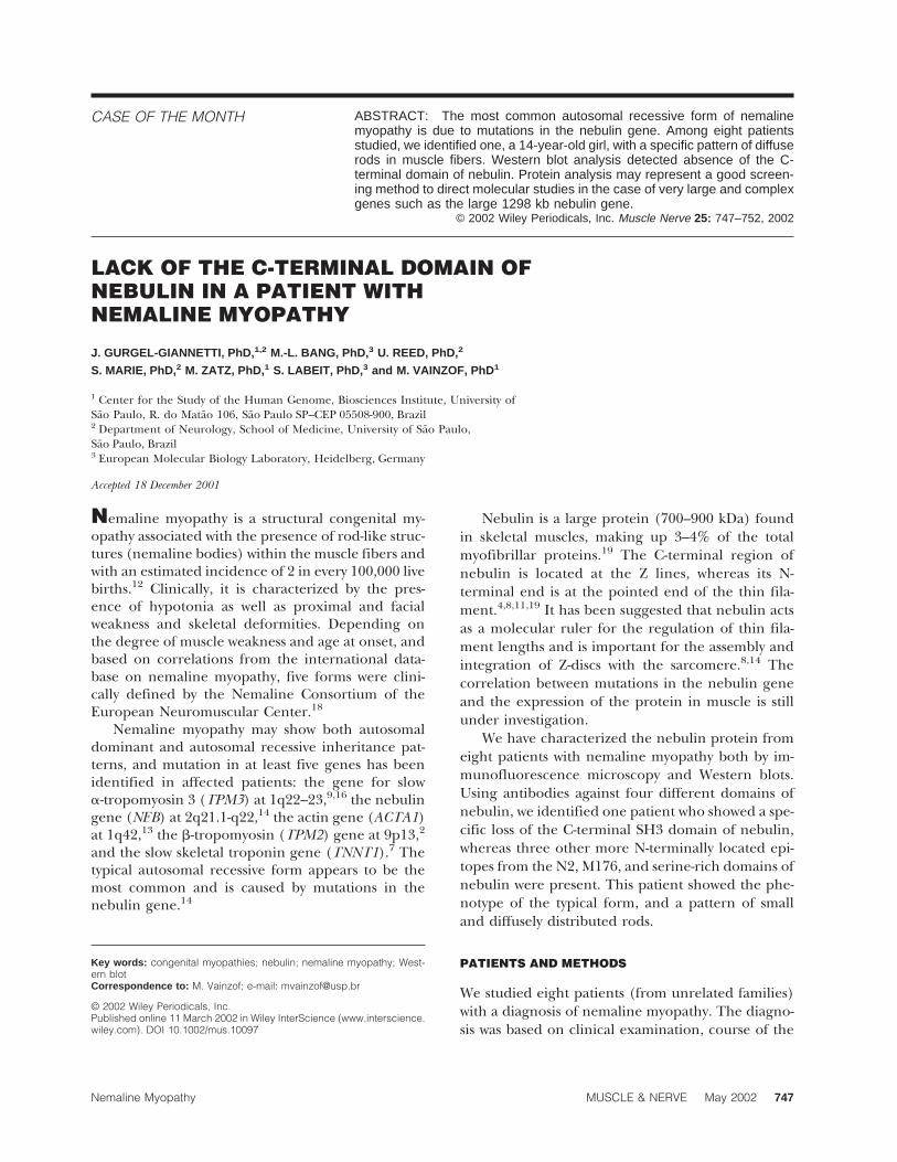
started during the neonatal period with severe hypo-tonia and proximal weakness. During the first yearsof life, she had repeated respiratory infections. Hermotor milestones were delayed. Thus, she sat at 3years of age, stood up at 3.5 years, and walked at 4years of age. Physical examination revealed facialand proximal muscle weakness, reduced tendon re-flexes, severe hypotonia, and moderate muscle atro-phy. She had a high-arched palate and pectum es-cavatum, but there was no tendinous retraction.Serum creatine level was mildly elevated. The elec-tromyographic study revealed a myopathic patterncharacterized by short-duration, small-amplitudepolyphasic motor unit potentials; motor and sensi-tive conduction velocity were normal. In the last 2months, she has experienced severe respiratory in-sufficiency requiring mechanical ventilation. Themuscle biopsy was performed at 6 years of age andshowed a typical myopathic pattern, fiber size varia-tion, and a slight degree of connective tissue infiltra-tion. ATPase reaction showed total type I fiber pre-dominance, and Gomori staining revealed thepresence of diffuse rods in 83% of the fibers (Fig. 1).
RESULTS
Immunohistochemical analysis in six of the eight pa-tients was reported previously.5 Briefly, muscle fromall patients showed the presence of nebulin in myo-fibrils. Some differences relating to the rod structurewere observed. The majority of the largest subsarco-lemmal rods were not labeled by the N2 nebulinantibody (I band epitope), and an indistinct patternof labeling with the two antibodies directed to theZ-band portion of nebulin (epitopes M176–M181and serine-rich domain) was observed. Diffuse rodswere not revealed using the three antibodies.5
Western blot analysis showed a band of the ex-pected molecular weight in eight patients with the
Table 1. Data on nebulin using four different antibodies and obtained through immunofluorescence and Westernblot methodologies.
Patient Sex Age (at biopsy)
Immunofluorescence Western blot
N2 101–102 176–181 N2 SH3 Dystrophin
1 F 3 + + + + + +2 F 6 + + + + − +3 M 14 + + + + + +4 F 9 + + +/m + + +5 F 3 + + + + + +6 F 18 + + + + + +7 M 5 + + + + + +8 M 4 + +/* +/* + + +
*Rare negatively labeled fibers; m, mosaic of positive/negative fibers; −, negative labeling; +, positive labeling.
748 Nemaline Myopathy MUSCLE & NERVE May 2002

FIGURE 1. Patient 2. Characteristic face (A) and phenotype (B), and pattern of rods in the modified Gomori reaction (C) and usingantibody against a-actinin 2 (D). Original magnification in (C) and (D), ×400.
FIGURE 3. Schematic representation of the nebulin protein, and the positive pattern of immunofluorescence in Patient 2, using antibodiesagainst the N2, the M176–181, and the serine-rich domains of nebulin. The position of the epitopes in the protein is shown at the top.
Nemaline Myopathy MUSCLE & NERVE May 2002 749

N2 antibody (Fig. 2). Interestingly, in one patient, animmunoreactive band was detected with the N2 an-tibody which did not react with the C-terminal SH3antibody (Patient 2, Fig. 2). The same blot was incu-bated with the dystrophin antibody and a strongband was observed in this and other tested patients.The blot with the reaction for the SH3 antibody wasre-reacted with antibody for the N2 domain of nebu-lin, and the patient with the deficient SH3 patternshowed the presence of the N2 band, thus confirm-ing the first result. The intensity of the N2 band inthis patient was slightly reduced.
On immunofluorescence, the sarcomeric patternof labeling was observed with the antibodies raised tothe N2, M176, and serine-rich domain. Unfortu-nately, the SH3 C-terminal antibody failed to workon sections, so that we were not able to confirm thelack of this epitope with immunofluorescence meth-odology (Fig. 3).
DISCUSSION
Nemaline myopathy is a congenital muscle disease,which presents with high clinical heterogeneity. In1999, a new clinical classification was proposed.Based on age of onset and severity of weakness, fiveclinical forms were defined: severe congenital, inter-mediate, typical, juvenile, and adult.18 The typicalform is the most common and is frequently causedby mutations in the nebulin gene.14 In this report,we have described a patient with clinical character-istics of the typical form, and therefore a strong can-didate for mutations in the nebulin gene.
Nebulin is a skeletal muscle–specific giant sarco-meric protein, which accounts for 3–4% of the totalmyofibrillar proteins.19 The C-terminal region is partof the Z-disc structure, whereas its N-terminal end isan integral component of the thin filament. About97% of the nebulin sequence consists of 185 copiesof a 35-repeats module (called M1 to 185). The N-terminal region of nebulin consists of a 10 kDaacidic domain which interacts with tropomodulinand therefore is likely to be involved in thin filamentlength regulation.10 Similarly, nebulin’s C-terminalSH3 domain may have assembly regulatory func-tions, as suggested by sequence homology to otherSH3 domains.8,11,15 Antibodies against these N- andC-terminal domains have been developed, allowingthe immunohistochemical study of nebulin-specificregions.5,14
In 1999, the first mutations in the nebulin genewere described.14 With the exception of one patient,patients with mutations in the M165–181 segmentsof the nebulin gene presented the nebulin proteinin the fibers, as determined by studies with two dif-ferent antibodies, M176–181 and SH3 nebulin do-mains.14
Among the eight patients here studied, one(Case 2) revealed a lack of the epitope in the SH3domain of nebulin, through the Western blot study.A band with the expected size was present, usingantibody for the N2 domain, suggesting no total pro-tein deficiency. Immunohistochemical analysis con-firmed the presence of the epitopes at N2, repeatsM176–181, the serine-rich domain, suggesting that a
FIGURE 2. Double Western blot analysis for dystrophin (DYS) and nebulin (NEB), using the N-terminal N2 and SH3 C-terminal antibodiesin three of the patients with nemaline myopathy. Observe the lack of the nebulin band with the SH3 terminal antibody in Patient 2. C =control, MYOS = myosin band in the Ponceau S prestained blot.
750 Nemaline Myopathy MUSCLE & NERVE May 2002

probable mutation in the nebulin gene would bepresent after this region. Unfortunately, the anti-body for the SH3 domain failed to work appropri-ately on immunofluorescence, so we were unable toconfirm a lack of this region by this means.
To exclude a possible degradation artifact, weconfirmed the preservation of high molecularweight proteins in the muscle extract through theconcomitant use of antibodies for dystrophin, indi-cating that our failure to detect the nebulin SH3epitope in Patient 2 was not the result of nonspecificmuscle protein degradation during extraction or inthe myofibril. Based on our results, this patient is astrong candidate for mutations in the nebulin gene,specifically in the SH3 domain. DNA analysis is cur-rently being performed to investigate this possibility.
In a previous study, a patient with a lack of theSH3 domain was detected. However, this patientshowed a homozygous mutation in the ninth codonof exon 185. The mutation changes a glutamic acid(GAG) to a stop codon (TAG) and should result inloss of the last 134 amino acids from the beginningof the serine-rich domain in the C-terminal region ofnebulin.14 It remains to be seen whether future stud-ies with specific antibodies for this region will show aprotein with a smaller size.
Our patient presented a homogeneous immuno-histochemical reaction with the three other nebulinantibodies, showing that the possible mutation wasnot related to any abnormalities in these domains. Amutation 38 of the serine-rich domain can cause alack of expression only in the nebulin C-terminalregion. Consistent with a mutation very close to theC-terminal end of nebulin, the Western blot analysisshowed no abnormalities in the size of the protein.The nebulin band detected with the N2 antibody wasslightly fainter, however, which suggests increaseddegradation of the proteolysis-sensitive nebulin. Inboth cases, i.e., primary loss of the SH3 domain by amutation or secondary loss by proteolysis, an im-proved understanding of the physiological roles ofthe nebulin SH3 domain is likely to be relevant to anappreciation of the molecular basis of nemaline my-opathy.
The normal pattern of immunofluorescence re-action using the M176–181 antibody shows slow fi-bers more intensely labeled than fast fibers.14 Yet,our patient showed homogeneous M176–181 reac-tion in all fibers. This feature is explained by thepredominance of type I fibers in her muscle.
In nemaline myopathy, the organization of therods within fibers can show different patterns. In themajority of our patients, both large subsarcolem-mally localized rods and small diffuse rods were de-
tected in the fiber in a high proportion of fibers.5 Atotal predominance of diffuse rods was observed in83% of the fibers of our Case 2, in a pattern detectedonly in this patient. This suggests a possible correla-tion of rod distribution with a specific type or regionof mutations in the nebulin gene.
Our results show the importance of performingnebulin immunohistochemical studies associatedwith Western blot analysis, because they may becomplementary. Molecular studies of the nebulingene are very complex. The gene is quite large, withmutations distributed along the whole coding re-gion, which makes the DNA study difficult, time-consuming, and expensive. The initial characteriza-tion of nebulin at the protein level may be helpful todirect further screenings for mutations on the geno-mic level. Additional studies on this patient may pro-vide further insights into the role(s) of nebulin’sC-terminal SH3 domain, which appears to be in-volved in linking together alpha-actinin and nebulinfilaments.1
The antibody for ACTN2 was kindly donated by Dr. Alan Beggs,and the antibody for dystrophin by Dr. L.V.B. Anderson, to whomwe are grateful. We also thank Marta Canovas and Fabiana da Silvafor technical assistance, Valdir Caldeiras and the Department ofBiology of the Biosciences Institute, University of Sao Paulo, forassistance with the confocal microscopy, and the physicians whoreferred patients for study. M.-L.B. was supported by a Marie-Curie Fellowship from the European Union. This work was sup-ported by grants from the Fundacao de Amparo a Pesquisa doEstado de Sao Paulo, Conselho Nacional de Pesquisas, and theAssociacao Brasileira de Distrofia Muscular.
REFERENCES
1. Bang ML, Mudry RE, McElhinny AS, Trombitas K, Geach AJ,Yamasaki R, Sorimachi H, Granzier H, Gregorio CC, Labeit S.Myopalladin, a novel 145-kilodalton sarcomeric protein withmultiple roles in Z-disc and I-band protein assemblies. J CellBiol 2001;153:413–427.
2. Donner K, Ollikaine M, Pelin K, Gronholm M, Carpen O,Wallgren-Pettersson C, Ridanpaa. Mutations in the b-tropo-myosin gene in rare cases of autosomal dominant nemalinemyopathy. Neuromuscul Disord 2000;10:342–343.
3. Dubowitz V. Muscle biopsy. 2nd ed. London: Balliere Tindall;1985. 720 p.
4. Furst DO, Osborn M, Nave R, Weber K. The organization oftitin filaments in the half-sarcomere revealed by monoclonalantibodies in immunoelectron microscopy: a map of ten non-repetitive epitopes starting at the Z line extends close to theM line. J Cell Biol 1988;106:1563–1572.
5. Gurgel-Giannetti J, Reed UC, Bang ML, Pelin K, Donner K,Marie SKN, Carvalho M, Fireman MAT, Zanoteli E, OliveiraASB, Zatz M, Wallgren-Pettersson C, Labeit S, Vainzof M.Nebulin expression in nemaline myopathy. Neuromuscul Dis-ord 2001;11:154–162.
6. Ho-Kim MA, Bedard A, Vincent M, Rogers PA. Dystrophin: asensitive and reliable immunochemical assay in tissue and cellculture homogenates. Biochem Biophys Res Commun 1991;181:1164–1172.
7. Johnston JJ, Kelley RI, Crawford TO, Morton DH, Agarwala R,Koch T, Schaffer AA, Francomano CA, Biesecker LG. A novel
Nemaline Myopathy MUSCLE & NERVE May 2002 751

nemaline myopathy in the Amish caused by a mutation introponin T1. Am J Hum Genet 2000;67:814–821.
8. Labeit S, Lolmerer B. The complete primary structure of hu-man nebulin and its correlation to muscle structure. J MolBiol 1995;248:308–315.
9. Laing NG, Wilton SD, Akkari PA, Dorosz S, Boundy K, Knee-bone C, Blumbergs P, White S, Watkins H, Love DR, Haan E.A mutation in the alpha tropomyosin gene TPM3 associatedwith autosomal dominant nemaline myopathy NEM1. NatGenet 1995;9:75–79.
10. McElhinny AS, Kolmerer B, Fowler VM, Labeit S, GregorioCC. The N-terminal end of nebulin interacts with tropomodu-lin at the pointed ends of the thin filaments. J Biol Chem2001;276:583–592.
11. Millevoi S, Trombitas K, Lolmerer B, Kostin S, Schaper J,Pelin K, Granzier H, Labeit S. Characterization of nebuletteand nebulin and emerging concepts of their roles for Z-discs.J Mol Biol 1998;282:111–123.
12. North KN, Laing NG, Wallgren-Pettersson CJ, and ENMC In-ternational Consortion on Nemaline Myopathy. Nemalinemyopathy: currents concepts. Med Genet 1997;34:705–713.
13. Nowak KJ, Wattanasirichaigoon D, Goebel HH, Wilce M, Pe-lin K, Donner K, Jacob RL, Hubner C, Oexle K, Anderson JR,Verity CM, North KN, Iannaccone St, Muller CR, Nurnberg P,Muntoni F, Sewry C, Hughes I, Sutphen R, Lacson AG, Swo-boda KJ, Vigneron J, Wallgren-Pettersson C, Beggs AH, LaingNG. Mutations in the skeletal muscle alpha actin gene inpatients with actin myopathy and nemaline myopathy. NatGenet 1999;23:208–212.
14. Pelin K, Hilpela P, Donner K, Sewry C, Akkary PA, Wilton SD,Wattanasirichaigoon D, Bang ML, Centner T, Hanefeld F,Odent S, Fardeau M, Urtizberea JA, Muntoni F, Dubowitz V,Beggs AH, Laing NG, Labeit S, Chapelle A, Wallgren-Pettersson C. Mutations in the nebulin gene associated withautosomal recessive nemaline myopathy. Proc Natl Acad SciUSA 1999;96:2305–2310.
15. Pfuhl M, Winder SJ, Castiglione Morelli MA, Labeit S, PastoreA. Correlation between conformational and binding proper-ties of nebulin repeats. J Mol Biol 1996;257:367–384.
16. Tan P, Briner J, Boltshauser E, Davis MR, Wilton SD, North K,Wallgren-Pettersson C, Laing NG. Homozygosity for a non-sense mutation in the alpha-tropomyosin slow gene TPM3 ina patient with severe infantile nemaline myopathy. Neuro-muscul Disord 1999;9:573–579.
17. Vainzof M, Zubrzycka-Gaarn EE, Rapaport D, Passos-BuenoMR, Pavanello RCM, Pavanello I, Zatz M. Immunofluores-cence dystrophin study in Duchenne dystrophy through theconcomitant use of two antibodies directed against the car-boxy-terminal and the amino-terminal region of the protein.J Neurol Sci 1991;101:141–147.
18. Wallgren-Pettersson C, Laing NG. Report of the 83rd ENMCInternational Workshop: 4th Workshop on Nemaline Myop-athy, 22–24 September 2000, Naarden, the Netherlands. Neu-romuscul Disord 2001;11:589–595.
19. Wang K, Wright J. Architecture of the sarcomere matrix ofskeletal muscle: immunoelectron microscopic evidence thatsuggests a set of parallel inextensible nebulin filaments an-chored at Z line. J Cell Biol 1988;6:2199–2212.
752 Nemaline Myopathy MUSCLE & NERVE May 2002