lactogenic hormones stimulate expression of lipogenic
TRANSCRIPT

Author's personal copy
Lactogenic hormones stimulate expression of lipogenic genes butnot glucose transporters in bovine mammary gland
Y. Shaoa, E.H. Walla, T.B. McFaddenb, Y. Misraa, X. Qiana, R. Blauwiekela, D. Kerra,F.-Q. Zhaoa,*
a Department of Animal Science, University of Vermont, Burlington, VT 05405, USAb Department of Agricultural, Food and Nutritional Sciences, University of Alberta, Edmonton, Alberta T6G 2P5, Canada
Received 29 June 2012; received in revised form 14 August 2012; accepted 10 September 2012
Abstract
During the onset of lactation, there is a dramatic increase in the expression of glucose transporters (GLUT) and a group ofenzymes involved in milk fat synthesis in the bovine mammary gland. The objective of this study was to investigate whether thelactogenic hormones mediate both of these increases. Bovine mammary explants were cultured for 48, 72, or 96 h with thefollowing hormone treatments: no hormone (control), IGF-I, insulin (Ins), Ins � hydrocortisone � ovine prolactin (InsHPrl), orIns � hydrocortisone � prolactin � 17�-estradiol (InsHPrlE). The relative expression of �-casein, �-lactalbumin, sterolregulatory element binding factor 1 (SREBF1), fatty acid synthase (FASN), acetyl-CoA carboxylase � (ACACA), stearyol-CoAdesaturase (SCD), GLUT1, GLUT8, and GLUT12 were measured by real-time PCR. Exposure to the lactogenic hormonecombinations InsHPrl and InsHPrlE for 96 h stimulated expression of �-casein and �-lactalbumin mRNA by several hundred-foldand also increased the expression of SREBF1, FASN, ACACA, and SCD genes in mammary explants (P � 0.01). However, thosehormone combinations had no effect on GLUT1 or GLUT8 expression and inhibited GLUT12 expression by 50% after 72 h oftreatment (P � 0.05). In separate experiments, the expression of GLUTs in the mouse mammary epithelial cell line HC11 or inbovine primary mammary epithelial cells was not increased by lactogenic hormone treatments. Moreover, treatment of dairy cowswith bovine prolactin had no effect on GLUT expression in the mammary gland. In conclusion, lactogenic hormones clearlystimulate expression of milk protein and lipogenic genes, but they do not appear to mediate the marked up-regulation of GLUTexpression in the mammary gland during the onset of lactation.© 2013 Elsevier Inc. All rights reserved.
Keywords: Gene expression; Glucose transporters; Mammary gland; Lactogenic hormones
1. Introduction
Glucose is an important nutrient in general, but it isabsolutely essential for milk production in the mam-mary gland of lactating animals because it is an energy
source as well as a key substrate for synthesis of milkprotein and lipids, and, especially, of lactose. The mam-mary gland does not synthesize glucose; thus, it needsto take up glucose from blood. Glucose uptake bymammary epithelial cells may play a rate-limiting rolein milk production [1].
Glucose transport across the plasma membrane ofmammary epithelial cells is mainly mediated by facil-itative glucose transporters (GLUTs). The family ofGLUTs consists of 13 isoforms, which are designated
* Corresponding author. Department of Animal Science, Universityof Vermont, 211 Terrill Hall, 570 Main Street, Burlington, VT 05405,USA. Tel.: �1 802 656 0786; fax: �1 802 656 8196.
E-mail address: [email protected] (F.-Q. Zhao).
Available online at www.sciencedirect.com
Domestic Animal Endocrinology 44 (2013) 57–69www.domesticanimalendo.com
0739-7240/13/$ – see front matterhttp://dx.doi.org/10.1016/j.domaniend.2012.09.001

Author's personal copy
as GLUT1 to GLUT12 and H�/myo-inositol cotrans-porter [2]. These transporters are structurally conservedand have different tissue distributions, transport kinet-ics, and regulatory properties. The lactating mammarygland mainly expresses GLUT1, GLUT8, and GLUT12isoforms [3–5].
The demand for glucose by the mammary glandincreases dramatically at the onset of lactation. Theglucose transport activity increases 40-fold from thevirgin state to the midlactating state [6,7]. Conse-quently, there is a coordinated increase in the expres-sion of GLUTs in the mammary gland during the onsetof lactation. From late pregnancy to early lactation, themRNA abundance of GLUT1, GLUT8, and GLUT12in bovine mammary gland increases markedly, by sev-eral-fold to several hundred-fold [1].
Expression of genes that code for enzymes and pro-teins required for de novo lipogenesis is also strikinglyupregulated in the mammary gland during lactogenesis.For example, expression of sterol regulatory elementbinding factor 1 (SREBF1), the master regulator oflipid synthesis [8], increases approximately 2-fold inboth mouse and cow mammary gland from late preg-nancy to early lactation [9,10]. Expression of its knowntargets, fatty acid synthase (FASN) and acetyl-CoAcarboxylase � (ACACA), also increases during thesame period [9,10]. In addition, stearoyl-CoA desatu-rase (SCD) is upregulated more than 40-fold from latepregnancy to lactation [9].
Mammary development and milk synthesis are reg-ulated both by systemic hormones and by local factors.It has been established that prolactin (Prl) and gluco-corticoids (GCs) are the main mediators of secretorycell differentiation and lactogenesis [11,12]. In cows,concentrations of Prl, GCs, and GH in blood increaseduring late pregnancy and peak near parturition [11].Concentrations of estrogens (Es) also increase gradu-ally during late pregnancy, then surge several daysbefore parturition. It has been shown that Prl-inducedsecretion of �-lactalbumin is markedly enhanced byadding 17�-estradiol to mammary explants [11]. Lo-cally produced IGF-I is believed to mediate the effectsof GH on the mammary gland, and the numbers ofIGF-I receptors on mammary epithelial cells increasemarkedly during late gestation [11]. In addition, dis-ruption of Prl signaling in the mammary gland de-creased GLUT1 expression [13,14]. On the basis ofthese facts, we hypothesized that some combination ofPrl, GCs, E, and/or IGF-I is responsible for the upregu-lation of GLUTs and lipogenic genes in the bovinemammary gland during onset of lactation. Our objec-
tive was to investigate the effects of those hormones onthe expression of GLUTs and lipogenic genes in themammary gland.
2. Materials and methods
2.1. Mammary tissue biopsy and explant culture
All animal use was approved by the University ofVermont Institutional Animal Care and Use Commit-tee. Mammary tissues were obtained via biopsy fromthe rear quarters of 2 Holstein heifers and 2 multiparousHolstein dry cows approximately 37 d prepartum (37 �5 d). The biopsy procedures were performed as de-scribed previously [15].
Immediately after biopsy, a small piece (5 mg) oftissue was fixed in 4% paraformaldehyde for 4 h at 4°C.After fixation, the tissue was rinsed with 3 changes ofcold PBS (2.67 mM potassium chloride, 1.47 mM po-tassium phosphate monobasic, 137.93 mM sodiumchloride, and 8.06 mM sodium phosphate dibasic), im-mersed in 0.5 M sucrose in PBS overnight at 4°C,preserved in optimal cutting temperature compound(Tissue-Tek; Sakura Finetek, Torrance, CA, USA), andthen frozen in liquid nitrogen chilled isopentane. Fro-zen tissues were stored at �80°C until use. The remain-ing tissue (�1 g) was cut into small pieces (�50 mg perpiece). Two pieces of fresh tissue were immediatelyfrozen in liquid nitrogen for RNA isolation. The re-maining tissue was placed in 50-mL tubes containingbasic medium [Medium 199 with Earle’s salts andL-glutamine (Sigma, St. Louis, MO, USA), containing26.2 mM sodium bicarbonate, 10 mM sodium acetate,15 mM HEPES, 100 U/mL penicillin, 100 �g/mLstreptomycin, and 0.25 �g/mL amphotericin B; roomtemperature] and transported to the laboratory within30 min. In a sterile, laminar flow hood, tissue waswashed 3 times with PBS containing 100 U/mL peni-cillin, 100 �g/mL streptomycin, and 0.25 �g/mL am-photericin B and then diced into explants (�1 mm3).Explants were washed 3 times with basic mediumwhich was removed by use of a strainer after eachwash. Explants were placed on siliconized lens paper(Whatman, Piscataway, NJ, USA) floating on culturemedia supplemented with various hormone treatments.Lens paper was siliconized as described previously[16]. There were 5 hormone treatments: NH (basicmedium without hormone), IGF-I (basic medium �200 ng/mL IGF-I), Ins (basic medium � 5 �g/mLinsulin), InsHPrl (basic medium � 5 �g/mL Ins � 5�g/mL ovine Prl � 1 �g/mL hydrocortisone), andInsHPrlE (basic medium � 5 �g/mL Ins � 5 �g/mL
58 Y. Shao et al. / Domestic Animal Endocrinology 44 (2013) 57–69

Author's personal copy
ovine Prl � 1 �g/mL hydrocortisone � 500 ng/mL17�-estradiol). Insulin-like growth factor I was pur-chased from PeproTech (Rocky Hill, NJ, USA), and allother hormones were products of Sigma. The IGF-Igroup was treated for 48 or 72 h. Other groups weretreated for 48, 72, or 96 h. The doses of individualhormones used were based on previous studies [17–20].There were 4 replicate cultures for each treatment, ateach time point. After hormone treatment, a small piece(�5 mg) of explants was fixed and preserved for im-munostaining as described above. The remaining ex-plants were stored in liquid nitrogen for RNA isolation.The experiment was repeated with mammary tissuesfrom 4 individual animals.
2.2. Culture of HC11 cells and primary bovinemammary epithelial cells
HC11 (mouse mammary epithelial cell line) cellswere grown in complete growth medium [RPMI 1640(Invitrogen, Carlsbad, CA, USA) supplemented with10% fetal bovine serum, 50 �g/mL gentamicin, 5�g/mL Ins, and 10 ng/mL epidermal growth factor] andkept confluent for 3 d. Then, the cells were primed inRPMI 1640 medium supplemented with 10% charcoal-stripped horse serum (Cocalico Biologicals, Ream-stown, PA, USA) and 5 �g/mL Ins for 2 d. For lacto-genic hormone induction, ovine Prl (5 �g/mL) anddexamethasone (0.1 �M) were added to priming me-dium, and incubations were continued for an additional48 h. Each experiment was repeated 3 times.
Bovine primary mammary epithelial cells (BMECs)were obtained as described [21]. Collagen was preparedfrom rat tail tendons, and cell culture dishes werecoated with collagen gel according to the proceduresdescribed by Imagawa et al [22]. BMECs were seededon collagen gels, grown in Dulbecco modified Eaglemedium (Invitrogen) supplemented with 10% fetal bo-vine serum and 5 �g/mL Ins and allowed to grow untilreaching confluence. The collagen gels were then re-leased by rimming each gel with a sterile scalpel blade.For lactogenic hormone induction, ovine Prl (5 �g/mL)and hydrocortisone (1 �g/mL) were added to culturemedium, and incubations continued for 48 h. Eachexperiment was repeated 3 times.
2.3. In vivo Prl administration
Five Holstein multiparous cows were injected intra-venously with bovine Prl (provided by John Byatt,Monsanto Co., St. Louis, MO, USA; 1 �g/kg of bodyweight) twice daily during the first 3 wk of lactation,whereas 5 additional cows served as controls [23].
After 7 d of treatment, mammary tissue was obtainedby biopsy from all 10 cows and was used for RNAisolation as described by Wall et al [23].
2.4. Quantitative real-time PCR
RNA was isolated from cells or mammary explantswith the use of Trizol reagent (Invitrogen). The con-centrations of isolated RNA were measured with aNanoDrop spectrophotometer (Thermo Scientific, Wil-mington, DE, USA). Reverse transcription (RT) wasperformed with 1 �g of total RNA and Moloney murineleukemia virus reverse transcriptase (USB, Cleveland,OH, USA) according to the manufacturer’s protocol.TaqMan Gene Expression Assays (Applied Biosys-tems, Carlsbad, CA, USA) were used to quantifymRNA for �-actin (Bt03279175), �-casein (Bt03217428),�-lactalbumin (Bt03213964), GLUT1 (Bt03215313),GLUT8 (Bt03217728), and GLUT12 (Bt03210346)with the use of a Bio-Rad (Hercules, CA, USA) CFX96Real-Time PCR Detection System. Reactions were per-formed in duplicate in a 10-�L volume containing 5 �Lof Universal PCR Master Mix (Applied Biosystems),0.5 �L of TaqMan Assay mixture containing primersand probe, and 4.5 �L of diluted cDNA (correspondingto 50 ng of reverse-transcribed total RNA). The relativeexpression of target genes was normalized to that of�-actin and calculated by the 2���C
T method [24].Expression of lipogenic gene mRNA was measured byRT-PCR with the use of SYBR Green. Reactions wereperformed in duplicate in a 20-�L volume containing10 �L of SsoFast EvaGreen Supermix (Bio-Rad), 1 �Lof forward primer, 1 �L of reverse primer and 8 �L ofdiluted cDNA (corresponding to 25 ng of reverse-tran-scribed total RNA). All primers were designed to spanthe junction of 2 exons to avoid amplification ofgenomic DNA during quantitative PCR (primer se-quences are shown in Table 1). The relative expressionof target genes was normalized to �-actin and calcu-lated by the 2���C
T method.
2.5. Immunohistochemistry
Fixed and embedded mammary tissues were cut into10-�m sections on a Cryostat HM 505 N (Microm,Walldorf, Germany) and mounted onto gelatin-coatedslides. Tissue sections were immersed in goat serum(10% in PBS) for 1 h at room temperature to blocknonspecific binding. After 3 PBS washes, tissue sec-tions were incubated with a GLUT1 antibody (Milli-pore, Billerica, MA, USA) diluted 1:500 in PBS con-taining 1% bovine serum albumin for 1 h at roomtemperature, followed by 3 PBS washes for 5 min each
59Y. Shao et al. / Domestic Animal Endocrinology 44 (2013) 57–69

Author's personal copy
time. Sections were then incubated with goat anti-rabbitsecondary antibody conjugated with Alexa Fluor 555(Invitrogen) diluted 1:500 in PBS at room temperaturein the dark. After 1-h incubation, sections were washed3 times for 5 min each in PBS, stained with 4,6-diamidino-2-phenylindole (Invitrogen) diluted 1:10,000 inPBS for 5 min, and then washed twice in PBS for15 min. Finally, slides were covered with a coverslip,and immunofluorescence was observed with a Zeiss(Thornwood, NY, USA) LSM 510 META Laser Scan-ning Microscope.
2.6. Statistical analysis
Statistical analyses were performed with JMP soft-ware (SAS Institute, Cary, NC, USA). The means ofdifferent hormone treatments were compared withANOVA and the Tukey-Kramer honestly significantdifference method was used for comparing individualmeans. For both studies (mammary explants and cellculture), hormone treatment was included in the modelas fixed effect. For the explant study, the cow effectwas used as block.
Before our in vivo experiment, we ran power calcu-lations with the use of gene expression data from ourprevious, similar experiments. On the basis of thosedata, and using a 2-tailed t-test and P � 0.05, wecalculated that we would have 80% to 90% power todetect a 1.5-fold difference in GLUT expression be-tween treatment groups with 3 to 4 per group.
3. Results
3.1. Lactogenic hormones stimulate �-casein and�-lactalbumin gene expression in bovine mammaryexplants
Mammary explants were treated with or without Ins,IGF-I, InsHPrl, or InsHPrlE for 48, 72, and 96 h.Expression of �-casein mRNA in cultured explants, aswell as in fresh tissue (FT), was measured by quanti-tative RT-PCR. Compared with FT, �-casein expres-
sion in explants exposed to the Ctrl, IGF-I, or Instreatment decreased sharply (P � 0.001), whereas theInsHPrl and InsHPrlE treatments increased the expres-sion of �-casein (P � 0.05) (Fig. 1A). In addition,�-casein expression did not differ between the InsHPrland InsHPrlE treatments. We also measured �-lactal-bumin expression in explants as an indicator of lactosesynthesis. As shown in Fig. 1B, expression of �-lact-albumin in the InsHPrl and InsHPrlE treatments was10-to several hundred-fold higher than that in the Ctrl,IGF-I, and Ins groups (P � 0.01). However, �-lactal-bumin expression in explants was lower than that in FT(P � 0.05).
3.2. Lactogenic hormones regulate lipogenic geneexpression in bovine mammary explants
In addition to milk protein and lactose synthesis,lactogenesis also involves induction of milk lipid syn-thesis, so we also investigated the regulation ofSREBF1, FASN, ACACA, and SCD gene expressionby lactogenic hormones in bovine mammary explants(Fig. 2). At 48 h, no difference was observed inSREBF1 mRNA expression among the Ctrl, Ins, andInsHPrl treatments. However, the InsHPrl treatmentincreased SREBF1 expression 2-fold (P � 0.001) at96 h compared with Ctrl, whereas Ins had no effect(Fig. 2A). The expression of FASN in explants treatedwith InsHPrl was about 4 times higher than that of Ctrlexplants throughout the experiment (P � 0.001). Re-gardless of treatment, expression of FASN in all cul-tured explants was lower than that in FT (P � 0.001;Fig. 2B). Incubation in InsHPrl increased the expres-sion of ACACA relative to that of the Ctrl groupthroughout the experiment (P � 0.001), whereas Insalone had no effect (Fig. 2C). However, Ins alonesupported an increase of approximately 3.5-fold inSCD expression, relative to Ctrl (P � 0.001 at 48 and72 h; P � 0.05 at 96 h; Fig. 2D). Incubation in theInsHPrl group further increased SCD expression by5.7-fold (48 h), 7.4-fold (72 h), and 7.9-fold (96 h)
Table 1Sequences of primers used in quantitative reverse transcription PCR for quantifying expression of lipogenic genes.
Gene Forward Reverse
ACTB GATCTGGCACCACACCTTCT CCAGAGGCATACAGGGACAGSREBF1 ACCGCTCTTCCATCAATGAC GCTGAAGGAAGCGGATGTAGFASN CTGAGTCGGAGAACCTGGAG CGAAGAAGGAAGCGTCAAACACACA TGGTCTGGCCTTACACATGA TGCTGGAGAGGCTACAGTGASCD ACAATTCCCGACGTGGCTT GGCATAACGGAATAAGGTGGC
Abbreviations: ACACA, acetyl-CoA carboxylase �; ACTB, �-actin; FASN, fatty acid synthase; SCD, stearoyl-CoA desaturase; SREBF1, sterolregulatory element binding factor 1
60 Y. Shao et al. / Domestic Animal Endocrinology 44 (2013) 57–69

Author's personal copy
compared with Ctrl, at the same times (P � 0.01).Expression of SCD also differed between the Ins andInsHPrl treatments (P � 0.01).
3.3. Effects of lactogenic hormones on GLUTexpression in bovine mammary explants
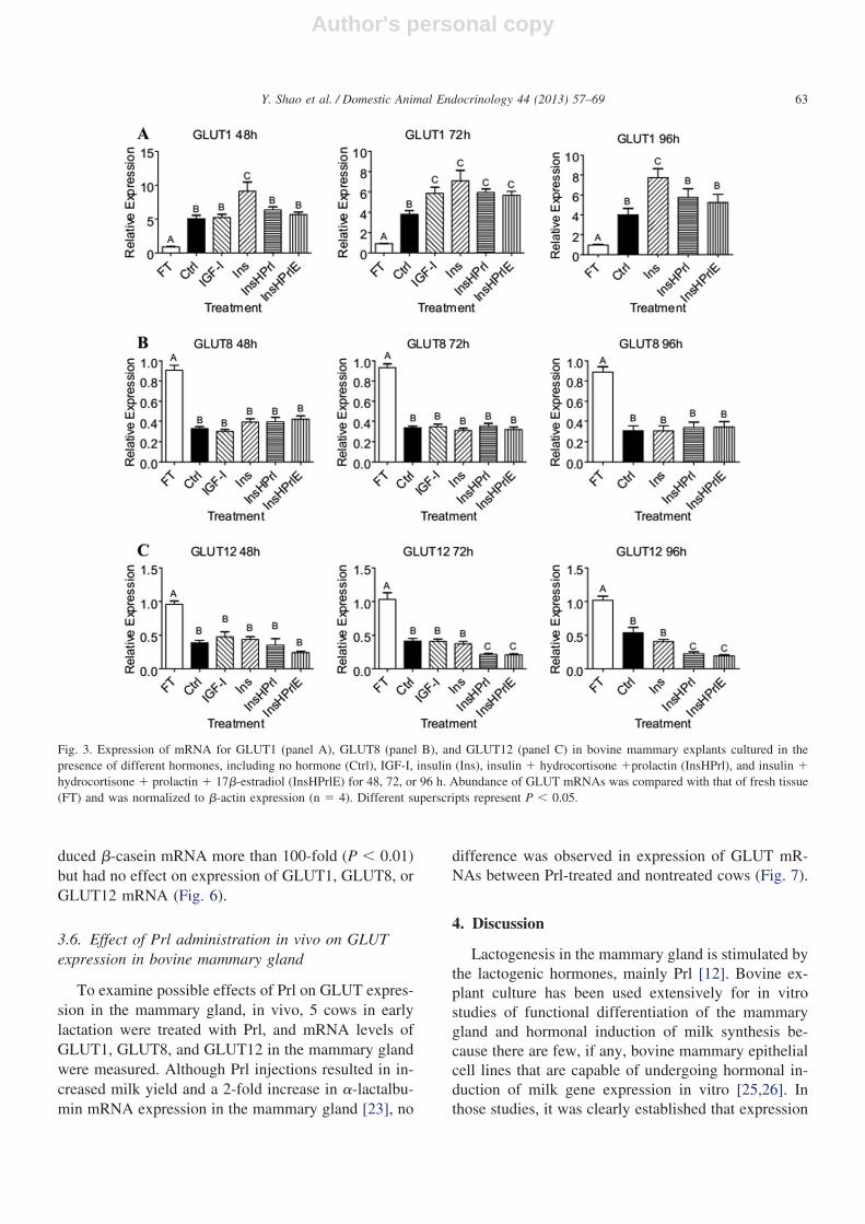
We next investigated the effect of lactogenic hor-mones on the expression of GLUT1, GLUT8, andGLUT12 in these explants (Fig. 3). At 48 and 96 h,GLUT1 mRNA expression was 50% to 80% higher inIns alone relative to Ctrl (P � 0.05), but other treat-ments had no effect on GLUT1 expression (Fig. 3A).At 72 h, although all hormone treatments increasedGLUT1 expression compared with Ctrl (P � 0.05), nodifference was observed among the hormone treat-ments. None of the hormone treatments affectedGLUT8 mRNA levels (Fig. 3B), nor did they affectGLUT12 expression at 48 h. But in explants incubatedin InsHPrl or InsHPrlE, GLUT12 expression was 50%lower at 72 h and 96 h (P � 0.05; Fig. 3C). Comparedwith FT, explants from all treatments had higherGLUT1 expression and lower GLUT8 and GLUT12expressions (P � 0.05). These results indicate thatlactogenic hormones and IGF-I apparently do not stim-
ulate expression of GLUT1, GLUT8, and GLUT12 inthe bovine mammary gland.
3.4. Effect of lactogenic hormones on GLUT1localization in bovine mammary explants
The subcellular localization of GLUT1 in the bovinemammary explants was examined by immunofluores-cence. As shown in Fig. 4, GLUT1 was mostly local-ized to the plasma membrane of epithelial cells in alltreatments, and no apparent difference was observed insubcellular localization of GLUT1. Explants did notexhibit fluorescence above background when normal(nonimmune) rabbit IgG replaced the first antibody norwhen either the first or second antibody was omitted(data not shown).
3.5. Effects of lactogenic hormones on GLUTexpression in bovine primary mammary cells andmouse mammary epithelial cell line HC11 cells
To verify the results obtained from explants, we alsocultured BMECs on collagen gel with various hormonetreatments (Fig. 5). Relative to Ins, expression of �-ca-
Fig. 1. Expression of mRNA for �-casein (panel A) and �-lactalbumin (panel B) in bovine mammary explants cultured in the presence of varioushormones, including no hormone (Ctrl), IGF-I, insulin (Ins), insulin � hydrocortisone � prolactin (InsHPrl), and insulin � hydrocortisone �prolactin � 17�-estradiol (InsHPrlE) for 48, 72, or 96 h. Abundance of �-casein and �-lactalbumin mRNA transcripts was compared with thatof fresh tissue (FT) and normalized to �-actin expression (n � 4). Different superscripts represent P � 0.05.
61Y. Shao et al. / Domestic Animal Endocrinology 44 (2013) 57–69

Author's personal copy
sein mRNA in these cells was increased 20-fold by theInsHPrl treatment for 48 h (P � 0.01); however, noeffect was observed of InsHPrl on expression ofGLUT1, GLUT8, or GLUT12 mRNA.
We further examined the effects of these hormoneson the HC11 mouse mammary epithelial cell line (Fig.6). Similar to the explants and the primary mammaryepithelial cells, the lactogenic complex, InsHPrl, in-
Fig. 2. Expression of mRNA for sterol regulatory element binding protein 1 (SREBF1; panel A), fatty acid synthase (FASN; panel B), acetyl-CoAcarboxylase � (ACACA; panel C), and stearoyl-CoA desaturase (SCD; panel D) in bovine mammary explants cultured in the presence of nohormone (Ctrl), insulin (Ins), or insulin � hydrocortisone � prolactin (InsHPrl) for 48, 72, or 96 h. The mRNA abundance of target genes wascompared with that of fresh tissue (FT and normalized to �-actin expression (n � 4). Different superscripts represent P � 0.05.
62 Y. Shao et al. / Domestic Animal Endocrinology 44 (2013) 57–69
Author's personal copy
duced �-casein mRNA more than 100-fold (P � 0.01)but had no effect on expression of GLUT1, GLUT8, orGLUT12 mRNA (Fig. 6).
3.6. Effect of Prl administration in vivo on GLUTexpression in bovine mammary gland
To examine possible effects of Prl on GLUT expres-sion in the mammary gland, in vivo, 5 cows in earlylactation were treated with Prl, and mRNA levels ofGLUT1, GLUT8, and GLUT12 in the mammary glandwere measured. Although Prl injections resulted in in-creased milk yield and a 2-fold increase in �-lactalbu-min mRNA expression in the mammary gland [23], no
difference was observed in expression of GLUT mR-NAs between Prl-treated and nontreated cows (Fig. 7).
4. Discussion
Lactogenesis in the mammary gland is stimulated bythe lactogenic hormones, mainly Prl [12]. Bovine ex-plant culture has been used extensively for in vitrostudies of functional differentiation of the mammarygland and hormonal induction of milk synthesis be-cause there are few, if any, bovine mammary epithelialcell lines that are capable of undergoing hormonal in-duction of milk gene expression in vitro [25,26]. Inthose studies, it was clearly established that expression
Fig. 3. Expression of mRNA for GLUT1 (panel A), GLUT8 (panel B), and GLUT12 (panel C) in bovine mammary explants cultured in thepresence of different hormones, including no hormone (Ctrl), IGF-I, insulin (Ins), insulin � hydrocortisone �prolactin (InsHPrl), and insulin �hydrocortisone � prolactin � 17�-estradiol (InsHPrlE) for 48, 72, or 96 h. Abundance of GLUT mRNAs was compared with that of fresh tissue(FT) and was normalized to �-actin expression (n � 4). Different superscripts represent P � 0.05.
63Y. Shao et al. / Domestic Animal Endocrinology 44 (2013) 57–69

Author's personal copy
of �-casein and �-lactalbumin mRNA can be inducedby the lactogenic complex of Ins, Prl, and GCs[25,26] and that Prl-induced secretion of �-lactalbu-min can be markedly enhanced by addition of Es[11]. These results were verified in our explant cul-
tures, except that 17�-estradiol did not enhance theinduction of milk protein gene expression stimulatedby the lactogenic complex in our study. Reasons forthis inconsistency are unknown, but Barrington et al[27] reported similar findings.
Fig. 4. Immunofluorescent staining (red) of GLUT1 subcellular localization in bovine mammary explants cultured in the presence of differenthormones, including no hormone (Ctrl), IGF-I, insulin (Ins), insulin � hydrocortisone � prolactin (InsHPrl), and insulin � hydrocortisone �prolactin � 17�-estradiol (InsHPrlE) for 48 h. Nuclei of epithelial cells were stained with 4,6-diamidino-2-phenylindole (blue).
64 Y. Shao et al. / Domestic Animal Endocrinology 44 (2013) 57–69

Author's personal copy
In addition to the milk protein genes, a set of genesencoding enzymes involved in milk lipid synthesispathways are also under tight regulation in the mam-mary gland. The mRNA levels of several key enzymesin fatty acid synthesis were increased at secretory ac-tivation in both mouse [28] and cow [9]. We hypothe-sized that upregulation of lipogenic genes during thistime is also mediated by lactogenic hormones. Indeed,SREBF1, FASN, ACACA, and SCD were upregulatedby lactogenic hormones in our explant cultures, sup-porting our hypothesis. In hepatocytes and adipocytes,Ins promotes lipid synthesis by upregulating SREBF1,FASN, and ACACA as one of the mechanisms formaintaining blood glucose homeostasis [29]. However,in our mammary explant cultures, Ins alone had noeffect on expression of these 3 genes. This may indicatetissue specificity in response to Ins. However, Ins alonestimulated SCD expression in our explant cultures, andthe addition of Prl and hydrocortisone further increased
its expression, indicating that SCD expression is regu-lated by mechanisms that differ from those controllingexpression of SREBF1, FASN, and ACACA. In addi-tion, among the lipogenic genes assayed in our ex-plants, SCD had the strongest response to lactogenichormones. This is consistent with the findings ofBionaz and Loor [9] who examined the expression of45 genes associated with lipid synthesis and secretionin the bovine mammary gland from late pregnancy tolactation and reported that SCD was one of the mostabundantly expressed genes during lactation and wasupregulated more than 40-fold from late pregnancy topeak lactation.
During the transition from late pregnancy to earlylactation the mammary gland undergoes dramatic func-tional differentiation, leading to the onset of lactation.This is accompanied by a markedly increased demandfor glucose by the mammary gland. This demand is metmainly by increased expression of GLUTs. The mRNA
Fig. 5. Expression of mRNA for �-casein and glucose transporters in bovine primary mammary epithelial cells. Cells were treated with insulin(Ins) or insulin � hydrocortisone � prolactin (InsHPrl) for 48 h. Gene expression of the InsHPrl group was compared with that of the Ins treatmentand was normalized to �-actin expression (n � 3). **P � 0.01.
65Y. Shao et al. / Domestic Animal Endocrinology 44 (2013) 57–69

Author's personal copy
levels of GLUT1, GLUT8, and GLUT12 increase bymore than 100-fold, 10-fold, and 10-fold, respectively,in bovine mammary gland from 40 d before lactation to7 d of lactation [1]. We hypothesized that this increasedGLUT expression was stimulated by lactogenic hor-
mones because expression of GLUT genes has similarpatterns to the expression of milk protein genes. Inaddition, in CIT3 mouse mammary epithelial cells,treatment with Prl, hydrocortisone, and Ins caused a15-fold induction of GLUT1 [30]. Three mouse models
Fig. 6. Expression of mRNA for �-casein and glucose transporters in cells of the HC11 mouse mammary epithelial cell line. Cells were treatedwith insulin (Ins) or insulin � hydrocortisone � prolactin (InsHPrl) for 48 h. Gene expression of the InsHPrl group was compared with that ofthe Ins treatment and was normalized to �-actin expression (n � 3). **P � 0.01.
Fig. 7. Expression of mRNA for glucose transporters in early lactating cows injected intravenously with bovine prolactin twice daily for 7 d. Geneexpression of prolactin-treated cows was compared with that of control cows and was normalized to �-actin expression (n � 5).
66 Y. Shao et al. / Domestic Animal Endocrinology 44 (2013) 57–69

Author's personal copy
with disrupted Prl signaling pathways exhibited a fail-ure of secretory activation and decreased expression ofGLUT1 [13]. In a similar vein, use of bromocriptine toinhibit Prl secretion in lactating rats caused a 37%decrease in GLUT1 expression. A more profound inhi-bition (up to 90% decrease) was observed in responseto treatment with bromocriptine plus a growth hor-mone-neutralizing antibody [14]. Contrary to these pre-vious findings and our hypothesis, our study showedthat lactogenic hormones did not stimulate the expres-sion of GLUT1, GLUT8, or GLUT12 in bovine mam-mary explants, BMECs, or in the mouse HC11 cell line.Furthermore, GLUT expression was not affected inearly lactating cows that were treated with Prl intrave-nously, even though expression of the milk proteingene, �-lactalbumin, was induced [23]. Thus, our datasuggest that the lactogenic complex does not mediatethe marked upregulation in expression of GLUTs in thebovine mammary gland during the onset of lactation.Reasons for the discrepancy between our results andthose of previous studies in rodents [13,14,30] are notclear. It may reflect species differences between cattleand rodents, but expression of GLUTs was also notaffected by the lactogenic hormones in the mouse mam-mary epithelial cell line HC11 in this study. It is pos-sible that the lactogenic hormones do not regulateGLUT1 expression directly. For example, Prl is essen-tial for mammary secretory activation and lactogenesis,and, if Prl secretion is disrupted, the mammary glandmay display a lower than normal metabolic rate, whichcould, in turn, reduce GLUT1 expression indirectly.Along these lines, one of the main challenges in study-ing regulation of lactogenesis and milk secretion hasbeen the universal inability to elicit lactose synthesis byin vitro mammary model systems, despite clear induc-tion of the A and B components of the lactose synthaseenzyme. Thus, it may be that the failure to inducelactose synthesis uncouples the expected demand forglucose and so increased GLUT expression is not re-cruited. Our data also present a novel possible expla-nation for the failure to induce lactose synthesis invitro; perhaps the inability to recruit heightened GLUTexpression deprives the biosynthetic machinery of thesole substrate required for lactose synthesis. In addi-tion, we cannot rule out the possibility that the lacto-genic hormones may induce GLUT1 expression in themammary gland specifically in lactogenesis stage 2because the main increase of GLUT expression in bo-vine mammary gland occurs around parturition [1], andthe explant tissues used in our study were taken frompregnant animals about 1 mo before parturition.
Transcriptional regulation of GLUT1 gene expres-sion has been studied in some detail because of its rolein cancer development and the response of cells tostress. Increased GLUT1 expression is elicited by avariety of factors and stressful stimuli, including hyp-oxia, glucose deprivation, hyperosmolarity, growth fac-tors, and transformation and inhibition of oxidativephosphorylation [31–36]. Of all of these factors, hyp-oxia is of most interest. It is well established thatGLUT1 expression is increased in many cell types inresponse to hypoxia [37]. Mammary consumption ofoxygen increases steadily during late pregnancy andpeaks during lactation [38]. This may well result in achronic local hypoxia. In addition, it has been shownthat mice in which the hypoxia-inducible factor 1� hasbeen deleted from the mammary gland exhibit impairedmammary differentiation and lipid secretion, strikingchanges in milk composition, and failure to lactate [39].Thus, hypoxia may also be an important regulator ofGLUT1 expression in the mammary gland.
Progesterone is another hormone that may regulateGLUT expression in the mammary gland during thetransition period. Concentrations of progesterone inblood remain high throughout pregnancy but drop dra-matically shortly before parturition [11]. High concen-trations of progesterone may inhibit expression ofGLUTs, and progesterone withdrawal prepartum maybe permissive for induction of GLUT expression in themammary gland. However, others have reported thatprogesterone upregulates GLUT1 expression inIshikawa endometrial cancer cells [40] and primarymurine and human endometrial stromal cells [41].Therefore, the role of progesterone in regulating GLUTexpression in the mammary gland needs to be investi-gated.
Little research has been done on the regulation ofGLUT8 and GLUT12 expression, and, to our knowl-edge, this is the first report on the regulation of GLUT8and GLUT12 expression by lactogenic hormones. EachGLUT isoform serves specific physiological functions,and each is regulated by distinct mechanisms. It is notsurprising that different factors may be responsible forupregulating expression of individual GLUTs in themammary gland. This is supported by our observationsthat, although Ins alone increased GLUT1 expression inbovine mammary explants, it had no effect on GLUT8or GLUT12 expression. Likewise, exposure to the lac-togenic complex of hormones reduced expression ofGLUT12, but not GLUT1 or GLUT8, in bovine mam-mary explants.
67Y. Shao et al. / Domestic Animal Endocrinology 44 (2013) 57–69

Author's personal copy
We conclude that IGF-I, 17�-estradiol, and lacto-genic hormones do not elicit the upregulation ofGLUTs in bovine mammary epithelial cells during thetransition period from late pregnancy to early lactation.Hypoxia, or other factors, may play a role, but needfurther investigation.
Acknowledgments
We thank Brian Trudell and Coral Kent-Dennis fortheir help with cow management and mammary glandbiopsies. This investigation was supported by NationalResearch Initiative Competitive grant 2007-35206-18037 from the USDA National Institute of Food andAgriculture (to F.-Q.Z. & T.B.M.).
The authors declare that there is no conflict of in-terest that could be perceived as prejudicing the impar-tiality of the research reported.
This article is part of Y.S.’s PhD thesis. Y.M., X.Q.,E.H.W., R.B., and T.B.M. helped with the animal ex-periment. Y.S., E.H.M, T.B.M., and F.-Q.Z. conceivedand designed the project and wrote the manuscript.D.K. provided primary mammary epithelial cells. Allauthors read and approved the final manuscript.
References
[1] Zhao F-Q, Keating AF. Expression and regulation of glucosetransporters in the bovine mammary gland. J Dairy Sci 2007;90(suppl 1):E76–E86.
[2] Zhao F-Q, Keating AF. Functional properties and genomics ofglucose transporters. Curr Genomics 2007;8:113–28.
[3] Miller PJ, Finucane KA, Hughes M, Zhao F-Q. Cloning andexpression of bovine glucose transporter GLUT12. Mamm Ge-nome 2005;16:873–83.
[4] Zhao F-Q, Glimm DR, Kennelly JJ. Distribution of mammalianfacilitative glucose transporter messenger RNA in bovine tis-sues. Int J Biochem 1993;25:1897–903.
[5] Zhao F-Q, Miller PJ, Wall EH, Zheng YC, Dong B, NevilleMC, et al. Bovine glucose transporter GLUT8: cloning, expres-sion, and developmental regulation in mammary gland.Biochim Biophys Acta 2004;1680:103–13.
[6] Prosser CG, Topper YJ. Changes in the rate of carrier-mediatedglucose transport by mouse mammary epithelial cells duringontogeny: hormone dependence delineated in vitro. Endocrinol-ogy 1986;119:91–6.
[7] Threadgold LC, Kuhn NJ. Monosaccharide transport in themammary gland of the intact lactating rat. Biochem J 1984;218:213–9.
[8] Eberlé D, Hegarty B, Bossard P, Ferré P, Foufelle F. SREBPtranscription factors: master regulators of lipid homeostasis.Biochimie 2004;86:839–48.
[9] Bionaz M, Loor JJ. Gene networks driving bovine milk fatsynthesis during the lactation cycle. BMC Genomics 2008;9:366.
[10] Rudolph MC, Monks J, Burns V, Phistry M, Marians R, FooteMR, et al. Sterol regulatory element binding protein and dietary
lipid regulation of fatty acid synthesis in the mammaryepithelium. Am J Physiol Endocrinol Metab 2010;299:E918–27.
[11] Akers RM. Lactation and the Mammary Gland Ames, IA: IowaState Press; 2002.
[12] Neville MC, McFadden TB, Forsyth I. Hormonal regulation ofmammary differentiation and milk secretion. J Mammary GlandBiol Neoplasia 2002;7:49–66.
[13] Naylor MJ, Oakes SR, Gardiner-Garden M, et al. Trans-criptional changes underlying the secretory activation phaseof mammary gland development. Mol Endocrinol 2005;19:1868–83.
[14] Fawcett HA, Baldwin SA, Flint DJ. Hormonal regulation of theglucose transporter GLUTI in the lactating rat mammary gland.Biochem Soc Trans 1992;20:17S.
[15] Wall EH, Auchtung TL, Dahl GE, Ellis SE, McFadden TB.Exposure to short day photoperiod during the dry period en-hances mammary growth in dairy cows. J Dairy Sci 2005;88:1994–2003.
[16] Ginsburg E, Vonderhaar BK. Whole organ culture of the mousemammary gland. In: Ip MM, Asch BB, editors. Methods inMammary Gland Biology and Breast Cancer Research. NewYork: Kluwer Academic/Plenum Press; 2000, p. 147–54.
[17] Cohick WS, Wang B, Verma P, Boisclair YR. Insulin-likegrowth factor I (IGF-I) and cyclic adenosine 3’,5’-monophos-phate regulate IGF-binding protein-3 gene expression by tran-scriptional and posttranscriptional mechanisms in mammaryepithelial cells. Endocrinology 2000;141:4583–91.
[18] Djiane J, Delouis C, Denamur R. Lactogenesis in organ culturesof heifer mammary tissue. J Endocrinol 1975;65:453–4.
[19] Collier RJ, Bauman DE, Hays RL. Lactogenesis in explantcultures of mammary tissue from pregnant cows. Endocrinology1977;100:1192–200.
[20] Nickerson SC, Heald CW, Bibb TL, McGilliard ML. Cytolog-ical effects of hormones and plasma on bovine mammary tissuein vitro. J Endocrinol 1978;79:363–8.
[21] Wellnitz O, Kerr DE. Cryopreserved bovine mammary cells tomodel epithelial response to infection. Vet Immunol Immuno-pathol 2004;101:191–202.
[22] Imagawa W, Yang J, Guzman RC, Nandi S. Collagen gelmethod for the primary culture of mouse mammary epithelium.In: Ip MM, Asch BB, editors. Methods in Mammary GlandBiology and Breast Cancer Research. New York: Kluwer Aca-demic/Plenum Press; 2000, p. 111–23.
[23] Wall EH, Crawford HM, Ellis SE, Dahl GE, McFadden TB.Mammary response to exogenous prolactin or frequent milkingduring early lactation in dairy cows. J Dairy Sci 2006;89:4640–8.
[24] Livak KJ, Schmittgen TD. Analysis of relative gene expressiondata using real-time quantitative PCR and the 2(-Delta deltaC(T)) method. Methods 2001;25:402–8.
[25] Gertler A, Weil A, Cohen N. Hormonal control of casein syn-thesis in organ culture of the bovine lactating mammary gland.J Dairy Res 1982;49:387–98.
[26] Goodman GT, Akers RM, Friderici KH, Tucker HA. Hormonalregulation of alpha-lactalbumin secretion from bovine mammarytissue cultured in vitro. Endocrinology 1983;112:1324–30.
[27] Barrington GM, Besser TE, Gay CC, Davis WC, Reeves JJ,McFadden TB. Effect of prolactin on in vitro expression of thebovine mammary immunoglobulin G1 receptor. J Dairy Sci1997;80:94–100.
68 Y. Shao et al. / Domestic Animal Endocrinology 44 (2013) 57–69

Author's personal copy
[28] Anderson SM, Rudolph MC, McManaman JL, Neville MC. Keystages in mammary gland development. Secretory activation inthe mammary gland: it’s not just about milk protein synthesis!Breast Cancer Res 2007;9:204.
[29] Saltiel AR, Kahn CR. Insulin signalling and the regulation ofglucose and lipid metabolism. Nature 2001;414:799–806.
[30] Haney PM. Localization of the GLUT1 glucose transporter tobrefeldin A-sensitive vesicles of differentiated CIT3 mousemammary epithelial cells. Cell Biol Int 2001;25:277–88.
[31] Behrooz A, Ismail-Beigi F. Dual control of GLUT1 glucosetransporter gene expression by hypoxia and by inhibition ofoxidative phosphorylation. J Biol Chem 1997;272:5555–62.
[32] Boado RJ, Pardridge WM. Glucose deprivation and hypoxiaincrease the expression of the GLUT1 glucose transporter via aspecific mRNA cis-acting regulatory element. J Neurochem2002;80:552–4.
[33] Chen C, Pore N, Behrooz A, Ismail-Beigi F, Maity A. Regulationof GLUT1 mRNA by hypoxia-inducible factor-1. Interaction be-tween h-RAS and hypoxia. J Biol Chem 2001;276:9519–25.
[34] Hwang DY, Ismail-Beigi F. Stimulation of GLUT-1 glucosetransporter expression in response to hyperosmolarity. Am JPhysiol Cell Physiol 2001;281:C1365–72.
[35] Murakami T, Nishiyama T, Shirotani T, Shinohara Y, Kan M,Ishii K, et al. Identification of two enhancer elements in the gene
encoding the type 1 glucose transporter from the mouse whichare responsive to serum, growth factor, and oncogenes. J BiolChem 1992;267:9300–6.
[36] Sánchez-Feutrie M, Viñals F, Palacín M, Zorzano A. Identifi-cation of a novel proliferation-dependent c-rich element thatmediates inhibition of the rat GLUT1 promoter. Gene 2003;322:47–55.
[37] Cassavaugh J, Lounsbury KM. Hypoxia-mediated biologicalcontrol. J Cell Biochem 2011;112:735–44.
[38] Reynolds M. Mammary respiration in lactating goats. Am JPhysiol 1967;212:707–10.
[39] Seagroves TN, Hadsell D, McManaman J, Palmer C, Liao D,McNulty W, et al. HIF1alpha is a critical regulator of secretorydifferentiation and activation, but not vascular expansion, in themouse mammary gland. Development 2003;130:1713–24.
[40] Medina RA, Meneses AM, Vera JC, Gúzman C, Nualart F,Rodriguez F, et al. Differential regulation of glucose transporterexpression by estrogen and progesterone in Ishikawa endome-trial cancer cells. J Endocrinol 2004;182:467–78.
[41] Frolova A, Flessner L, Chi M, Kim ST, Foyouzi-Yousefi N,Moley KH. Facilitative glucose transporter type 1 is differen-tially regulated by progesterone and estrogen in murine andhuman endometrial stromal cells. Endocrinology 2009;150:1512–20.
69Y. Shao et al. / Domestic Animal Endocrinology 44 (2013) 57–69