lafora disease: insights into neurodegeneration from plant
TRANSCRIPT

TIBS-707; No of Pages 12
Lafora disease: insights intoneurodegeneration from plantmetabolismMatthew S. Gentry1, Jack E. Dixon2,3 and Carolyn A. Worby2
1 Department of Molecular and Cellular Biochemistry and Center for Structural Biology, University of Kentucky, Lexington,
KY 40536-0509, USA2 Departments of Cellular and Molecular Medicine, Pharmacology, and Chemistry and Biochemistry, University of California,
San Diego, La Jolla, CA 92093, USA3 Howard Hughes Medical Institute, University of California, San Diego, La Jolla, CA 92093, USA
Review
Glossary
CBM: a carbohydrate-binding module. CBMs are defined by their tertiary fold,
which allows them to bind to one or many types of carbohydrates. They are
classified into one of 53 families on the basis of amino acid similarity, substrate
binding preferences, polypeptide folds, and evolutionary relationships.
CX5R: the catalytic active site motif of the protein tyrosine phosphatase (PTP)
superfamily.
DSP: the dual specificity phosphatases are a heterogeneous group of
phosphatases that are more evolutionarily diverse than the classical PTPs.
They dephosphorylate pTyr, pSer, pThr, and non-proteinaceous substrates
(e.g. phosphoinositols, RNA, and glucans).
Glucan: a polymer of glucose monomers linked by glycosidic bonds, e.g.
starch, glycogen, amylopectin, cellulose, Lafora body.
GWD: a-glucan, water dikinase. A plant kinase that transfers the b-phosphate of
ATP to the C6 position of glucose in starch.
Lafora body: insoluble glucan that closely resembles plant amylopectin and
accumulates in the cytoplasm of most cells in LD patients.
Protist: a diverse group of eukaryotic organisms with a unicellular level of
organization.
PTP: the protein tyrosine phosphatase superfamily, which is encoded by the
largest family of phosphatase genes. PTPs are defined by the active-site motif
CX5R, in which the cysteine functions as a nucleophile and is essential for
activity. They are divided into the classical PTPs that dephosphorylate pTyr, and
the DSPs that dephosphorylate pTyr, pSer, pThr, and non-proteinaceous
substrates.
PWD: phosphoglucan, water dikinase. A plant kinase that transfers the
Reversible phosphorylation modulates nearly every stepof glycogenesis and glycogenolysis. Multiple metabolicdisorders are the result of defective enzymes that controlthese phosphorylation events, enzymes that were ident-ified biochemically before the advent of the molecularbiology era. Lafora disease is a metabolic disorder result-ing in accumulation of water-insoluble glucan in thecytoplasm, and manifests as a debilitating neurodegen-eration that ends with the death of the patient. Unlikemost metabolic disorders, the link between Lafora dis-ease and metabolism has not been defined in almost 100years. The results of recent studies with mammaliancells, mouse models, eukaryotic algae, and plants havebegun to define the molecular mechanisms that causeLafora disease. The emerging theme identifies a newphosphorylation substrate in glycogen metabolism, theglucan itself.
Nearly a century of Lafora disease historyIn 1911, Dr. Gonzalo Lafora, a student of Dr. Alois Alzhei-mer, reported autopsy results from patients with ‘teenage-onset myoclonus epilepsy with dementia’ and described‘amyloid bodies in the protoplasm of the ganglion cells’[1,2]. Although amyloid was later shown to be protein-aceous, the term originally referred to any material thatstained in a manner similar to that of starch, which is amixture of amylose and amylopectin [3]. The deposit thatDr. Lafora described was later shown to be an accumu-lation of water-insoluble glucans, i.e. polymers of glucoselinked by glycosidic bonds, and named a Lafora body (LB)[4–6]. Like the ‘amyloid deposits’, the disease that Dr.Lafora described now bears his name, and is called Laforadisease (LD) (OMIM 254780).
LD is an autosomal recessive neurodegenerative dis-order resulting in severe epilepsy and death. It is one of fivemajor progressive myoclonus epilepsies (PMEs) [7–10].Unlike most other forms of epilepsy, LD is only moderatelymanaged by medication for a brief period of time. LDcommonly presents as a single seizure in the second decadeof the patient’s life; this single event is followed by pro-gressive central nervous system degeneration and ends
Corresponding author: Gentry, M.S. ([email protected]).
0968-0004/$ – see front matter � 2009 Elsevier Ltd. All rights reserved. doi:10.1016/j.tibs.2009.
with the death of the patient within ten years of the firstseizure [8,11–13].
LD is unique among the PMEs because of the patient’srapid neurological deterioration and the accumulation ofcytoplasmic LBs, which contain 80–93% polyglucans [1,5].LD is unique among neurodegenerative diseases in that itinvolves formation of an inclusion body that is largely non-proteinaceous. Whereas LBs are found in the cytoplasm ofcells from most tissues, clinical features of LD are confinedto the CNS and non-neurologic symptoms are rare [12]. LDpatients exhibit increased neuronal cell death, numerousseizures, and LB accumulation as they age; thus, it ishypothesized that LBs trigger these symptoms and ulti-mately the death of the patient [5].
Two groups independently identified epilepsy, progress-ive myoclonus 2A (EPM2A) as a gene mutated in approxi-mately 48% of LD cases [14,15]. EPM2A encodes thebimodular protein laforin that contains a canonical dual-specificity phosphatase (DSP) active site motif,HCXXGXXRS/T (Cx5R), and a carbohydrate-binding
b-phosphate of ATP to the C3 position of glucose in starch subsequent to
phosphorylation of the C6 position.
08.002 Available online xxxxxx 1
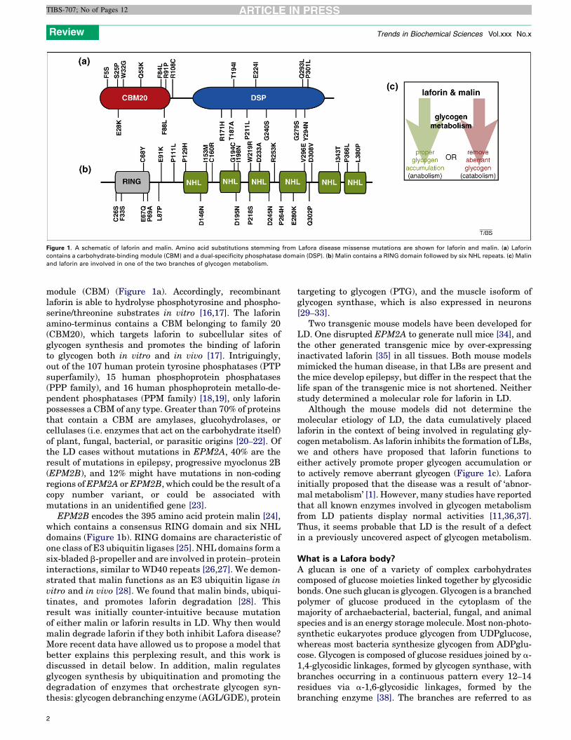
Figure 1. A schematic of laforin and malin. Amino acid substitutions stemming from Lafora disease missense mutations are shown for laforin and malin. (a) Laforin
contains a carbohydrate-binding module (CBM) and a dual-specificity phosphatase domain (DSP). (b) Malin contains a RING domain followed by six NHL repeats. (c) Malin
and laforin are involved in one of the two branches of glycogen metabolism.
Review Trends in Biochemical Sciences Vol.xxx No.x
TIBS-707; No of Pages 12
module (CBM) (Figure 1a). Accordingly, recombinantlaforin is able to hydrolyse phosphotyrosine and phospho-serine/threonine substrates in vitro [16,17]. The laforinamino-terminus contains a CBM belonging to family 20(CBM20), which targets laforin to subcellular sites ofglycogen synthesis and promotes the binding of laforinto glycogen both in vitro and in vivo [17]. Intriguingly,out of the 107 human protein tyrosine phosphatases (PTPsuperfamily), 15 human phosphoprotein phosphatases(PPP family), and 16 human phosphoprotein metallo-de-pendent phosphatases (PPM family) [18,19], only laforinpossesses a CBM of any type. Greater than 70% of proteinsthat contain a CBM are amylases, glucohydrolases, orcellulases (i.e. enzymes that act on the carbohydrate itself)of plant, fungal, bacterial, or parasitic origins [20–22]. Ofthe LD cases without mutations in EPM2A, 40% are theresult of mutations in epilepsy, progressive myoclonus 2B(EPM2B), and 12% might have mutations in non-codingregions ofEPM2A orEPM2B, which could be the result of acopy number variant, or could be associated withmutations in an unidentified gene [23].
EPM2B encodes the 395 amino acid protein malin [24],which contains a consensus RING domain and six NHLdomains (Figure 1b). RING domains are characteristic ofone class of E3 ubiquitin ligases [25]. NHL domains form asix-bladed b-propeller and are involved in protein–proteininteractions, similar to WD40 repeats [26,27]. We demon-strated that malin functions as an E3 ubiquitin ligase invitro and in vivo [28]. We found that malin binds, ubiqui-tinates, and promotes laforin degradation [28]. Thisresult was initially counter-intuitive because mutationof either malin or laforin results in LD. Why then wouldmalin degrade laforin if they both inhibit Lafora disease?More recent data have allowed us to propose a model thatbetter explains this perplexing result, and this work isdiscussed in detail below. In addition, malin regulatesglycogen synthesis by ubiquitination and promoting thedegradation of enzymes that orchestrate glycogen syn-thesis: glycogen debranching enzyme (AGL/GDE), protein
2
targeting to glycogen (PTG), and the muscle isoform ofglycogen synthase, which is also expressed in neurons[29–33].
Two transgenic mouse models have been developed forLD. One disrupted EPM2A to generate null mice [34], andthe other generated transgenic mice by over-expressinginactivated laforin [35] in all tissues. Both mouse modelsmimicked the human disease, in that LBs are present andthe mice develop epilepsy, but differ in the respect that thelife span of the transgenic mice is not shortened. Neitherstudy determined a molecular role for laforin in LD.
Although the mouse models did not determine themolecular etiology of LD, the data cumulatively placedlaforin in the context of being involved in regulating gly-cogenmetabolism. As laforin inhibits the formation of LBs,we and others have proposed that laforin functions toeither actively promote proper glycogen accumulation orto actively remove aberrant glycogen (Figure 1c). Laforainitially proposed that the disease was a result of ‘abnor-mal metabolism’ [1]. However, many studies have reportedthat all known enzymes involved in glycogen metabolismfrom LD patients display normal activities [11,36,37].Thus, it seems probable that LD is the result of a defectin a previously uncovered aspect of glycogen metabolism.
What is a Lafora body?A glucan is one of a variety of complex carbohydratescomposed of glucose moieties linked together by glycosidicbonds. One such glucan is glycogen. Glycogen is a branchedpolymer of glucose produced in the cytoplasm of themajority of archaebacterial, bacterial, fungal, and animalspecies and is an energy storagemolecule. Most non-photo-synthetic eukaryotes produce glycogen from UDPglucose,whereas most bacteria synthesize glycogen from ADPglu-cose. Glycogen is composed of glucose residues joined by a-1,4-glycosidic linkages, formed by glycogen synthase, withbranches occurring in a continuous pattern every 12–14residues via a-1,6-glycosidic linkages, formed by thebranching enzyme [38]. The branches are referred to as

Figure 2. Models of glycogen and amylopectin. The three-dimensional structure of glycogen and starch cannot be determined experimentally due to their polydispersity,
but these are the widely accepted models [98–100]. In each model, unbroken lines represent glucan chains. (a) Glycogen production is initiated when glycogenin (G)
covalently attaches glucose to itself at Tyr194 and continues with the autocatalytic addition of about ten glucosyl residues. This protein–glucosyl complex serves as the
starting point that glycogen synthase and branching enzyme utilize to link glucose by a-1,4-glycosidic linkages with branches linked by a-1,6-glycosidic linkages every 12–14
residues. Glycogen synthase and branching enzyme construct up to 12 tiers of branches, five of which are depicted here. These tiers are organized in a continuous manner,
rendering glycogen water soluble. (b) Amylopectin is also composed of a-1,4-glycosidic linkages with a-1,6-glycosidic branches, but with branches arranged in clusters at
regular intervals. The glucan chains within the clusters interact and this is represented by intersection of the adjacent chains, which form double helices and organize into
crystalline lamellae. Between each cluster is a non-branched region that makes up the amorphous lamellae. The decreased branching and the crystalline lamellae render
amylopectin and starch insoluble in water.
Review Trends in Biochemical Sciences Vol.xxx No.x
TIBS-707; No of Pages 12
tiers, with a single glycogenmolecule being composed of upto 12 tiers [39,40]. These characteristics make glycogen awater-soluble polymer (Figure 2a and Table 1). Interest-ingly, two groups reported that glycogen contains smallamounts of phosphate, but the purpose of the phosphate orthe enzymes responsible for the phosphate have not beendetermined [41–43].
Starch is the functional equivalent of glycogen for photo-synthetic eukaryotes. In green algae and higher plants,starch is produced in a plastid, one type of which is achloroplast [44]. In contrast to glycogen, starch is awater-insoluble, semi-crystalline mixture of <10% w/wamylose and >80% w/w amylopectin produced in diurnalcycles in Arabidopsis thaliana leaves [45]. Amylose is alinear molecule with very few a-1,6-glycosidic linkages.Amylopectin, like glycogen, is composed of a-1,4-glycosidiclinkages with a-1,6-glycosidic branches, but with branchesarranged in clusters at regular intervals (Figure 2b andTable 1). Within the clusters, adjacent chains form doublehelices and the clusters organize into crystalline lamellae.The decreased branching and the crystalline lamellaerender amylopectin, and thus starch, insoluble in water.
Although glycogen is the normal glucan storage mol-ecule for animals, LBs are accumulations of poorlybranched, insoluble glucans and are not defined as glyco-gen. In fact, studies from the 1960s defined the biochemicalcomposition of LBs and characterized them asmore closely
Table 1. Biochemical and physical properties of glucans
Glucan Residues/branch Branching p
Eukaryotic glycogen 12–14106,99 Continuous
Bacterial glycogen 10–15111,44 Continuous
Amylopectin 12–2598,109 Discontinuo
Floridean starch 12–20110,77 Discontinuo
Lafora body 12–30+47,66 Discontinuo
resembling plant amylopectin than glycogen [5,46,47].Therefore, a LB is an aberrantly formed glucan. In thissense, LBs are similar to the misfolded proteinaceousaccumulations seen in multiple neurodegenerative dis-eases. The biochemical characterization of LBs in the1960s was largely overlooked, but this work demonstratedclearly and convincingly that LBs aremore similar to plantamylopectin than animal glycogen [5,46,47]. This workspurred us on to examine the literature on the compositionof another insoluble glucan called floridean starch.
Floridean starch is synthesized from UDPglucose in thecytoplasm of a group of eukaryotic, non-photosyntheticorganisms and photosynthetic red algae, all of which arederivatives of kingdom Plantae/Archaeplastida [48–52].Floridean starch was isolated originally from the multi-cellular red alga Florideophycidae and is composed ofamylopectin and amylose [50,53]. The major differencesbetween floridean starch and other plant starches are (1)that floridean starch is generated in the cytoplasm andstarch is generated in plastids in other plants and (2)floridean starch is generated fromUDP-glucose and starchfrom ADP-glucose [44,52]. Upon probing the genome oforganisms that generate floridean starch, we found thatlaforin is conserved in a subset of protozoans, Toxoplasmagondii, Eimeria tenella, Tetrahymena thermophila, Para-mecium tetraurelia, and Cyanidioschyzon merolae [54].These organisms synthesize floridean starch and utilize
attern Water soluble Phosphate content91,105,106 Yes 0.064–0.25% w/w41–43,66
111,44 Yes N.D.
us98,109 No 0.1–0.5% w/w107,108,84
us110,77 No N.D.
us65 No 0.35–1.0% w/w66,47
3

Review Trends in Biochemical Sciences Vol.xxx No.x
TIBS-707; No of Pages 12
it during the hibernation stage of their life-cycle. Thus,laforin is conserved in all vertebrates and in a small,defined group of protists. This finding prompted us to re-examine the molecular role of laforin in Lafora disease andto investigate unique possibilities.
The substrate is the keyAlthough mouse models existed that faithfully mimickedLafora disease, the molecular etiology of LD remained amystery largely because the function of laforin wasunknown; i.e. the substrate was not identified. Two veryplausible hypotheses dominated the LD field.
As glycogen metabolism is driven by the coordinatedactivity of glycogen synthase (GS) and branching enzyme(BE), one hypothesis suggested that LBs formed as a resultof misregulation of one of these enzymes [12,34,55–57].This hypothesis postulated that GS and BE were at oppo-site ends of a fulcrum and that misregulation of either onewould lead to an accumulation of a glucan with decreasedbranching and decreased solubility, the biochemical hall-marks of LBs. The best evidence for this model came fromthe surprising finding that glycogen synthase over-expres-sion in mouse muscle resulted in aberrant glycogen thatresembled an LB [56,57]. However, Roach and colleagueslater demonstrated definitively that both arms of thispathway (GS and BE) are normal in a mouse lackinglaforin [58]. In addition, many earlier studies of patienttissue came to similar conclusions [11,36,37].
A second hypothesis was that laforin was involved in‘destroying’ LBs, possibly by targeting them to lysosomes[12,34,35,58–60]. Thismodel was proposed byGanesh et al.and Minassian and co-workers and was based on theobservation that laforin binds LBs in preference to glyco-
Figure 3. Model of Lafora body formation caused by loss of laforin. Glucose moieties are
via a-1,6-glycosidic linkages. Glycogen contains small amounts of covalently linked phos
43,65]. Phosphate is represented by red filled circles, with phosphomonesters adjacen
nascent glycogen molecules are being synthesized by glycogen synthase and branchin
mechanism. Laforin removes phosphomonoesters so that glycogen production proc
negatively impact glycogen branching and lead to formation of the Lafora body (LB). LBs
characteristics make LBs insoluble in water.
4
gen. Later, this hypothesis was bolstered by the workdescribed above, which demonstrated no change in glyco-gen metabolizing enzymes in the LD mouse [23,58,59].These groups suggested that laforin’s function begins afterthe appearance of ‘nascent LBs’ and that laforin is involvedin monitoring and preventing the accumulation of LBs[23,59]. Additionally, they speculated that laforin mightpromote the transport or destruction of ‘nascent LBs’before they become detrimental. This hypothesis is sup-ported by recent work demonstrating that deletion ormutation of laforin exacerbates the unfolded proteinresponse due to endoplasmic reticulum stress [61,62].
We proposed a third hypothesis when we discoveredthat laforin possesses the unique ability to dephosphory-late phospho-glucans [63], dephosphorylating glycogenmolecules as they are synthesized. We postulated thatin the absence of laforin glycogen becomes hyperpho-sphorylated, phosphate molecules disrupt and decreasenormal branching, and a LB forms (Figure 3). Surprisingly,two groups reported almost 40 years ago that LBs fromhuman patients contain more phosphate and less branch-ing compared to glycogen [46,47]. Although LBs containincreased phosphate, no data exist that would place laforinon the anabolism or catabolism side of glycogen metab-olism (Figure 1c). Thus, a theme similar to that describedabove, but placing laforin on the catabolism side of glyco-gen metabolism, is equally as likely. In this scenario,glycogen metabolism enzymes would release glucose fromglycogen and laforin would remove phosphate from glucoseas it was exposed. During glycogen metabolism, the outerglucose tiers (Figure 2a) are released and the inner tiersserve as the foundation for subsequent rounds of glycogenanabolism. One could envision that phosphate groups
depicted as hexagons. Glucose is linked by a-1,4-glycosidic linkages with branches
phate (0.25% w/w), present as both phosphomonoesters and phosphodiesters [41–
t to glucose hexagons and phosphodiesters between two glucose hexagons. As
g enzyme, phosphomonoesters and phosphodiesters accumulate by an unknown
eeds normally. In the absence of laforin, phosphomonoesters accumulate and
contain more phosphate and less branching compared to glycogen, and these two

Review Trends in Biochemical Sciences Vol.xxx No.x
TIBS-707; No of Pages 12
could block the action of glycogen catabolism enzymes,similar to that described recently in plants and discussedbelow (Figure 6a) [64]. In the absence of laforin, each roundof glycogen metabolism would result in a slightly morephosphorylated glucan andwould eventually result in a LDmouse model. Roach and colleagues have suggested asimilar model in which laforin acts as part of a ‘‘repair
Figure 4. Structural and bioinformatic properties of laforin. (a) Slices of the active
phosphoinositol phosphatase MTMR2 in blue; (2) the deep and narrow active site of the
site of the dual-specificity phosphatase VHR in orange. Reproduced with permission fro
VHR (hVHR) were generated using PROMALS [102]. The accepted phosphatase motifs a
Similar amino acids are boxed in light grey and identical amino acids are boxed in dark
respectively. (c) A phylogeny built using the catalytic domain of the dual-specificity pho
highlighted with a grey background. The more ancient and divergent ‘atypical’ DSPs fall
substrates and this group has no highlighted background. The second group is more div
within this clade dephosphorylate non-proteinaceous substrates (e.g. phosphoinositol
have undefined in vivo substrates, but they have activity against non-proteinaceous su
generated from a PROMALS multiple sequence alignment using PROTDIST and FITCH
[102,103].
or corrective mechanism’’ and in the absence of laforin,glycogen gradually accumulates ‘structural defects’ thateventually develop into LBs [65].
In support of the above glucan phosphatase models,Roach and colleagues confirmed the earlier reports ofhyperphosphorylated LBs by demonstrating that micelacking laforin have increased glucan phosphate and
site surface of three classes of PTPs: (1) the deep and wide active site of the
pTyr-specific phosphatase PTP1B in green; and (3) the shallow and narrow active
m [101]. (b) An alignment and secondary structure prediction of human laforin and
re indicated above each segment and the secondary structure is indicated below.
grey. The recognition domain and variable loop are highlighted in green and red,
sphatases. The more recently evolved MAPK phosphatases or ‘classical’ DSPs are
into two groups. One group is relatively tightly clustered and utilizes proteinaceous
ergent and has a tan background, and includes laforin and SEX4. Most of the DSPs
s, RNA, and glucans), highlighted in green boxes. Some of the DSPs in this clade
bstrates in vitro, highlighted with a green broken line. The phylogenetic tree was
from the PHYLIP 3.65 software package and displayed using HYPERTREE 1.0.0
5

Review Trends in Biochemical Sciences Vol.xxx No.x
TIBS-707; No of Pages 12
decreased branching compared to wild-type controls[65,66]. In addition, they demonstrated that wild-typeglycogen contains measurable amounts of phosphate andthat laforin releases phosphate from glycogen. Therefore,the above two models are supported by both biochemicaland patient data and have been recapitulated in an LDmouse model.
This hypothesis is bolstered by bioinformatic data andstructural properties of laforin. The catalytic cleft of mostdual-specificity phosphatases (DSPs) is shallow andnarrow, to accommodate both pSer/pThr and pTyr(Figure 4a). This architecture is typical of the DSPs thatdephosphorylate proteinaceous substrates. Alternatively,this region is deep and narrow in protein tyrosine phos-phatases so that they only accommodate pTyr and are notcapable of dephosphorylating pSer/pThr (Figure 4a). Phos-phoinositol phosphatases exhibit a deep and wide catalyticcleft that can accommodate the large phosphoinositol headgroups (Figure 4a) [71,67–69]. The architecture of this cleftis largely due to the length of the recognition region andvariable loop of the phosphatase, with DSPs possessingfewer amino acids in these regions [71,70]. The recognitiondomain and variable loop of laforin are both twice as longas that of human VHR, a prototypical proteinaceous DSP(Figure 4b). Thus, laforin is predicted to have a deeper andwider cleft, more similar to that of the phosphoinositolphosphatases (e.g. PTEN and the myotubularins) than toproteinaceous DSPs. This deeper and wider cleft couldaccommodate phosphorylated glucans more easily.Furthermore, when one generates a phylogeny of all DSPsusing only the phosphatase domain there are three distinctclusters (Figure 4c). The more evolutionarily recent ‘clas-sical’ DSPs cluster together tightly, away from the moreancient and divergent ‘atypical’ DSPs [71]. Within theatypical DSPs is a very divergent group that includeslaforin. Many of the DSPs within this clade dephosphor-ylate non-proteinaceous substrates (e.g. phosphoinositols,RNA, and glucans), whereas others have non-defined sub-strates and/or have activity against non-proteinaceoussubstrates in vitro. Collectively, these structural qualitiessuggest that laforin does not dephosphorylate a protein-aceous substrate and support our finding that laforin isindeed a glucan phosphatase. However, the definitivestructural data will come from a crystal structure oflaforin.
Although the above model resolves many questionsabout laforin and LD, a fourth hypothesis was proposedrecently. A mouse expressing simian virus 40 large tumorantigen was engineered with a transgenic rearranged T-cell receptor (TCR) [72]. These mice are immunocompro-mised and develop a high rate of lymphoma [73]. Zhangand colleagues later showed that the TCR transgene isserendipitously inserted into the laforin gene locus [73].They presented convincing data that laforin suppressestumor growth in these immunocompromised mice. Inaddition, they presented data and stated that laforindephosphorylates glycogen synthase kinase 3-beta(GSK3b), but did not recapitulate this finding in vitro usingrecombinant laforin. Instead, they over-expressed laforinin HEK293 cells, immunoprecipitated it, and showed thatthis mixture of proteins dephosphorylated a 20-mer pep-
6
tide containing pSer9 of GSK3b. Subsequently, both weand Roach and colleagues demonstrated that laforin doesnot dephosphorylate GSK3b at Ser9 in vitro, nor is there anincrease in phosphorylation of GSK3b at Ser9 in multipletissues from laforin-deficient mice [63,66]. In contrast tothese findings, using wild-type and laforin-deficient MEFs,Zheng and Minassian subsequently reported that laforindephosphorylates Ser9 of GSK3b [74]. Surprisingly, theydid not examine the state of GSK3b Ser9 in tissue fromwild-type versus laforin-deficient mice.
Although there is no consensus concerning the endogen-ous substrate(s) of laforin, it is striking that no one hasreported an increase of tumors in either LD patients orlaforin-deficient mice. In addition, Roach and colleaguesreported a convincing correlative study where they tookmultiple lines of transgenic mice that accumulated moreglycogen than normal and found that laforin protein levelsincrease with glycogen levels, suggesting that more laforinis ‘needed’ as glycogen levels increase to presumably depho-sphorylate the excess glycogen [75]. The exact molecularetiology of Lafora disease is still unclear and each of theabove models could contribute to the pathology of the dis-ease. However, we feel the data presented above stronglysupport laforin as a glucan phosphatase and that this roleexplains, at least partially, how laforin inhibits LD.
Lessons from plants and protistsIt is rare and very informative when fields as diverse asneuroscience and plant starch metabolism intersect. Niit-tyla et al. discovered a gene in plants they called starchexcess 4 (SEX4), which contains a DSP followed by a CBM,the same domains as laforin but in the opposite orientation(Figure 5a) [76]. Strikingly, SEX4 mutations result in acellular phenotype similar to that seen in LD patients;namely, an increase in insoluble glucans. We went on todemonstrate that SEX4 has the same biochemical proper-ties as laforin (i.e. it binds glucans, possesses phosphataseactivity, and releases phosphate from glucans) and showedthat laforin is a functional equivalent of SEX4 by rescuingthe plant phenotype with human laforin [54].
The Arabidopsis experiments were initiated when wediscovered that the gene encoding laforin is not confined tovertebrates, as previously thought, but that it is presentalso in a small set of protists (Figure 5b) [54]. Theseprotists all produce floridean starch, which is very similarto LBs and plant amylopectin (Table 1) [48,77]. We foundthat each protist that contains laforin produces florideanstarch. Conversely, protists that do not produce florideanstarch or a similar glucan lack laforin. Thus, laforin isabsent from the majority of protists, as well as yeast.Although laforin is conserved in vertebrates and a smallsubset of non-vertebrate organisms, SEX4 is found in allorganisms of green algal descent (Figure 5c) [78]. The factthat SEX4 is conserved in all members of kingdom Archae-plastida/Plantae argues for a conserved necessary functionfrom unicellular alga to multicellular plants.
Our understanding of the role of glycogen phosphoryl-ation is still in its infancy, but plant researchers haveelucidated a mechanistic cause and effect for starch phos-phorylation. Arabidopsis has two kinases that phosphor-ylate starch directly. Glucan water dikinase (GWD)

Figure 5. Evolutionary conservation of laforin and SEX4. (a) Schematic of SEX4, which is composed of a chloroplast-targeting peptide (cTP), dual-specificity phosphatase
domain (DSP), and carbohydrate-binding module family 21 (CBM21). (b) Unrooted phylogeny of the small subunit ribosomal RNA (SSU rRNA) from organisms
representing many evolutionary niches (modified [78]). Organisms containing laforin are boxed in yellow and those containing SEX4 are boxed in green. Alveolates are
shaded with a grey background and vertebrates are shaded with a brown background. Bootstrap values are indicated by colour coding in the inset. (c) Unrooted phylogeny
of all SEX4 orthologs. Bootstrap values are as in (b). The phylogenetic trees were generated as in Figure 4. Double hash marks indicate a place where the intervening
segment was removed due to space limitations.
Review Trends in Biochemical Sciences Vol.xxx No.x
TIBS-707; No of Pages 12
transfers the b-phosphate of ATP onto the C6 position ofglucose in starch. Similarly, phosphoglucan water dikinase(PWD) phosphorylates the C3 position after GWD hasphosphorylated the C6 position [79–84]. Mutations ineitherGWD or PWD result in a starch excess accumulationsimilar to that seen in plants with mutations in SEX4. Theemerging theme of starch phosphorylation is described indetail in Figure 6a but, simply stated, it appears thatglucan phosphorylation solubilizes the outer surface andallows access to the degradation machinery, then phos-phate is removed at the C3 and/or C6 position by SEX4 sothat another round of degradation can begin [64].
Although much progress has been made regarding glu-can phosphorylation in plants, the very existence of phos-phate in glycogen remained unproven until the 1980swhentwo groups showed definitively that phosphate is presentin both a mono- and diester form [41,43,85]. As glycogen,like starch, contains both phosphate and a phosphatase to
remove phosphate, one could envision a theme for glycogensimilar to that described in Figure 6A for starch. Towardsthis end, we and others have performed bioinformaticssearches to identify vertebrate homologues of GWD and/or PWD, but none have been identified to date. However,one group identified an activity in rabbit skeletal musclethat positions glucose 1-phosphate on the C6 position ofglucose residues in glycogen and is likely to account for thephosphodiester in glycogen [43]. They named this enzmeUDPglucose:glycogen glucose 1-phosphotransferase, but ithas not been purified. They proposed that phosphomonoe-ster groups in glycogen could arise by removal of glucosemoieties originally transferred as glucose 1-phosphate.The phosphatase activity of laforin could be necessary tocounter-balance these events.
Collectively, it seems that laforin and SEX4 are involvedin degrading insoluble glucans. We propose that in protistsand plants, laforin and SEX4 dephosphorylate glucans
7
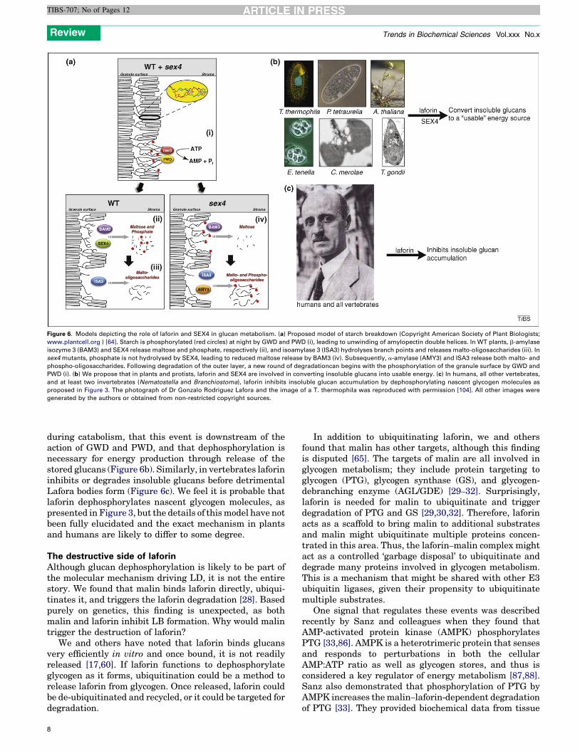
Figure 6. Models depicting the role of laforin and SEX4 in glucan metabolism. (a) Proposed model of starch breakdown (Copyright American Society of Plant Biologists;
www.plantcell.org ) [64]. Starch is phosphorylated (red circles) at night by GWD and PWD (i), leading to unwinding of amylopectin double helices. In WT plants, b-amylase
isozyme 3 (BAM3) and SEX4 release maltose and phosphate, respectively (ii), and isoamylase 3 (ISA3) hydrolyses branch points and releases malto-oligosaccharides (iii). In
sex4 mutants, phosphate is not hydrolysed by SEX4, leading to reduced maltose release by BAM3 (iv). Subsequently, a-amylase (AMY3) and ISA3 release both malto- and
phospho-oligosaccharides. Following degradation of the outer layer, a new round of degradationcan begins with the phosphorylation of the granule surface by GWD and
PWD (i). (b) We propose that in plants and protists, laforin and SEX4 are involved in converting insoluble glucans into usable energy. (c) In humans, all other vertebrates,
and at least two invertebrates (Nematostella and Branchiostoma), laforin inhibits insoluble glucan accumulation by dephosphorylating nascent glycogen molecules as
proposed in Figure 3. The photograph of Dr Gonzalo Rodriguez Lafora and the image of a T. thermophila was reproduced with permission [104]. All other images were
generated by the authors or obtained from non-restricted copyright sources.
Review Trends in Biochemical Sciences Vol.xxx No.x
TIBS-707; No of Pages 12
during catabolism, that this event is downstream of theaction of GWD and PWD, and that dephosphorylation isnecessary for energy production through release of thestored glucans (Figure 6b). Similarly, in vertebrates laforininhibits or degrades insoluble glucans before detrimentalLafora bodies form (Figure 6c). We feel it is probable thatlaforin dephosphorylates nascent glycogen molecules, aspresented in Figure 3, but the details of thismodel have notbeen fully elucidated and the exact mechanism in plantsand humans are likely to differ to some degree.
The destructive side of laforinAlthough glucan dephosphorylation is likely to be part ofthe molecular mechanism driving LD, it is not the entirestory. We found that malin binds laforin directly, ubiqui-tinates it, and triggers the laforin degradation [28]. Basedpurely on genetics, this finding is unexpected, as bothmalin and laforin inhibit LB formation. Why would malintrigger the destruction of laforin?
We and others have noted that laforin binds glucansvery efficiently in vitro and once bound, it is not readilyreleased [17,60]. If laforin functions to dephosphorylateglycogen as it forms, ubiquitination could be a method torelease laforin from glycogen. Once released, laforin couldbe de-ubiquitinated and recycled, or it could be targeted fordegradation.
8
In addition to ubiquitinating laforin, we and othersfound that malin has other targets, although this findingis disputed [65]. The targets of malin are all involved inglycogen metabolism; they include protein targeting toglycogen (PTG), glycogen synthase (GS), and glycogen-debranching enzyme (AGL/GDE) [29–32]. Surprisingly,laforin is needed for malin to ubiquitinate and triggerdegradation of PTG and GS [29,30,32]. Therefore, laforinacts as a scaffold to bring malin to additional substratesand malin might ubiquitinate multiple proteins concen-trated in this area. Thus, the laforin–malin complex mightact as a controlled ‘garbage disposal’ to ubiquitinate anddegrade many proteins involved in glycogen metabolism.This is a mechanism that might be shared with other E3ubiquitin ligases, given their propensity to ubiquitinatemultiple substrates.
One signal that regulates these events was describedrecently by Sanz and colleagues when they found thatAMP-activated protein kinase (AMPK) phosphorylatesPTG [33,86]. AMPK is a heterotrimeric protein that sensesand responds to perturbations in both the cellularAMP:ATP ratio as well as glycogen stores, and thus isconsidered a key regulator of energy metabolism [87,88].Sanz also demonstrated that phosphorylation of PTG byAMPK increases themalin–laforin-dependent degradationof PTG [33]. They provided biochemical data from tissue

Review Trends in Biochemical Sciences Vol.xxx No.x
TIBS-707; No of Pages 12
culture models, and they confirmed these findings by usingLD patient data, significantly strengthening this pre-viously disputed result. The signals that regulate thesedegradation events and the timing of the events are cur-rently being elucidated.
Why do only neurons die?Whereas glycogen is generated in virtually all liver andskeletal muscle cells, glycogen is generated only in astro-cytes and not in neurons in the mature brain [89]. Para-doxically, whereas neurons do not produce or storeglycogen, neurons of LD patients accumulate LBs andare the only cells in LD patients reported to exhibit acellular phenotype [34]. Recent work by Guinovart and co-workers solved the perplexing problem of how neuronswere capable of generating LBs without generating gly-cogen. They demonstrated that neurons express lowlevels of muscle glycogen synthase (MGS) and thatMGS in neurons is kept in an inactive, hyperphosphory-lated state [30]. In addition, they demonstrated that amalin–laforin complex utilizes ubiquitination to ensurelow levels of MGS in neurons and they elegantly showedthat when MGS is dephosphorylated by protein phospha-tase 1 (PP1) an aberrant poorly branched polyglucanforms, eventually leading to LBs in LD patients. Thiswork has greatly expanded our knowledge of neuronalmetabolism, but it does not define why only neuronsundergo cell death in LD patients. The molecular mech-anism that triggers neuronal apoptosis in LD patients isunknown, but we present four hypotheses that are notmutually exclusive. Given that LD symptoms take 15years to manifest, it may be that multiple mechanismscontribute to the pathogenesis.
Glial cells outnumber neurons approximately 10:1 andprovide them with most energy needs. Neurons storeminimal to no glycogen and have increased energy needsdue to numerous ion channels, and inherently live in an‘‘energy crisis.’’ Thus, glial glycogen is thought of as a safetynet that ensures neurons maintain an energy sourceduring periods of intense activation (reviewed in Refs[89,90]). The degree of branching and the length of glucosechains in glycogen are optimized for maximal glucosestorage in the smallest volume and maximal energyrelease [39,91]. As neurons have very limited energy storesand are both dependent on glial glycogen and utilize largeamounts of energy, they are hypersensitive to energyperturbations. LBs are likely to accumulate a significantamount of ‘trapped’ and ‘unusable’ energy. If this trappedenergy causes a temporary disruption in energy release,then cells that lack their own energy stores, e.g. neurons,would be the first to undergo apoptosis. In this scenario,neurons are the ‘canary in the coal mine’ and are respond-ing to decreased energy availability. Neuronal apoptosisleads to an early death of the patient, so no other cells havea chance to undergo apoptosis or cell death.
A second possibility is that LBs present a major traffick-ing problem. Whereas a neuron is 4–100 mm in diameter,neuronal LBs range from 3 to 40 mm [13]. Therefore, LBsmight form a blockade in the cytoplasm. In addition, theneurons transport intracellular cargos over considerablyfurther distances than other cells. Thus, a trafficking
defect could first present in them and later result inneuronal apoptosis.
Third, neuronal death in LD patients could be the onlycellular phenotype because of the age of neurons. Neuronsabolish mitotic division and have a markedly increasedlifespan compared to most other cell types. Neurons in LDpatients might be the only cells undergoing apoptosisbecause of their advanced ‘age’, and other cell types mightnot live long enough to experience the detrimental affects.This hypothesis would explain why neurons in murinemodels that exhibit LBs do not undergo widespread apop-tosis and why LD mouse and dog models do not die at ayoung age [92–94]. The lifespan of murine and canineneurons is not as long as that of human neurons and wouldnot be long enough for LBs to cause massive apoptosis,which takes 15+ years in humans.
Lastly, it is possible that LBs are not the cause of LD,but rather a cellular defense mechanism to sequester anddispose of aberrantly folded glucans. This hypothesis hasgained support among researchers studying the multipleneurodegenerative diseases involving proteinaceousaccumulations. Corroborating this hypothesis in LD isthe fact that not all neurons that undergo cell death inthe laforin-deficient mouse model have visible LBs. Thisresult could mean that LBs are the end result of multipleaberrant steps of glycogen synthesis, and that the earlier,non-visible products are the pathogenic cause of neuronalapoptosis. Alternatively, LBs might not be the pathogeniccause of LD. Ganesh and colleagues have suggested that adefect in autophagy or the ubiquitin proteasome system isa driving force in LD and LD pathology [95,96]. However,their studies examining autophagy in cell models utilizeover-expressed proteins and treatment with proteasomalinhibitors; thus, the data needed to support this hypothesisare not yet entirely convincing. Nonetheless, it is strikingthat, like the proteinopathies, mutations in an E3 ubiqui-tin ligase, malin, result in LD.
Concluding remarks and future perspectivesCollectively, biochemistry, mouse models, cell biology, andLD patient data suggest two essential roles for laforin: (1)dephosphorylation of glycogen, or nascent glucans, to inhi-bit excess glycogen phosphorylation and LB formation; and(2) recruitment of malin to the site of glycogen synthesis sothat malin can ubiquitinate PTG, GS, AGL, laforin, andpossibly other proteins to inhibit LB formation. Thus,laforin performs two essential functions, and malin one,in maintaining proper glycogen metabolism. This idea oflaforin having two roles also is supported by LD patientdata. LD patients with malin mutations live 25% longerthan patients with laforin mutations [97]. Thus, these datasuggest that the function of laforin could be downstream ofmalin, or that laforin has a disproportionate role in glyco-gen metabolism.
As many pathways are regulated by ubiquitination, it isnot overly surprising that ubiquitylation also regulatesglycogen metabolism. However, we now must identifyand define both the auxiliary proteins mediating theseevents and the signals that regulate these proteins atthe cellular, tissue and organismal levels. As discussedabove, an emerging regulator of these events is AMPK;
9

Review Trends in Biochemical Sciences Vol.xxx No.x
TIBS-707; No of Pages 12
however, the extent to which AMPK orchestrates thisregulation is still being determined.
A surprising discovery from theLafora diseasefield is theidentification of a glucan phosphatase activity that is con-served from plants to humans. Although the picture isbecoming increasingly clear as to how plants utilize glucanphosphorylation anddephosphorylation to storeand releaseenergy, respectively, it is not clear how or why vertebrateglucans become phosphorylated. Is there a glucan kinase invertebrates, similar to GWD and PWD in plants? Manygroups have performed bioinformatic searches but haveyet to identify a similar kinase in vertebrates.Alternatively,the actions of a phospho-glucotransferase could result inglycogen phosphorylation, but this enzyme has not beenidentified. Is glycogen phosphorylation the result of anevolutionary remnant, i.e. a mistake, or does it have anundefined purpose? Lastly, as yeast, flies, and worms alllack laforin and malin, how do they deal with insolubleglucan accumulations and can we identify an alternativepathway in these model organisms? The answers to thesequestions will further define Lafora disease at a molecularlevel, are likely to uncover potential therapies, will furtheridentify similarities and differences between glycogen andstarch metabolism, and may provide mechanisms to modu-late glucan (i.e. energy) production ina variety of organisms.
AcknowledgementsWe apologize to colleagues whose work we were not able to cite due tospace constraints. The authors acknowledge the support of NIH grantsNS061803, RR0202171, and University of Kentucky College of Medicinestartup funds (to M.S.G.); and NIH grant DK0118849, the WaltherCancer Institute, and the Howard Hughes Medical Institute (to J.E.D.).
References1 Lafora, G. and Glick, G. (1911) Beitrag zur histopathologie der
myoklonischen epilepsie. Z. Ges. Neurol. Psychiatr. 6, 1–142 Lafora, G.R. (1911) Uber des Vorkommen amyloider KJrperchen im
innern der Ganglienzellen. Virchows Arch. Path. Anat. 205, 2953 Virchow, R.L.K. (1858). Die Cellularpathologie inihrer Begrundung
auf physiologische and pathologische Gewebelehre. Hirschwald4 Yokoi, S. et al. (1967) Isolation and characterization of Lafora bodies
in two cases of myoclonus epilepsy. J. Neuropathol. Exp. Neurol. 26,125–127
5 Yokoi, S. et al. (1968) Studies in myoclonus epilepsy (Lafora bodyform). I. Isolation and preliminary characterization of Lafora bodies intwo cases. Arch. Neurol. 19, 15–33
6 Harriman, D.G. et al. (1955) Progressive familialmyoclonic epilepsy inthree families: its clinical features and pathological basis. Brain 78,325–349
7 Hodskins, M.B. et al. (1930) Anatomico-clinical observations onmyclonus in epileptics and on related symptom complexes. Am. J.Psychiatry 86, 827–848
8 Berkovic, S.F. et al. (1991) Progressive myoclonus epilepsies: clinicaland neurophysiological diagnosis. J. Clin. Neurophysiol. 8, 261–274
9 Harriman, D.G. and Miller, J.H.D. (1955) Progressive familialmyclonic epilepsy in three families: its clinical features andpatholgical basis. Brain 78, 325–349
10 Berkovic, S.F. et al. (1986) Progressive myoclonus epilepsies: specificcauses and diagnosis. N. Engl. J. Med. 315, 296–305
11 Janeway, R. et al. (1967) Progressive myoclonus epilepsy with Laforainclusion bodies. I. Clinical, genetic, histopathologic, and biochemicalaspects. Arch. Neurol. 16, 565–582
12 Minassian, B.A. (2001) Lafora’s disease: towards a clinical, pathologic,and molecular synthesis. Pediatr. Neurol. 25, 21–29
13 Van Heycop Ten Ham, M.W. (1975) In Handbook of ClinicalNeurology: Lafora disease, a form of progressive myoclonus epilepsy(Vol. 15), North Holland, pp. 382–422
10
14 Minassian, B.A. et al. (1998) Mutations in a gene encoding a novelprotein tyrosine phosphatase cause progressive myoclonus epilepsy.Nat. Genet. 20, 171–174
15 Serratosa, J.M. et al. (1999) A novel protein tyrosine phosphatasegene is mutated in progressive myoclonus epilepsy of the Lafora type(EPM2). Hum. Mol. Genet. 8, 345–352
16 Ganesh, S. et al. (2000) Laforin, defective in the progressivemyoclonus epilepsy of Lafora type, is a dual-specificityphosphatase associated with polyribosomes. Hum. Mol. Genet. 9,2251–2261
17 Wang, J. et al. (2002) A unique carbohydrate binding domain targetsthe lafora disease phosphatase to glycogen. J. Biol. Chem. 277, 2377–
238018 Alonso, A., Sasin, J., Bottini, N., Friedberg, I., Friedberg, I.,
Osterman, A., Godzik, A., Hunter, T., Dixon, J. and Mustelin, T.(2004) Protein tyrosine phosphatases in the human genome. Cell117, 699
19 Almo, S.C. et al. (2007) Structural genomics of protein phosphatases.J. Struct. Funct. Genomics 8, 121–140
20 Boraston, A.B. et al. (2004) Carbohydrate-binding modules: fine-tuning polysaccharide recognition. Biochem. J. 382, 769–781
21 Coutinho, P.M. and Henrissat, B. (1999). Recent Advances inCarbohydrate Bioengineering: Carbohydrate-active enzymes: anintegrated database approach (Svensson, B. et al. eds) pp. 3-12,The Royal Society of Chemistry
22 Rodriguez-Sanoja, R. et al. (2005) Microbial starch-binding domain.Curr. Opin. Microbiol. 8, 260–267
23 Chan, E.M. et al. (2004) Progressive myoclonus epilepsy withpolyglucosans (Lafora disease): evidence for a third locus.Neurology 63, 565–567
24 Chan, E.M. et al. (2003) Mutations in NHLRC1 cause progressivemyoclonus epilepsy. Nat. Genet. 35, 125–127
25 Hershko, A. and Ciechanover, A. (1998) The ubiquitin system. Annu.Rev. Biochem. 67, 425–479
26 Edwards, T.A. et al. (2003) Model of the brain tumor-Pumiliotranslation repressor complex. Genes Dev. 17, 2508–2513
27 Slack, F.J. and Ruvkun, G. (1998) A novel repeat domain that is oftenassociated with RING finger and B-box motifs. Trends Biochem. Sci.23, 474–475
28 Gentry, M.S. et al. (2005) Insights into Lafora disease: malin is an E3ubiquitin ligase that ubiquitinates and promotes the degradation oflaforin. Proc. Natl Acad. Sci. USA 102, 8501–8506
29 Solaz-Fuster, M.D. et al. (2008) Regulation of glycogen synthesis bythe laforin-malin complex is modulated by the AMP-activated proteinkinase pathway. Hum. Mol. Genet. 17, 667–678
30 Vilchez, D. et al. (2007) Mechanism suppressing glycogen synthesis inneurons and its demise in progressive myoclonus epilepsy. Nat.Neurosci. 10, 1407–1413
31 Cheng, A. et al. (2007) A role for AGL ubiquitination in the glycogenstorage disorders of Lafora and Cori’s disease. Genes Dev. 21, 2399–
240932 Worby, C.A. et al. (2008) Malin decreases glycogen accumulation by
promoting the degradation of protein targeting to glycogen (PTG). J.Biol. Chem. 283, 4069–4076
33 Vernia, S. et al. (2009) AMP-activated protein kinase phosphorylatesR5/PTG, the glycogen targeting subunt of the R5/PTG-PP1holoenzyme and accelerates its downregulation by the laforin-malin complex. J. Biol. Chem. 284, 8247–8255
34 Ganesh, S. et al. (2002) Targeted disruption of the Epm2a gene causesformation of Lafora inclusion bodies, neurodegeneration, ataxia,myoclonus epilepsy and impaired behavioral response in mice.Hum. Mol. Genet. 11, 1251–1262
35 Chan, E.M. et al. (2004) Laforin preferentially binds the neurotoxicstarch-like polyglucosans, which form in its absence in progressivemyoclonus epilepsy. Hum. Mol. Genet. 13, 1117–1129
36 Coleman, D.L. et al. (1974) Muscle in Lafora disease. Arch. Neurol. 31,396–406
37 Gambetti, P. et al. (1971) Myoclonic epilepsy with lafora bodies. Someultrastructural, histochemical, and biochemical aspects.Arch. Neurol.25, 483–493
38 Roach, P.J. et al. (2001) Regulation of glycogen metabolism. In TheEndocrine Pancreas and Regulation of Metabolism (Jefferson, L.S.and Cherrington, A.D., eds), pp. 609–647, Oxford University Press

Review Trends in Biochemical Sciences Vol.xxx No.x
TIBS-707; No of Pages 12
39 Melendez, R. et al. (1997) How did glycogen structure evolve to satisfythe requirement for rapid mobilization of glucose? A problem ofphysical constraints in structure building. J. Mol. Evol. 45, 446–455
40 Shearer, J. and Graham, T.E. (2004) Novel aspects of skeletal muscleglycogen and its regulation during rest and exercise. Exerc. Sport Sci.Rev. 32, 120–126
41 Fontana, J.D. (1980) The presence of phosphate in glycogen. FEBSLett. 109, 85–92
42 Lomako, J. et al. (1994) The role of phosphate in muscle glycogen.Biofactors 4, 167–171
43 Lomako, J. et al. (1993) Glycogen contains phosphodiester groups thatcan be introduced by UDPglucose: glycogen glucose 1-phosphotransferase. FEBS Lett 329, 263–267
44 Ball, S.G. and Morell, M.K. (2003) From bacterial glycogen to starch:understanding the biogenesis of the plant starch granule. Ann. Rev.Plant Biol. 54, 207–233
45 Zeeman, S.C. et al. (2007) The diurnal metabolism of leaf starch.Biochem. J. 401, 13–28
46 Schnabel, R. and Seitelberger, F. (1968) Histophysical andhistochemical investigations of myoclonus bodies. Pathol. Eur. 3,218–226
47 Sakai, M. et al. (1970) Studies in myoclonus epilepsy (Lafora bodyform). II. Polyglucosans in the systemic deposits of myoclonusepilepsy and in corpora amylacea. Neurology 20, 160–176
48 Coppin, A. et al. (2005) Evolution of plant-like crystalline storagepolysaccharide in the protozoan parasite Toxoplasma gondii arguesfor a red alga ancestry. J. Mol. Evol. 60, 257–267
49 Guerardel, Y. et al. (2005) Amylopectin biogenesis andcharacterization in the protozoan parasite Toxoplasma gondii, theintracellular development of which is restricted in theHepG2 cell line.Microbes Infect. 7, 41–48
50 Meeuse, B.J.D. et al. (1960) Floridean starch. J. Exp. Bot. 11, 129–14051 Nyvall, P. et al. (1999) Purification and characterisation of a novel
starch synthase selective for uridine 5’-diphosphate glucose from thered alga Gracilaria tenuistipitata. Planta 209, 143–152
52 Viola, R. et al. (2001) The unique features of starch metabolism in redalgae. Proc. R. Soc. B 268, 1417–1422
53 McCracken, D.A. and Cain, J.R. (1981) Amylose in floridean starch.New Phytol. 88, 67–71
54 Gentry, M.S. et al. (2007) The phosphatase laforin crossesevolutionary boundaries and links carbohydrate metabolism toneuronal disease. J. Cell Biol. 178, 477–488
55 Minassian, B.A. (2002) Progressive myoclonus epilepsy withpolyglucosan bodies: Lafora disease. Adv. Neurol. 89, 199–210
56 Pederson, B.A. et al. (2003) Overexpression of glycogen synthase inmouse muscle results in less branched glycogen. Biochem. Biophys.Res. Commun. 305, 826–830
57 Raben, N. et al. (2001) Surprises of genetic engineering: a possiblemodel of polyglucosan body disease. Neurology 56, 1739–1745
58 Wang, W. et al. (2007) Glycogen metabolism in tissues from a mousemodel of Lafora disease. Arch. Biochem. Biophys. 457, 264–269
59 Ganesh, S. et al. (2004) The carbohydrate-binding domain of Laforadisease protein targets Lafora polyglucosan bodies. Biochem. BiophysRes. Commun. 313, 1101–1109
60 Wang, W. and Roach, P.J. (2004) Glycogen and relatedpolysaccharides inhibit the laforin dual-specificity proteinphosphatase. Biochem. Biophys. Res. Commun. 325, 726–730
61 Liu, Y. et al. (2009) Deletions and missense mutations of EPM2Aexacerbate unfolded protein response and apoptosis of neuronal cellsinduced by endoplasm reticulum stress. Hum. Mol. Genet. 18, 2622–
263162 Vernia, S. et al. (2009) Increased endoplasmic reticulum stress and
decreased proteasomal function in lafora disease models lacking thephosphatase laforin. PLoS One 4, e5907
63 Worby, C.A. et al. (2006) Laforin: a dual specificity phosphatase thatdephosphorylates complex carbohydrates. J. Biol. Chem. 281, 30412–
3041864 Kotting, O. et al. (2009) STARCH-EXCESS4 is a laforin-like
phosphoglucan phosphatase required for starch degradation inArabidopsis thaliana. Plant Cell 21, 334–346
65 Tagliabracci, V.S. et al. (2008) Abnormal metabolism of glycogenphosphate as a cause for lafora disease. J. Biol.Chem. 283, 33816–
33825
66 Tagliabracci, V.S. et al. (2007) Laforin is a glycogen phosphatase,deficiency of which leads to elevated phosphorylation of glycogen invivo. Proc. Natl Acad. Sci. USA 104, 19262–19266
67 Barford, D. et al. (1994) Crystal structure of human protein tyrosinephosphatase 1B. Science 263, 1397–1404
68 Lee, J-O. et al. (1999) Crystal structure of the PTEN tumorsuppressor: implications for its phosphoinositide phosphataseactivity and membrane association. Cell 99, 323–334
69 Guan, K.L. et al. (1991) A Tyr/Ser protein phosphatase encoded byvaccinia virus. Nature 350, 359–362
70 Yuvaniyama, J. et al. (1996) Crystal structure of the dual specificityprotein phosphatase VHR. Science 272, 1328–1331
71 Alonso, A. et al. (2003). The dual-specific protein tyrosine phosphatasefamily. In Topics Currents in Genetics (Vol. 5) pp. 333–358, Springer
72 Geiger, T. et al. (1992) T-cell responsiveness to an oncogenicperipheral protein and spontaneous autoimmunity in transgenicmice. Proc. Natl Acad. Sci. USA 89, 2985–2989
73 Wang, Y. et al. (2006) Epm2a suppresses tumor growth in animmunocompromised host by inhibiting Wnt signaling. Cancer Cell10, 179–190
74 Liu, R. et al. (2008) Laforin negatively regulates cell cycle progressionthrough GSK-3b dependent mechanisms. Mol. Cell Biol. 28, 7236–
724475 Wang, W. et al. (2006) Relationship between glycogen accumulation
and the laforin dual specificity phosphatase. Biochem. Biophys. Res.Commun. 350, 588–592
76 Niittyla, T. et al. (2006) Similar protein phosphatases control starchmetabolism in plants and glycogen metabolism in mammals. J. Biol.Chem. 281, 11815–11818
77 Peat, S. et al. (1959) The structure of floridean starch. Part II. Enzymichydrolysis and other studies. J. Chem. Soc 3341–3344
78 Gentry, M.S. and Pace, R.M. (2009) Conservation of the glucanphosphatase laforin is linked to rates of molecular evolution andthe glycogen metabolism of the organism. BMC Evol. Biol. 9, 138
79 Lorberth, R. et al. (1998) Inhibition of a starch-granule-bound proteinleads to modified starch and repression of cold sweetening. Nat.Biotechnol. 16, 473–477
80 Baunsgaard, L. et al. (2005) A novel isoform of glucan, water dikinasephosphorylates pre-phosphorylated alpha-glucans and is involved instarch degradation in Arabidopsis. Plant J. 41, 595–605
81 Kotting, O. et al. (2005) Identification of a novel enzyme required forstarch metabolism in Arabidopsis leaves. The phosphoglucan, waterdikinase. Plant Physiol 137, 242–252
82 Ritte, G. et al. (2006) Phosphorylation of C6- and C3-positions ofglucosyl residues in starch is catalysed by distinct dikinases. FEBSLett. 580, 4872–4876
83 Ritte, G. et al. (2002) The starch-related R1 protein is an alpha-glucan,water dikinase. Proc. Natl Acad. Sci. USA 99, 7166–7171
84 Blennow, A. et al. (2002) Starch phosphorylation: a new front line instarch research. Trends Plant Sci. 7, 445–450
85 Kirkman, B.R. and Whelan, W.J. (1986) Glucosamine is a normalcomponent of liver glycogen. FEBS Lett. 194, 6–11
86 Solaz-Fuster, M.C. et al. (2008) Regulation of glycogen synthesis bythe laforin-malin complex is modulated by the AMP-activated proteinkinase pathway. Hum. Mol. Genet. 17, 667–678
87 Hardie, D.G. (2007) AMP-activated/SNF1 protein kinases:conserved guardians of cellular energy. Nat. Rev. Mol. Cell. Biol.8, 774–785
88 McBride, A. et al. (2009) The glycogen-binding domain on the AMPKbeta subunit allows the kinase to act as a glycogen sensor. Cell Metab.9, 23–34
89 Brown, A.M. and Ransom, B.R. (2007) Astrocyte glycogen and brainenergy metabolism. Glia 55, 1263–1271
90 Swanson, R.A. (1992) Physiologic coupling of glial glycogenmetabolism to neuronal activity in brain. Can. J. Physiol.Pharmacol. 70 (Suppl.), S138–S144
91 Melendez-Hevia, E. et al. (1993) Optimization of molecular design inthe evolution of metabolism: the glycogen molecule. Biochem. J. 295,477–483
92 Cavanagh, J.B. (1999) Corpora-amylacea and the family ofpolyglucosan diseases. Brain Res. Brain Res. Rev. 29, 265–295
93 Lohi, H. et al. (2005) Expanded repeat in canine epilepsy. Science307, 81
11

Review Trends in Biochemical Sciences Vol.xxx No.x
TIBS-707; No of Pages 12
94 Yamanami, S. et al. (1992) Comparative study of intraneuronalpolyglucosan bodies in brains from patients with Lafora diseaseand aged dogs. Acta Pathol. Jpn 42, 787–792
95 Garyali, P. et al. (2009) The malin-laforin complex suppresses thecellular toxicity of misfolded proteins by promoting their degradationthrough the ubiquitin-proteasome system.Hum. Mol. Genet. 18, 688–
70096 Mittal, S. et al. (2007) Lafora disease proteins malin and laforin are
recruited to aggresomes in response to proteasomal impairment.Hum. Mol.Genet. 16, 753–762
97 Gomez-Abad, C. et al. (2005) Lafora disease due to EPM2Bmutations:a clinical and genetic study. Neurology 64, 982–986
98 Buleon, A. et al. (1998) Starch granules: structure and biosynthesis.Int. J. Biol. Macromol. 23, 85–112
99 Gunja-Smith, Z. et al. (1970) A revision of theMeyer-Bernfeldmodel ofglycogen and amylopectin. FEBS Lett. 12, 101–104
100 Myers, A.M. et al. (2000) Recent progress toward understandingbiosynthesis of the amylopectin crystal. Plant Physiol. 122, 989–998
101 Begley, M.J. and Dixon, J.E. (2005) The structure and regulation ofmyotubularin phosphatases. Curr. Opin. Struct. Biol. 15, 614–620
102 Pei, J. et al. (2008) PROMALS3D: a tool for multiple protein sequenceand structure alignments. Nucleic Acids Res. 36, 2295–2300
12
103 Bingham, J. and Sudarsanam, S. (2000) Visualizing large hierarchicalclusters in hyperbolic space. Bioinformatics 16, 660–661
104 Robinson, R. (2006) Ciliate genome sequence reveals unique featuresof a model eukaryote. PLoS Biol. 4, e304
105 Goldsmith, E. et al. (1982) Structure of maltoheptaose by differenceFourier methods and a model for glycogen. J. Mol. Biol. 156, 411–427
106 Ryman, B.E. and Whelan, W.J. (1971) New aspects of glycogenmetabolism. Adv. Enzymol. Relat. Areas Mol. Biol. 34, 244–285
107 Yu, T.S. et al. (2001) The Arabidopsis sex1 mutant is defective in theR1 protein, a general regulator of starch degradation in plants, andnot in the chloroplast hexose transporter. Plant Cell 13, 1907–1918
108 Tabata, S. and Hizukuri, S. (1971) Studies on starch phosphate. Part2. Isolation of glucose 3-phosphate and maltose phosphate by acidhydrolysis of potato starch. Starch 23, 267–272
109 Gallant, D.J. et al. (1997) Microscopy of starch: evidence of a new levelof granule organization. Carbohydr. Polymers 32, 177–191
110 Manners, D.J. and Wright, A. (1962) Alpha-1,4-glucosans. Part XIV.The interaction of concanavalin-A with glycogens. J. Chem. Soc.4592–4595
111 Manners, D.J. et al. (1957). The molecular structure of glycogens. In:Advances in Carbohydrate Chemistry (Vol. 12), pp. 261-298, AcademicPress