lange biochemistry and genetics flash cards
DESCRIPTION
Lange Biochemistry and Genetics Flash CardsTRANSCRIPT


Lange FlashCards Biochemistry & GeneticsSecond Edition
Suzanne J. Baron, MDMassachusetts General HospitalBoston, Massachusetts
Christoph I. Lee, MD, MSHSUniversity of Washington School of MedicineSeattle, Washington
New York / Chicago / San Francisco / Lisbon / London / Madrid / Mexico CityMilan / New Delhi / San Juan / Seoul / Singapore / Sydney / Toronto

Notice
Medicine is an ever-changing science. As new research and clinical experience broaden our knowledge,
changes in treatment and drug therapy are required. The authors and the publisher of this work have
checked with sources believed to be reliable in their efforts to provide information that is complete
and generally in accord with the standards accepted at the time of publication. However, in view of
the possibility of human error or changes in medical sciences, neither the authors nor the publisher nor
any other party who has been involved in the preparation or publication of this work warrants that the
information contained herein is in every respect accurate or complete, and they disclaim all responsi-
bility for any errors or omissions or for the results obtained from use of the information contained in
this work. Readers are encouraged to confirm the information contained herein with other sources. For
example and in particular, readers are advised to check the product information sheet included in the
package of each drug they plan to administer to be certain that the information contained in this work
is accurate and that changes have not been made in the recommended dose or in the contraindications
for administration. This recommendation is of particular importance in connection with new or infre-
quently used drugs.

Copyright © 2013 by The McGraw-Hill Companies, Inc. All rights reserved. Except as permitted under the United States Copyright Act of 1976, no part of this publication may be reproduced or distributed in any form or by any means, or stored in a database or retrieval system, without the prior written permission of the publisher.
ISBN: 978-0-07-182050-9
MHID: 0-07-182050-7
The material in this eBook also appears in the print version of this title: ISBN: 978-0-07-176580-0, MHID: 0-07-176580-8.
All trademarks are trademarks of their respective owners. Rather than put a trademark symbol after every occurrence of a trademarked name, we use names in an editorial fashion only, and to the benefi t of the trademark owner, with no intention of infringement of the trademark. Where such designations appear in this book, they have been printed with initial caps.
McGraw-Hill eBooks are available at special quantity discounts to use as premiums and sales promotions, or for use in corporate training programs. To contact a representative please e-mail us at [email protected].
TERMS OF USE
This is a copyrighted work and The McGraw-Hill Companies, Inc. (“McGraw-Hill”) and its licensors reserve all rights in and to the work. Use of this work is subject to these terms. Except as permitted under the Copyright Act of 1976 and the right to store and retrieve one copy of the work, you may not decompile, disassemble, reverse engineer, reproduce, modify, create derivative works based upon, transmit, distribute, disseminate, sell, publish or sublicense the work or any part of it without McGraw-Hill’s prior consent. You may use the work for your own noncommercial and personal use; any other use of the work is strictly prohibited. Your right to use the work may be terminated if you fail to comply with these terms.
THE WORK IS PROVIDED “AS IS.” McGRAW-HILL AND ITS LICENSORS MAKE NO GUARANTEES OR WARRANTIES AS TO THE ACCURACY, ADEQUACY OR COMPLETENESS OF OR RESULTS TO BE OBTAINED FROM USING THE WORK, INCLUDING ANY INFORMATION THAT CAN BE ACCESSED THROUGH THE WORK VIA HYPERLINK OR OTHERWISE, AND EXPRESSLY DISCLAIM ANY WARRANTY, EXPRESS OR IMPLIED, INCLUDING BUT NOT LIMITED TO IMPLIED WARRANTIES OF MERCHANTABILITY OR FITNESS FOR A PARTICULAR PURPOSE. McGraw-Hill and its licensors do not warrant or guarantee that the functions contained in the work will meet your requirements or that its operation will be uninterrupted or error free. Neither McGraw-Hill nor its licensors shall be liable to you or anyone else for any inaccuracy, error or omission, regardless of cause, in the work or for any damages resulting therefrom. McGraw-Hill has no responsibility for the content of any information accessed through the work. Under no circumstances shall McGraw-Hill and/or its licensors be liable for any indirect, incidental, special, punitive, consequential or similar damages that result from the use of or inability to use the work, even if any of them has been advised of the possibility of such damages. This limitation of liability shall apply to any claim or cause whatsoever whether such claim or cause arises in contract, tort or otherwise.

This page intentionally left blank

iii
Contents
Preface ivAcknowledgments vAbout the Authors vAbbreviations vi
1 Cellular Energy 1-42 Carbohydrate Metabolism 5-253 Lipid Metabolism 26-544 Amino Acid Metabolism 55-685 Nucleotide Metabolism 69-786 Heme Metabolism 79-877 Steroid Hormone Synthesis 88-958 Nutrition 96-1149 Genetics 115-184
References 185Index 186-189

This page intentionally left blank

iv
Preface
When we began to review the biochemistry and genetics material covered in the USMLE Step 1 at the end of our second year at Yale Medical School, we realized that most of the practice questions were approaching the material from a clinical perspective and not from the basic science perspective in which we had learned these topics. Although we had taken introductory biochemistry and genetics courses back in college and covered the material again during the first few months of medical school, we found ourselves studying the clinical aspects of biochemical and genetic diseases for the first time. Flipping through the highly rated biochemistry and genetics review sources, we realized that there was no gold standard review source for these high-yield topics that make up 15% of USMLE Step 1 questions.
Lange FlashCards: Biochemistry and Genetics is the result of our struggles in studying these topics for Step 1 with the clinical slant that the boards demand. These cards offer the most complete, concise, and high-yield information for the major biochemical and genetic diseases tested on Step 1 and in medical school basic science courses. We are confident that the content covered in the second version of our cards includes the most current and board-relevant information that cannot be found in any other single biochemistry and genetics review text.
We are pleased to present this information in a format modeled after Lange FlashCards: Pathology, our first publication in this series. Each card provides a structured presentation of a specific disease and allows students to easily compare and contrast diseases. The introductory cards in each chapter describe the basic principles of biochemistry and genetics that are board relevant and high yield. Each disease-specific card contains a clinical vignette on one side and important characteristics on the reverse side. These characteristics are organized into sections entitled biochemical or genetic defect, pathophysiology, clinical manifestations, treatment, and addi-tional pearls. The most salient features of each disease are highlighted in bold for ease of rapid review.

iv
We suggest using these cards as an adjunct to your biochemistry and genetics courses in medical school. Being familiar with these cards early on will be very helpful during your Step 1 review. We also encourage you to jot down your own notes in the margins and to make these cards your personal biochemistry and genet-ics review for the boards.
We are confident that the newly revised second edition of Lange FlashCards: Biochemistry and Genetics will be one of the most powerful tools to help prepare you for the boards and will serve as a resource that will bridge your basic science knowledge with the clinical aspects of disease. We wish you the best of luck on Step 1 and welcome your comments on how to improve this study tool in the next edition.
Suzanne J. BaronBoston, Massachusetts
Christoph I. LeeSeattle, Washington
[email protected] 2012

v
Acknowledgments
We would like to thank the many editors at McGraw-Hill for all their support and hard work on this project. We would especially like to thank Michael Weitz, who has wholeheartedly supported the expansion of the Lange FlashCards series and continues to be its driving force.
We acknowledge the many basic and clinical science teaching faculty of the Yale University School of Medicine who provided much of the foundation for the relevant content in these cards. Their input and contribu-tions were invaluable to the quality of our final product. We also thank our mentors throughout medical school, residency, and fellowship for their continuing encouragement and dedication to our professional development.
To our family and friends, we thank you for your continuing support and love that have made this process even more meaningful. Special thanks to John and Jay Lee, Fran and Joe Baron, Elena Paul, Bettina Lee, Monique Mogensen and Steven Fay.

v
About the Authors
Suzanne J. Baron, MD, earned an AB magna cum laude in psychology and biology from Harvard University and an MD from Yale University School of Medicine. She is an elected member of Alpha Omega Alpha Honors Society. Dr Baron completed her residency in internal medicine and fellowship in cardiology at Massachusetts General Hospital. She is currently pursuing additional subspecialty training in interventional cardiology also at Massachusetts General Hospital. Dr Baron is an accomplished pianist.
Christoph I. Lee, MD, earned an AB cum laude in English from Princeton University and an MD cum laude from Yale University School of Medicine. He completed his residency in diagnostic radiology at Stanford University and fellowship in breast imaging at UCLA. Dr Lee has completed a health policy fellowship in the prestigious Robert Wood Johnson Clinical Scholars program. He has previously managed a global tuberculosis initiative for Ralph Nader.

vi
1,2-DAG: 1,2-diacylglycerol2,3-BPG: 2,3-bisphosphoglycerateα-t: α-thalassemiasβ-t: β-thalassemiasABG: arterial blood gasAC: adenylate cyclaseACE: angiotensin-converting-enzymeACTH: adrenocorticotropic hormoneADA: adenosine deaminaseADP: adenosine diphosphateALA: aminolevulinic acidALL: acute lymphoblastic leukemiaALT: alanine aminotransferaseAMP: adenosine monophosphateApo: apoproteinATP: adenosine triphosphate AST: aspartate aminotransferaseATP: adenosine triphosphateAUG: adenine uracil guanineBAL: British AntiLewisite, dimercaprolBCKD: branched-chain α-ketoacid dehydrogenase BSS: Bernard-Soulier syndromeBtk: Bruton tyrosine kinaseBUN: blood urea nitrogenCAG: cytosine adenine guanine
cAMP: cyclic adenosine monophosphateCBC: complete blood countCDP: cytidine diphosphateCFTR: cystic fibrosis transmembrane conductor regulatorCGG: cytosine guanine guanineCHF: congestive heart failureChr: chromosomeCl–: chloride ionCMT: Charcot-Marie-Tooth (disease)CNS: central nervous systemCoA: coenzyme ACOPRO: coproporphyrinogenCT: computed tomographyCTG: cytosine thymidine guanineCTP: cytosine-5′-triphosphatedADP: deoxyadenosine diphosphatedATP: deoxyadenosine triphosphatedCDP: deoxycytidine diphosphatedGDP: deoxyguanosine diphosphateDHEA: dehydroepiandrosteroneDHT: dihydrotestosteroneDMD: Duchenne muscular dystrophyDNA: deoxyribonucleic acidDNAO: DNA polymeraseDNAP: DNA polymerase
Abbreviations

vi
DOPA: dihydroxyphenylalaninedTMP: deoxythymidylateDTRs: deep tendon reflexesdUDP: deoxyuridine 5′-diphosphatedUMP: deoxyuridylateECG: electrocardiogramEDS: Ehlers-Danlos syndromeESR: erythrocyte sedimentation rateESRD: end-stage renal diseaseF1,6BP: fructose 1,6 bisphosphateF6P: fructose-6-phosphateFAD: flavin adenine dinucleotideFAMN: flavin adenine mononucleotideFEP: free erythrocyte protoporphyrinFEV1: forced expiratory volume in 1 secondFGFR3: fibroblast growth factor receptor 3FMN: flavin mononucleotideFMR-1: familial mental retardationFSH: follicle-stimulating hormoneFVC: functional vital capacityG3P: glyceraldehyde-3-phosphateG6P: glucose-6-phosphateG6PD: glucose-6-phosphate dehydrogenaseGAA: guanine adenine adenineGAG: glycosaminoglycanG-CSF: granulocyte colony–stimulating factorFGFR3: fibroblast growth factor receptor 3GDP: guanosine diphosphate
GFR: glomerular filtration rateGI: gastrointestinalGMP: guanosine monophosphateGT: glanzmann thrombastheniaGTP: guanosine triphosphateHD: Huntington diseaseHDL: high-density lipoproteinHgb: hemoglobinHGPRT: hypoxanthine-guanine phosphoribosyltransferaseHIV: human immunodeficiency virusHMG-CoA: 5-hydroxy-3-methylglutaryl coenzyme AHMP: hexose monophosphateIBD: inflammatory bowel diseaseIDL: intermediate-density lipoproteinIMP: inosine monophosphateIV: intravenous IVP: intravenous pyelogramKUB: kidneys, ureter, bladder (x-ray)LDL: low-density lipoproteinLFTs: liver function testsLH: luteinizing hormoneLHON: Leber hereditary optic neuropathyLPL: low-density lipoproteinMCHC: mean corpuscular hemoglobin concentrationMCV: mean corpuscular volume MELAS: mitochondrial encephalomyopathy with lactic acidosis and strokelike episodesMEN: multiple endocrine neoplasia

MERRF: myoclonic epilepsy with ragged red fibersMI: myocardial infarctionMRI: magnetic resonance imagingmRNA: messenger RNAmtDNA: mitochondrial DNAMTP: metatarsophalangealNAD: nicotinamide adenine dinucleotide NADP: nicotinamide adenine dinucleotide phosphateNADPH: nicotinamide adenine dinucleotide phosphate hydrogenNF1: neurofibromatosis 1NPD: Niemann-Pick diseaseNPTHM: N5-methyl tetrahydrofolate homocysteine methyltransferaseNSAID: nonsteroidal anti-inflammatory drugOMP: orotidine-5′-monophosphatePBG: porphobilinogen PPD: purified protein derivativePRPP: phosphoribosylpyrophosphatePT: prothrombin timePTH: parathyroid hormonePTT: partial thromboplastin timeRBC: red blood cellRFLP: restriction fragment length polymorphismRNA: ribonucleic acidRNAP: RNA polymeraserRNA: ribosomal ribonucleic acid
RUQ: right upper quadrantSAM: S-adenosylmethionineSCID: severe combined immunodeficiencySSB: single-strand DNA bindingTB: tuberculosisTGF-β: tissue growth factor βTIBC: total iron-binding capacityTLC: total lung capacityTMP-SMX: trimethoprim-sulfamethoxazoleTPP: thiamine pyrophosphateTTP: thymidine triphosphatetRNA: transfer ribonucleic acidUA: urinalysisUDP: uridine 5′-diphosphateUDPGT: uridine diphosphoglucuronosyl transferaseUMP: uridine-5′-monophosphateURO: uroporphyrinogenUTI: urinary tract infectionUTP: uracil-5′-triphosphateUV: ultravioletVLDL: very-low-density lipoproteinVMA: vanillylmandelic acidvWF: von Willebrand factorWAS: Wiskott-Aldrich syndromeXR: x-ray
vii

This page intentionally left blank

1
1
CELLULAR ENERGY
GENERAL CONCEPTS
Citric acid cycle
Electron transport chain
NADH shuttles

This page intentionally left blank

CITRIC ACID CYCLE
1
2
Citrate
Succinate
Isocitrate
Fumarate
Succinyl CoA
α-Ketoglutarate
Oxaloacetate
Malate
Acetyl CoA
Citrate Synthase
Malate
Dehyd
rogen
ase
Succinyl CoA Synthase
NAD+
NADH
FADH2
FAD
NAD+
NAD+
NADH
NADH
CO2
CO2
GDPGTP
Succinate
Dehydrogenasea-K
etoglutarate
Dehydrogenase
AconitaseIsocitrate
DehydrogenaseF
umar
ase

2
CITRIC ACID CYCLE
The citric acid cycle occurs in the mitochondrial matrix. Functions include the oxidation of acetyl CoA to CO 2 , the formation of NADH and FADH 2 for entrance into the electron transport chain and subsequent ATP genera-tion, and the synthesis of several important molecules, including succinyl CoA (precursor molecule of heme), oxaloacetate (early intermediate molecule in gluconeogenesis and substrate for amino acid synthesis), α-ketoglutarate (substrate for amino acid synthesis), and citrate (substrate for fatty acid synthesis).
YIELD OF THE CITRIC ACID CYCLE
Each molecule of acetyl CoA entering the citric acid cycle yields the following:
• Two CO 2 • Three NADH • One FADH 2 • One GTP
Because each NADH will eventually produce 2.5 ATP and each FADH 2 will produce 1.5 ATP through the electron transport chain, the overall ATP yield from 1 acetyl CoA is 10 ATP (7.5 from NADH, 1.5 from FADH 2 , and 1 from GTP).
REGULATION OF THE CITRIC ACID CYCLE
Enzyme Inhibitors Activators
Citrate synthase ATPNADHSuccinyl CoA
—
Isocitrate dehydrogenase ATPNADH
ADP
α-Ketoglutaratedehydrogenase
ATP or GTPNADHSuccinyl CoA
—

3
1
ELECTRON TRANSPORT CHAIN
I
II
III IV VCoQ
CC
NADH
NAD+
FAD
FADH2
H2O
1/2 O2ATP
ADP
H+H+
H+
H+
H+
H+
H+
H+ H+
H+ H+
H+H+
H+
H+
Intermembrane Space
Mitochondrial Matrix
e− e−
e−
e−
e−

3
ELECTRON TRANSPORT CHAIN
COMPONENTS OF THE ELECTRONTRANSPORT CHAIN
Complex I (NADH dehydrogenase): Contains FMN, which accepts 2 e − and H + from 2 NADH to become the reduced form of FMNH 2 ; also contains iron atoms, which assist in the transfer of the e − and H + to coenzyme Q. Inhibited by amobarbital and rotenone.
Complex II (succinate dehydrogenase): Contains iron and succinate, which oxidizes FAD to form FADH 2 . Inhibited by antimycin A.
Coenzyme Q : Accepts e − from FMNH 2 (complex I) and FADH 2 (complex II). Transfers e − to complex III.
Complex III (cytochrome b): Contains heme group , in which the Fe 3+ accepts the e − from coenzyme Q to become Fe 2+ . Transfers e − to cytochrome c.
Cytochrome c : Contains heme group , in which the Fe 3+
accepts the e − from complex III to become Fe 2+ . Transfers e− to complex IV.
Complex IV (cytochrome a): Contains heme group , in which the Fe 3+ accepts e − from cytochrome c to become
Fe 2+ . Transfers e − to O 2 , which is combined with hydrogen to form H 2 O. Inhibited by cyanide, CO, and sodium azide.
Complex V (ATP synthase): Contains a proton channel that allows for protons to cross into the matrix, using the proton gradient energy to form ATP. Inhibited by oligomycin(blocks H + channel).
Each NADH yields 2.5 ATP ; each FADH 2 yields 1.5 ATP .
THE CHEMIOSMOTIC HYPOTHESIS
Electron transport causes H + ions to be pumped from the mitochondrial matrix into the intermembrane space, thereby resulting in the formation of an electrical and pH gradient across the inner mitochondrial membrane. The energy cre-ated by the formation of this gradient is then harnessed to form ATP as the protons travel down their gradient into the matrix through ATP synthase channel (complex V). 2, 4-dinitrophenol acts to uncouple ATP formation from electron transport by dissipating the proton gradient.
4
1
Cytoplasm
Mitochondrial Matrix
Aspartate
Aspartate Oxaloacetate
Oxaloacetate
Malate
Malate
Inner Mitrochondrial Membrane
NADH NAD+
NADH NAD+
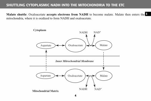
Malate shuttle : Oxaloacetate accepts electrons from NADH to become malate. Malate then enters the mitochondria, where it is oxidized to form NADH and oxaloacetate.
SHUTTLING CYTOPLASMIC NADH INTO THE MITOCHONDRIA TO THE ETC

4
Cytoplasm
Mitochondrial Matrix
α-Glycerol-P
Inner Mitrochondrial Membrane
FADH2 FAD+
NADH NAD+
Dihydroxyacetone-P
Dihydroxyacetone-P
α-Glycerol-P
α-Glycerol phosphate shuttle : DHAP accepts electrons from NADH to become α-Glycerol phosphate. α-Glycerol phosphate enters the mitochondria, where it is oxidized to form FADH 2 and DHAP. Here, only 1.5 ATPs are formed for each cytoplasmic NADH oxidized since FADH 2 is produced.
SHUTTLING CYTOPLASMIC NADH INTO THE MITOCHONDRIA TO THE ETC

5
2
CARBOHYDRATE METABOLISM
GENERAL CONCEPTS
GlycogenesisGlycogenolysis
GlycolysisPyruvate metabolism
Pentose phosphate pathwayFructose metabolismGalactose metabolism
GluconeogenesisCori cycle
DISEASES
Glycogen Storage DiseasesVon Gierke disease
Pompe diseaseCori disease
McArdle diseaseLiver phosphorylase deficiency
Andersen diseaseTarui disease
Pyruvate Dehydrogenase Complex DeficiencyGlucose-6-Phosphate Dehydrogenase Deficiency
Disorders of Fructose Metabolism
Essential fructosuriaFructose intolerance
Disorders of Galactose MetabolismClassic galactosemia
Galactokinase deficiency
Disorders of Lactose MetabolismLactase deficiency

This page intentionally left blank

2
GLYCOGENESIS
6
α-1,4-glycosidicbond
α-1,6-glycosidicbond
O
O
O
OH OHUDP+
O
UDP
GlycogenSynthase
+
Glucosyl(4:6)Transferase
UDP-Glucose
O
O
O
O
O
O
O
O
O
O
O
O
OH
O
O
O
O
O
OH
O
O
O
O
O
• Location: Glycogenesis takes place in the cyto-plasm of cells in muscle, liver, and adipose tissue.
• Substrate: UDP-glucose.• Enzymes: Glycogen synthase adds glucose units
to the nonreducing ends of existing chains in α-1,4 linkages. Glucosyl (4:6) transferase transfers seven-glucose-residue-long pieces from the non-reducing ends of the chains to create internal branches with α-1,6 linkages.
• Stimulator: Insulin stimulates glycogenesis via dephosphorylation and thus activation of glyco-gen synthase.
• Inhibitors: Glucagon (liver) and epinephrine (liver and muscle) inhibit glycogenesis via the cAMP protein kinase A phosphorylation cascade, which results in phosphorylation and thus deactivation of glycogen synthase.

6
GLYCOGENOLYSIS
+
DebranchingEnzyme
+
Glucose-1-Phosphate
Glucose-6-Phosphate Glycolysis
Phosphoglucomutase
Glycogen
Glycogen
Phosphorylase
O
O
O
O
O
OO
O
O
OH
O
O
O
O
O
OH
O
O
O
OH
O
O
O
OH
• Location: Glygogenolysis takes place in the cyto-plasm of cells in muscle, liver, and adipose tissue.
• Substrate: Glucose-1-phosphate is released from the nonreducing ends of glycogen chains.
• Enzymes: Glycogen phosphorylase breaks α-1,4linkages and debranching enzyme breaks α-1,6linkages to release single units of glucose-1-phosphate. Phosphoglucomutase converts glucose- 1-phosphate to glucose-6-phosphate, which is then shuttled into the glycolytic pathway.
• Stimulators: Glucagon (liver) and epinephrine (liver and muscle) stimulates glycogenolysis via the cAMP protein kinase A phosphorylation cas-cade, which results in the phosphorylation and thus activation of glycogen phosphorylase.
• Inhibitors: Insulin inhibits glycogenolysis via dephosphorylation and thus results in inactivation of glycogen phosphorylase.

7
2
GLYCOLYSIS
Glucose Glucose-6-phosphate Fructose-6-phosphate Fructose-1,6-bisphosphate
Glyceraldehyde-3-phosphateDihydroxyacetone phosphate
1,3-Bisphosphoglycerate3-Phosphoglycerate2-Phosphoglycerate
Phosphoenolpyruvate Pyruvate
Hexokinase orGlucokinase
Phosphohexoseisomerase Phosphofructokinase 1
Aldolase
Glyceraldehyde-3-phosphateDehydrogenase
Triose PhosphateIsomerase
Phosphoglycerate Kinase
Enolase
PhosphoglycerateMutase
Pyruvate Kinase
ATP
ADP
ATP
ADP
NAD+
NADH
ATP
ADP
ATP
ADP
Phase I
Phase II

7
GLYCOLYSIS
GENERAL INFORMATION
• Location: Glycolysis takes place in the cyto-plasm of cells in most body tissues.
• Phase I: Energy investment phase.� Converts one glucose to two G3P.� Consumes two ATP.� Includes rate-limiting step of the conversion
of fructose-6-phosphate to fructose-1,6-bisphosphonate as catalyzed by phosphofruc-tokinase.
• Phase II: Energy production phase.� Converts two G3P to two pyruvate.� Produces four ATP and two NADH.
• Diseases: Deficiency in any of the glycolytic enzymes leads to hemolytic anemia because RBCs depend on glycolysis for energy production and will lyse if their energy demands are not met as a result of faulty glycolysis.
Enzyme Inhibitors Activators
Hexokinase (found throughout the body)
Glucose-6-phosphateGlucagon
Insulin
Glucokinase (found in liver & pancreas)
Fructose-6-phosphateGlucagon
Insulin
Phosphofructo-kinase (rate-limiting enzyme)
GlucagonATP Citrate
Fructose-2,6-bisphophateInsulinAMP
Pyruvate kinase Glucagon ATP Alanine
InsulinFructose-1,6-bisphosphonate
REGULATION OF GLYCOLYSIS

8
2
PYRUVATE METABOLISM
The Fates of Pyruvate
Pyruvate
Oxaloacetate
Acetyl CoALactate
EthanolNAD+
H+
NADH
CO2
CO2
CO2 NAD+
NAD+
NADHH+
NADHH+
Cytosol Mitochondria
Pyruvate Decarboxylase
Lact
ate
Deh
ydro
gena
se
Pyruvate CarboxylasePyruvate D
ehydrogenase

8
ENZYMES OF PYRUVATE METABOLISM
PYRUVATE CARBOXYLASE
• Location: Mitochondria • Cofactors: Biotin• Products: Oxaloacetate• Regulation: Stimulated by acetyl CoA• Purpose: Produce oxaloacetate for use in citric
acid cycle and gluconeogenesis• Reaction: Irreversible
LACTATE DEHYDROGENASE
• Location: Cytosol• Products: Lactate; NAD+
• Regulation: Stimulated by high NADH-NAD+
ratio• Purpose: Replenish NAD+ stores• Reaction: Reversible in liver, heart, and muscle
PYRUVATE DEHYDROGENASE
• Location: Mitochondria• Cofactors: Thiamine pyrophosphate; FAD; NAD+;
CoA; lipoic acid• Products: Acetyl CoA; CO2; NADH• Regulation: Inhibited by acetyl CoA, NADH,
and ATP• Purpose: Produce acetyl CoA for entry into citric
acid cycle and fatty acid synthesis• Reaction: Irreversible
PYRUVATE DECARBOXYLASE
• Location: Cytosol of yeast and bacteria• Cofactors: Thiamine pyrophosphate• Products: Ethanol; NAD+; CO2
• Purpose: Replenish NAD+ stores

9
2
PENTOSE-PHOSPHATE PATHWAY
Ribulose-5-phosphate
Fructose-6-phosphate
Xylulose-5-phosphate
Glyceraldehyde-3-phosphate
NonoxidativePortion
Glucose-6-phosphate
OxidativePortion
NADP+
NADPH
Transketolase
Transketolase
Erythrose-4-phosphate
Sedoheptulose-7-phosphate
Ribose-5-phosphate
6-Phosphogluconate
Glyceraldehyde-3-phosphate
Xylulose-5-phosphate
Glycolysis
NucleotideSynthesis
Transaldolase
NADP+
NADPH
Glucose-6-PhosphateDehydrogenase

9
PENTOSE-PHOSPHATE PATHWAY
• Location: Cytoplasm of cells of the liver, adrenal cortex, and lactating mammary glands.• Substrate: Glucose-6-phosphate.• Oxidative portion: Irreversible.
� Generates two NADPH, which can then be used in fatty acid synthesis and cholesterol synthesis and for maintaining reduced glutathione inside RBCs.
• Nonoxidative portion: Reversible.� Generates intermediate molecules (ribose-5-phosphate; glyceraldehyde-3-phosphate; fructose-6-
phosphate) for nucleotide synthesis and glycolysis.• Regulation: Key enzyme in the pentose-phosphate pathway is glucose-6-phosphate dehydrogenase. Levels
of glucose-6-phosphate dehydrogenase are increased in the liver and adipose tissue when large amounts of carbohydrates are consumed. Glucose-6-phosphate dehydrogenase is stimulated by NADP+ and inhibited by NADPH and by palmitoyl-CoA (part of the fatty acid synthesis pathway).
• Purpose: Functions as an alternative route for glucose oxidation that does not directly consume or produce ATP.

10
2
METABOLISM OF FRUCTOSE
Pathway of Fructose Metabolism
Fructose Fructose-1-Phosphate
DihydroxyacetonePhosphate
Glyceraldehyde
Glycolysis
ATPADP
FructokinaseFructose-1-Phosphate
Aldolase
Fructose-1-PhosphateAldolaseATP
ADP
TrioseKinase
Glyceraldehyde-3Phosphate
• Location: Fructose metabolism takes place primarily in the cytoplasm of cells of the liver.• Substrate: Fructose (which is derived from breakdown of sucrose in small intestine).• Purpose: Allows fructose to be converted into intermediate molecules in the glycolysis pathway. Since this
pathway bypasses the rate-limiting step in glycolysis, fructose is metabolized to pyruvate more rapidly than glucose.
• Results: Generates 2 intermediate molecules of glycolysis for each molecule of fructose. Requires 2 ATP.

METABOLISM OF GALACTOSE
10
Pathway of Galactose Metabolism
Galactose Galactose-1-Phosphate
Glucose-1-Phosphate
Glycolysis orGluconeogenesis
ATPADP
GalactokinaseGalactose-1-Phosphate
Uridyl Transferase
UDP-GalactoseUDP-Glucose
UDP-Glucose-4 Epimerase
Glucose-6-Phosphate
Phospho-glucomutase
• Location: Galactose metabolism takes place primarily in the cytoplasm of cells of the liver.• Substrate: Galactose (which is derived from breakdown of lactose in small intestine).• Purpose: Allows galactose to be converted into intermediate molecules in the glycolysis or gluconeogenesis
pathway.• Results: Generates 1 intermediate molecule of glycolysis or gluconeogenesis for each molecule of galactose.
Requires 1 ATP.

11
2
GLUCONEOGENESIS
Pyruvate
Oxaloacetate
Phosphoenolpyruvate
Fructose-1,6-bisphosphate
Glucose-6-phosphate
Fructose-6-phosphate
Glucose
Fructose-1,6-bisphosphatase
PyruvateCarboxylase
PEP
Carboxylase
Gluconeogenesis
Gly
coly
sis
Glucose-6-phosphatase
GlycolyticEnzymes
GlycolyticEnzymes
• Location: Liver, kidney, and intestine; not in skel-etal muscle. The first reaction (catalyzed by pyruvate carboxylase) takes place in the mitochondria, whereas the rest of the reactions occur in the cytosol.
• Requirements to make one glucose:� Two pyruvate.� Four ATP and two GTP.� Two NADH.� Six H2O.
• Key enzymes:� Pyruvate carboxylase; requires biotin.
� Activators: acetyl CoA.� Inhibitors: ADP.
� PEP carboxylase.� Inhibitors: ADP.
� Fructose-1,6-bisphosphatase.� Activators: cAMP; glucagon.� Inhibitors: AMP; insulin; fructose-2,6-
bisphosphate.� Glucose-6-phosphatase.
• Diseases: Deficiency in any of the gluconeogenic enzymes leads to hypoglycemia.

11
MUSCLE
LIVER
Glucose Lactate
Lactate
LactateGlucose
GlucoseSERUM
CORI CYCLE
• Description: This biochemical cycle describes the transport of substrates, generated by gluco-neogenesis, between the liver and the skeletal muscle.
• Function: Lactate created by active muscle is taken up by the liver and converted to glucose through gluconeogenesis. The liver releases resynthesized glucose back into the bloodstream for use by the active muscles.
• Purpose: This transfer of excess reducing equiva-lents from the muscle to the liver allows the muscle to function anaerobically, netting two ATP mole-cules per glycolytic cycle.

12
2
A 3-year-old boy is brought to your pediatric clinic because of restlessness and fatigue. The mother
is concerned that he becomes fidgety between meals. On physical examination, you notice that the
child has very fat cheeks, making his face appear “doll-like,” and his abdomen is quite protuberant.
The boy is short for his age, and his arms and legs are thin in comparison to his trunk. You order a
series of laboratory studies, expecting to find marked hypoglycemia, elevated serum uric acid, and
elevated serum lipids. If your hypothesis is correct, you believe that the child will benefit from
frequent meals with cornstarch supplementation and restriction of fructose and galactose in his
diet.

Von Gierke Disease (Type I)
12
BiochemicalDefect
An autosomal recessive disorder that results in a defective glucose-6-phosphataseenzyme in the liver, kidney, and intestinal mucosa.
Pathophysiology Glucose-6-phosphatase is required for conversion of G6P into glucose during gluconeogenesis. Defective G6P results in the buildup of G6P, thereby resulting in accu-mulation of structurally normal glycogen in the liver and kidney, leading to hepato-megaly. Patients are also deficient in the production of glucose from gluconeogenesis and thus become susceptible to fasting hypoglycemia resulting from glucose deficiency.
Clinical Manifestations
Affected patients present at 3-4 months of age with hepatomegaly or hypoglycemia.Patients often have “doll-like facies” (fat cheeks), thin extremities, short stature, and a protuberant abdomen resulting from hepatomegaly.
Lab findings: Hypoglycemia; lactic acidosis; hyperuricemia; hyperlipidemia.
Treatment Continuous nasogastric infusion of glucose or oral administration of cornstarch; restrict-ed dietary intake of fructose and galactose because these molecules cannot be converted to glucose; dietary supplements of multivitamins and calcium; allopurinol to lower levels of uric acid.
Notes The hepatic glycogen storage diseases that are characterized by hepatomegaly and hypo-glycemia include Von Gierke (type I) as well as liver phosphorylase deficiency (Hers disease, type VI) and phosphorylase kinase deficiency (type IX).

13
2
A 5-year-old girl is brought to your pediatric clinic for a routine physical. On examination, you
notice that the child has not met her expected growth milestones. You also note evidence of hepa-
tomegaly on examination. A series of initial laboratory studies reveal mild hypoglycemia, mildly
elevated liver enzymes, and mild hyperlipidemia. You explain to the mother that you suspect that her
daughter has a deficiency of a specific enzyme that is involved in glycogen metabolism, and you
reassure her that this abnormality will likely resolve by puberty.
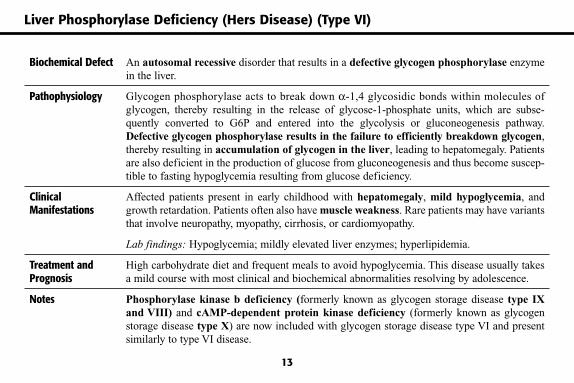
Liver Phosphorylase Deficiency (Hers Disease) (Type VI)
13
Biochemical Defect An autosomal recessive disorder that results in a defective glycogen phosphorylase enzyme in the liver.
Pathophysiology Glycogen phosphorylase acts to break down α-1,4 glycosidic bonds within molecules of glycogen, thereby resulting in the release of glycose-1-phosphate units, which are subse-quently converted to G6P and entered into the glycolysis or gluconeogenesis pathway.Defective glycogen phosphorylase results in the failure to efficiently breakdown glycogen,thereby resulting in accumulation of glycogen in the liver, leading to hepatomegaly. Patients are also deficient in the production of glucose from gluconeogenesis and thus become suscep-tible to fasting hypoglycemia resulting from glucose deficiency.
Clinical Manifestations
Affected patients present in early childhood with hepatomegaly, mild hypoglycemia, and growth retardation. Patients often also have muscle weakness. Rare patients may have variants that involve neuropathy, myopathy, cirrhosis, or cardiomyopathy.
Lab findings: Hypoglycemia; mildly elevated liver enzymes; hyperlipidemia.
Treatment and Prognosis
High carbohydrate diet and frequent meals to avoid hypoglycemia. This disease usually takes a mild course with most clinical and biochemical abnormalities resolving by adolescence.
Notes Phosphorylase kinase b deficiency (formerly known as glycogen storage disease type IX and VIII) and cAMP-dependent protein kinase deficiency (formerly known as glycogen storage disease type X) are now included with glycogen storage disease type VI and present similarly to type VI disease.

14
2
A 3-month-old girl is brought to your pediatric clinic because she is not feeding well and has poor
weight gain. On physical examination, you find that she has a large tongue and mild hepatomegaly.
She is also significantly flaccid and hypotonic. Fearing an autosomal recessive glycogen storage
disease, you order an ECG, which shows a short PR interval and a wide QRS interval. You order a
series of serum studies, expecting to see elevated creatine kinase, aspartate transaminase, and lactate
dehydrogenase. You fear that the child has a poor prognosis, and will likely suffer cardiopulmonary
failure and death before her first birthday.

Pompe Disease (Type II)
14
BiochemicalDefect
An autosomal recessive disorder caused by a deficiency of lysosomal acid α-1,4-glucosidase.
Pathophysiology Lysosomal acid α-1,4-glucosidase (acid maltase) is an enzyme responsible for the degra-dation of glycogen in lysosomal vacuoles. If lysosomal acid α-1,4-glucosidase is defective, there is resulting accumulation of lysosomal glycogen in the skeletal muscle, cardiac muscle, liver, and kidneys, which leads to myopathies, cardiomyopathy, and hepatic dysfunction.
Clinical Manifestations
There are several forms of Pompe disease. In infantile-onset disease (most severe), patients present with hypertrophic cardiomyopathy, hypotonia and myopathy, failure to thrive, macroglossia (large tongue), and hepatomegaly. Death occurs by 1 year of age. The juvenile form typically presents as delayed motor milestones and difficulty in walking followed by swallowing difficulties and proximal muscle weakness with death by the third decade. The adult form presents as a slowly progressive proximal muscle weakness with truncal involvement. There is no cardiac involvement in the juvenile or adult form.
Lab findings: Elevated serum creatine kinase; elevated aspartate transaminase; elevated lactate dehydrogenase; muscle biopsy shows vacuoles staining positively for glycogen.
Treatment No effective treatment for the infantile form. High-protein diet for the juvenile and adult forms. Ventilatory support as needed.
Notes Other glycogenosis diseases are characterized by cytoplasmic accumulation of glycogen, whereas Pompe disease is the only glycogenosis disease that is characterized by lysosomal accumulation of glycogen.

15
2
A 7-year-old boy presents to your pediatric clinic complaining of muscle weakness of 1-year duration.
His parents are concerned because he gets tired very quickly when playing with his classmates in
the yard. He is an only child, and his mother’s pregnancy was full-term with no complications. On
physical examination, you note that he has moderate hepatosplenomegaly, is of short stature, and
has marked muscle wasting. At his last clinic visit, serum studies revealed hypoglycemia, hyperlip-
idemia, a fasting ketosis, and elevated liver transaminases. You consider the possibility of a glyco-
gen storage disease and suggest a high-carbohydrate, high-protein diet while awaiting definitive
DNA-based analyses.

Cori Disease (Type III)
15
Biochemical Defect An autosomal recessive disorder that is caused by a deficiency of glycogen debranching enzyme, α-1,6-glucosidase.
Pathophysiology Glycogen debranching enzyme, α-1,6-glucosidase, is an enzyme that aids in glycogen degradation by breaking α-1,6 bonds. When this debranching enzyme is defective, glycogen breakdown is incomplete, and abnormal glycogen with short outer chains accu-mulates in the liver and muscle, leading to hepatomegaly. Hypoglycemia results from inef-fective glycogen breakdown.
Clinical Manifestations
Patients present with hepatosplenomegaly, hypoglycemia, hyperlipidemia, and growth retardation. Other symptoms include short stature, skeletal myopathy (progressive muscle wasting), and cardiomyopathy. Hepatomegaly improves with age and disappears after puberty.
Lab findings: Hypoglycemia; hyperlipidemia; elevated liver enzymes in childhood; fasting ketosis.
Treatment High-carbohydrate meals with cornstarch or nocturnal gastric drip feedings for hypoglyce-mia. High-protein diet during the day plus overnight protein infusion for myopathy.
Notes Cori disease is of high prevalence in non-Ashkenazi Jews of North African descent.

16
2
An 8-month-old boy is brought to your office by his parents, who are concerned that he is not feed-
ing well. Examination reveals a listless child, who is small for his age. You also notice an enlarged
liver and spleen on abdominal examination as well as an enlarged heart on chest x-ray. An electro-
cardiogram shows frequent premature ventricular contractions. When laboratory testing reveals a
deficiency in the glycogen branching enzyme, you inform the parents that their son is afflicted with
an autosomal recessive disorder of metabolism and recommend a consultation with a pediatric
hepatologist.

Andersen Disease (Type IV)
16
BiochemicalDefect
An autosomal recessive disorder that is caused by a deficiency of the glycogen branch-ing enzyme, transglucosidase.
Pathophysiology Glycogen branching enzyme, transglucosidase, is an enzyme that aids in the produc-tion of glycogen. When this enzyme is defective, long, unbranched glucose chains are formed and result in the deposition of this defective glycogen in the liver, heart, and nervous system.
ClinicalManifestations
Patients present in infancy with hepatosplenomegaly and failure to thrive. End-organ damage can lead to liver failure and cirrhosis. There are some rare neuromuscular variants, which present with muscle weakness and wasting.
Lab findings: Elevated liver enzymes in childhood.
Treatment andPrognosis
Symptomatic treatment of liver failure, including possible liver transplantation. Prognosis is poor.
Notes

17
2
A 22-year-old man presents to your ambulatory clinic complaining of painful muscle cramps when
walking eight flights of stairs to his new apartment. On further investigation, you learn that he also
experiences these severe muscle cramps after lifting weights in the gym. On several occasions, he
has had reddish-purple urine after exercising. You suspect that this patient may suffer from an
autosomal recessive glycogen storage disease, and you obtain a serum sample to test for elevated
serum creatine kinase at rest. While awaiting the test results, you suggest that the patient avoid
strenuous exercise and eat a high-protein diet.

McArdle Disease (Type V)
17
Biochemical Defect An autosomal recessive disorder caused by a deficiency of muscle glycogenphosphorylase.
Pathophysiology Muscle glycogen phosphorylase is responsible for breaking the α-1,4 linkages of glycogen in muscle. Deficiency of muscle phosphorylase results in deficient glycogen breakdown, leading to glycogen accumulation in muscle. Without effective glycogen breakdown, the body has to use other means to generate ATP (often through the break-down of muscle fibers), thereby resulting in eventual muscle degradation.
Clinical Manifestations Patients present in adulthood with exercise intolerance and muscle cramps. Symptoms are triggered by brief exercise of great intensity (eg, sprinting) or less intense but sustained activity (eg, climbing stairs). Half of patients report burgundy-colored urine after exer-cise (myoglobinuria as caused by muscle breakdown).
Lab findings: Elevated serum creatine kinase even at rest.
Treatment Avoid strenuous exercise. Augment exercise tolerance by aerobic training or by prior ingestion of glucose or sucrose. A high-protein diet may increase exercise endurance.
Notes

18
2
A 6-year-old girl is brought to an urgent care clinic by her parents, complaining of severe muscle
cramps in both legs after her weekly soccer game. Upon speaking further with the patient, you
discover that she has been suffering from muscle cramps as well as nausea after every soccer prac-
tice for the last year. Her mother also reports that the patient’s urine is reddish in color at times.
When laboratory studies demonstrate evidence of hyperuricemia, hemolytic anemia, and elevated
creatine kinase levels, you begin to suspect that the patient may be suffering from a deficiency of
an enzyme in the glycolysis pathway.

Tarui Disease (Type VII)
18
Biochemical Defect An autosomal recessive disorder caused by a deficiency of muscle phosphofructokinase.
Pathophysiology Phosphofructokinase catalyzes the conversion of fructose-6-phosphate to fructose-1, 6-diphosphate during glycolysis. When phosphofructokinase is absent, glycolysis is significantly impaired.
Clinical Manifestations Patients present in childhood with exercise intolerance and muscle cramps and weakness after exercise. Many patients report burgundy-colored urine after exercise (myoglobinuria as caused by muscle breakdown) as well as nausea and vomiting.
Lab findings: Hyperuricemia that is worsened with exercise; hemolytic anemia; elevatedcreatine kinase levels.
Treatment and Prognosis
Avoid strenuous exercise. Consider a high-protein diet. Usually does not progress to severe disability, although a rare infantile form has been reported and is usually fatal.
Notes Tarui disease is more prevalent among people of Ashkenazi Jewish descent.

19
2
A 4-year-old boy is brought to your pediatric clinic because his mother is concerned about a pos-
sible developmental delay. On physical examination, you find the patient to have marked hypotonia,
an ataxic gait, and choreoathetosis. Ophthalmologic examination reveals poor visual tracking,
grossly disconjugate eye movements, and poor pupillary response bilaterally. You order a series of
laboratory tests, which reveal the presence of a lactic acidosis. You immediately become concerned
that this patient may suffer from a defect of carbohydrate metabolism, and you decide to check
serum levels of pyruvate to confirm your suspicion.

Pyruvate Dehydrogenase Complex Deficiency
19
Biochemical Defect An autosomal recessive disorder that is caused by a deficiency in the pyruvate dehydro-genase complex.
Pathophysiology The pyruvate dehydrogenase complex is responsible for converting pyruvate to acetyl CoA during carbohydrate metabolism. Because acetyl CoA is necessary for citrate production, a deficiency in this enzymatic complex will limit citrate production. Since citrate is the first substrate in the citric acid cycle, the cycle cannot proceed, and an energy deficit develops in the CNS, leading to neurologic dysfunction. A backup of substrates also develops (including lactate and pyruvate) and results in a lactic acidosis.
Clinical Manifestations
Progressive neurologic symptoms usually start in infancy but may be evident at birth or in late childhood. These symptoms may include developmental delay, intermittent ataxia,poor muscle tone, abnormal eye movements, and seizures. It is often exacerbated in alcoholics because of thiamine deficiency.
Lab findings: High blood lactate and pyruvate levels; lactic acidosis.
Treatment Increase the intake of high-fat foods with ketogenic nutrients.
Notes Lysine and leucine are the two purely ketogenic amino acids, the catabolism of which leads to products that can be used in the citric acid cycle without having to be routed through the pyruvate dehydrogenase complex first.

20
2
A 36-year-old Mediterranean man presents to your clinic with increased fatigue and weakness of
2 days duration. He was recently tested for tuberculosis exposure and was PPD positive with a
normal chest x-ray film. He just started anti-TB prophylaxis medications the week before. On
physical examination, he is tachycardic, appears jaundiced, and has mild splenomegaly. You order
serum studies, which show low hemoglobin, low hematocrit, and elevated indirect bilirubin. You
begin to suspect that this patient suffers from an X-linked recessive disorder triggered by his current
TB prophylaxis, and you decide to consult an infectious disease specialist about alternative regimens
that will not cause his current symptoms.

Glucose-6-Phosphate Dehydrogenase Deficiency
20
Genetic Defect An X-linked recessive disorder caused by a deficiency in G6PD.
Pathophysiology G6PD catalyzes the oxidation of glucose-6-phosphate to 6-phosphogluconate in the HMP shunt pathway while concomitantly reducing NADP+ to NADPH. Deficiency in G6PD leads to decreased NADPH, a required cofactor in many biosynthetic reactions. NADPH maintains glutathione in its reduced form, which, in turn, acts as a scavenger for dangerous oxidative metabolites in the cell and converts harmful hydrogen peroxide to water. Patients with G6PD deficiency have increased hemolysis of red blood cells, which rely heavily on G6PD activity as the only source of NADPH for protecting against oxidative stresses.
Clinical Manifestations
Most patients are asymptomatic until undergoing an oxidative stress (causes include fava beans, sulfamethoxazole, primaquine, anti-TB drugs), which results in hemolytic anemia. Patients may also report a history of jaundice, gallstones (from increased hemolysis), fatigue, and splenomegaly.
Lab findings: Normocytic, normochromic anemia; hemoglobinemia; indirect bilirubinemia; decreased serum haptoglobin levels; peripheral smear shows spherocytes and Heinz bodies(hemoglobin precipitates within red blood cells).
Treatment Discontinuation of precipitating agent; oxygen and bed rest.
Notes G6PD deficiency is more prevalent among African-Americans and those of Mediterranean descent and has been associated with protection against malaria.

21
2
A 14-year-old boy presents for a routine physical examination to participate in high school athletics.
He has no known medical history, and his family history is significant for type 2 diabetes in a
maternal uncle. His physical examination is completely unremarkable. His laboratory studies are
within normal limits except for positive reducing sugar found in both serum and urine. Nevertheless,
his blood glucose level was normal at 85. When you inform the patient about this finding, he is
concerned about possible diabetes. You assure him that it is unlikely and that further testing will
most likely show a benign condition for which he needs no treatment.

Essential Fructosuria
21
Biochemical Defect An autosomal recessive disorder caused by a deficiency in fructokinase.
Pathophysiology Fructokinase converts fructose to fructose-1-phosphate in the fructose metabolism pathway. The deficiency of fructokinase activity in the liver and intestine significantly reduces the capacity to assimilate fructose into cells.
Clinical Manifestations Patients are usually asymptomatic, and the disease comes to light as an incidental finding.
Lab findings: Positive reducing sugar in the blood and urine after meals rich in fructose.
Treatment No treatment is necessary.
Notes Essential fructosuria may be confused with diabetes mellitus if the nature of the reducing sugar in the urine is not defined.

22
2
A 6-month-old girl is brought to your pediatric clinic because her mother noticed that the baby has
seemed lethargic and irritable for the last several weeks. The mother had just begun feeding the
baby fruit juices several weeks before this visit. On physical examination, the child is slow in her
movements, mildly jaundiced, and small for her age. She also has mild hepatomegaly. You are
concerned that the symptoms started after the ingestion of fruit juices, and you order serum and
urine studies, which you suspect will show hypoglycemia and fructosemia. While you await the
results of the laboratory tests, you tell the mother to stop giving the child any fruit juices in her diet
because you believe that the patient may be suffering from an inherited enzyme deficiency that
alters her metabolism of certain carbohydrates.

Fructose Intolerance
22
Biochemical Defect An autosomal recessive disorder caused by a deficiency of fructose-1,6-bisphosphatealdolase B in the liver, kidney, and intestine.
Pathophysiology Fructose-1,6-bisphosphate aldolase catalyzes the hydrolysis of fructose-1-phosphate and fructose-1,6-bisphosphate into three-carbon sugars (dihydroxyacetone phosphate, glyceraldehyde-3-phosphate, glyceraldehyde) in the fructose metabolism pathway. Deficiency of this enzyme causes the rapid accumulation of fructose-1-phosphate. Fructose-1-phosphate has a toxic effect on the liver, impeding hepatic function (eg, impaired glycolysis, glycogenolysis, and gluconeogenesis).
Clinical Manifestations Patients are asymptomatic until fructose or sucrose is ingested (usually from fruit, fruit juice, table sugar, or sweetened cereal). Clinical manifestations include lethargy, irritability, jaundice, hepatomegaly, vomiting, and convulsions. Complications include cirrhosis and kidney failure.
Lab findings: Hypoglycemia; fructosemia; prolonged clotting time; hypoalbuminemia; elevated bilirubin and transaminases.
Treatment Dietary elimination of all sources of sucrose, fructose, and sorbitol.
Notes

23
2
A 1-month-old foreign-born boy is brought to your pediatric clinic by his mother, who tells you that
the child is vomiting after feedings and has been gaining weight poorly. His mother did not receive
proper prenatal care, and the child did not have screening tests before or after delivery. On physical
examination, the infant is small for his age, appears jaundiced, and has mild hepatomegaly. While
normally you would think of possible intestinal obstruction, you notice on physical examination that
the child’s lenses are clouded as though he were developing cataracts. You order serum and urine
studies, specifically looking for hypoglycemia, galactosuria, and aminoaciduria. You believe that
the child is suffering from a deficiency of galactose-1-phosphate uridyl transferase and that the
patient’s diet should be limited in intake of milk and other foods rich in lactose or galactose.

Classic Galactosemia
23
Biochemical Defect An autosomal recessive disorder caused by a deficiency of galactose-1-phosphate uridyl transferase.
Pathophysiology Galactose-1-phosphate uridyl transferase aids in the conversion of galactose-1-phosphateinto glucose-1-phosphate in the galactose metabolism pathway. A deficiency of galactose-1-phosphate uridyl transferase results in the buildup of galactose-1-phosphate, galactose, and galactitol. These substances are toxic to the parenchymal cells of the kidney, liver, lens, spleen, and brain.
Clinical Manifestations
Infants present with several nonspecific findings, including lethargy, irritability, feeding difficulties, poor weight gain, jaundice, hepatomegaly, ascites, splenomegaly, convulsions, cataracts, and mental retardation.
Lab findings: Hypoglycemia; aminoaciduria; galactosuria; markedly reduced galactose-1-phosphate uridyl transferase activity.
Treatment Elimination of galactose from the diet.
Notes Neonates are routinely screened for galactosemia. The test consists of a demonstration of a reducing substance in urine specimens collected while the patient is receiving milk or formula containing lactose.

24
2
A 2-year-old foreign-born girl is brought to your pediatric clinic for a routine checkup. Her mother
tells you that the child has not seen a doctor since she was born. The child’s physical examination is
normal, except for clouding of her eye lenses consistent with cataracts. She is of normal height and
weight and has met all of her developmental milestones up to this point. While you ask the nurse to
draw up a battery of immunizations, you also ask for serum studies, including a serum galactose
level. While you await the results of the laboratory testing, you tell the parents to restrict galactose
in the child’s diet and reassure them that cataracts are the only manifestations of their child’s
hereditary disorder.

Galactokinase Deficiency
24
Biochemical Defect A benign autosomal recessive disorder caused by deficiency of galactokinase.
Pathophysiology Galactokinase is required to phosphorylate galactose into galactose-1-phosphate dur-ing galactose metabolism. When galactokinase is deficient, galactose builds up. In the lens of the eye, galactose reductase and aldose reductase convert this excess galactose into galacti-tol. Galactitol causes the entry of water into the eye by osmosis, leads to the development of cataracts.
Clinical Manifestations
In contrast to the multiple organ systems affected in classic galactosemia, infant cataractsare usually the sole manifestation of galactokinase deficiency.
Lab findings: Elevated blood galactose levels.
Treatment Dietary restriction of galactose.
Notes

25
2
A 14-year-old Asian-American boy presents to your pediatric clinic concerned about diarrhea that
has persisted for more than 1 month. He reports that the diarrhea occurs about 30 minutes after he
eats his bowl of cereal with milk each morning. He states that the stools are bulky and frothy, but
there is no gross blood. He also has a lot of gas and bloating after eating breakfast. He reports
resolution of symptoms for the remainder of the day. On further questioning, the patient does not
eat yogurt, ice cream, or any other dairy products throughout the day. You believe that the patient
has a common enzyme deficiency and that his intestinal lining is not absorbing an ingested sugar
properly. You suggest that he try a commercial enzyme substitute with each bowl of cereal or a
different brand of milk that has the enzymes necessary for its digestion.

Lactase Deficiency
25
Biochemical Defect Caused by reduced genetic expression of the enzyme lactase-phlorhizin hydrolase.
Pathophysiology Lactase-phlorhizin hydrolase is involved in the rate-limiting step of lactose digestion. Lactose is hydrolyzed by intestinal lactase to glucose and galactose on the microvillus membrane of the intestinal adsorptive cells. Lactose that is not absorbed by the small bowel, because of the absence or deficiency of the lactase enzyme, is passed rapidly into the colon, thereby leading to the entry of water into the colon by osmosis and the develop-ment of diarrhea.
Clinical Manifestations After ingestion of lactose-containing products (eg, milk), patients have diarrhea,abdominal pain, and flatulence. Stools are often bulky, frothy, and watery.
Treatment Reduced dietary lactose intake; commercial enzyme substitute; alternative calcium and nutrient sources.
Notes This common problem has a prevalence of 10%-20% among Caucasians, 80%-95% among Native Americans, 65%-75% among African-Americans, 90% among Asian-Americans, and 50% among Hispanics.

26
3
LIPID METABOLISM
GENERAL CONCEPTS
Fatty acid synthesisCitrate shuttle
Fatty acid oxidationCarnitine shuttleLipid transport
Lipoprotein and apolipoprotein functionCholesterol synthesis
Sphingolipid synthesisSphingolipid degradationPhospholipid synthesis
DISEASES
Inherited HyperlipidemiasFamilial hypercholesterolemia
HypertriglyceridemiaFamilial hyperchylomicronemia
Mixed hypertriglyceridemiaCombined hypercholesterolemia and
hypertriglyceridemiaDysbetalipoproteinemia
Sphingolipid Storage DiseasesHurler diseaseHunter disease
Sanfilippo syndromeSly syndrome
Tay-Sachs diseaseSandhoff disease
Fabry diseaseGaucher disease
Niemann-Pick diseaseFarber diseaseI-cell disease
Krabbe diseaseMetachromatic leukodystrophy

This page intentionally left blank

3
FATTY ACID SYNTHESIS
27
Palmitate (16C)
H2O
CO2NADPH
NADP+
NADPH
NADP+
After 7 Cycles
MalonylCoA
Fatty AcidSynthaseComplex
Fatty AcidSynthaseComplex
MalonylMoiety
SCO[CH2]nCH3
SCO[CH2](n+1)CH3
CoA
Malonyl CoATransferase
AcetylCoA
ATP
ADPAcetyl CoACarboxylasewith Biotin

27
FATTY ACID SYNTHESIS
• Location: Fatty acid synthesis takes place in the cytosol and is carried out by a multienzyme complex called FAS. • Substrates (to make one palmitate):
� 8 acetyl CoA � 14 NADPH � 7 ATP
• Products:� 1 molecule of palmitate (16-carbon fatty acid) � 7 H 2 O
• Pathway: Acetyl CoA is converted to malonyl CoA by acetyl CoA carboxylase. Malonyl CoA is transferred to FAS. Through a series of condensation, reduction, and dehydration reactions, the two carbons of malonyl CoA are added to the growing fatty acyl moiety on FAS. FAS is then recharged with another malonyl moiety, and the cycle continues. Each turn of the cycle results in the addition of a two-carbon group to the fatty acid moiety as well as the use of one ATP, one acetyl CoA, and two NADPH. When the cycle has completed seven turns, the 16-carbon fatty acid (palmitate) is released from FAS.
• Important enzymes:� Acetyl CoA carboxylase : Transforms acetyl CoA to malonyl CoA with the use of biotin and bicarbonate as
cofactors. Requires one ATP. � Malonyl CoA transferase : Transfers the malonyl CoA molecule to FAS. � FAS : This collection of enzymes transfers the two carbons of malonyl CoA to the carboxyl end of the growing
chain of the fatty acyl moiety. Requires two NADPH. • Activators: Insulin stimulates fatty acid synthesis by dephosphorylating and, therefore, activating acetyl CoA
carboxylase . • Inhibitors: Glucagon and epinephrine inhibit fatty acid synthesis by inactivation of acetyl CoA carboxylase .

28
3
FATTY ACID OXIDATION
R[CH2]n-1CO--CoA
H2O
FAD
FADH2
NADH
NAD+
CoA
Acetyl-CoA
Citric AcidCycle
ElectronTransport
Chain
NADH + FADH2
Fatty AcidR[CH2]nCO--CoA
Acyl CoADehydrogenase
Enoyl CoAHydratase
3-Hydroxy-Acyl CoADehydrogenase
Acyl CoAAcyltransferase

28
FATTY ACID OXIDATION
• Location: β-Oxidation takes place in the mitochondria. • Substrates: Free fatty acids; H 2 O. • Products: One acetyl CoA, one NADH, and one FADH 2 for every removal of a two-carbon group from the fatty acid chain. • Pathway: In the mitochondria, the fatty acid undergoes a series of oxidation and hydration reactions, which results in the
removal of a two-carbon group (in the form of acetyl CoA) from the fatty acid chain as well as the formation of one NADH and one FADH 2 , which enter the electron transport chain to form five ATP. The acetyl CoA formed will enter the citric acid cycle and then the electron transport chain, leading to the formation of another 12 ATP. The cycle continues, with each turn of the cycle removing another two-carbon group, until the formerly long-chain fatty acid has been reduced to acetyl CoA or propionyl CoA. Propionyl CoA can be converted to succinyl CoA through three enzymatic events, which require biotin and vitamin B 12 as cofactors, and then succinyl CoA can enter the citric acid cycle.
• Important enzymes:� Acyl CoA dehydrogenase : Forms a double bond between the α and β carbon atoms in the fatty acid chain. Produces
one FADH 2 . � Enoyl CoA hydratase : Incorporates a water molecule into the fatty acid chain, thereby breaking the double bond
between the α and β carbon atoms. � 3-Hydroxy-acyl CoA dehydrogenase : Dehydrogenates the fatty acid chain again, thereby forming a double bond
between the β carbon and the oxygen molecule. Produces one NADPH. � Acyl CoA acyltransferase : Cleaves acetyl CoA off the end of the fatty acid chain with the addition of CoA to the β carbon.
• Activators: Epinephrine stimulates β-oxidation by activating a cAMP–dependent protein kinase, which leads to the phosphorylation and thus activation of HSL. When activated, HSL releases fatty acids and glycerol from adipose tissue for β-oxidation.
• Inhibitors: Insulin inhibits β-oxidation by dephosphorylating HSL and thus inhibiting the release of fatty acids from adipose tissue.

29
3
CITRATE SHUTTLE
AcetylCoA
Citrate Citrate
Oxaloacetate
Oxaloacetate
Malate
Pyruvate Pyruvate
Cytosol MitochondrialMatrix
ATP
ADP
ATP
ADP
NADH
NAD+
NADP+
NADPH
CO2 CO2
AcetylCoA
• Description: The citrate-malate-pyruvate shuttle functions to transport acetyl CoA from the mito-chondria into the cytosol for use during fatty acid synthesis.
• Function: Acetyl CoA combines with oxaloace-tate to form citrate, which then crosses the mito-chondrial membrane. Once in the cytosol, citrate is broken down to reform acetyl CoA and oxalo-acetate. Oxaloacetate is then transported back into the mitochondria in the form of pyruvate. During this process, two ATP and one NADH are used, while one NADPH is formed.
• Purpose: Fatty acid synthesis takes place in the cytosol and uses acetyl CoA as a substrate. Because acetyl CoA is primarily formed in the mitochondria (usually by pyruvate dehydroge-nase ), the citrate shuttle is necessary to transport acetyl CoA into the cytosol, where fatty acid syn-thesis occurs. Furthermore, the citrate shuttle produces NADPH, which is subsequently used in the process of fatty acid synthesis.

29
CARNITINE SHUTTLE
Fatty-acylCoA
Carnitine
CoA Fatty-acylCarnitine
Fatty-acylCarnitine
CoA
Fatty-acylCoACarnitine
Cytosol Mitochondrial Matrix
CarnitineAcyl
Transferase I
CarnitineAcyl
Transferase II
b-Oxidation
• Description: The carnitine transport system shut-tles long-chain fatty acids from the cytosol into the mitochondria.
• Function: The fatty acyl chain is enzymatically attached to carnitine in the cytosol via carnitineacyl transferase I , and then shuttled across the mitochondrial membrane. Once inside the mito-chondria, the fatty acids are released from carni-tine by the enzyme, carnitine acyl transferase II .
• Purpose: Because long-chain fatty acids are unable to enter the mitochondria, the carnitine transport system allows for the movement of fatty acids in the mitochondrial matrix where fatty acid oxidation occurs.
• Diseases: Inherited defects in the carnitine shuttle present with hypoglycemia, muscle pain, and muscle atrophy because of the accumulation of fat in muscle tissue. Affected infants benefit from being fed fat with medium-chain triacylglycerols (eg, butter fat) because medium-sized fatty acids can bypass the carnitine shuttle.

30
3
EXOGENOUS LIPID TRANSPORT
Cholesterol
TriglyceridesChylomicron
Free Fatty Acids
Glycerol
Chylomicron Remnant(rich in cholesterol)
HDL
LiverPeripheral
TissuesAdipose
Cells
Intestine Lipoprotein
Lipase
• Pathway: Dietary cholesterol and triglycerides are absorbed from the intestinal lumen into the mucosal cells of the small intestine, where they are incorpo-rated into chylomicrons and released into the blood-stream. Chylomicrons are degraded by LPL, which is present on the capillary endothelium of muscle and adipose tissue, into glycerol, free fatty acids, and a chylomicron remnant. The free fatty acids are either stored in adipose cells or taken up by muscle or other peripheral tissues. The glycerol is transferred to the liver. The remnant of the chylomicron, which is rich in cholesterol molecules, is then either directly taken up by the liver through endocytosis or transported to the liver by HDL.
• Fates of dietary cholesterol and triglycerides:� Free fatty acids: Either used as fuel in muscle or
other peripheral tissues or stored as triacylgycer-ols in adipose tissue.
� Glycerol: Transferred to the liver where it is used in glucose synthesis.
� Chylomicron remnant: Absorbed by the liver where the cholesterol within the remnant is either converted to bile acids or transformed to VLDLs.
30
ENDOGENOUS LIPID TRANSPORT
Cholesterol
TriglyceridesChylomicron
Free Fatty Acids
Glycerol
Chylomicron Remnant(rich in cholesterol)
HDL
LiverPeripheral
TissuesAdipose
Cells
Intestine Lipoprotein
Lipase
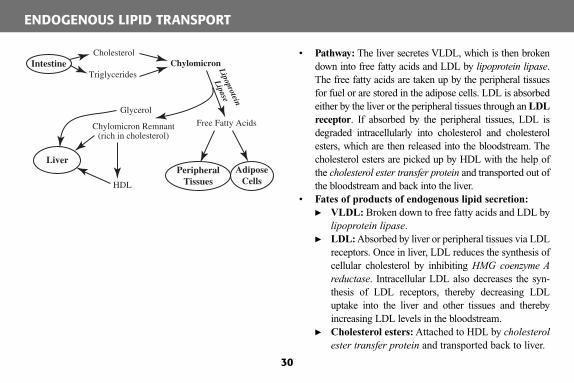
• Pathway: The liver secretes VLDL, which is then broken down into free fatty acids and LDL by lipoprotein lipase . The free fatty acids are taken up by the peripheral tissues for fuel or are stored in the adipose cells. LDL is absorbed either by the liver or the peripheral tissues through an LDL receptor . If absorbed by the peripheral tissues, LDL is degraded intracellularly into cholesterol and cholesterol esters, which are then released into the bloodstream. The cholesterol esters are picked up by HDL with the help of the cholesterol ester transfer protein and transported out of the bloodstream and back into the liver.
• Fates of products of endogenous lipid secretion:� VLDL: Broken down to free fatty acids and LDL by
lipoprotein lipase . � LDL: Absorbed by liver or peripheral tissues via LDL
receptors. Once in liver, LDL reduces the synthesis of cellular cholesterol by inhibiting HMG coenzyme A reductase . Intracellular LDL also decreases the syn-thesis of LDL receptors, thereby decreasing LDL uptake into the liver and other tissues and thereby increasing LDL levels in the bloodstream.
� Cholesterol esters: Attached to HDL by cholesterol ester transfer protein and transported back to liver.

31
3
LIPOPROTEIN FUNCTION
Lipoprotein Source FunctionAssociated Enzymes or Receptors
Consequencesof Excess Lipoprotein
Chylomicron Secreted by intestine
Transports exogenous lipids to liver, adipose tissue, and peripheral tissues
Broken down by lipoproteinlipase into glycerol, free fatty acids, and chylomicronremnants
Pancreatitis EruptivexanthomasLipemia retinalis
VLDL Secreted by liver Transports endogenous triglyc-erides, LDL, and cholesterol esters from the liver to other tissues
Broken down by lipoproteinlipase into free fatty acids and LDL particles
Pancreatitis
LDL Formed from breakdown of VLDL particles
LDL particles are absorbed by tissues when cellular choles-terol is needed
Absorbed into cells via the LDLreceptor, which is down-regu-lated by increased LDL levels within the cell
AtherosclerosisArcus corneae Xanthomas
HDL Secreted by liver Transports cholesterol to liver where it is secreted into bile or to steroid hormone-producing tissues where it is utilized in steroid hormone production
Works in conjunction with cho-lesterol ester transfer protein,which binds free cholesterol esters in the bloodstream, to transport cholesterol back to the liver
None

APOLIPOPROTEIN FUNCTIONS
31
Apolipoprotein Function in Lipid MetabolismAssociated with Lipoproteins Associated Metabolic Diseases
A (Apo A-I, A-II, A-IV)
A-I: activates LCAT , which acts to trap cholesterol esters within HDL A-II and A-IV: interact with PLTP to transfer phospholipids to HDL
All Apo As are found in HDL
A-I: defects lead to HDL deficiencies A-II and A-IV: defects lead tohypercholesterolemia
B(Apo B-48, B-100)
B-48: involved in synthesis and secretion of chylomicrons from small intestine B-100: binds LDL receptor to facili-tate LDL binding
B-48: chylomicrons B-100: VLDL; LDL
B-48: deficiency leads to abetalipopro-teinemia (unable to absorb dietary fats) B-100: defective B-100 leads to increased LDL levels
C(Apo C-I, C-II, C-III)
C-I: inhibits cholesterol estertransfer proteinC-II: activates lipoprotein lipaseC-III: inhibits lipoprotein lipase
C-I: VLDL, HDL;chylomicronsC-II: VLDL;chylomicronsC-III: VLDL
C-I: increased levels lead tohypercholesterolemiaCII: deficiencies lead tohyperlipoproteinemia type Ib C-III: increased levels lead tohypertriglyceridemia
E(Apo E2, E3, E4)
Synthesized in liver; act totransport triglycerides andcholesterol to the liver
Found in chylomi-crons and VLDLs
Deficiency results indysbetalipoproteinemia

32
3
CHOLESTEROL SYNTHESIS
Acetyl CoA + Acetoacetyl CoA
HMG CoA
Mevalonic Acid
Cholesterol
HMG-CoASynthase
HMG-CoAReductase
2 NADPH
2 NADP+
CoA
• Location: Cholesterol synthesis takes place in the liver and intestinal mucosa.
• Substrates: Acetyl CoA; acetoacetyl CoA. • Products:
� Cholesterol: Oxidized to bile acids in liver; precursor for steroid hormones.
� Mevalonic acid: Precursor for terpenes (eg, vitamins A and K, coenzyme Q).
• Regulation: HMG CoA reductase is inhibited by high levels of cholesterol. This enzyme is the pharmacologic target of lovastatin and other drugs in that class.
• Circulation: Two-thirds of plasma cholesterol is esterified by LCAT, an enzyme activated by apo A. Cholesterol esterification by LCAT traps choles-terol in HDL and prevents membrane cholesterol uptake, which can lead to alterations in membrane permeability.

32
SPHINGOLIPID SYNTHESIS
• Location: Sphingolipid synthesis occurs in the cytosol. • Substrates: Serine; palmitoyl CoA. • Products:
� Sphingomyelin: Principal lipid of nervous tissue membranes.
� Gangliosides: Acidic glycosphingolipids found in ganglion cells of the nervous system.
� Sulfatides: Acidic glycosphingolipids found pri-marily in nervous tissue.
� Cerebrosides: Neutral glycosphingolipids found primarily in the CNS myelin.
• General pathway: There are two phases in sphingo-lipid synthesis. The first phase involves the formation of the ceramide core, which is produced by the combi-nation of palmitoyl-CoA, serine, and a fatty acyl-CoA molecule. There are several possible pathways in the second phase of sphingolipid synthesis, which result in the formation of the different sphingolipids. In general though, the second phase involves the addition of a specific compound (eg, phosphocholine, glucose, galactose, sulfate, etc) to the hydroxyl group on the terminal carbon of the ceramide molecule.
• Degradation: Normally degraded by lysosomes.
Serine + Palmitoyl-CoA
Sphingosine
Ceramide
Galactocerebroside+
Glucocerebroside
Sphingomyelin
Gangliosides+
Sulfatides
UDP-galactoseor
UDP-glucose
UDP
Phosphatidylcholine
Diacylglycerol
Acylation Step

33
3
SPHINGOLIPID DEGRADATION
GM1 ganglioside
GM2 ganglioside
GM3 ganglioside
Lactosylceramide
Globoside
Globotriaosylceramide
Digalactosylceramide
Galactosylceramide
Sulfatide
Ceramide
Sphingosine
Glucosylceramide
Sphingomyelin
GM1-β-galactosidase
Hexosaminidase A
Neuraminidase
GalCer-β-galactosidase
α-galactosidase A
β-hexosaminidase A and B
Glucocerebrosidase
Arylsulfatase AAcid Ceramidase
β-galactosylceramidaseSphingomyelinase
α-galactosidase A

33
IMPORTANT ENZYMES IN SPHINGOLIPID DEGRADATION
Enzyme Action
Associated Sphingolipid Storage Disease
Accumulated Substance Seen with Deficiency
GM1-β-galactosidase Breaks down G M1 ganglioside to G M2ganglioside
GM1 gangliosidosis G M1 ganglioside
Hexosaminidase Breaks down G M2 ganglioside to G M3ganglioside
Tay-Sachs disease G M2 ganglioside
Neuraminidase Breaks down G M3 ganglioside tolactosylceramide
Sialidosis G M3 ganglioside
GalCer-β-galactosidase Breaks down lactosylceramide to glucosylcer-amide
No clear associatedsyndrome
Lactosylceramide
Glucocerebrosidase Breaks down glucosylceramide to ceramide Gaucher disease Glucosylceramide
β-Hexosaminidase A and B
Breaks down globoside to globotriaosylcer-amide
Sandhoff disease Globoside
α-Galactosidase A Breaks down globotriaosylceramide to lactosyl-ceramideBreaks down digalactosylceramide to galacto-sylceramide
Fabry disease GlobotriaosylceramideDigalactosylceramide
Sphingomyelinase Breaks down sphingomyelin to ceramide Niemann-Pick disease Sphingomyelin
Acid ceramidase Breaks down ceramide to sphingosine Farber disease Ceramide
Arylsulfatase A Breaks down sulfatide to galactosylceramide Metachromaticleukodystrophy
Sulfatide
β-Galactosylceramidase Breaks down galactosylceramide to ceramide Krabbe disease Galactosylceramide

34
3
Glycerol-3-Phosphate
PhosphatidateTriacylglycerol
1,2-Diacylglycerol
Ethanolamine
Ethanolamine-PPhosphatidylEthanolamine
PhosphatidylCholine
Phosphatidylserine
Phosphatidylinositol
Inositol
EthanolamineSerine
3 SAM
CO2
ATP
ADP
Cytidine-P-P-P
P-P
CDP-Ethanolamine
H2O
CDP-Choline
CMP
1,2-Diacylglycerol-PATP
ADP
CDP-Diacylglcerol
CMP
Acyl CoA
2 Acyl CoA
2 CoA
Pi
CoA
CMP
Cytidine-P-P-P
P-P
PHOSPHOLIPID SYNTHESIS

34
PHOSPHOLIPID SYNTHESIS
• Location: Phospholipid synthesis occurs in the cytosol in the cells of the liver, intestine, and adiposetissue.
• Main substrate: (1,2-DAG) (which is derived from glycerol-3-phosphate). • Products:
� Phosphatidylinositol: Negatively charged phospholipid; when phosphorylated, it plays a major role in cell signaling.
� Phosphatidyl choline: Most abundant phospholipid; it is neutral; acts as key component of lipoproteins, as well as membranes of cells in several types of tissues; may have a role in cell-signaling.
� Phosphatidyl ethanolamine: Found in membranes in the cells of the nervous tissue (particularly white matter of the brain).
� Phosphatidylserine: Acidic phospholipid found mostly in membranes of myelin cells. • General pathway: All phospholipids are derived from 1, 2-DAG. The addition of a phosphorylated ethanol-
amine group to 1,2-DAG results in phosphatidyl ethanolamine. The addition of an activated choline group to 1,2-FAG results in phosphatidyl choline. The addition of an inositol group to phosphorylated 1,2-DAG results in phosphatidylinositol. Phosphatidylserine is formed by the exchange of a serine group for the etha-nolamine group on phosphatidyl ethanolamine.
• Degradation: Normally degraded by a family of enzymes known as phospholipases .

35
3 A 31-year-old man presents with complaints of sharp retrosternal chest pain when lifting heavy
objects or walking up several flights of stairs. The pain lasts for about 2 minutes at a time and radi-
ates up to his left jaw. He reports that the pain is relieved by rest. The patient reports that his father
and two paternal uncles had heart attacks during their 30s. His symptoms and ECG are consistent
with stable angina. While you prescribe nitroglycerin to treat the angina, you also order a serum
lipid panel because you are concerned that this patient may have a codominant genetic disorder that
is putting him at a very high risk for early-onset atherosclerotic heart disease.

Familial Hypercholesterolemia (Type IIa)
35
Biochemical Defect Causes include a codominant genetic disorder resulting from mutations in the gene forthe LDL receptor or an autosomal dominant genetic disorder associated with defective apoprotein B-100, a protein that facilitates binding of LDL to the LDL receptor.
Pathophysiology When LDL receptors or apoprotein B-100 are deficient, LDL is unable to be taken into the liver for processing. This results in increased plasma LDL levels, leading to intracellular and extracellular deposits of cholesterol, which result in xanthomas and xanthelasmas as well as premature atherosclerotic disease. Also, because the liver senses decreased LDL levels, it responds by secreting more IDL and VLDL (LDL precursors), resulting in increased LDL production.
Clinical Manifestations
Homozygotes develop severe atherosclerosis with heart disease in early or middle age . Other symptoms include tendon xanthomas of the Achilles and knuckle extensor tendons, tuberous xanthomas (soft, painless nodules) on elbows and buttocks, and xanthelasmas(barely elevated deposits of cholesterol on eyelids).
Lab findings: Elevated serum cholesterol level of 275-500 mg/dL and elevated LDL level; normal plasma triglycerides; normal HDL and VLDL levels.
Treatment Low-fat, low-cholesterol diet and exercise; HMG-CoA reductase inhibitor (atorvastatin or simvastatin) in combination with cholestyramine ; nicotinic acid can be added as a third agent.
Notes The homozygous form of the LDL receptor mutation is rare (1:1 000 000); however, HMG-CoA reductase inhibitors are ineffective in treating the homozygous form of this disorder.

36
3 A 34-year-old man presents to your clinic complaining of chronic, recurrent abdominal pain. He
has a history of pancreatitis, but his current symptoms do not suggest an acute process. Upon taking
a family history, you discover that several members of his family, including his mother and two sisters
suffer from an inherited metabolic disease. On funduscopic examination, you discover the presence
of lipemia retinalis. Abdominal examination shows moderate hepatosplenomegaly. You are con-
cerned that this patient may have a genetic disorder predisposing him to attacks of abdominal pain.
You order serum studies, looking for elevated fasting plasma triglycerides, along with tests for
elevated amylase and lipase. In addition to a low-fat diet and exercise, you also start the patient on
gemfibrozil.

Familial Hypertriglyceridemia
36
Biochemical Defect Familial hypertriglyceridemia (type IV) is an autosomal dominant disorder, but the underlying mutation has not been identified.
Pathophysiology Pathophysiology involves both reduced catabolism of triglyceride-rich lipopro-teins and overproduction of VLDL in the liver.
Clinical Manifestations Usually asymptomatic, but eruptive xanthomas (small nonpainful orange-red papules) and lipemia retinalis may appear with triglyceride levels >1000 mg/dL. Also associated with an increased risk of vascular disease.
Lab findings: Elevated fasting plasma triglycerides (200-750 mg/dL), elevated VLDL, elevated total cholesterol.
Treatment Fat-free diet; niacin, gemfibrozil, and/or fish oil supplements.
Notes Obesity, inactivity, alcohol use, and insulin resistance are associated with hyper-triglyceridemia.

37
3 A 45-year-old man with a medical history significant for obesity and diabetes mellitus presents to
your clinic complaining of an abnormal rash on his body. Physical examination reveals small
orange-red papules on his scalp, elbows, and knees that are not painful to the touch. You order
serum studies, which demonstrate elevated triglyceride levels as well as elevated levels of chylo-
microns and VLDL. You explain to the patient that he likely has a partial genetic deficiency of an
enzyme involved in lipid metabolism, and you start the patient on gemfibrozil and fish oil supple-
ments.

Familial Hyperchylomicronemia and Mixed Hypertriglyceridemia
37
Biochemical Defect Familial hyperchylomicronemia (type I) is an autosomal recessive disordercaused by an absence of either LPL (type Ia) or apoprotein C-II (type Ib).
Mixed hypertriglyceridemia (type V) is a heterogeneous disorder caused by a partial deficiency (as opposed to total deficiency) in either LPL or apoprotein C-II.
Pathophysiology Apo C-II activates LPL, which then hydrolyzes chylomicrons. A deficiency or absence in either apo C-II or LPL results in the accumulation of chylomicrons in the plasma.
Clinical Manifestations Usually asymptomatic, but eruptive xanthomas (small nonpainful orange-red papules), pancreatitis, and lipemia retinalis may appear with triglyceride levels >1000 mg/dL.
Lab findings : Elevated fasting plasma triglycerides (>750 mg/dL) associated with chylomicronemia . Type V will also have evidence of elevated VLDL.
Treatment Lifelong fat-free diet; niacin, gemfibrozil, or fish oil supplements.
Notes Obesity, inactivity, alcohol use, and insulin resistance are associated withhypertriglyceridemia.

38
3 A 38-year-old woman presents to your clinic 1 month after having been admitted to the hospital and
treated for a myocardial infarction. Although she is feeling well during this visit, she is concerned
about what may have led to a heart attack so early in her life. She eats a regular diet and exercises
twice a week. On physical examination, you notice cholesterol deposits in the palmar creases of
both her hands. She also has two very small tuberous xanthomas near her buttocks. You believe that
this patient suffers from an autosomal recessive disease and you order serum lipid studies, expect-
ing to find elevated VLDL and IDL levels in the presence of normal LDL and HDL levels. You
believe that the patient may benefit from niacin and clofibrate treatment.

Dysbetalipoproteinemia
38
Biochemical Defect Dysbetalipoproteinemia (type III) is an autosomal recessive disorder resulting from homozygosity for apo E-2, the binding-defective form of apo E.
Pathophysiology Apo E is involved in the hepatic uptake of chylomicron, VLDL, and IDL remnants. Defective apo E results in elevations in both VLDL triglyceride and VLDL cholesterol levels.
Clinical Manifestations Tuberous xanthomas; striae palmaris (deposits of cholesterol in palmar creases) are pathognomonic; vascular disease is present usually by the fifth decade.
Lab findings: Elevated VLDL and IDL ; chylomicron remnants in plasma; normal LDL and HDL.
Treatment Niacin and fibrates.
Notes

39
3 A 52-year-old man presents to your primary care office for establishment of care. He tells you that
his past medical history includes peripheral arterial disease, borderline diabetes mellitus, and high
cholesterol. Family history is significant for early-onset coronary artery disease as well as hyper-
lipidemia in several family members. Routine laboratory studies reveal moderately elevated total
cholesterol levels as well as moderately elevated triglyceride levels. You begin to suspect that this
patient’s early vascular disease may be related to an overproduction of apolipoprotein B-100 and
you suggest that he begin treatment with an HMG-CoA reductase inhibitor.

Familial Combined Hyperlipidemia
39
Biochemical Defect Familial combined hyperlipidemia (type IIb) is an autosomal dominant disorder; the underlying defect is currently unknown although a defective locus has been discovered on chromosome 1q21. The proband (initial case discovered within a fam-ily) typically has combined hyperlipidemia or isolated hypertriglyceridemia.
Pathophysiology The defect results in the overproduction of apo B-100 lipoproteins, which results in the increased circulation of VLDL particles in the blood.
Clinical Manifestations Type IIb: Usually asymptomatic until premature vascular disease appears by fifth decade; insulin resistance ; patients usually do not have xanthomas.
Lab findings: Moderately elevated triglyceride and total cholesterol levels .
Treatment HMG-CoA reductase inhibitors (ie, statins), cholestyramine (ie, bile sequestrants), and/or niacin.
Notes

40
3 A 1-year-old boy is brought to the pediatric ophthalmology clinic because his pediatrician noticed
corneal clouding on a routine physical examination. While evaluating the patient, you notice that he
is small for his age, has a large tongue, and has mild coarsening of his facial features. On ophthalmo-
logic examination, you find bilateral corneal opacities and papilledema. You believe that the patient
may be suffering from an autosomal recessive trait caused by a deficiency in a lysosomal enzyme.
Although the patient may benefit from symptomatic treatment for his eyes, you fear that his prog-
nosis is grim and that he will likely die in childhood.

Hurler Disease
40
Biochemical Defect An autosomal recessive disorder caused by deficiency in the enzyme α - L-iduronidase .
Pathophysiology α-L-iduronidase is a lysosomal enzyme necessary for the breakdown of GAGs. When this enzyme is deficient, there is a buildup of dermatan sulfate and heparan sulfate(GAGs that are linked to proteins in connective tissue). Dermatan sulfate and heparan sulfate tend to accumulate in the skin and bones (leading to physical deformities), and in the heart, liver, and brain (leading to abnormal functioning of these organs).
Clinical Manifestations Affected infants are normal at birth but exhibit mild coarsening of facial features and growth retardation in the first year. Distinguishing features include coarse facies , jointstiffness , short stature, and valvular heart disease . Other symptoms include hepatosple-nomegaly, corneal clouding , large tongue, developmental delay, dwarfism, hearing loss, and mental retardation.
Lab findings: Dermatan sulfate and heparan sulfate in the urine.
Treatment and Prognosis
Symptomatic therapies currently include corneal transplantation, heart valve replacement, and physical therapy for joint contractures. Bone marrow transplantation and enzyme replacement are experimental. Death usually occurs before age 10.
Notes Scheie syndrome is an adult variant of Hurler disease that is also caused by a deficiency of α-L-iduronidase.
Hurler disease, Scheie syndrome, Hunter disease, Sly syndrome, and Sanfilippo disease are considered mucopolysaccharidoses.

41
3 An 8-year-old boy is referred to your rheumatologic clinic by his pediatrician, who had noticed prom-
inent joint stiffness during a recent visit. On physical examination, you find that the patient has coarse
facial features, a large tongue, a small jaw, and marked hepatosplenomegaly. He also has a distinctive
nonpainful, pebbly skin lesion on his upper back. When asked to perform movements, he exhibits
remarkable joint stiffness for his age and is unable to touch his toes from a standing position. You order
a urinalysis, looking for dermatan sulfate and heparan sulfate, which you suspect will be present in the
urine if the patient has a particular X-linked recessive disorder. If this turns out to be the case, you will
inform the parents of their child’s rare disorder and you plan to invite them to join a promising clinical
trial for enzyme replacement therapy, which may improve the child’s symptoms.

Hunter Disease
41
Biochemical Defect An X-linked recessive disorder caused by deficiency in the enzyme iduronatesulfatase .
Pathophysiology Iduronate sulfatase is a lysosomal enzyme that is involved in the breakdown of GAGs. When this enzyme is deficient, there is a buildup of dermatan sulfate and heparan sulfate (GAGs that are linked to proteins in connective tissue) as in Hurler disease. Dermatan sulfate and heparan sulfate tend to accumulate in the skin and bones, leading to physical deformities, and in the heart, liver, and brain, leading to abnormal function-ing of these organs.
Clinical Manifestations As in Hurler disease, affected Hunter disease infants are normal at birth but develop coarse facies , growth retardation, joint stiffness, hepatosplenomegaly, large tongue, small jaw, mental retardation, and valvular heart disease as they age. Unlike Hurler disease, patients with Hunter disease have retinal degeneration but no corneal cloud-ing , mild or no mental retardation, and distinctive pebbly skin lesions .
Lab findings: Dermatan sulfate and heparan sulfate in the urine.
Treatment Symptomatic therapies currently include heart valve replacement and physical therapy for joint contractures. Bone marrow transplantation has been unsuccessful, and enzyme replacement therapy is experimental.
Notes Hurler disease, Scheie syndrome, Hunter disease, Sly syndrome, and Sanfilippo disease are considered mucopolysaccharidoses.

42
3 A mother returns to your pediatric clinic with her 3-year-old son to receive test results from the
previous visit. The patient was last seen 2 weeks ago for progressive behavioral problems, including
lashing out at other children in the playground. During your initial history and physical examina-
tion, you learned that the patient is an only child and the mother’s pregnancy was full term and
noncomplicated. The child’s medical history is significant for a seizure disorder and a develop-
mental delay. On physical examination, you noticed that the child had mildly coarse facial features.
Laboratory testing demonstrates elevated heparan sulfate in the patient’s serum. When the mother
asks you about her child’s condition, you tell her that he suffers from a rare autosomal recessive
disorder caused by defective breakdown of glycosaminoglycans.

Sanfilippo Disease
42
Biochemical Defect An autosomal recessive disorder caused by a deficiency in a number of lysosomal enzymes (usually heparan N -sulfatase ) involved in GAG catabolism.
Pathophysiology Heparan N -sulfatase is a lysosomal enzyme that is involved in the breakdown of GAGs. When this enzyme is deficient, there is a buildup of heparan (a GAG that is linked to proteins in connective tissue). Heparan sulfate tends to accumulate in the skin and bones (leading to physical deformities), and in the liver and brain (leading to abnor-mal functioning of these organs).
Clinical Manifestations Affected infants present with severe mental retardation, mild coarse facies, progressive behavioral problems , and CNS disease in the form of seizures.
Treatment and Prognosis Psychotropic drugs to control behavior. Patients can survive into the third or fourth decade, although they will suffer from progressive CNS disease.
Notes Hurler disease, Scheie syndrome, Hunter disease, Sly syndrome, and Sanfilippo disease are considered mucopolysaccharidoses.

43
3 A 6-year-old boy is brought to your office for establishment of care. The patient’s mother states that
her child has been generally well, although he has had two cases of walking pneumonia in the last
2 years, which were successfully treated with antibiotics. On physical examination, you note that
the child is short for his age and that he has coarse facies. Spinal examination reveals mild kyphosis.
When you ask the mother how the child has been doing in school, she admits that he is behind his
classmates in reading and writing and that his teachers have wondered if he has some form of a
learning disability. You begin to suspect that the child has a rare autosomal recessive disorder caused
by a deficiency in β-glucuronidase.

Sly Syndrome
43
Biochemical Defect An autosomal recessive disorder caused by a deficiency of β-glucuronidase.
Pathophysiology β-Glucuronidase is a lysosomal enzyme that is involved in the breakdown of GAGs.In particular, β-glucuronidase is responsible for the removal of β-D-glucuronic acid moieties from the nonreducing end of GAGs. When β-glucuronidase is deficient, there is a buildup of GAGs in the skin, bones, and brain leading to skeletal deformities as well as mental retar-dation and bone marrow malfunctioning.
Clinical Manifestations
Patients present in infancy with short stature , coarse facies , and spinal deformities (kyphosis or scoliosis). CNS disease is manifested as mental retardation . Bone marrow malfunctioning is manifested as recurrent infections as well as hepatosplenomegaly. In its most severe form, hydrops fetalis can result prior to birth.
Treatment and Prognosis
Supportive treatment with physical therapy to treat skeletal deformities. Survival to middle age is possible with milder forms of the disease.
Notes Hurler disease, Scheie syndrome, Hunter disease, Sly syndrome, and Sanfilippo disease are considered mucopolysaccharidoses.

44
3 A 7-month-old boy of Ashkenazi Jewish descent is brought to your pediatric clinic for lethargy. On
physical examination, you notice that the child has an exaggerated startle reaction to noise while
otherwise appearing quite limp and sleepy. He has a fixed gaze and a larger-than-normal head size.
On funduscopic examination, you identify macular pallor with a distinctive cherry-red spot. You
begin to suspect that this patient may suffer from an autosomal recessive disorder that results in a
progressive neurologic disease and death by age 3.

Tay-Sachs Disease
44
Biochemical Defect An autosomal recessive disorder caused by a deficiency in the enzyme hexosaminidase A .
Pathophysiology Hexosaminidase A is a lysosomal enzyme that is involved in the breakdown of gan-gliosides , a type of glycolipid that contains neuraminic acid and is found in high concen-tration in the ganglion cells of the CNS. When this enzyme is deficient, there is the accumulation of GM2 gangliosides , which are toxic to neuronal cells and lead to progres-sive neurologic damage.
Clinical Manifestations There are several different clinical forms of this disorder. The infantile form is a neurode-generative disease characterized by macrocephaly, loss of motor skills, increased startle reaction to noise (hyperacusis), hepatosplenomegaly, and macular pallor with cherry-red spot on retinal examination . The juvenile-onset form presents with dementia and ataxia. The adult-onset form is characterized by childhood clumsiness, progressive motor weakness in adolescence, spinocerebellar or lower motor neuron symptoms in adulthood, and the eventual development of psychosis.
Treatment and Prognosis
Supportive treatment for symptoms. The infantile form usually results in death by age 3; the juvenile form results in death by age 15.
Notes Screening for Tay-Sachs disease carriers is recommended among Ashkenazi Jews because 1 in 30 people of this descent carries the allele for this disease.
Tay-Sachs disease and Sandhoff disease are considered GM 2 gangliosidoses.

45
3 A 7-year-old boy presents to your pediatric clinic with an unspecified history of seizures. The par-
ents are concerned that the seizures have affected their child’s ability to function physically. They
tell you that, over the last 2 years, the patient’s motor skills have progressively degenerated. On
physical examination, you note that the child has an ataxic gait and is unable to write without shak-
ing. Ophthalmologic examination is significant for macular pallor with a cherry-red spot. You tell
the parents that you believe that their son is suffering from an autosomal recessive disorder caused
by defects in two lysosomal enzymes.

Sandhoff Disease
45
Biochemical Defect An autosomal recessive disorder caused by defects in β - hexosaminidase A and B .
Pathophysiology Hexosaminidase A and B are lysosomal enzymes, which are involved in the break-down of gangliosides , a type of glycolipid that contains neuraminic acid and is found in high concentration in the ganglion cells of the CNS. When this enzyme is deficient, there is the accumulation of GM2 gangliosides , which are toxic to neuronal cells and lead to progressive neurologic damage.
Clinical Manifestations Sandhoff disease is nearly identical to Tay-Sachs disease in that it is a fatal neurodegen-erative disorder. In its infantile form, it is characterized by macrocephaly, loss of motor skills, seizures, and macular pallor with cherry-red spot on retinal examination . In its later-onset variants, patients suffer from progressive visceral and degenerative CNS disease. Unlike Tay-Sachs disease, Sandhoff disease patients do not have hepatosplenomegalyor bony dysplasias, and the course of the disease is more rapid.
Treatment Supportive treatment for symptoms.
Notes Tay-Sachs disease and Sandhoff disease are considered GM 2 gangliosidoses.

46
3 You receive a call to your pediatric genetics clinic from a colleague, who works in an adult genetics
clinic. He tells you that she is caring for a 30-year-old patient, who has developed the slow onset of
dementia as well as gait ataxia over the last 2 years. Neurologic workup for causes of early-onset
Parkinson disease has been unrevealing, and your colleague is beginning to suspect that his patient
may suffer from a genetic disease. You suspect that this patient may suffer from a disorder that is
related to an enzyme deficiency involved in the breakdown of gangliosides that can present with
one of three clinical subtypes, and you recommend that your colleague assess the levels of
β-galactosidase activity in his patient.

GM1 Gangliosidosis
46
Biochemical Defect An autosomal recessive disorder caused by a deficiency in β - galactosidase .
Pathophysiology β - Galactosidase is a lysosomal enzyme that is involved in the breakdown of ganglio-sides, a type of glycolipid that contains neuraminic acid and is found in high concentration in the ganglion cells of the CNS. When this enzyme is deficient, there is the accumulation of GM1 gangliosides , which are toxic to neuronal cells and lead to progressive neurologic damage.
Clinical Manifestations There are three clinical subtypes of GM 1 gangliosidosis. Type 1 ( infantile ) manifests at birth with hepatosplenomegaly, coarse facies, macular cherry-red spots, and CNS dysfunc-tion. Type 2 ( juvenile ) presents within the first 3 years of life and is not marked by orga-nomegaly or macular spots, but does have coarse facies and skeletal deformities. Type 3 (adult ) is marked by a normal childhood; however, dementia and CNS degeneration with gait ataxia develop by middle age.
Treatment and Prognosis
No effective treatment is currently available. The infantile subtype is usually fatal by age 2.
Notes

47
3 A 13-year-old boy presents to your pediatric clinic complaining of episodic burning pain in his
hands, feet, arms, and legs after playing soccer. These painful episodes occur only after strenuous
exercise or when he is sick with the flu. On physical examination, you notice telangiectatic skin
lesions on his back that are dark red, punctate, and nonblanching with pressure. He states that these
lesions have grown in size and become more numerous over the years. His neurologic examination
is normal. You order a battery of serum studies, which show elevated BUN and creatinine, suggest-
ing that the patient’s kidneys are damaged. You believe that he may be suffering from an X-linked
recessive disorder that is associated with a lysosomal enzyme deficiency, and you prescribe pheny-
toin and carbamazepine for treatment of his painful burning episodes while you await further
genetic testing.

47
Fabry Disease
Biochemical Defect An X-linked recessive disorder that results from a deficiency of the enzyme α-galactosidase A .
Pathophysiology α-Galactosidase A is a lysosomal enzyme that is involved in cleaving the terminal α-galactosyl moiety from globotriaosylceramide (trihexosylceramide), which is a key step in glycosphingolipid metabolism . When this enzyme is deficient, globotriaosylceramide (ceramide trihexoside) accumulates in the skin, heart, kidneys, and CNS, leading to abnor-mal functioning of these organs.
Clinical Manifestations
The disease presents in childhood with angiokeratomas (telangiectatic skin lesions), acro-paresthesia, and hypohidrosis (sweating less than usual). Angiokeratomas are small, punctate, and dark red to blue-black, do not blanch with pressure, and increase in size and number with age. The acroparesthesia presents as episodic burning pain of the hands, feet, and proximal extremities that is precipitated by exercise, fatigue, or fever. Corneal and lenticular lesions are detectable on slit-lamp examination, with tortuosity of conjunctival and retinal vessels. Patients may eventually develop heart failure and renal failure as well.
Lab findings: Elevated serum BUN and creatinine.
Treatment Phenytoin and carbamazepine diminish acroparesthesia; dialysis and kidney transplantation for renal failure.
Notes Fabry disease, Gaucher disease, and Niemann-Pick disease are classified as neutralglycosphingolipidoses.

48
3 A 13-year-old Swedish boy presents to your clinic complaining of increased forgetfulness. He has
been held back in school several times and has just recently begun forgetting common, everyday
facts. On further questioning, he informs you that he has also become more injury prone, sustaining
small fractures over the last 2 years while playing sports. On physical examination, you find mod-
erate hepatosplenomegaly. Neurologic examination shows defects in his lateral gaze tracking.
These findings lead you to suspect that he may suffer from an autosomal recessive disorder that is
associated with a lysosomal enzyme deficiency. Given the patient’s history, you believe that the
definitive diagnosis will require a bone marrow biopsy, which may demonstrate characteristic
“wrinkled tissue paper”–appearing lipid-laden macrophages in the bone marrow.

48
Gaucher Disease
Biochemical Defect An autosomal recessive disorder that results from a deficiency of the enzyme acidβ-glucosidase (β-glucocerebrosidase).
Pathophysiology Acid β-glucosidase is responsible for cleaving glucosylceramide into ceramide during gly-cosphingolipid catabolism . When this enzyme is deficient, glucosylceramide (glucocere-broside) accumulates in the brain, liver, spleen, and bone marrow, causing damage to those organs. In the bone marrow specifically, there is the infiltration of Gaucher cells (lipid-laden macrophages), which leads to infarction, necrosis, and cortical bone destruction.
Clinical Manifestations
There are multiple clinical forms of this disorder. Patients exhibit hepatosplenomegaly as well as variable manifestations in the CNS and viscera. Type I (adult form) presents in early adulthood with rapidly progressive, myoclonic seizures and aseptic necrosis with fractures of the femoral head. Type II (infantile form) presents early with slowly progressive CNS involvement and mental retardation. Type III (juvenile form) presents in adolescence with dementia .
Lab findings: Mild anemia and thrombocytopenia; bone marrow biopsy reveals Gaucher cells (characteristic wrinkled tissue paper– appearing macrophages).
Treatment Cerezyme, a recombinantly produced acid β-glucosidase, is the treatment of choice. Symptomatic management of the blood cytopenias; joint replacement surgeries.
Notes Gaucher disease type I is the most common and most compatible with life.Fabry disease, Gaucher disease, and Niemann-Pick disease are classified as neutral glycosphingolipidoses.

49
3 A 1-year-old boy and his mother return to your pediatric genetics clinic to receive results from a
bone marrow biopsy performed several days earlier. The patient had initially been referred to you
by his pediatrician, who was concerned about the patient’s seizures and general spastic movements.
Your initial physical examination demonstrated possible diminished vision in both eyes, dyspnea,
hepatosplenomegaly, and a general failure to thrive. The bone marrow biopsy showed characteristic
“foam cells” containing sphingomyelin and cholesterol. With this information, you inform the
mother that her child has an autosomal recessive genetic disorder associated with a lysosomal
enzyme deficiency. Although there is no specific treatment, you inform the mother about possible
clinical trials involving bone marrow transplantation and enzyme replacement therapy.

49
Niemann-Pick Disease
Biochemical Defect An autosomal recessive disorder caused by deficiency of the enzyme sphingomyelinase.
Pathophysiology Sphinogomyelinase is a lysosomal enzyme that is responsible for converting sphingomy-elin to ceramide during glycosphingolipid catabolism . When this enzyme is deficient, sphingomyelin accumulates in the histiocytic lysosomes (foam cells) of the brain, liver, spleen, bone marrow, and lung, leading to dysfunction of these organs.
Clinical Manifestations Two clinical variants exist. NPD type A presents in the first 6 months of life with rap-idly progressive CNS deterioration (seizures), spasticity, and failure to thrive. NPDtype B has a later onset. In both types, patients develop mental retardation , hepatosple-nomegaly , osteoporosis, and macular degeneration. There is also progressive pulmo-nary disease , which eventually leads to the development of pulmonary hypertension and cor pulmonale.
Imaging: Reticular infiltrative pattern on chest x-ray film.
Treatment and Prognosis
Experimental treatments include hepatic or bone marrow transplantation and enzyme therapy. Death usually occurs during adolescence from pulmonary disease.
Notes Fabry disease, Gaucher disease, and Niemann-Pick disease are classified as neutral glycosphingolipidoses.

50
A 3-month-old girl and her mother present to your pediatrics clinic for an urgent care visit. The
mother states that she has noted that her child has developed swollen glands and joints over the last
few weeks. She also states that the child often chokes when feeding. Upon physical examination,
you note the presence of fatty nodules in multiple joints on the child’s body as well as mild hepa-
tosplenomegaly. While examining the child, she begins to cry and you appreciate a hoarseness to
the child’s cry that is unusual. You begin to worry that this child may suffer from a rare autosomal
recessive disorder that is related to an abnormal accumulation of ceramide in the patient’s mac-
rophages.
3

50
Farber Disease
Biochemical Defect An autosomal recessive disorder caused by deficiency of the enzyme , acid ceramidase .
Pathophysiology Acid ceramidase is a lysosomal enzyme that is responsible for converting ceramide to sphingosine during glycosphingolipid catabolism . When this enzyme is deficient, cer-amide accumulates in the histiocytic lysosomes (foam cells) of the musculoskeletal system, throat, liver, and central nervous system, leading to dysfunction of these organs.
Clinical Manifestations
Patients tend to present with the disease within the first few months of life , although phe-notypic variability has had some patients presenting later in childhood. Symptoms include developmental delay, arthritis with joint swelling and contracture , hoarseness , dys-phagia, and hepatosplenomegaly.
Treatment and Prognosis
Supportive treatment for symptoms. There are multiple clinical forms with variable progno-ses, although most patients die of the disease by age 3.
Notes

51
3 A 6-month-old boy is admitted to your pediatric service for evaluation of failure to thrive. The child
has been small for his age and quite lethargic since birth. He has not met his developmental mile-
stones. On initial physical examination, you notice the child has significantly coarse facial features
as well as significant deterioration of his gums. Fundoscopic examination reveals corneal clouding.
When serum studies reveal highly elevated levels of lysosomal enzymes in the plasma, you imme-
diately become concerned that the child may suffer from an autosomal recessive disorder caused
by a deficiency in a lysosomal phosphotransferase.

51
I-Cell Disease
Biochemical Defect Anautosomal recessive disorder caused by a deficiency in the enzyme N -acetylglucosamine- 1-phosphotransferase .
Pathophysiology N -acetylglucosamine-1-phosphotransferase is involved in the development of the mannose-6-phosphate signal, which serves to sort lysosomal enzymes into the lyso-somes during enzyme production. When this enzyme is deficient, there is defective cell targeting of lysosomal hydrolases, which leads to numerous enzymes being secreted out-side the cell and accumulation of their substrate mucopolysaccharides outside the cell.
Clinical Manifestations Small, lethargic infants with mental retardation, corneal clouding, coarse facies, and gingival hypoplasia .
Lab findings: Greatly elevated serum levels of lysosomal enzymes ; absence of mucopoly-sacchariduria.
Treatment Symptomatic treatment.
Notes I-cell disease is categorized as a mucolipidosis.

52
3 A 22-year-old man presents to your neurology clinic for an initial evaluation of seizures. He states
that he developed his first seizure 6 months ago. He describes the seizures as whole-body jerks that
last a few seconds. On neurologic examination, you note macular cherry-red spots as well as mild
gait ataxia. You tell the patient that his symptoms of myoclonic seizures in conjunction with his
findings on neurologic examination may be consistent with a metabolic disorder that is caused by
an abnormality in the degradation of glycoproteins.

52
Sialidosis
Biochemical Defect An autosomal recessive disorder caused by a deficiency in the enzyme, sialidase .
Pathophysiology Sialidase is involved in the degradation of glycoproteins with sialic acid moieties . When this enzyme is deficient, there is defective degradation of sialyloligosaccharides, resulting in the abnormal accumulation of these glycoproteins in lysosomes in the cells of the mus-culoskeletal system, central nervous system, and reticuloendothelial system.
Clinical Manifestations
There are two clinical forms of the disorder. Type I presents in early adulthood with myo-clonic seizures , gait instability, and macular cherry-red spots . Type II presents in early childhood with coarse facies, skeletal abnormalities, hepatosplenomegaly, and developmen-tal delay. Only some patients with type II disease will have macular cherry-red spots.
Treatment and Prognosis
Supportive treatment of symptoms. Type I form of the disease is usually fatal by age 35, whereas type II form of the disease is fatal by age 2.
Notes

53
3 A 9-month-old girl is brought to your pediatric clinic by her parents, who noticed that she has been
increasingly irritable. Her mother also tells you that she has noticed some stiff and jerky movements
of the child’s extremities. On physical examination, you discover that the patient is small for her
age and has hyperactive deep tendon reflexes and marked hamstring rigidity. She does not have
much of a startle reflex, suggesting possible diminished visual or hearing acuity. Her suck reflex is
also quite weak. Laboratory serum studies are within normal limits. You begin to wonder whether
this child might suffer from a leukodystrophy disorder associated with demyelination in the CNS,
and you refer the child and her family to a medical geneticist.

53
Krabbe Disease
Biochemical Defect An autosomal recessive disorder caused by a deficiency in the enzyme galactosylcerami-dase (galactosylceramide β-galactosidase).
Pathophysiology Galactosylceramidase is a lysosomal enzyme that catalyzes the conversion of galactosyl-ceramide (galactocerebroside) to ceramide during glycosphingolipid catabolism . When this enzyme is deficient, there is an accumulation of galactosylceramide (galactocerebro-side) and galactosyl sphingosine in the brain , leading to neuronal damage (white matter globoid cells on gross pathology) and demyelination .
Clinical Manifestations
Optic atrophy (blindness) ; deafness; spasticity or paralysis; mental retardation; seizures.
Treatment and Prognosis
Symptomatic treatment.This disease is usually fatal in childhood.
Notes Krabbe disease and metachromatic leukodystrophy are classified as the leukodystrophies.

54
3 A 30-month-old white boy is brought to your pediatric clinic because of deterioration in his ability
to stand and walk. The infant was born full term without any complications during pregnancy. He
had met all developmental milestones up to this point. On standing the patient up, you find a wide-
based gait and ataxia. The child also has mildly hyperreflexive deep tendon reflexes. The parents
were also concerned about possible seizure-like activity 1 week ago. A lumbar puncture shows
increased protein, thereby ruling out cerebral palsy. You become concerned about an autosomal
recessive leukodystrophy that tends to present in this fashion, and you set out to diagnose this dis-
order by demonstrating a deficiency of arylsulfatase A in nucleated cells.

54
Metachromatic Leukodystrophy
Biochemical Defect An autosomal recessive disorder caused by deficiency of the lysosomal enzyme, arylsul-fatase A .
Pathophysiology Arylsulfatase A is involved in the conversion of galactosylceramide sulfate to galactosylce-ramide during glycosphingolipid catabolism . When this enzyme is deficient, there is an accumulation of galactosylceramide sulfate or sulfatide in the nervous system (especially CNS white matter and myelinated peripheral nervous system tracts), kidney, and liver.
Clinical Manifestations
There are several clinical variations of this disorder. The infantile form presents by age 2 with regression of developmental milestones and mental retardation . The juvenile and adult forms present with ataxia (gait disturbances), mental regression, optic atrophy, peripheral neuropathy, and seizures. In adults, behavioral disturbances such as psychosis and demen-tia are common.
Lab findings: Metachromasia of nerves with staining on microscopic examination.
Treatment and Prognosis
Later-onset diseases respond to bone marrow transplantation. The infantile and juvenile forms of the disease are usually fatal by age 10.
Notes Krabbe disease and metachromatic leukodystrophy are classified as the leukodystrophies. Adrenal leukodystrophy is a rare, fatal X-linked recessive disorder characterized by the defective breakdown of very long-chain fatty acids, resulting in the accumulation of choles-terol esters in the CNS white matter, peripheral nerves, adrenal cortex, and testes. Patients present in early childhood with gait deterioration, spasticity resulting from demyelination, seizures, loss of vision, and Addison disease caused by adrenal gland degeneration.

55
AMINO ACID METABOLISM
GENERAL CONCEPTS
Amino acid function and structure Overview of amino acid degradation
Deamination of amino acids Transport of ammonium to liver
Urea cycle Derivatives of amino acid carbon skeletons Breakdown of branched-chain amino acids
Metabolism of phenylalanine Metabolism of methionine Derivatives of amino acids
DISEASES
Alkaptonuria Cystinuria
Hartnup disease Homocystinuria
Maple syrup urine disease Phenylketonuria
4

This page intentionally left blank

4
AMINO ACIDS
56
NONPOLAR VERSUS POLAR AMINO ACIDS AMINO ACID FUNCTION
• Amino acids are required for synthesis of pro-teins and function as a nitrogen source for several important substances.
• The breakdown of both dietary and tissue proteins yields nitrogen-containing substrates and carbon skeletons.
• The nitrogen-containing substrates are used in the biosynthesis of purines, pyrimidines, neurotrans-mitters, hormones, porphyrins, and nonessential amino acids.
• Some amino acids cannot be synthesized by the body and must be ingested via food. These amino acids are called essential amino acids and include phenylalanine, valine, tryptophan, threonine, iso-leucine, methionine, histidine, leucine, and lysine.
• The carbon skeletons are used as a fuel source in the citric acid cycle, used for gluconeogenesis, or used in fatty acid synthesis.
Side Chain Amino Acid
Non
pola
r
Aliphatic Glycine LeucineAlanine IsoleucineValine Proline
Aromatic PhenylalanineTryptophan
Sulfur containing Methionine
Pola
r
Basic LysineArginineHistidine
Acidic Aspartic acidGlutamic acid
Uncharged Serine GlutamineThreonine AsparagineCysteine (contains sulfur)Tyrosine (aromatic)

56
AMINO ACIDS
OVERVIEW OF AMINO ACID DEGRADATION
• There are several steps involved in the degradation of amino acids. 1. The α-amino group must be removed from the amino acid. This can be done either via transamination or
oxidative deamination (see card 57). 2. Once the α-amino group is removed from the amino acid, the ammonia moiety must then be transported
to the liver for metabolism into urea. This transport occurs with the help of alanine and glutamine (see card 58).
3. Once in the liver, the ammonia moiety is transformed into urea via the urea cycle (see card 59). 4. The remaining carbon skeleton are then degraded into intermediates of the citric acid cycle or can be used
as building blocks for other molecules (see card 60). 5. The branched-chain amino acids (valine, isoleucine, leucine) require specific enzyme complexes in order to be
fully degraded (see card 61).

4
57
DEAMINATION OF AMINO ACIDS
• Location: Many tissues, especially liver, skeletal muscle, and kidney.
• Cofactors: Pyridoxal phosphate (vitamin B 6 ). • Purpose: Deamination is the first step in amino
acid metabolism and can occur either through trans-amination or oxidative deamination. In transamina-tion, enzymes, known as aminotransferases, act to remove the amino group from specific amino acids and transfer them to another molecule. Almost every amino acid has a specific aminotransferase. ALT and AST are shown here. � ALT: Transfers amino group from alanine to
α-ketoglutarate, resulting in the formation of glutamate and pyruvate.
� AST: Transfers amino group from glutamate to oxaloacetate, resulting in the formation of aspartate and α-ketoglutarate.
TRANSAMINATION
α-Ketoglutarate
Glutamate
AlanineAminotransferase NH3
+
NH3+
Alanine
Pyruvate
AsparateAminotransferase
Oxaloacetate
Aspartateα-Ketoglutarate
Glutamate

57
DEAMINATION OF AMINO ACIDS
OXIDATIVE DEAMINATION
• Location: Liver and kidney. • Cofactors: NAD + or NADP + . • Purpose: Deamination is the first step in amino acid
metabolism and can occur either through transami-nation or oxidative deamination. Oxidative deamina-tion acts to remove the amino group from glutamate and release it as ammonia.
• Regulation:� Stimulators: ADP and GDP activate glutamate
dehydrogenase. � Inhibitors: ATP and GTP inhibit glutamate
dehydrogenase.
NH4+
NAD(P)+
NAD(P)H
α-KetoglutarateGlutamate
GlutamateDehydrogenase

4
58
TRANSPORT OF AMMONIUM TO LIVER FOR UREA SYNTHESIS
Glutamine Shuttle
Alanine Shuttle
Blood
LiverMuscle
Blood
Liver and KidneyMuscle and Brain
Pyruvate
Alanine-(NH3)
GlucoseGlucose
Glutamate + NH4+
Glutamine
ATP
NH4+
Glutamate-(NH3)
Amino Acid-(NH3)
α-Ketoacid
α-Ketoglutarate
Pyruvate Glutamate-(NH3)
α-Ketoglutarate
AlanineAminotransferase
AlanineAminotransferase
GlutamineSynthetase
Glutamine
Glutamate
Glutaminaseα-Ketoglutarate
andNH4
+
α-Ketoglutarateand
NH4+
OxidativeDeamination
OxidativeDeamination
Alanine-(NH3)

58
TRANSPORT OF AMMONIUM TO LIVER FOR UREA SYNTHESIS
GLUTAMINE SHUTTLE
• Location: Skeletal muscle, brain, kidney, and liver. • Important enzymes: Glutamine synthetase; glutaminase. • Purpose: To transport free ammonium moieties formed
in the tissues to the liver for metabolism. • Pathway:
� In the skeletal muscle or brain:� Glutamate combines with the ammonia moiety to
form glutamine. This reaction requires one ATP. � Glutamine then travels through the bloodstream to
the liver or kidney. � In the liver or kidney:
� The ammonia moiety is removed from glutamine by glutaminase, to form glutamate and an ammo-nium ion. This ammonium ion can then be directly excreted by the kidney.
� Through the process of oxidative deamination, another ammonia moiety is then released from glutamate and can enter the urea cycle in the liver.
ALANINE SHUTTLE
• Location: Skeletal muscle and liver. • Important enzyme: Alanine transaminase. • Purpose: To transport α-amino groups that have been
removed from amino acids to the liver for metabolism from the skeletal muscle.
• Pathway:� In the skeletal muscle:
� The ammonia moiety is transferred from the amino acid to α-ketoglutarate to form glutamate with the help of the enzyme family of transaminases.
� The ammonia moiety is then subsequently trans-ferred from glutamate to pyruvate to form alanine with the help of alanine transaminase.
� Alanine then travels from skeletal muscle through the bloodstream to the liver.
� In the liver:� Alanine combines with α-ketoglutarate to form
pyruvate and glutamate, thereby transferring the ammonia moiety to glutamate.
� Through the process of oxidative deamination, the ammonia moiety is then released from gluta-mate and enters the urea cycle.

4
59
UREA CYCLE
Citrulline
Argininosuccinate
Arginine
Argininosuccinate
LyaseOrnithine
Aspartate
ATP
AMP + PPi
Fumarate
UreaCarbamoylphosphate
NH4+ + CO2
Carbamoyl PhosphateSynthetase I
Mitochondria Cytosol2 ADP
2 ATP
Argin
inos
uccin
ate
Synt
heta
se
Ornithine
Transcarbamoylase
Argin
ase

59
UREA CYCLE
• Location: Cytosol and mitochondria of hepatocytes. • Substrates: NH 3 (as derived from oxidative deamination of glutamate); CO 2 ; aspartate; three ATP. • Products: Urea; fumarate; H 2 O. • Purpose: The urea cycle allows for the excretion of NH 4
+ by transforming ammonia into urea, which is then excreted by the kidneys.
• Important enzymes:� Carbamoyl phosphate synthetase I: Converts ammonium and bicarbonate into carbamoyl phosphate. This
is the rate-limiting step in the urea cycle. This reaction requires two ATP and occurs in the mitochondria. � Ornithine transcarbamoylase: Combines ornithine and carbamoyl phosphate to form citrulline. Located
in mitochondria. � Argininosuccinate synthetase: Condenses citrulline with aspartate to form arginosuccinate. This reaction
occurs in the cytosol and requires one ATP. � Argininosuccinate lyase: Splits argininosuccinate into arginine and fumarate. Occurs in the cytosol. � Arginase: Cleaves arginine into one molecule of urea and ornithine in the cytosol. The ornithine is then
transported back into the mitochondria for entry back into the cycle. • Regulation: Carbamoyl phosphate synthetase I catalyzes the rate-limiting step of the cycle and is stimu-
lated by N -acetylglutamate. • Diseases: Hyperammonemia occurs when there is a deficiency in one of more of the urea cycle enzymes,
causing insufficient removal of NH 4+ . Ammonia intoxication leads to CNS deterioration in the form of men-
tal retardation, seizure, coma, and death.

4
60
DEGRADATION OF AMINO ACID CARBON SKELETONS
• After the ammonia moiety has been removed from the amino acid, the carbon skeleton remains. The amino acid carbon skeletons undergo a series of reactions in order to be used as a fuel source or they can be used as building blocks to make other molecules.
• The different amino acids can thus be grouped by what citric acid cycle intermediate they become. • An amino acid carbon skeleton can be considered ketogenic, glucogenic, or both. If an amino acid is keto-
genic, its carbon skeleton will be used for ketogenesis. If an amino acid is glucogenic, its carbon skeleton will be used for glucogenesis. Lysine and leucine are strictly ketogenic. Isoleucine, phenylalanine, tryptophan, and tyrosine are both glucogenic and ketogenic. All other amino acids are strictly glucogenic.
Acetyl CoA `-Ketoglutarate Fumarate Oxaloacetate Pyruvate Succinyl CoA
Isoleucine Arginine Phenylalanine Asparagine Alanine Isoleucine
Leucine Glutamate Tyrosine Aspartate Cysteine Methionine
Lysine Glutamine Glycine Threonine
Phenylalanine Histidine Serine Valine
Tryptophan Proline Threonine
Tyrosine Tryptophan

DERIVATIVES OF AMINO ACIDS
60
Amino Acid Derivatives
Tyrosine Thyroid hormones
Dopa (eventually gives rise to dopamine, norepinephrine, epinephrine, and melanin)
Methionine S-Adenosylmethionine
Homocysteine
Cysteine
Tryptophan Niacin
Serotonin
Melatonin
Glutamate GABA
Glycine Porphyrin (heme synthesis)
Arginine Creatine
Nitric oxide
Urea
Histidine Histamine
Phenylalanine Tyrosine

4
61
DEGRADATION OF BRANCHED-CHAIN AMINO ACIDS
ValineIsoleucineLeucine
Branched-chain α-ketoacids(derivatives of valine, isoleucine, leucine)
Branched-chainacyl-CoA of valine
Branched-chainacyl-CoA of leucine Branched-chain acyl-CoA
of isoleucine
Succinyl CoAPropionyl CoAAcetoacetyl CoA Acetyl CoA
Citric Acid CycleFatty Acid Synthesis
NAD++ CoA
NADH + CO2
Branched-chainaminotransferase
withpyridoxalphosphate
Branched-chainα-ketoacid dehydrogenase
withlipoate and
thiamine-pyrophosphate
α-Ketoglutarate
Glutamate

61
DEGRADATION OF BRANCHED-CHAIN AMINO ACIDS
• Location: Mitochondria of skeletal muscle cells. • Substrates: Valine/isoleucine/leucine; α-ketoglutarate; NAD + ; CoA. • Products: Succinyl CoA; propionyl CoA; acetyl CoA; acetoacetyl CoA; NADH; CO 2 ; glutamate. • Overview of pathway: Branched-chain amino acids (valine, isoleucine, leucine) require the use of two common
enzyme complexes to catalyze their degradation. The products of degradation are intermediates in the pathways for fatty acid synthesis or the citric acid cycle and hence are converted to energy.
• Important enzymes:� Branched-chain aminotransferase: Transfers the α-amino group from the branched-chain amino acid to
α-ketoglutarate, thereby forming a molecule of glutamate as well as a branched-chain α-ketoacid. This enzyme uses pyridoxal phosphate as a cofactor.
� Branched-chain α-ketoacid dehydrogenase: A multienzyme complex that decarboxylates the branched- chain α-ketoacid into a branched-chain acyl-CoA analog of the respective branched-chain amino acid through a series of reactions. This enzyme complex requires lipoate and thiamine pyrophosphate as cofactors. These reactions result in the formation of an NADH as well as a CO 2 .
• Diseases: When branched-chain α-ketoacid dehydrogenase is deficient, maple syrup urine disease results (see card 67).

4
62
• Overview of pathway: Phenylalanine can be converted into one of two products. If the α-amino group is removed from phenylalanine, it is transformed into phenylpyruvate, which is eventually broken down into fumarate and entered into the citric acid cycle. Alternatively, phenylalanine can be used to synthesize tyro-sine. The conversion of phenylalanine to tyrosine is catalyzed by phenylalanine hydroxylase .
• Important enzymes:� Phenylalanine hydroxylase: Hydroxylates phenylalanine to tyrosine. Requires tetrahydrobiopterin and O 2
as cofactors. � Dihydrobiopterin reductase: Regenerates tetrahydrobiopterin by reducing dihydrobiopterin to form tetra-
hydrobiopterin. Requires a molecule of NADPH. • Diseases: When phenylalanine hydroxylase or dihydropteridine reductase is deficient, phenylketonuria
results (see card 68).
METABOLISM OF PHENYLALANINE
Phenylalanine Tyrosine
NADP+ NADPH
Phenylalanine Hydroxylase
DihydropteridineReductase
DihydrobiopterinTetrahydrobiopterin
Phenylpyruvate
Aminotransferase
α-Ketoglutarate
Glutamate

62
METABOLISM OF METHIONINE
• Overview of pathway: Methionine is the precursor for the formation of SAM, which is used as a methylating agent in many reactions. Furthermore, after several more reactions, methionine can be transformed into homocysteine, which can then either be converted back to methionine (with the help of N5 -methyl-tetrahydrofolate and methyltransferase with vitamin B 12 ) or can be converted into cysteine with the help of cystathionine synthase.
• Important enzymes:� S-Adenosylmethionine synthetase: Converts methionine to SAM. Uses one ATP.
� Methyltransferase: Converts excess homocysteine back into methionine. Requires vitamin B 12 as cofactor as well as a molecule of N5 -methyl-tetrahydrofolate.
� Cystathionine synthase: Combines serine and homocysteine to form cystathionine, which is eventually converted to cysteine. Requires pyridoxal phosphate as a cofactor.
• Diseases: When either cystathionine synthase or methyltransferase is deficient or there is a decreased affinity of cystathionine synthase for pyridoxal phosphate (which results in decreased function of the enzyme), homocystinuria results (see card 66).
Methionine S-Adenosyl-methionine
S-AdenosylHomocysteine
ATPPi + PPi
Methyltransferase
R-CH3
R
S-AdenosylmethionineSynthetase
CystathionineSynthase
(with PyridoxalPhosphate)
Homocysteine Methionine
Cystathionine Cysteine
Serine
H2O
Methyltransferase(with Vitamin B12)
N5-methyl-tetrahydrofolate
Tetrahydrofolate

4
63
A 25-year-old man presents to your office for an initial visit complaining of pain and swelling in
his knee joints. He has just moved to the area and tells you that has avoided physicians most of his
young life. He reports that his knee pain is a chronic issue along with chronic back pain. On
physical examination, he has limited range of motion of both his spine and knees. He also has dark
spots in his conjunctiva and nasal bridge. On further questioning, you learn that the water in his
toilet bowl turns black if he forgets to flush it after urination. You order x-ray films of his back and
knees and send his urine for analysis, expecting to find premature arthritic changes on x-ray film
and elevated urine homogentisic acid in the urinalysis.

Alkaptonuria
63
Biochemical Defect An autosomal recessive disorder associated with a mutation on chromosome 3 that results in the defective formation of homogentisic acid oxidase .
Pathophysiology Homogentisic acid oxidase is responsible for the degradation of tyrosine. When this enzyme is defective, there is a buildup of tyrosine, phenylalanine (precursor to tyrosine), and homo-gentisic acid (intermediate in tyrosine breakdown). The accumulation of homogentisic acid causes degeneration of cartilage, leading to a dark blue discoloration of connective tissue(ochronosis) and degenerative joint disease.
Clinical Manifestations
Increased pigmentation of ears, nasal bridge, conjunctiva, neck, and anterior thorax; arthralgias and incapacitating arthritis of knee joints, spine, and fingers.
Lab findings: Elevated urine homogentisic acid; dark urine caused by polymers of homo-gentisate.
Imaging: Premature arthritic changes and cartilaginous calcifications seen on x-ray film.
Treatment Symptomatic treatment of arthritis.
Notes

4
64
A 14-year-old boy presents to the emergency room complaining of sudden severe, intermittent right
flank pain associated with nausea and vomiting. Physical examination is remarkable for a slight
fever, tachycardia, and tenderness in the right upper quadrant of the abdomen and right flank. His
routine urinalysis shows hematuria. After three-way films of the abdomen revealing a radiopaque
stone in the area of the right kidney, you begin treating him for kidney stones. When further urinalysis
testing reveals the presence of cysteine crystals, you begin to wonder whether this patient might suffer
from an autosomal recessive disorder that is associated with abnormal renal and intestinal transport
of four amino acids.

Cystinuria
64
Biochemical Defect An autosomal recessive disorder that results in the formation of a defective amino acid transporter in the renal tubule and intestinal epithelial cells.
Pathophysiology The amino acid transporter is responsible for transporting cysteine, ornithine, lysine, and arginine . Defective tubular reabsorption of these amino acids in the kidneys results in increased cysteine in the urine , which can precipitate and cause kidney stones.
Clinical Manifestations
Cysteine kidney stones presenting with severe, intermittent flank pain and hematuria.
Lab findings: Increased urinary excretion of cysteine, ornithine, arginine, and lysine on urine amino acid chromatography; hematuria and cysteine crystals (hexagonal) on cooling of acidified urine sediment.
Imaging: Radiopaque kidney stones on CT scan.
Treatment Low-methionine diet; increased fluid intake; acetazolamide to alkalinize the urine.
Notes Cystinosis is a rare disorder characterized by the intralysosomal accumulation of free cys-teine in body tissues. One variant of cystinosis is the infantile (nephropathic) autosomal recessive form, which manifests as Fanconi syndrome . Fanconi syndrome is characterized by renal proximal tubular dysfunction, leading to hypophosphatemia, renal glycosuria, generalized amino aciduria, and hypokalemia. Clinical manifestations include growth retar-dation, vomiting, rickets, polyuria, dehydration, metabolic acidosis, and photophobia. Death usually occurs as a result of uremia or infection by age 10.

4
65
A 32-year-old man presents to your primary care practice complaining of a rash on his face and
neck. He reports that the rash is usually worse after he spends the day outside. On further question-
ing, he also reveals that he has been feeling more irritable than usual. Physical examination is sig-
nificant for mild photophobia, an ataxic gait, and the presence of a scaly, red rash on the face, back
of the neck, and extensor surfaces of his limbs. You feel as though his symptoms are rather consis-
tent with niacin (vitamin B 6 ) deficiency, although he reports eating a healthy and well-balanced diet.
You prescribe nicotinic acid supplements and concurrently refer him to a geneticist for workup of
a rare autosomal recessive disease that causes defective transport of certain amino acids in the
intestine and kidney.

Hartnup Disease
65
Genetic Defect A rare autosomal recessive disorder that results in the mutation of a sodium-dependent transport channel of neutral amino acids (ie, tryptophan).
Pathophysiology The neutral amino acid transport channel is present in both the proximal tubule of the nephron and the brush border of the small intestine . If this transport channel is defec-tive, neutral amino acids cannot be absorbed in the intestine or reabsorbed by the kidney after filtration, thereby resulting in a relative deficiency of the neutral amino acids, such as tryptophan . If the body is deficient in tryptophan (a precursor for niacin), symptoms can arise that mimic niacin deficiency (ie, pellagra).
Clinical Manifestations
Symptoms appear intermittently and tend to decrease with age. Symptoms include a pho-tosensitive dermatitis that affects face, neck, and extensor surfaces of limbs and neuro-logic signs (headaches; personality disturbances ; photophobia; mental retardation; cere-bellar ataxia ).
Lab findings: Renal aminoaciduria ; indoles in the urine.
Treatment Nicotinic acid supplements.
Notes

4
66
A 7-year-old boy presents to your pediatric clinic complaining of diminished visual acuity. He was
born in Southeast Asia and immigrated to the United States 1 year ago. On physical examination,
you find that he has lenticular dislocation on ophthalmologic examination and abnormally long
fingers. His concerned parents also report that he was far behind his peers in terms of developmental
milestones and seems to suffer from some mild mental retardation. While you await an emergent
pediatric ophthalmology consult, you order serum and urine studies, expecting elevated serum
methionine and urine homocysteine levels. You suspect that the patient will need high-dose pyridoxine,
cysteine, folate supplements, and a methionine-restricted diet.

Homocystinuria
66
Biochemical Defect An autosomal recessive disorder that is caused by a defect in cystathionine synthase orin NPTHM. The disorder can be caused by either a deficiency of either enzyme or decreased affinity of cystathionine synthase for pyridoxal phosphate (vitamin B 6) , a necessary cofactor for the enzyme.
Pathophysiology Homocysteine is converted to either methionine by NPTHM or to cystathionine (and eventually cysteine) by cystathionine synthase (with vitamin B 6 ). If either NPTHM or cystathionine syn-thase is defective, homocysteine will accumulate. Homocysteine is a toxin to the vascular endo-thelium, leading to increased atherosclerosis and increased platelet adhesiveness to the vessel wall and thus increased incidence of thrombus formation. Elevated homocysteine levels also interfere with normal collagen formation, thereby resulting in ocular and skeletal malformations.
Clinical Manifestations
Marfanoid appearance with elongated limbs; can cause mental retardation, neuropsychiatric dysfunction, osteoporosis , and characteristic lens dislocation (ectopia lentis). Patients with this disorder are at increased risk for thromboembolism and coronary artery disease.
Lab findings: Increased methionine in serum; excess homocysteine in urine .
Treatment Enzyme deficiency: Decreased methionine and increased cysteine and folate in diet.
Decreased affinity of synthase for pyridoxal phosphate: high-dose vitamin B 6 in diet.
Notes Homocystinuria can also result in response to a deficiency of vitamin B 12 , which is needed for normal functioning of NPTHM.

4
67
A concerned mother brings her 4-day-old boy to the pediatric emergency room because he is vom-
iting her breast milk. On further questioning, she tells you that the child is urinating regularly, but
that the urine has a strange odor reminiscent of pancake syrup. On physical examination, the child
is afebrile; however, you note that his Moro reflex is absent and that his muscle tone is rigid. When
laboratory studies suggest the presence of a metabolic acidosis, you decide to admit the child to the
pediatric intensive care unit and tell his mother that the child’s symptoms are likely associated with
a rare genetic disorder associated with defective breakdown of certain amino acids.

Maple Syrup Urine Disease
67
Biochemical Defect An autosomal recessive disorder resulting in a defect of the BCKD.
Pathophysiology BCKD is the second enzyme in the pathway of the breakdown of the three branched-chain amino acids: isoleucine, leucine, and valine . When this enzyme is defective, branched-chain ketoacids build up, resulting in a metabolic acidotic state. Also, the elevated levels of these ketoacids are toxic to the brain and lead to brain edema with gliosis and white matter demyelination.
Clinical Manifestations
Symptoms include those associated with metabolic acidosis , psychomotor retardation (muscular rigidity, loss of Moro reflex), brain damage , and a maple syrup odor of the urine (caused by branched-chain amino acids in urine).
Lab findings: Increased serum and urine levels of branched-chain amino acids (isoleucine, leucine, valine).
Treatment and Prognosis
Protein-modified diet restricting intake of branched-chain amino acids; dialysis; thiamine supplementation.
Associated with high mortality rate.
Notes

4
68
A 2-year-old boy with blonde hair and blue eyes presents to your pediatric clinic after just having
immigrated from outside the United States. The child appears small for his age and has slight micro-
cephaly. His parents report some concern about possible developmental delays. Physical examination
is significant for hypertonia and hyperreflexia in all limbs. In addition to ordering a CBC and uri-
nalysis, you order a Guthrie test, which you suspect will be positive. While you await the results of
the laboratory testing, you tell the parents that the patient will likely need to avoid foods containing
Nutrasweet.

Phenylketonuria
68
Biochemical Defect An autosomal recessive disorder caused by multiple loss-of-function mutations in phenyl-alanine hydroxylase or decreased tetrahydrobiopterin, a cofactor for the enzyme.
Pathophysiology Phenylalanine hydroxylase is responsible for converting phenylalanine into tyrosine .When this enzyme is deficient, phenylalanine builds up. High levels of phenylalanine lead to severe brain damage by competitively inhibiting amino acid transport required for pro-tein synthesis, impairing polyribosome stabilization, reducing myelin production, and decreasing the formation of norepinephrine and serotonin. Phenylalanine is also a com-petitive inhibitor of tyrosinase, a key enzyme in the pathway of melanin synthesis, and thereby leads to hypopigmentation of the hair and skin.
Clinical Manifestations
Mental and growth retardation ; microcephaly; decreased pigmentation (blonde and blue-eyed) ; eczema; “mousy” body odor; heavy perspiration; musty urine odor; hypertonia, hyperreflexia.
Lab findings: Phenylketones detected in urine (phenylacetate, phenyllactate, and phenyl-pyruvate); positive Guthrie test (measures phenylalanine in blood) at birth.
Treatment Decreased intake of phenylalanine (avoid aspartame, which is found in Nutrasweet) and increased dietary tyrosine (essential amino acids for patients with this disorder).
Notes Histidinemia is an autosomal recessive disorder caused by a defect in histidine-α-deaminase, which results in defective breakdown of histidine. Thus, there are elevated levels of histidine in the blood. The disorder is characterized by both hearing and speech deficits.

69
5
NUCLEOTIDE METABOLISM
GENERAL CONCEPTS
Nucleotide and base structureOrigin of atoms of purine and pyrimidine rings
Purine and pyrimidine nucleotide synthesisPurine and pyrimidine nucleotide degradation
Deoxyribonucleotide synthesis Thymidylate synthesis
DISEASES
Adenosine deaminase deficiencyLesch-Nyhan syndrome
Orotic aciduria

This page intentionally left blank

5
NUCLEOTIDES AND BASES
70
• A nucleotide contains a sugar (deoxyribose or ribose), a base (purine or pyrimidine), and at least one phosphate group.
• A nucleotide is a nucleoside (sugar with a base in glycosidic linkage to C1) with phosphate group(s) in an ester linkage to C5.
• Purines include adenine and guanine and have two rings; pyrimidines include cytosine, thiamine, and uracil and have one ring.
• Adenine has an ammonia group on its rings, whereas guanine has a ketone group.
• Thymine (found in DNA) and uracil (found in RNA) are similar in that they both have ketone groups, but thymine has an extra methyl group on its ring.
• Bonds between guanine and cytosine (three hydrogen bonds) are stronger than bonds between adenine and thymine (two hydrogen bonds).
Purines
Pyrimidines
NO
H
NH2
N
NO
H
NH
O
NO
H
NH
OCH3
NO
H
NH2
NHN
NN
O
H
H2N
NHN
N
Cytosine Uracil Thymine
Adenine Guanine

70
ORIGINS OF ATOMS IN THE PURINE AND PYRIMIDINE RINGS
C4, C5, C6, N1: Aspartate
C2, N3: Carbamoylphosphate
N
N
C4, C5, N7: Glycine
C6: Respiratory CO2
N1: Aspartate
N10-formylTetrahydrofolate
N10-formylTetrahydrofolate
N3, N9: Glutamine
N
N
N
N
PYRIMIDINE RING PURINE RING

71
5
DE NOVO PURINE NUCLEOTIDE SYNTHESIS
Ribose-5-Phosphate
PRPP
5-Phosphoribosylamine
IMP
AMPGMP
PRPPSynthetase
Glutamine PRPPAmidinotransferase
AdenylosuccinateSynthetase
IMP
Dehydrogenase
GTPGDP
ATP
ADP
AMP
ATP
Glutamine
Glutamate

71
• Location: Purine synthesis occurs in all tissues. • Substrates: Ribose-5-phosphate; glycine; glutamine; H 2 O; ATP; CO 2 ; aspartate. • Products: GMP; AMP; glutamate; fumarate; H 2 O. • Overview of pathways:
� Ribose-5-phosphate (as provided by the pentose-phosphate pathway) is converted into PRPP by PRPPsynthetase, in a step requiring one ATP .
� In the committed step in the process, an α-amino group is then added to PRPP from glutamine to form 5-phosphoribosylamine. This reaction is catalyzed by glutamine PRPP amidinotransferase.
� A series of nine reactions results in the formation of IMP. � IMP can then be transformed either to GMP by IMP dehydrogenase , or to AMP by adenylosuccinate
synthetase . • Regulation of important enzymes:
� PRPP synthetase: Inhibited by AMP, IMP, and GMP. � Glutamine PRPP amidinotransferase: Inhibited by AMP, IMP, and GMP. � IMP dehydrogenase: Inhibited by GMP. � Adenylosuccinate synthetase: Inhibited by AMP.
• Pharmacologic inhibitors: Although not shown, tetrahydrofolate is involved in two reactions of de novo purine synthesis. Folic acid analogs, such as methotrexate, inhibit the formation of tetrahydrofolate and thus interfere with purine synthesis.
DE NOVO PURINE NUCLEOTIDE SYNTHESIS

72
5
PURINE SALVAGE PATHWAYS
Hypoxanthine
Guanine GMP
IMP
AMP GMP
Hypoxanthine-GuaninePhosphoribosyltransferase
PRPPPPi
PRPPPPi
Hypoxanthine-GuaninePhosphoribosyltransferase
Adenine AMP
PRPPPPi
AdeninePhosphoribosyltransferase

72
• Location: Purine synthesis via the salvage pathways occurs in all tissues. • Substrates: Hypoxanthine; PRPP; guanine; adenine. • Products: GMP; AMP; IMP. • Overview of pathways:
� Bases from degraded nucleic acids can be converted back into purine nucleotides via the salvage pathways.
� Hypoxanthine can be combined with PRPP (which acts as the donor of ribose-5-phosphate) to form IMP in a reaction catalyzed by HGPRT . IMP can subsequently be transformed into AMP or GMP via the last few steps of the pathway of de novo purine synthesis.
� HGPRT also catalyzes the reaction which combines PRPP with guanine to form GMP. � Adenine phosphoribosyltransferase converts adenine and PRPP to form AMP.
• Regulation of important enzymes:� HGPRT: Inhibited by IMP and GMP. � Adenine phosphoribosyltransferase: Inhibited by AMP.
• Diseases: Deficiency of HGPRT leads to Lesch-Nyhan syndrome (see card 77), which is characterized by self-mutilation and CNS deterioration.
PURINE SALVAGE PATHWAYS

73
5
CO2
Carbamoyl phosphate
Carbamoyl aspartate
Dihydroorotate
Orotic acid
Orotidine monophosphate
UMP UTP
Glutamine + 2 ATP
Glutamate + 2 ADP
Aspartate
OMPDecarboxylase
CarbamoylPhosphate
Synthetase II
AspartateTranscarbamoylase
DihydroorotaseCTP
Dih
ydro
orot
ate
Deh
ydro
gena
seCTP Synthetase
OrotatePhosphoribosyl
Transferase
2 ATP2 ADP
NAD+
NADH
PRPP
CO2
PPi
Glutamine+ ATP Glutamate
+ ADP
H2O
PYRIMIDINE NUCLEOTIDE SYNTHESIS

73
PYRIMIDINE NUCLEOTIDE SYNTHESIS
• Location: De novo pyrimidine synthesis occurs in the cytosol of cells in all tissues. • Substrates: CO 2 ; glutamine; ATP; aspartate; H 2 O; NAD + ; PRPP. • Products: UTP; CTP; glutamate; NADH; CO 2 . • Overview of Pathways:
� CO2 and glutamine are combined to form carbamoyl phosphate. This reaction is catalyzed by carbamoyl phosphate synthetase II , which is the major regulated step for this pathway.
� Carbamoyl phosphate is then combined with water and aspartate before being subsequently dehydrogenated in a series of reactions to form orotic acid.
� The ribose-5-phosphate ring is then attached to orotic acid by orotate phosphoribosyl transferase , to form OMP.
� OMP is decarboxylated to form UMP by OMP decarboxylase . � UMP can then be phosphorylated to form UTP. � UTP can subsequently be converted to CTP with the addition of an amino group that is donated by
glutamine. The conversion of UTP to CTP is catalyzed by CTP synthetase . • Regulation of important enzymes:
� Carbamoyl phosphate synthetase II: Inhibited by UTP; activated by ATP and PRPP. � Orotidylate (OMP) decarboxylase: Inhibited by UMP and CMP. � CTP synthetase: Inhibited by CTP.
• Pyrimidine synthesis via salvage pathways: Pyrimidines can be salvaged from orotic acid, uracil, and thymine but not from cytosine. Salvage is accomplished by the enzyme pyrimidine phosphoribosyl transferase.
• Diseases: Deficiencies in orotate phosphoribosyl transferase or OMP decarboxylase can lead to orotic aciduria (see card 78), which is characterized by growth retardation and anemia.

74
5
PURINE NUCLEOTIDE DEGRADATION
GMP AMP
Guanosine Adenosine
Inosine
HypoxanthineGuanine
Xanthine
Uric Acid
AdenosineDeaminase
XanthineOxidase
Xanth
ine
Oxi
dase
PurineNucleoside
Phosphorylase
Aminohydrolase
NH3
NH3
PurineNucleoside
PhosphorylaseRibose-
1-Phosphate
• Location: Purine degradation takes place in most tissues.
• Substrates: AMP; GMP. • Product: Uric acid (excreted in the urine). • Overview of pathways:
� AMP and GMP are dephosphorylated to form their respective nucleosides, adenosine and guanosine.
� Adenosine is then deaminated by adenosinedeaminase to form inosine.
� The sugar ring, ribose-1′-phosphate is then removed from inosine and guanosine by purine nucleoside phosphorylase to form hypoxanthine and gua-nine, respectively.
� Guanine is then deaminated by aminohydrolaseto form xanthine, while hypoxanthine is oxidized to form xanthine.
� Xanthine is further oxidized by xanthine oxidaseto form uric acid, which is excreted in the urine.
• Diseases and treatments:� Deficiency of adenosine deaminase results in
severe combined immune deficiency (see card 76). � Allopurinol is a xanthine oxidase inhibitor that is
used to treat hyperuricemia and gout.

PYRIMIDINE NUCLEOTIDE DEGRADATION
74
Surplus Nucleotides
Uracil or Cytosine Thymine
CO2 + NH4+
β-AlanineCO2 + NH4
+
β-Aminoisobutyrate
reduction, ring-opening,deamination-decarboxylation
Acetyl CoA Succinyl CoA
Citric Acid CycleFatty Acid Synthesis
• Location: Pyrimidine degradation can take place in many tissues.
• Substrates: UTP; CTP; TTP. • Products: CO 2 ; NH 4
+ ; β-alanine; β-aminobutyrate. • Purpose: Unlike purine nucleotides, pyrimidine
nucleotides can be completely degraded into precur-sors for intermediates of other metabolic processes, such as the citric acid cycle.
• Overview of pathways:� Nucleotides are degraded to their base forms of
uracil, cytosine, and thymine. � Through a three-step process of reduction, ring-open-
ing and deamination-decarboxylation that requires a series of enzymes, the bases are broken down into a ammonium ion, carbon dioxide, and a carbon skel-eton (either β-alanine or β-aminoisobutyrate).
� Subsequent degradation of these carbon skele-tons results in molecules of acetyl CoA and suc-cinyl CoA, which can subsequently be entered into the pathways for fatty acid synthesis or the citric acid cycle.

75
5
DEOXYRIBONUCLEOTIDE SYNTHESIS
• Location: Deoxyribonucleotide synthesis occurs in all tissues.
• Substrates: ADP; CDP; UDP; GDP; NADPH. • Products: dADP; dCDP; dUDP; dGDP. • Overview of pathways:
� Ribonucleotide reductase acts to reduce the ribonucleotide to a deoxyribonucleotide. The two sulfhydryl groups of thioredoxin provide reducing power for ribonucleotide reductase , and thioredoxin becomes oxidized in the pro-cess.
� In a reaction requiring NADPH, thioredoxin reductase converts the oxidized thioredoxin back to its reduced state so that it can be reused by ribonucleotide reductase .
• Regulation of ribonucleotide reductase : Allosterically inhibited by dATP and other deoxy-nucleotide triphosphates.
Ribonucleoside Diphosphate
Deoxyribonucleoside Diphosphate
RibonucleotideReductase
Thioredoxin(reduced)
Thioredoxin(oxidized)
ThioredoxinReductase
NADPH
NADP+

75
THYMIDYLATE SYNTHESIS
dUMP dTMPThymidylate Synthase
N5,N10-MethyleneTetrahydrofolate Dihydrofolate
TetrahydrofolateSerine
Glycine NADP+
NADPH
DihydrofolateReductase
• Location: Deoxythymidylate (dTMP) synthesis occurs in all body tissues.
• Substrates: dUMP; NADPH; serine. • Products: dTMP; glycine. • Overview of pathways:
� Thymidylate synthase catalyzes a reaction in which a one-carbon unit from N 5 , N 10 -methylene tetrahydrofolate (FH 4 ) is transferred to C5 on the uracil ring of dUMP.
� Concurrently, FH 4 is oxidized to dihydrofolate to reduce the methylene group on dUMP to a methyl group, thereby forming dTMP.
� FH4 must be regenerated from dihydrofolate for this process to continue. Dihydrofolate reductaseconverts dihydrofolate to tetrahydrofolate with the aid of NADPH. Tetrahydrofolate is then remethyl-ated with the aid of serine to form FH 4, which can subsequently be used again by thymidylate synthase .
• Pharmacologic inhibitors: Folic acid analogs (such as methotrexate) act to inhibit dihydrofolate reduc-tase , which results in a lack of tetrahydrofolate and thus inhibition of this pathway.

76
5
A 2-month-old male infant has been referred to your pediatric genetics practice for failure to thrive
and repeated infections. Over the last month, the child was admitted to the hospital twice for the
treatment of bacterial and viral pneumonias as well as for positive fungal growth from past stool
samples. While hospitalized, the child was found to have a significantly deficient lymphocyte
count. The child’s older brother and older sister are in good health. The patient’s mother notes that
her father’s sister died before the age of 1 as a result of repeated illnesses. You suspect that the child
has an immune deficiency and will not be able to survive past his first birthday without extraordi-
nary treatment. You inform the concerned parents that the child may need a bone marrow transplan-
tation and raise the possibility of experimental gene therapy.

Adenosine Deaminase Deficiency
76
Biochemical Defect An autosomal recessive disorder that results in a defect of ADA .
Pathophysiology ADA is involved in converting adenosine to inosine during purine degradation . When ADA is deficient, adenosine accumulates. This accumulation of adenosine even-tually results in an excess of dATP, which serves to inhibit ribonucleotide reductase , a key enzyme in the synthesis of DNA. Defective DNA synthesis results in deficient lymphoid differentiation and results in dysfunctional T and B cells .
Clinical Manifestations Adenosine deaminase deficiency accounts for half of the autosomal recessive cases of SCID . SCID is associated with severe and repeated fungal, bacterial, viral, and pro-tozoal infections during the first year of life. The disorder commonly presents with Pneumocystis carinii pneumonia and failure to thrive. Graft-versus-host disease will often develop after transfusions.
Treatment and Prognosis Bone marrow transplantation as a source of stem cells. Administration of exogenous ADA prognosis (ADA conjugated to polyethylene glycol) may improve immunologic function and clinical status. ADA gene therapy is also used with limited success. Affected infants rarely survive beyond 1 year without treatment.
Notes SCID can also be inherited by autosomal recessive RAG-1 or RAG-2 mutations, by mutations in the DNA-dependent tyrosine kinase gene, or as an X-linked recessive disorder that results in defective IL-2 receptors on T cells .

77
5
A 2-year-old boy presents to your pediatric clinic without having been seen by a pediatrician since
6 months of age. His parents are concerned about his propensity for biting himself and strange
writhing body movements. On physical examination, the patient is quite spastic in his movements
with marked hyperreflexia in all limbs. His fingers are notably disfigured from his constant self-
biting. When questioned further, the mother admits that the child produces reddish-orange urine in
his diapers but states that this has been a constant phenomenon since birth. You ask the nurse to
collect urine from the child, suspecting marked hyperuricemia and uric acid crystals on analysis.
You start the child on allopurinol and suggest that the parents see a dentist about removing the
child’s newly forming front teeth.

Lesch-Nyhan Syndrome
77
Biochemical Defect An X-linked recessive disease that results from a deficiency of HGPRT .
Pathophysiology HGPRT is an enzyme involved in the salvage purine synthesis pathway , which catalyzes the reaction that combines PRPP with either hypoxanthine or guanine to form IMP or GMP, respectively. When HGPRT is deficient, hypoxanthine and guanine are degraded to form uric acid instead of IMP and GMP, leading to hyperuricemia . Hyperuricemia then leads to nephrolithiasis and arthritis. IMP and GMP levels are decreased and PRPP levels are increased, leading to stimulation of the de novo purine synthesis pathway. Excessive levels of purines can lead to CNS damage and neurologic problems.
Clinical Manifestations Self-mutilative behavior ; aggression; spasticity; choreoathetosis (involuntary writhing); kidney stones; arthritis; hyperreflexia; gout ; mental retardation ; orange or red urine.
Lab findings: Hyperuricemia ; uric acid crystals in urine.
Treatment Allopurinol (xanthine oxidase inhibitor) can prevent problems related to hyperuricemia but has no effect on behavior or neurologic abnormalities.
Notes Primary gout is caused by inherited errors of metabolism that result in increased levels in uric acid. Although many of these mutations are not characterized, some cases of primary gout are caused by a partial deficiency of HGPRT. Symptoms of gout include acute arthri-tis (especially in the big toe, or podagra) and obstructive nephropathy. Gout is treated with colchicines and NSAIDs for acute relief and allopurinol for prevention.

78
5
A 2-month-old girl presents to the pediatric clinic for a follow-up visit. She was seen 2 weeks ago
for a routine physical examination and was found to have a hypochromic megaloblastic anemia at that
time. She was started on iron, folic acid, and vitamin B 12 . According to her serum studies, she has been
unresponsive to this treatment regimen. On physical examination, she is small for her age and has
sparse hair. She looks rather pale and lethargic. When a urine study shows a specific crystalluria, you
begin to suspect that this child may suffer from a rare autosomal recessive disorder of pyrimidine
synthesis.

Orotic Aciduria
78
Biochemical Defect An autosomal recessive disorder of pyrimidine synthesis that is caused by mutations in the bifunctional enzyme, UMP synthase .
Pathophysiology UMP synthase is composed of two different enzymes: orotate phosphoribosyl transferaseand orotidylate (OMP) decarboxylase . UMP synthase is involved in the conversion of orotic acid into UMP by the addition of a ribose-5′-monophosphate ring during de novo pyrimidine synthesis . When UMP synthase is defective, orotic acid builds up and the syn-thesis of nucleic acids is impaired, leading to deficient hematopoiesis and growth .
Clinical Manifestations
Symptoms of anemia (lethargy, weakness , pallor); growth retardation ; neurologic abnor-malities.
Lab findings: Orotic acid crystalluria in urine; peripheral blood smear shows hypochro-mic megaloblastic anemia .
Treatment Supplementation of synthetic uridine and cytidine (supplies pyrimidine nucleotides needed for RNA and DNA synthesis).
Notes
6
79
GENERAL CONCEPTS
Heme synthesisHeme degradation
Hemoglobin structure and function
DISEASES
Acute intermittent porphyriaCongenital erythropoietic porphyria
Porphyria cutaneous tardaHereditary coproporphyria
Lead poisoning
HEME METABOLISM

This page intentionally left blank

6
HEME SYNTHESIS
80
Succinyl CoA + glycine
Aminolevulinic Acid
Porphobilinogen
Preuroporphyrinogen
Uroporphyrinogen III
Coproporphyrinogen III
Protoporphyrinogen IX
Protoporphyrin IX
Heme
Mitochondria
Cytosol
FerrochelataseALASynthase
ALADehydrase
Uroporphyrinogen ISynthetase
Uroporphyrinogen III
Cosynthetase
UroporphyrinogenDecarboxylase
CoproporphyrinogenOxidase
Fe2+

80
HEME SYNTHESIS
• Location : Heme synthesis takes place in the cytosol and mitochondria of cells of the liver and bone marrow. • Substrates : Succinyl A CoA; glycine; Fe 2+ . • Product : Heme. • Overview of pathways :
� Succinyl CoA and glycine are combine by ALA synthase to form aminolevulinic acid. � Aminolevulinic acid is subsequently dehydrated by ALA dehydrase to form porphobilinogen. � A series of reactions in the cytosol of the cell results in the transformation of porphobilinogen to proto-
porphyrinogen IX, which is subsequently transferred back into the mitochondria of the cell. � In the mitochondria of the cell, protoporphyrinogen IX is converted to protoporphyrin IX. � Iron is added to protoporphyrin IX by the enzyme, ferrochelatase , to form heme.
• Regulation : ALA synthase , the primary regulating enzyme of heme synthesis, is inhibited by high levels of hemin (a heme derivative).
• Diseases and toxicities:� Deficiencies in any of the heme synthesis enzymes leads to a porphyria. � Lead inhibits ferrochelatase and ALA dehydrase , leading to deficient heme synthesis with a resulting
anemia and other symptoms of lead poisoning. � Barbiturates and other drugs that induce the cytochrome P-450 system can lead to the activation of ALA
synthase . Activation of the cytochrome P-450 system leads to decreased heme levels, which results in enhanced ALA synthase activity. Use of these drugs can precipitate clinical manifestations of porphyrias.

6
81
HEME DEGRADATION
Heme Biliverdin Bilirubin
BilirubinBilirubin
Diglucuronide
Urobilinogen
Stercobilin
BilirubinDiglucuronide
(in Bile)Bilirubin
HemeOxygenase
BiliverdinReductase
Bilirubin GlucuronylTransferase
NADPH
NADP+ CO + Fe2+
2 UDP-glucuronicacid2 UDP
NADPH
NADP+
Macrophage
Liver
Intestine

81
HEME DEGRADATION
• Location: Various components of heme degradation occur in the cells of the reticuloendothelial system, liver, and intestine.
• Substrates: Heme; NADPH; 2 UDP-glucuronic acid. • Products : Urobilinogen (excreted in urine); stercobilin (excreted in feces); carbon monoxide (CO); Fe 2+ . • Overview of pathway :
� RBCs are engulfed by cells of the reticuloendothelial system. The globin is recycled into amino acids, which in turn are catabolized into intermediates of the citric acid cycle and fatty acid oxidation.
� Heme is oxidized; the heme ring is opened by heme oxygenase . The oxidation occurs on a specific car-bon, producing the linear tetrapyrrole biliverdin, ferric iron (Fe 3+ ), and CO.
� In the next reaction, a second bridging methylene is reduced by biliverdin reductase , producing bilirubin. � Bilirubin is then transported in the serum by albumin to the liver, where it is conjugated with glucuronate
by bilirubin glucuronyl transferase and excreted in the bile. � In the intestine, bilirubin is deconjugated and converted to urobilinogen and stercobilin. Some urobilino-
gen is reabsorbed and excreted as urobilin in the urine. Most urobilinogen is oxidized in the feces to stercobilin.
• Diseases: In patients with abnormally high red cell lysis or obstructive liver damage, bilirubin can accumu-late, leading to the clinical manifestation of jaundice.

6
82
HEMOGLOBIN
HGB-O2 DISSOCIATION CURVE HEMOGLOBIN STRUCTURE AND FUNCTION
• Hgb is a heme protein that is found in erythrocytes and is responsible for binding oxygen in the lung and transporting the bound oxygen throughout the body where it is used in aerobic metabolism.
• Hgb is composed of four polypeptide subunits (two α and two β).
• Oxygen binds at each of the four heme groups of the Hgb molecule.
• Hgb exists in two forms: � In the T (taut) form, the protein has a low affin-
ity for oxygen and promotes oxygen unloading. � In the R (relaxed) form, the protein has a high
affinity for oxygen (up to 300 times higher than in T form).
PO2 (mm Hg)
Myoglobin
Hemoglobin
CO poisoning
PO2 in tissues(Hgb releases O2)
%O
2 sa
tura
tion
PO2 in lungs(Hgb saturated
with O2)

82
DISCUSSION OF HGB-O 2 DISSOCIATION CURVE
REGULATION
• The T (taut) form of hemoglobin, and thus oxygen unloading, is activated by the following: � Increased Cl − . � Decreased pH (increased H + ). � Increased CO 2 . � Increased temperature. � Increased levels of 2,3-BPG, seen in chronic
hypoxemia or anemia (eg, emphysema or high altitude).
• Carbon dioxide binding favors the T (taut) form of Hgb and binds to the amino acids in the globin chain at the N terminus rather than to heme.
• Hgb, unlike myoglobin, exhibits positive coopera-tivity and negative allostery. This accounts for the sigmoid shape of the oxygen dissociation curve.
CO POISONING
• CO poisoning shifts the oxygen dissociation curve to the left and plateaus the curve because it has 200 times greater affinity for hemoglobin compared with oxygen.
• CO poisoning is reversible with the administration of large quantities of oxygen.
• CO poisoning causes headaches, dizziness, weak-ness, nausea and vomiting, chest pain, confusion, loss of consciousness, and death.
METHEMOGLOBIN
• Methemoglobin is an oxidized form of hemoglobin (ferric, Fe 3+ ) that cannot bind oxygen as readily.
• Formation of methemoglobin by amyl nitrite (oxi-dizes Fe 2+ to Fe 3+ ) compound is used as a treatment for cyanide poisoning because methemoglobin can bind and sequester cyanide.

6
83
A 26-year-old woman presents to the emergency room complaining of severe abdominal pain. This
is her fourth visit for the same complaint over the last calendar year. She has a history of epilepsy
for which she takes several medications, including valproic acid, which was added to her regimen
within the last year. Recent testing has included a normal CT scan of the abdomen, a normal endos-
copy, and a negative colonoscopy. Upon taking a more detailed history, you learn that she has no
vomiting or diarrhea but has not taken in food for the last 2 days because of pain. Your physical
examination is significant for hypoactive deep tendon reflexes and a prominent left footdrop. You
begin to wonder whether she may suffer from an autosomal dominant disorder involving a defect
in the biosynthesis of heme, and you decide to test her urine for elevated porphobilinogen and ALA.

AIP
83
Biochemical Defect An autosomal dominant disorder that results from a defect in uroporphyrinogen I synthetase.
Pathophysiology Uroporphyrinogen I synthetase is involved in the biosynthesis of heme. Specifically, it catalyzes the conversion of PBG to preuroporphyrinogen. When this enzyme is deficient, PBG and ALA accumulate, resulting in neurologic damage. Although the mechanism of disease is not well understood, PBG is thought to be neurotoxic and ALA may promote oxidative damage to the CNS.
Clinical Manifestations
Symptoms include intermittent, recurrent abdominal pain (resulting from autonomic dys-regulation), neuropsychiatric signs and symptoms (blurred vision, hallucinations, hypore-flexia; peripheral neuropathy), and urine that darkens on exposure to air. Patients are notphotosensitive.
Lab findings: Elevated urine and plasma porphobilinogen and aminolevulinic acid; hypo-natremia.
Treatment Hemin (acts to decrease the synthesis of ALA synthase and thus decrease PBG and ALA accumulation); discontinuation of precipitating factors (exogenous and endogenous gonadal steroids; alcohol; barbiturates; valproate; low-calorie diets).
Notes Symptomatic AIP results in patients who have the defective enzyme as well as exposure to drugs or environmental situations (such as fasting) that stimulate heme synthesis.

6
84
A 32-year-old woman presents to your primary care clinic for an urgent care visit. She reports that
she has had multiple episodes of crampy, episodic abdominal pain over the last 6 months. The
abdominal pain is associated with nausea and constipation. She denies any new medications or
significant change in her diet, although she does note that she has been trying to lose weight as of
late and will go on 3- to 4-day stretches of fasting. Her husband, who has accompanied her to
the appointment, also notes that she is often hysterical and seems to hallucinate during the attacks
of abdominal pain. Physical examination is notable for hypoactive deep tendon reflexes. Her
abdominal examination is unremarkable, although you do make note of several blisters on her
extremities. You initiate routine testing for abdominal symptoms, which is unremarkable. Upon
reviewing her case with a specialist, you decide to send a stool sample to evaluate for elevated
levels of coproporphyrins as you suspect that her symptoms may be related to a defect in the pro-
cess of heme synthesis.

Hereditary Coproporphyria
84
Biochemical Defect An autosomal dominant disorder that results from a defect in coproporphyrinogen oxidase.
Pathophysiology Coproporphyrinogen oxidase is involved in the biosynthesis of heme. Specifically, it cata-lyzes the conversion of coproporphyrinogen III to proprotoporphyrinogen IX. When this enzyme is deficient, the precursors of heme (ie, porphobilinogen and ALA accumulate,resulting in central and peripheral neurologic damage as well as skin damage due to deposi-tion of porphyrin precursors in the skin.
Clinical Manifestations
Symptoms include episodic recurrent, colicky abdominal pain (resulting from autonomic dysregulation), psychiatric symptoms, and autonomic neuropathies, which can manifest as seizures, constipation, hypertension, or peripheral neuropathy. Patients are photosen-sitive and can develop blisters with long-term sun exposure.
Lab findings: Elevated stool and urinary coproporphyrins; hyponatremia.
Treatment Hemin (acts to decrease the synthesis of ALA synthase and thus decrease PBG and ALA accumulation); discontinuation of precipitating factors (exogenous and endogenous gonadal steroids; alcohol; barbiturates; valproate; low-calorie diets); seizure control as needed.
Notes Symptomatic hereditary coproporphyria results in patients who have the defective enzyme as well as exposure to drugs or environmental situations (such as fasting) that stimulate heme synthesis.

6
85
A mother brings her 6-month-old boy to your pediatric clinic for his first well-baby visit. The preg-
nancy was full term and uncomplicated; however, since the birth, the mother has not been able to
visit the pediatric clinic until now. The mother is concerned about several vesicular lesions on the
child’s face and hands that developed a few days earlier after a prolonged outing at a neighborhood
picnic. On physical examination, you notice several friable bullae on the child’s face and hands and
mild splenomegaly on abdominal examination. The child also has more hair than normal on the fore-
arms, face, and hands. You consult the pediatric geneticist, who suggests ordering special laboratory
studies to check for uroporphyrin or coproporphyrin in the serum and urine.

Congenital Erythropoietic Porphyria
85
Biochemical Defect An autosomal recessive disorder characterized by markedly deficient activity of URO III cosynthetase.
Pathophysiology URO III cosynthetase is involved in the conversion of preuroporphyrinogen to uroporphy-rinogen III during the biosynthesis of heme. When this enzyme is deficient, uroporphyrin I and coproporphyrin I (isomers/derivatives of preuroporphyrinogen) accumulate in the bone marrow, erythrocytes, teeth, plasma, and urine. Porphyrin deposition in the teeth leads to discoloration, whereas increased levels of erythrocyte porphyrins lead to increased hemolysis and splenomegaly. Porphyrins can also damage the bone marrow, leading to an increased susceptibility to infections. Porphyrin deposition in the skin results in the for-mation of oxygen-free radicals, which can then damage cells and lead to photosensitivity.
Clinical Manifestations
Severe cutaneous photosensitivity in early infancy with the appearance of friable bullae and vesicles. Other skin symptoms include skin thickening, focal pigmentation abnormali-ties, and facial and extremity hypertrichosis. Patients also suffer from disfigurement of face and hands, reddish-brown teeth, and splenomegaly.
Lab findings: Elevated levels of uroporphyrin I and coproporphyrin I in the urine.
Treatment Blood transfusion to suppress erythropoiesis; splenectomy to reduce hemolysis; β-carotenesupplementation (free radical scavenger); possible bone marrow transplantation in severe cases.
Notes

6
86
A 32-year-old man presents to your free clinic complaining of bullae formation on his forearms,
hands, and face. He is an intravenous drug user and has not seen a physician for years. He tells you
that he had similar problems as a child and that his brother has similar skin issues. On physical
examination, you note small white plaques interspersed among fluid-filled vesicles and bullae over
his face, hands, and forearms. The patient had recently become homeless and has been spending
more time in the sun. The neurologic examination is completely benign. Besides ordering a series
of laboratory studies, including tests for HIV and hepatitis, you also decide to order studies looking
for elevated porphyrins in the urine. You advise the patient to continue to return to the free clinic,
believing that his skin condition can be treated with repeated phlebotomy.

Porphyria Cutaneous Tarda
86
Biochemical Defect An autosomal dominant disorder that is characterized by a deficiency in hepatic URO decarboxylase. Sporadic cases of this disorder can also occur.
Pathophysiology URO decarboxylase is involved in the heme biosynthetic pathway. It is responsible for converting uroporphyrinogen III to coproporphyrinogen III. When hepatic URO decarbox-ylase is defective, uroporphyrinogen III accumulates, resulting in the deposition of excess porphyrins in the skin. Excess porphyrin deposition in the skin results in the formation of oxygen free radicals, which can then damage cells and lead to photosensitivity.
Clinical Manifestations
Cutaneous photosensitivity as evidenced by fluid-filled vesicles and bullae over sun-exposed areas of face, the dorsum of hands and feet, forearms, and legs. Often, small white plaques (milia) will precede vesicle formation. Other skin manifestations include hypertri-chosis, hyperpigmentation, and skin thickening. There are no neurologic manifestations.
Lab findings: Elevated porphyrins in the plasma and urine; urinary ALA slightly increased; urinary PBG level is normal.
Treatment Repeated phlebotomy to reduce hepatic iron; low-dose chloroquine; stop use of alcohol, iron supplements, and estrogen, all of which can exacerbate symptoms.
Notes HIV and hepatitis can precipitate symptom onset.

6
87
A 36-year-old man presents to your walk-in clinic complaining of recurrent abdominal pain, consti-
pation, muscle pain, and headaches over the last 3 months. He reports no medical history or signifi-
cant family history. After further questioning, you learn that he started working for a contractor
several months ago and has been stripping and remodeling old warehouses. On physical examina-
tion, you see a bluish tinge to the gum-tooth line in his mouth and a significant ankle drop on the
left side. You order serum studies and a peripheral blood smear, expecting to find microcytic hypo-
chromic anemia with basophilic stippling on smear. You advise the patient to pursue safer working
conditions and decide to inform the proper authorities of the patient’s working conditions.

Lead Poisoning
87
Biochemical Defect Increased levels of lead in the blood leads to the inhibition of sulfhydryl-dependent enzymes such as γ-aminolevulinic acid dehydratase and ferrochelatase, which are enzymes involved in heme synthesis.
Pathophysiology Inorganic lead is absorbed via lungs and GI tract. Elevated levels of lead disrupt hemoglobin synthesis, leading to an increase in FEP (eg, ALA), which contribute to oxidative damage to several organ systems, including demyelination and axonal degeneration in nervous system, decreased erythrocyte survival time, increased hemolysis, renal toxicity, and hyper-tension.
Clinical Manifestations
Abdominal pain (lead colic); constipation; irritability; difficulty concentrating; arthralgia; myalgia; encephalopathy; anorexia; decreased libido; “lead line” (a bluish pigmentation seen at the gum-tooth line); peripheral neuropathy (extensor weakness or wrist/ankle drop).
Lab findings: Elevated serum lead level, elevated FEP level, microcytic hypochromic anemia; basophilic stippling on peripheral blood smear.
Imaging: Lead lines on x-ray film of long bone epiphyses.
Treatment Reduction of lead exposure; chelation with succimer (2,3-dimercaptosuccinic acid) if blood lead levels <80 μg/dL; other pharmacologic treatments include vitamin C, calcium-edetic acid, penicillamine, and dimercaprol.
Notes

7
88
STEROID HORMONE SYNTHESIS
GENERAL CONCEPTS
Steroidogenesis in the adrenal cortexStructure, function, and regulation of adrenal hormones
DISEASES
21β-hydroxylase deficiency17α-hydroxylase deficiency11β-hydroxylase deficiency
3β-hydroxysteroid dehydrogenase deficiencyAndrogen insensitivity syndrome

This page intentionally left blank

7
STEROIDOGENESIS IN THE ADRENAL CORTEX
89
Cholesterol
Pregnenolone
Progesterone
11-deoxycorticosterone
Corticosterone
Aldosterone
17-hydroxypregnenolone
DHEA
17α-hydroxyprogesterone Androstenedione
11-deoxycortisol
CortisolTestosterone
DHT
Estrogens
Desmolase
17α-hydroxylase
17α-hydroxylase
21β-hydroxylase
11β-hydroxylase
AldosteroneSynthase
21β-hydroxylase
11β-hydroxylase
5α-reductase
Aromatase
Aromatase
3β-hydroxyster
oid dehydrogenase
3β-hydroxysteroiddehydrogenase
17, 20 lyase
17, 20 lyase
3β-hydroxysteroiddehydrogenase
17β-hydroxysteroiddehydrogenase

89
STEROIDOGENESIS IN THE ADRENAL CORTEX
Enzyme Action Pertinent Clinical Fact
Desmolase Converts cholesterol to pregnenolone N/A
3β-hydroxysteroid dehy-drogenase
Converts:• pregnenolone to progesterone• 17-hydroxypregnenolone to 17α-hydroxyprogesterone• DHEA to androstenedione
Defect leads to deficiency of aldosterone, cortisol, and adrenal androgens (see card 94)
17α–hydroxylase Converts:• pregnenolone to 17-hydroxypregnenolone• progesterone to 17α-hydroxyprogesterone
Defect leads to deficiency of cortisol and adrenal androgens (see card 92)
21β–hydroxylase Converts:• progesterone to 11-deoxycorticosterone• 17α-hydroxyprogesterone to 11-deoxycortisol
Defect leads to deficiency of aldosterone and cortisol (see card 91)
11β–hydroxylase Converts:• 11-deoxycorticosterone to corticosterone• 11-deoxycortisol to cortisol
Defect leads to deficiency of aldosterone and cortisol (see card 93)
17, 20 lyase Converts:• 17-hydroxypregnenolone to DHEA• 17α-hydroxyprogesterone to androstenedione
Defect leads to clinical syndrome similar to defect in17α–hydroxylase
17β-hydroxysteroid dehy-drogenase
Converts androstenedione to testosterone Defect leads to deficiency of testosterone and, hence, impaired virilization of males
Aldosterone synthase Converts corticosterone to aldosterone N/A
5α–reductase Converts testosterone to DHT Pharmacologic inhibitors of this enzyme are used to treat prostate cancer and benign prostatic hypertrophy
Aromatase Converts testosterone and androstenedione to estrogens Pharmacologic inhibitors of this enzyme are used to treat breast and ovarian cancer

7
90
STRUCTURE OF ADRENAL HORMONES
O
O
AndrostenedioneO
HO
Dehydroepiandrosterone
O
OH
CH2OH
O
OH
Cortisol
C
OH
OH
O
CH2OHAldosterone
OCC

90
FUNCTION AND REGULATION OF ADRENAL HORMONES
Hormone Site of Release Action Regulation
Aldosterone Zona glomerulosa of adrenal cortex
Increases renal Na+ reabsorption; increases renal K+ and H+ secretion
Stimulators: angiotensin II; hyperkalemia; ACTH
Cortisol Zona fasciculata ofadrenal cortex
Stimulates gluconeogenesis;immunosuppressant; anti-inflammatory; increases GFR; inhibits bone formation
Stimulator: ACTH inhibitor: cortisol
Androgens(dehydroepiandrosterone,androstenedione)
Zona reticularis of adrenal cortex
Females: pubic and axillary hair growth
Males: same as testosterone
Stimulator: ACTH inhibitor: cortisol

7
91
You deliver a baby girl to a 40-year-old woman. On examination of the child at birth, you notice
that her genitalia are abnormal. The child’s sex had been confirmed as female by karyotype from
an amniocentesis performed during the pregnancy; however, you observe that the child has a penis-
like clitoris and scrotum-like labia. When further evaluation reveals that the child is hypotensive and
hyponatremic, you begin to suspect that she may suffer from an autosomal recessive disorder that
leads to defective steroid synthesis in the adrenal gland.

21 a-Hydroxylase Deficiency
91
Genetic Defect An autosomal recessive mutation on chromosome 6 that results in a deficiency of 21 a-hydroxylase.
Pathophysiology 21β-hydroxylase is involved in the synthesis of aldosterone (converts progesterone to 11-deoxycorticosterone) and the synthesis of cortisol (converts 17-hydroxyprogesterone to 11-deoxycortisol). Without 21β-hydroxylase, there is a deficiency of aldosterone and cortisol as well as a buildup of progesterone and 17-hydroxyprogesterone. These intermedi-ate molecules will then be shunted toward the synthesis of DHEA and androstenedione, leading to an excess of adrenal androgens.
Clinical Manifestations
Masculinization of external genitalia of female fetuses; hypotension; precocious puberty with premature appearance of pubic and axillary hair; suppression of gonadal function in females; hypovolemia as a result of “salt wasting.”
Lab findings: Hyperkalemia; hyponatremia; low cortisol levels; elevated levels of ACTH.
Treatment Replacement of glucocorticoids and mineralocorticoids; symptomatic treatment of mascu-linization with antiandrogen therapy.
Notes 21β-hydroxylase deficiency is considered one of the congenital adrenal hyperplastic syndromes, along with 17α-hydroxylase deficiency and 11β-hydroxylase deficiency. In all of these disorders, there is hyperplasia of the adrenal cortex as a result of increased ACTH levels. Increased levels of ACTH are released from the pituitary in response to negative feedback from the decreased cortisol levels associated with these disorders.

7
92
A 17-year-old girl presents to your office expressing concern over the fact that she has not achieved
menarche. She also tells you that she has not developed breasts or grown axillary or pubic hair.
Physical examination is significant for a blood pressure of 180/100, normal-appearing female exter-
nal genitalia, and lack of breast development. You send for laboratory studies, which reveal
decreased K + levels and elevated HCO 3− levels. You tell the patient that she will need to be treated
for hypertension and will also need hormone replacement therapy.

17`-Hydroxylase Deficiency
92
Genetic Defect An autosomal recessive mutation that results in a deficiency of 17`-hydroxylase.
Pathophysiology 17α-hydroxylase is involved in the synthesis of adrenal androgens and cortisol (converts preg-nenolone to 17-hydroxypregenolone and converts progesterone to 17α-hydroxyprogesterone). Without 17α-hydroxylase, there is a deficiency of cortisol and adrenal sex hormones as well as a buildup of progesterone and pregnenolone. These intermediate molecules will then be shunted toward the synthesis of aldosterone and other mineralocorticoids, leading to an excess of mineralocorticoids.
Clinical Manifestations
Hypertension; fluid retention; lack of onset of puberty (no axillary/pubic hair growth; no secondary sex characteristics development); patients appear female.
Lab findings: Hypokalemia; metabolic alkalosis; low cortisol levels; elevated ACTH levels.
Treatment Replacement of glucocorticoids and adrenal sex hormones; treatment of hypertension.
Notes 17α-hydroxylase deficiency is considered one of the congenital adrenal hyperplastic syndromes, along with 21β-hydroxylase deficiency and 11β-hydroxylase deficiency. In all of these disorders, there is hyperplasia of the adrenal cortex as a result of increased ACTH levels. Increased levels of ACTH are released from the pituitary in response to negative feedback from the decreased cortisol levels associated with these disorders.

7
93
A 9-year-old girl is brought to your pediatric clinic for her annual checkup. She is a new patient,
and her mother tells you that the child has been healthy over the past year except for some occa-
sional complaints of headaches. On physical examination, you find that the patient has a blood
pressure of 176/104, pubic and axillary hair, and masculinized female external genitalia. You tell
the patient’s mother that you suspect that the girl may have a rare enzymatic deficiency, and you
refer the patient to a geneticist.

11a-Hydroxylase Deficiency
93
Genetic Defect An autosomal recessive mutation that results in a deficiency of 11a-hydroxylase.
Pathophysiology 11β-hydroxylase is involved in the synthesis of aldosterone (converts 11-deoxycorticosterone to corticosterone) and cortisol (converts 11-deoxycortisol to cortisol). Without 11β-hydroxylase, there is a deficiency of cortisol and aldosterone as well as a buildup of 11-deoxycorticoste-rone and other aldosterone and cortisol precursors (eg, progesterone and pregnenolone). These aldosterone and cortisol precursors will then be shunted toward the synthesis of DHEA and androstenedione, leading to an excess of adrenal androgens. 11-deoxycorticosterone has weak mineralocorticoid activity and will, therefore, lead to hypertension through salt and water retention.
Clinical Manifestations
Masculinization of external genitalia of female fetuses; hypertension; precocious puberty with premature appearance of pubic and axillary hair; suppression of gonadal function in females.
Lab findings: Low cortisol levels; elevated ACTH levels.
Treatment Replacement of glucocorticoids; treatment of masculinization with antiandrogen therapy; treatment of hypertension.
Notes 11β-hydroxylase deficiency is considered one of the congenital adrenal hyperplasticsyndromes, along with 21β-hydroxylase deficiency and 17α-hydroxylase deficiency. In all of these disorders, there is hyperplasia of the adrenal cortex as a result of increased ACTH levels. Increased levels of ACTH are released from the pituitary in response to negative feedback from the decreased cortisol levels associated with these disorders.

7
94
A baby boy is born to a 33-year-old woman. Within several hours of his birth, the child goes into
hypovolemic shock, and he is transferred to the neonatal intensive care unit. Despite your best
efforts at resuscitation, the child dies during his first week of life. In an attempt to understand why
the baby died, an autopsy and several genetic tests are performed. When one of the genetic tests
shows a mutation that would have resulted in the complete deficiency of aldosterone, cortisol, and
adrenal androgens, you explain to the grief-stricken parents that their child died from a rare enzyme
deficiency that interferes with normal steroid synthesis in the adrenal cortex.

3a-Hydroxysteroid Dehydrogenase Deficiency
94
Etiology An autosomal recessive mutation that results in a deficiency of 3a-hydroxysteroid dehydrogenase.
Pathophysiology 3β-hydroxysteroid dehydrogenase is involved in the synthesis of aldosterone, cortisol, and adrenal androgens (converts pregnenolone to progesterone and 17-hydroxypreg-nenolone to 17-hydroxyprogesterone). Without 3β-hydroxysteroid dehydrogenase, there is a deficiency of aldosterone, cortisol, and adrenal androgens. Without aldosterone or any molecules with mineralocorticoid activity, there is no salt and water retention, leading to extreme hypovolemia and hypotension.
Clinical Manifestations Severe “salt wasting” in urine, with resultant hypovolemia, usually leading to hypovole-mic shock early in life.
Lab findings: Hyponatremia; hyperkalemia; elevated ACTH levels; low cortisol levels.
Treatment and Prognosis Glucocorticoid, mineralocorticoid, and sex hormone replacement therapy. Most patients suffer an early death.
Notes

7
95
A 1-year-old baby girl is brought to your pediatric surgical clinic. The mother reports that she has
noticed a small lump in her child’s left groin region for the last 3 months. It is stable in size and is
nontender. She states that otherwise her child has been healthy and her physical examination (with
the exception of the mass in the child’s groin) is unremarkable. You decide to take the child to surgery
to excise the mass. When pathology reveals that the mass is consistent with a testes and laboratory
studies reveal normal testosterone levels, you begin to wonder if this patient may have an X-linked
genetic defect that results in the loss of function of a specific cell receptor.

Androgen Insensitivity Syndrome
95
Etiology An X-linked recessive mutation that results in a loss of function in the androgen receptor.
Pathophysiology Androgens are produced by the adrenal gland as well as the sex organs. In response to the binding of androgens to androgen receptors on cell surfaces, the cell undergoes activation and specific gene transcription (eg, for embryological gonadal development) ensues. In the case of a defective androgen receptor in XY patients, the cell does not respond to the andro-gen signals and embryo does not develop the normal external male sex organs and appears female.
Clinical Manifestations
Presence of inguinal mass noted in infancy, later found to be undescended testes in a phenotypic female; if the disorder is not discovered in childhood, it may come to light when the patient presents with primary amenorrhea.
Lab findings: XY on karyotype in a phenotypic female; normal levels of testosterone and dihydrotestosterone.
Treatment Hormone replacement therapy (primarily of estrogen if the patient identifies with the female gender); psychological support.
Notes

8
96
NUTRITION
GENERAL CONCEPTS
Function and sources of micronutrientsFunctions of macronutrients
Metabolism of ethanol
DISEASES
Macronutrient DeficienciesKwashiorkor and marasmus
Vitamin DeficienciesVitamin A deficiencyVitamin D deficiencyVitamin K deficiencyVitamin B1 deficiencyVitamin B2 deficiencyVitamin B3 deficiencyVitamin B6 deficiencyVitamin B12 deficiencyVitamin C deficiency
Biotin deficiencyFolic acid deficiency
Mineral DeficienciesIron deficiency
Calcium deficiencyIodine deficiency
Magnesium deficiencyPhosphorus deficiency
Zinc deficiency

This page intentionally left blank

8
FUNCTION AND SOURCES OF VITAMINS
97
Vitamin Solubility Source Function
A Fat Carrots; leafy greens (eg, spinach); sweet potato
Synthesis of rhodopsin; cell differentiation and growth; antioxidant
B1 Water Whole grains; breads; cereals Precursor for TPP; involved in nerve conduction
B2 Water Bread; cereals; milk Precursor for FAD and FMN
B3 Water Chicken; fish; whole grains; cereals Precursor for NAD and NADP
B5 Water Chicken; beef; cereals; potatoes Constituent of coenzyme A and fatty acid synthase
B6 Water Cereals; soy; liver Precursor for pyridoxal phosphate; involved in heme synthesis
B12 Water Liver; fruits; meats Cofactor in methionine metabolism and inpropionyl coenzyme A metabolism
C Water Citrus fruits; peppers; broccoli Facilitates collagen synthesis; increases iron absorption in GI tract
D Fat Fish; milk; cereals; skin production via sunlight
Increases Ca2+ through increased absorption in kidney and GI tract
E Fat Cereals; almonds; vegetable oils Antioxidant
K Fat Leafy green vegetables (eg, spinach); cabbage
Facilitates γ-carboxylation of clotting factors II, VII, IX, and X

97
FUNCTION AND SOURCES OF MINERALS
Micronutrient Source Function
Biotin Liver; fruits; meats Cofactor for pyruvate carboxylase, propionyl coenzyme A carboxylase, and acetyl coenzyme A carboxylase
Folic Acid Leafy green vegetables; cereals; whole grain breads
Cofactor for 1-carbon transfers (eg, in methionine and nucleotidesynthesis)
Iron Cereals; beans; beef; eggs Key component of heme synthesis as well as enzymes of the electron transport chain
Calcium Milk; cheese; yogurt; cereals; spinach
Component of skeletal system; involved in activation of coagulation cascade; necessary for muscle contraction and nerve function
Iodine Processed food; iodized salt Necessary for synthesis of thyroid hormone
Magnesium Leafy green vegetables; almonds; fish
Binds ATP to facilitate many ATP-mediated reaction; stabilizesmembrane function in heart and muscle cells
Phosphorus Milk; meat; eggs; cereals Component of nucleic acids, cell membranes and skeletal system
Zinc Red meat; cereals; fish Acts as cofactors for multiple enzyme complexes involved in immune and nerve function

8
98
FUNCTION OF MACRONUTRIENTS
Macronutrient kcal/g % Caloric Intake Function
Carbohydrates 4 50-60 Metabolized to glucose, which is used as fuel; fibers assist in bowel elimination
Fats 9 30 Precursor for prostaglandin and leukotriene synthesis; carrier molecules for fat-soluble vitamins
Proteins 4 10-20 Source of 9 essential amino acids for synthesizing proteins and other nitrogen-containing substances

98
METABOLISM OF ETHANOL
Ethanol
Acetaldehyde
Acetate
Acetyl CoA
CoA
ATP
AMP + PPi
NAD+
NADH2Alcohol
Dehydrogenase
AcetaldehydeDehydrogenase
Citric Acid Cycle
NAD+
NADH2
Acetyl-CoASynthetaseExcreted in urine
(majority)
• Location: Cells of the kidney and liver. • Substrates: Ethanol, two NAD + . • Products: Acetate, two NADH 2 . • Overview of pathway:
� Ethanol is oxidized to acetaldehyde by alcohol dehy-drogenase , in a reaction requiring one NAD + .
� Acetaldehyde is an unstable compound that is prone to forming free radical structures, and there-by damaging nearby tissues, it is subsequently oxidized to acetate by acetaldehyde dehydroge-nase . Again, this reaction requires one NAD + .
� Acetate can either be excreted in the urine (which occurs in the majority of cases) or can be trans-formed into acetyl CoA and entered into the citric acid cycle.
• Important enzymes: � Alcohol dehydrogenase: Requires NAD + and zinc
to catalyze the reaction; acts via zero-order kinetics. � Acetaldehyde dehydrogenase: Requires NAD + to
function; inhibited by disulfiram, which has been marketed as a drug to treat alcoholism.

8
99
On your first day of working at a refugee camp clinic in Southwest Asia, a 3-year-old boy is brought
to see you. His mother tells you that he has developed dark patches of flaky skin on his body. You
also notice that the boy has pitting edema of his lower extremities and a protuberant abdomen, while
his arms show signs of muscle wasting. You suspect that the boy’s edema and skin changes are likely
due to poor nutrition caused by his social situation.

Kwashiorkor and Marasmus
99
Etiology and Epidemiology
Kwashiorkor: Caused by the inadequate intake of protein in the setting of adequate calorie intake; typically seen in underdeveloped countries in children about 1 year of age, when weaning begins.
Marasmus: Caused by the inadequate intake of calories; typically seen in underdeveloped coun-tries in children younger than 1 year, when breast milk is supplemented with calorie-deficient cereals.
Pathophysiology Malnutrition affects every organ system in the human body. Initial effects include loss of body weight, fat stores, and muscle mass. Protein mass is lost from several organs, including the heart, liver, and kidneys. Respiratory function is depressed as respiratory muscles atrophy, leading to decreased tidal volume. Cardiac output is decreased. Hepatic function also suffers, leading to decreased albumin production (especially marked in kwashiorkor, thereby causing the character-istic edema). The immune system is also depressed with decreased numbers of T lymphocytes and impaired complement and granulocytic activity, leading to increased susceptibility to infection.
Clinical Manifestations Kwashiorkor: Skin changes (dark, flaky patches); diarrhea; stunted growth; increased suscep-tibility to infections; pitting edema; hepatomegaly.
Marasmus: Muscle wasting; increased susceptibility to infections; stunted growth; weakness; anemia.
Treatment Nutritious diet; mineral/electrolyte/vitamin supplementation.
Notes

8
100
A 75-year-old man presents to your evening clinic complaining of worsening night vision. Medical
history is significant for constipation, for which he uses mineral oil laxatives, and he notes that he
has suffered from at least three bouts of the common cold over the past 4 months. Physical examina-
tion reveals scattered white patches on his conjunctiva as well as two poorly healed cuts on his left
hand, which he states he incurred 2 weeks ago while cutting vegetables. You assure this man that
his night blindness will likely improve with treatment and recommend that he seek alternative
therapy for his constipation.

Vitamin A (Retinol) Deficiency
100
Etiology and Epidemiology
Causes include fat malabsorption syndromes (pancreatic insufficiency, cholestatic liver dis-ease, celiac sprue, inflammatory bowel disease, gastrectomy), mineral oil laxative abuse, and malnutrition. In the United States, vitamin A deficiency occurs most commonly in the elderly or urban poor.
Function There are several derivatives of vitamin A, which are involved in multiple different metabolic processes. 11-cis-retinol is involved in the synthesis ofrhodopsin, the visual pigment in reti-nal cells. Retinoic acid acts to regulate cell growth and differentiation. β-Carotene, a precur-sor for vitamin A, has been known to act as an antioxidant.
Clinical Manifestations
Early symptoms include night blindness, poor wound healing, increased susceptibility to infection, and Bitot spots (white patches on the conjunctiva). Later symptoms include hyper-keratinization and resulting skin dryness, ulceration and keratinization of the cornea, and complete blindness.
Treatment and Prognosis
Treat with vitamin A supplementation (30 000 IU daily for 1 week for early deficiency;20 000 IU/kg for 5 days for late deficiency).
Early signs of deficiency can often be reversed with supplementation.
Notes Vitamin A toxicity occurs with ingestion of more than 50 000 IU per day of vitamin A for longer than 3 months. Symptoms include scaly skin, nausea, diarrhea, headache, papill-edema, and hepatosplenomegaly, which may lead to eventual cirrhosis.

8
101
A 48-year-old man presents to your office complaining of generalized weakness and increased pain
on his left side as well as in his right thigh. His medical history includes a Billroth type II gastrec-
tomy more than 1 year ago. Physical examination reveals muscle strength of 4 out of 5 in all
extremities as well as pain on palpation of the fifth and sixth ribs on his left side. You order several
routine lab tests and x-ray films of the chest and right femur. The x-ray films reveal several healed
rib fractures as well as diffuse radiolucency with thinning of the cortical bone of his femur. You
begin to suspect that this patient may be suffering from a vitamin deficiency that is likely related to
his gastrectomy.

Vitamin D Deficiency
101
Etiology Causes include malnutrition, fat malabsorption syndromes, decreased exposure to the sun, liver disease, and chronic renal failure.
Function andSupply
Vitamin D can be either absorbed intestinally or synthesized in the skin by ultraviolet radiation and then hydroxylated by either the liver or the kidney to form potent metabolites, 25[OH]D3 or 1,25[OH]2D3, respectively. Vitamin D is involved in stimulating the synthesis of a calcium-binding protein found in the intestine and thus is involved in increasing intes-tinal calcium absorption. Vitamin D also acts in conjunction with PTH to stimulate osteo-blast activity, leading to bone demineralization and calcium release into the blood, and calcium reabsorption by the distal tubules of the kidney.
ClinicalManifestation
Rickets: Seen in young children; skeletal deformities (resulting from disruption of miner-alization at epiphyseal plates); shortened stature; pigeon breast (resulting from sternum protrusion); rachitic rosary (costochondral junction thickening); late closing of fonta-nelles; craniotabes (thinning of occipital and parietal bones).
Osteomalacia: Seen in adults; diffuse bone pain; muscle weakness; pathologic fractures; hypocalcemia; radiographs demonstrate diffuse radiolucency with thinning of cortical bone.
Treatment Vitamin D supplementation; adequate sunlight exposure.
Notes Clinical manifestations of vitamin D toxicity include hypercalcemia, calcification of soft tissues, kidney stones, and bone demineralization. Vitamin D toxicity can also be seen in sarcoidosis, in which abnormal cells convert vitamin D into active metabolites.

8
102
A 72-year-old woman presents to your clinic for follow-up of her ulcerative colitis. She also suffers
from diabetes, and she tells you that she recently had a foot ulcer, which is being treated with broad-
spectrum antibiotics. On physical examination, you notice that she has several bruises on her arms
and legs, which she attributes to clumsiness. You order routine lab tests, which reveal a prolonged
PT and PTT. You suspect that these abnormal lab values are due to her current medical conditions,
and they will likely be corrected rapidly with treatment.

Vitamin K Deficiency
102
Etiology Caused by fat malabsorption syndromes and use of broad-spectrum antibiotics, which serve to suppress bowel bacterial flora, thereby decreasing the synthesis of vitamin K.
Function and Supply Vitamin K is supplied to the body through diet and endogenous synthesis by intestinal bacteria. Vitamin K acts as a cofactor for glutamate carboxylase, which catalyzes the post-translational f-carboxylation of glutamic acid residues on clotting factors II, VII, IX, and X and thereby results in superior activation of the coagulation cascade.
Clinical Manifestations
May be asymptomatic in mild cases or may present with bleeding from mucous mem-branes, impaired blood clotting, and increased bruising.
Lab findings: Prolonged PT and PTT; normal bleeding time and thrombin time.
Treatment Vitamin K supplementation.
Notes Infants are always given a dose of vitamin K at birth because they are born with a mild vitamin K deficiency, resulting from poor diffusion of vitamin K across the placenta as well as decreased intestinal flora and subsequent decreased vitamin K synthesis.
Vitamin E is believed to act as an antioxidant, reacting with free radicals and thereby pro-tecting cellular membranes from damage. Deficiency occurs in fat malabsorption syn-dromes and abetalipoproteinemia. Vitamin E deficiency manifests clinically with vision disturbances, hemolytic anemia (because of the increased fragility of RBC membranes), neurologic dysfunction (ataxic gait, areflexia, decreased proprioception), and myopathies.

8
103
A 56-year-old man is brought to the emergency room by several concerned family members, who
report that the patient has been increasingly confused lately. The family tells you that the patient has
been forgetful over the past month, but has become even more confused in the last week and has
been inventing stories about people and places unknown to the family. Medical history is significant
for a 30-year history of alcohol abuse, although a current breathalyzer test reads 0. A neurologic
examination reveals that the patient is oriented only to name and that he has significant nystagmus,
decreased sensation to pinprick from the knees down bilaterally, and an ataxic gait. As you prepare
to admit this patient to the hospital for further evaluation, you suspect that he will need a thorough
cardiac workup as well as neurologic care.

Vitamin B1 (Thiamine) Deficiency
103
Etiology Most commonly associated with alcoholism, which leads to thiamine deficiency through poor dietary nutrition and impaired absorption of thiamine. Thiamine deficiency is also associated with malnutrition, malabsorption syndromes, and dialysis treatment.
Function Thiamine acts as a precursor molecule for TPP, which is a coenzyme for enzymes involved in carbohydrate and amino acid metabolism. These enzymes include pyruvate dehydrogenase (glycolysis), α-ketoglutarate dehydrogenase (citric acid cycle), transketolase (pentosephosphate pathway), and branched-chain keto acid dehydrogenase (amino acid metabolism). TPP has also been implicated in nerve conduction.
Clinical Manifestations
Early disease presents with muscle cramps, poor appetite, and peripheral motor and sen-sory neuropathy (dry beriberi). More advanced disease may present with wet beriberi (dilated cardiomyopathy resulting in high-output heart failure and pulmonary edema) or Wernicke-Korsakoff syndrome, which is a combination of Wernicke encephalopathy (characterized by the triad of confusion, ataxia, and ophthalmoplegia) and Korsakoff syndrome (amnesia and confabulation).
Treatment and Prognosis
Thiamine supplementation.About half of patients will only have partial or no resolution of their symptoms with treatment.
Notes

8
104
A 21-year-old homeless man presents to your clinic complaining of generalized weakness. He
reports that he has only been eating 3 to 4 days per week, when he is able to attend meals at a local
soup kitchen. You notice that he has cracked skin at the corners of his lips, and further physical
examination reveals vascularization of his corneas. Although you suspect that this patient likely has
other medical problems related to poor nutrition, you believe that these physical findings are due to
a deficiency in the precursor for enzymes involved in oxidation-reduction reactions of the cell.

Vitamin B2 (Riboflavin) Deficiency
104
Etiology Usually caused by dietary deficiency or other conditions associated with malnutrition, such as alcoholism.
Function Riboflavin is the precursor for the coenzymes FAD and FAMN, both of which act as electron acceptors in a variety of oxidation-reduction reactions (especially in the citric acid cycle and the electron transport chain).
Clinical Manifestations
Usually occurs in conjunction with other B vitamin deficiencies. Symptoms of riboflavin deficiency include dermatitis, glossitis, corneal vascularization, angular cheilitis (crack-ing at corners of lips), weakness, and anemia.
Treatment Vitamin B2 supplementation.
Notes Vitamin B5 (pantothenic acid) is a constituent of CoA and fatty acid synthase.Deficiency is rarely seen, but when it does occur (usually in conjunction with other B vita-min deficiencies), it may present with dermatitis, hair loss, and GI upset.

8
105
An 87-year-old woman is brought to the emergency room from her nursing home for increased
weakness. By report, she has been increasingly depressed and has recently admitted to secretly
throwing out her food trays. You are able to establish a good rapport with this patient, and she tells
you that she has not eaten a healthy meal in more than 2 months. She also reports that, over the past
3 weeks, she has developed a watery, nonbloody diarrhea and is having difficulty remembering
things. When her physical examination reveals several dark, scaly patches on her face, neck, and
dorsum of both hands, you begin to suspect that this patient’s presentation is related, in part, to a
nutritional deficiency.

Vitamin B3 (Niacin) Deficiency
105
Etiology Causes include dietary inadequacy, alcoholism, Hartnup disease (results in deficiency of tryptophan), isoniazid treatment, and carcinoid syndrome.
Function andSupply
Niacin is supplied to the body through dietary ingestion or by endogenous synthesis from the amino acid tryptophan. Niacin serves as a precursor for coenzymes NAD and NADP,both of which act as electron acceptors in a variety of oxidation-reduction reactions, espe-cially in the citric acid cycle and the electron transport chain.
Clinical Manifestations
Symptoms of mild deficiency include poor appetite with weight loss, weakness, and glossi-tis. More advanced deficiency results in pellagra, which consists of the triad of dermatitis (usually on sun-exposed areas), dementia, and diarrhea.
Treatment Niacin supplementation.
Notes High doses of niacin supplementation have been found to reduce LDL and VLDL levels and to increase HDL levels and thus can be used to treat hypercholesterolemia and hypertriglyc-eridemia. Side effects of high-dose niacin include peripheral flushing (caused by vasodila-tion) and GI upset.

8
106
A 28-year-old woman presents to the emergency room after having a generalized tonic-clonic sei-
zure. She has no history of a seizure disorder, but she is currently receiving isoniazid prophylaxis
after having a positive PPD test with no sign of active tuberculosis. Physical examination is sig-
nificant for a mild peripheral neuropathy, manifesting as decreased sensation to light touch and
pinprick in all distal extremities. When her blood tests reveal a sideroblastic anemia, you begin to
suspect that her symptoms are related to her isoniazid treatment.

Vitamin B6 (Pyridoxine) Deficiency
106
Etiology Caused by dietary malnutrition, alcoholism, pregnancy, certain metabolic diseases (eg, homo-cystinuria), or certain pharmacologic agents (isoniazid, penicillamine, oral contraceptives) that interfere with pyridoxine metabolism or act as competitive inhibitors at pyridoxine-binding sites.
Function Vitamin B6 is a precursor forpyridoxal phosphate, which is a coenzyme that acts as a carrier of amine groups during the transamination reaction in amino acid breakdown, as a cofactor for cystathionine synthase during methionine metabolism, and as a cofactor during other decarboxylation and trans-sulfuration reactions. Pyridoxal phosphate is also involved in the synthesis of heme.
Clinical Manifestations
Mild deficiency results in personality disturbances (irritability, depression), dermatitis, and glossitis. More severe deficiency manifests as a peripheral neuropathy, seizures, and a sideroblastic anemia.
Treatment Vitamin B6 supplementation.
Notes Vitamin B6 toxicity can occur in patients receiving large doses of vitamin B6 over a long time. It generally manifests as a sensory neuropathy, which can be irreversible.

8
107
A 24-year-old man presents to your office complaining of a gradual onset of diarrhea, generalized
weakness, and a feeling of numbness in both legs over the past week. He has no significant medical
history, although on social history he does tell you that he eats sushi five to six times a week. On
physical examination, you note that he has impaired proprioception and vibratory sensation in both
lower extremities, a smooth red tongue, and an ataxic gait. Laboratory testing reveals megaloblastic
anemia but a negative Schilling test. You decide to test a stool sample to look for a parasitic infection,
but in the meantime you begin the patient on empiric praziquantel as well as a specific nutritional
supplementation.

Vitamin B12 (Cobalamin) Deficiency
107
Etiology Causes include dietary deficiency (usually seen only in vegans), pancreatic insufficiency, decreased production of intrinsic factor (eg, pernicious anemia or gastrectomy), decreased ileal absorption of vitamin B12 (eg, Crohn disease, Diphyllobothrium latum infection, sprue, or surgi-cal resection of small intestine), or blind loop syndrome (leading to bacterial overgrowth and resulting competition for vitamin B12).
Function andSupply
When ingested, vitamin B12 becomes bound to intrinsic factor, a protein secreted by the parietal cells of the gastric mucosa. The vitamin B12-intrinsic factor complex is then absorbed in the distal ileum, and the vitamin B12 is stored in the liver. In methionine metabolism, vitamin B12 serves as a cofactor in the conversion of homocysteine to methionine. In the metabolism of propionyl CoA, a final product of fatty acid β-oxidation, vitamin B12 serves as a cofactor in the conversion of methylmalonyl CoA to succinyl CoA.
Clinical Manifestations Neurologic abnormalities (ataxia, impaired proprioception, and vibratory sensation); glossitis; diarrhea; symptoms of autoimmune gastritis (if vitamin B12 deficiency is caused by pernicious anemia).
Lab findings: Megaloblastic anemia (with hypersegmented neutrophils on peripheral blood smear); decreased serum vitamin B12. Possibly anti-intrinsic factor antibodies and abnormal Schilling test (tests for decreased absorption of vitamin B12) if vitamin B12 deficiency is caused by pernicious anemia.
Treatment Vitamin B12 supplementation.
Notes

8
108
A 17-year-old adolescent boy presents to your evening clinic complaining of generalized weakness.
He tells you that he had run away from home a year ago and has been living on the streets. On
further questioning, he tells you that he has not been eating well because of his financial constraints.
Physical examination reveals multiple purpura over his body, gingival swelling with bleeding gums,
and several cuts that look poorly healed. When laboratory tests demonstrate anemia, you suspect
that this patient’s condition is likely related to a nutritional deficiency and recommend that he take
supplements that the clinic can provide to treat his illness.

Vitamin C (Ascorbic Acid) Deficiency
108
Etiology and Epidemiology
Usually caused by dietary inadequacy.
More often seen in the elderly, alcoholics, the homeless, or patients with chronic illnesses such as cancer or chronic renal failure.
Function Vitamin C acts as a cofactor for several different oxidation-reduction reactions, including the hydroxylation of proline and lysine in the synthesis of collagen leading to decreased osteoid matrix synthesis, metabolism of tyrosine, conversion of dopamine to norepineph-rine, and synthesis of carnitine. Vitamin C also has antioxidant properties and facilitatesiron absorption in the intestine by keeping iron in a reduced state (Fe2+), which is more amenable to absorption.
Clinical Manifestations
Manifests as scurvy: subperiosteal hemorrhage; bleeding into joint spaces; purpura and petechiae; bleeding from gums; osteoporosis; gingival swelling; fatigue; weakness; ane-mia; impaired wound healing.
Treatment Vitamin C supplementation.
Notes

8
109
A 25-year-old woman presents to your genetics clinic for a workup of elevated cholesterol levels,
which were discovered during a routine physical. While you prepare to order several tests to check
for genetic abnormalities that might explain her elevated cholesterol levels, she reveals that she has
also been suffering from generalized muscle cramping, scaly skin, hair loss, and chronic diarrhea
over the past month. When you probe further and learn that she has been on a fad diet for the past
year that requires consumption of 25 raw eggs a day, you begin to suspect that her health problems
are related to a nutritional deficiency and you suggest that she discontinue her fad diet.

Biotin Deficiency
109
Etiology Associated with long-term antibiotic use, increased ingestion of raw egg whites (which contain avidin, a protein that interferes with biotin digestion), and long-term parenteral nutrition.
Function andSupply
Biotin can be ingested in the diet and is also synthesized in the bowel by intestinal flora. Biotin acts as a cofactor for three different carboxylase enzymes: pyruvate carboxylase (converts pyruvate to oxaloacetate during gluconeogenesis), propionyl CoA carboxylase (involved in breakdown of propionyl CoA, a product of β-oxidation in fatty acid metabolism, to methylmalonyl CoA), and acetyl CoA carboxylase (converts acetyl CoA to malonyl CoA in fatty acid synthesis).
Clinical Manifestations
Deficiency is rarely seen, but symptoms include alopecia, dermatitis, GI upset, muscle pain with paresthesias, and elevated cholesterol levels.
Treatment Biotin supplementation for patients requiring parenteral nutrition or long-term antibiotic use.
Notes

8
110
A 32-year-old woman presents to your neurology clinic for follow-up of her newly diagnosed sei-
zure disorder. She has been taking phenytoin for the last 6 months, which has been quite effective
in controlling her seizures. During the appointment, she tells you that she has been feeling more
tired than usual and that she has recently developed some watery diarrhea. On physical examina-
tion, you notice that she is rather pale, has a smooth red tongue, and is tachycardic. When laboratory
testing reveals a megaloblastic anemia, you suspect that the phenytoin is responsible for the nutri-
tional deficiency that is causing her symptoms.

Folic Acid Deficiency
110
Etiology Caused by inadequate dietary intake (especially seen in alcoholics), medications that decrease folate absorption in the intestine (eg, sulfasalazine, phenytoin, TMP-SMX), sprue, methotrexate use (inhibits conversion of folic acid to active form), or conditions in which folic acid requirements are increased (eg, pregnancy or chronic hemolytic anemia).
Function The reduced form of folic acid (tetrahydrofolate) acts as a cofactor for many one-carbontransfer reactions in nucleotide synthesis (especially the conversion of dUMP to dTMP in the synthesis of thymidylate), in methionine synthesis (especially the conversion of homocysteine to methionine), and in the conversion of serine to glycine and vice versa.
Clinical Manifestations
Glossitis and diarrhea; neural tube defects can result from maternal folate deficiency.
Lab findings: Megaloblastic anemia (with hypersegmented neutrophils on peripheral blood smear).
Treatment Folic acid supplementation.
Notes

8
111
A 75-year-old man presents to your clinic complaining of worsening shortness of breath with exer-
tion and fatigue over the past month. On further questioning, he reveals that his stools have become
darker (“like tar”) over the past month. When a rectal examination reveals stool positive for occult
blood and blood tests reveal a microcytic hypochromic anemia, you refer him to a gastroenterologist
immediately for an endoscopy and possible colonoscopy. You tell the patient that you suspect that
his symptoms of fatigue and dyspnea with exertion are partially related to a mineral deficiency
caused by his GI blood loss.

Iron Deficiency
111
Etiology Caused by dietary inadequacy, decreased absorption of iron from GI tract (eg, antacids can interfere with iron absorption; vitamin C deficiency), blood loss (eg, menstrual or GI tract), increased need for iron (eg, pregnancy or breastfeeding), or hemoglobinuria (usu-ally associated with hemolysis).
Function Iron is a key component of heme molecules, such as hemoglobin and myoglobin, and thereby plays a role in oxygen transport through the blood. Iron is also a constituent of the cytochrome molecules (complexes III and IV and cytochrome c) of theelectron transport chain.
ClinicalManifestations
Fatigue; pallor; tachycardia and dyspnea during exercise; smooth tongue; brittle nails; development of pica (craving for odd foods such as ice or dirt).
Lab findings: Microcytic, hypochromic anemia; decreased serum iron; decreased serum ferritin (storage form of iron); increased TIBC.
Treatment Iron supplementation; treatment of underlying cause.
Notes Iron toxicity can occur in people taking excessive amounts of iron. Excess iron may pro-mote the formation of reactive free radicals, which may lead to the oxidation of LDL and thereby promote the development of cardiovascular disease.

8
112
A 63-year-old man presents to your office for a follow-up visit regarding his chronic renal insuf-
ficiency. He tells you that he has been suffering from wrist spasms recently, has felt increasing pain
in his lumbar spine, and that his urine output has decreased significantly. During physical examina-
tion, you notice that he has carpal spasms 2 minutes after you inflate the blood pressure cuff. You
begin to suspect that his renal function has worsened since you saw him last and that he may have
developed a mineral deficiency as a result, which would account for his symptoms.

Calcium Deficiency
112
Etiology Causes include malnutrition, alcoholism, vitamin D deficiency, increased loss of calcium (renal failure; loop diuretic use), and endocrinologic disease (hypoparathyroidism; pseu-dohypoparathyroidism; medullary thyroid carcinoma with resulting calcitonin release).
Function Calcium circulates in the body in a free ionized form or bound to protein (usually albumin). Ionized calcium is necessary for normal muscle contraction and nerve function. Calcium is also a major component of the skeletal system and teeth and is involved in facilitatingthe coagulation cascade (activation of several clotting factors is calcium dependent).
Clinical Manifestations
Muscular cramps, tetany, paresthesias, or other signs of neuromuscular irritability; pro-longed QT interval on ECG, which may lead to ventricular arrhythmias; Trousseau sign (carpal spasm 2 minutes after inflation of blood pressure cuff above systolic blood pres-sure); Chvostek sign (twitching of the facial muscles on superficial tapping of the facial nerve); bone pain with pathologic fractures.
Treatment Calcium supplementation; vitamin D supplementation.
Notes Hypercalcemia is most commonly caused by hyperparathyroidism or malignancy (multiple myeloma; lung, ovary, or kidney neoplasm). Other causes include sarcoidosis, milk-alkali syndrome (increased calcium ingestion), vitamin D toxicity, and Paget disease of bone. Symptoms include constipation, polyuria, ventricular arrhythmias, and coma. Hypercalcemia can be treated with intravenous saline and furosemide to enhance renal calcium excretion.

8
113
A 45-year-old alcoholic man presents to the emergency room complaining of painful muscle
spasms in his arms and legs. He has had a long history of alcohol abuse and reports having poor
nutritional intake. His ECG shows ventricular arrhythmias, and laboratory testing reveals several
electrolyte abnormalities. You immediately begin fluid and electrolyte repletion because you sus-
pect that his presentation is likely due to a mineral deficiency.

113
Other Mineral Deficiencies
Iodine Deficiency Function: Necessary for thyroid hormone synthesis.
Clinical manifestations: Hypothyroidism manifesting as cretinism (mental retardation, stunted growth) in children and myxedema (periorbital edema, thick facial features) in adults.
MagnesiumDeficiency
Function: Binds to ATP to facilitate many ATP-dependent reactions.
Clinical manifestations: Increased excitability at neuromuscular junction leading to muscu-lar spasms and tetany; seizures; confusion; ventricular arrhythmias; decreased PTH release resulting in hypocalcemia; hypokalemia.
PhosphorusDeficiency
Function: Constituent of nucleic acids, cell membranes, and bone matrix.
Clinical manifestations: Rare but may include bone pain with skeletal malformations or fractures, hemolytic anemia, platelet dysfunction, and encephalopathy.
Zinc Deficiency Function: Cofactor for many metalloenzymes.
Clinical manifestations: Dermatitis; increased susceptibility to infection; stunted growth; altered mental status.
Notes

8
114
A 58-year-old man presents to the emergency room after being found lying on the sidewalk on a
busy city street. After you rouse the patient, you find that he is unable to provide any medical his-
tory, although you are struck by his slurred speech and ataxic gait. You order laboratory tests, which
reveal an elevated serum ethanol level and hypoglycemia. You suspect that this patient’s hypoglyce-
mia is related to impaired gluconeogenesis caused by ethanol ingestion.

114
Ethanol Intoxication
Etiology Caused by ethanol ingestion.
Pathophysiology Ethanol is converted to acetaldehyde by alcohol dehydrogenase, which is an enzyme that operates via zero-order kinetics. Hence, large amounts of ethanol can take significant time to metabolize given the fixed rate of oxidation by alcohol dehydrogenase. Acetaldehyde is then converted to acetate by acetaldehyde dehydrogenase. Acetaldehyde is an unstable molecule, which is prone to forming free radicals, which can be toxic to the liver (leading to cirrhosis). Acetaldehyde can also be damaging to embryological neural crest tissue and is thought to be involved in the neurologic manifestations of fetal alcohol syndrome.
Clinical Manifestations
Mild intoxication results in altered mental status with euphoria, ataxia, and slurred speech. More severe intoxication can lead to respiratory depression, bradycardia, hypotension, and coma. If significant amounts of ethanol are ingested during pregnancy, fetal alcohol syn-drome (as defined by growth deficiency, characteristic facies with a smooth philtrum, developmental delay, and neurologic impairments) can result.
Lab findings: Hypoglycemia; elevated serum ethanol levels.
Treatment Fluid resuscitation; airway protection if necessary.
Notes Alcoholics are at risk for hypoglycemia when they ingest ethanol. The increased ratio of NADH/NAD+, which results from the metabolism of ethanol, causes pyruvate and oxaloac-etate to be reduced to lactate and malate, respectively. Because pyruvate and oxaloacetate are intermediates in gluconeogenesis, gluconeogenesis is impaired and hypoglycemia can result in people with poor glycogen stores (ie, people who are malnourished, such as alcoholics).

9
115
GENETICS
DNA and RNA structureGenetic code
Techniques of biotechnologyDNA replication
Cell cycleTranscription and mRNA processing
Transfer RNATranslation
Autosomal dominant inheritanceAutosomal recessive inheritanceX-linked recessive inheritance
Mitochondrial inheritanceMeiotic nondisjunction and chromosomal disorders
Definitions of genetic conceptsTypes of mutations
Hardy-Weinberg population genetics
GENERAL CONCEPTS

115
Chromosomal DisordersDown syndrome
Edwards and Patau syndromesPrader-Willi and Angelman syndromes
Klinefelter syndromeTurner syndrome
Fragile X syndromeCri Du Chat syndrome
Genetic Disorders of theHematologic System
Factor V LeidenFanconi anemia
Hemophilia A and BHereditary spherocytosis
Glucose-6-phosphatedehydrogenase deficiency
Sickle cell anemiaThalassemias
von Willebrand diseaseGlanzmann thrombasthenia and
Bernard-Soulier syndrome
Genetic Disorders of the Cardiovascular System
Familial hypertrophiccardiomyopathy
Noonan syndromeHereditary hemorrhagic
telangiectasiavon Hippel-Lindau disease
Genetic Disorders of theRespiratory System
Cystic fibrosisKartagener syndrome
Genetic Disorders of the GI Systemα1-Antitrypsin deficiency
Congenital hyperbilirubinemiasPrimary hemochromatosis
Multiple polyposis syndromesWilson disease
Genetic Disorders of theRenal System
Alport syndromeAutosomal dominant and autosomal recessive polycystic kidney disease
Genetic Disorders of the Endocrinologic System
Multiple endocrine neoplasiasyndromes
Pseudohypoparathyroidism
Genetic Disorders of theNervous System
Ataxia-telangiectasiaFriedreich ataxia
Huntington diseaseNeurofibromatosis 1Tuberous sclerosis
Charcot-Marie-Tooth disease
DISEASES
Genetic Disorders of the EyeLeber hereditary optic neuropathy
Retinoblastoma
Genetic Disorders of the Musculoskeletal System
AchondroplasiaDuchenne muscular dystrophy
Ehlers-Danlos syndromeMarfan syndrome
Mitochondrial myopathiesMyotonic dystrophy
Osteogenesis imperfecta
Genetic Disorders of the SkinAlbinism
Xeroderma pigmentosum
Genetic Disorders of theImmune System
Bruton agammaglobulinemiaWiskott-Aldrich syndrome
Chronic granulomatous diseaseof childhood
Hyper IgM syndromeDiGeorge syndrome
Chediak-Higashi syndromeMediterranean familial fever
Genetic Disorders of the Cell CycleBloom syndrome
Li-Fraumeni syndrome

9
THE STRUCTURE OF DNA AND RNA
116
RNA STRUCTURE
• RNA is composed of ribonucleotides . • The nitrogenous bases that compose the ribonucleo-
tides include adenine, cytosine, uracil, and guanine. • The ribonucleotides are linked together by 3�-5�
phosphodiester bonds . • RNA is a single-stranded helix . • The strand has a 5′ end (with a phosphate group) and
a 3′ end (with a hydroxyl group). • There are three types of RNA.
� rRNA (ribosomal): Involved in protein synthesis. � tRNA (transfer): Involved in translation. � mRNA (messenger): Involved in transcription.
DNA STRUCTURE
• DNA is composed of deoxyribonucleotides. • The nitrogenous bases that compose the deoxyribo-
nucleotides include adenine, cytosine, thymine, and guanine.
• The deoxyribonucleotides are linked together by 3′-5′ phosphodiester bonds .
• DNA is a double-stranded helix .• Each strand has a 5′ end (with a phosphate group)
and a 3′ end (with a hydroxyl group). • The strands are antiparallel , meaning that one strand
runs in a 5′ to 3′ direction, while the other strand runs in a 3′ to 5′ direction.
• The strands are complimentary , meaning that the base adenine always interacts with a thymine (A-T) on the opposite strand via two hydrogen bonds and cytosine always interacts with guanine (C-G) viathree hydrogen bonds on the opposite strand.
• The shape of the helix is stabilized by hydrogen bonding and hydrophobic interactions between bases.

116
THE GENETIC CODE AND TECHNIQUES OF BIOTECHNOLOGY
TECHNIQUES OF BIOTECHNOLOGY
• RFLP analysis :� Each individual’s DNA contains variations known as
polymorphisms. � Restriction endonucleases cleave DNA at specific
sequences called restriction sites, thereby producing DNA fragments.
� Polymorphisms cause the restriction endonucleases to produce DNA fragments of different lengths in differ-ent individuals.
� These differently sized fragments result in a DNA “fingerprint” that is specific to an individual.
• Polymerase chain reaction :� Produces large numbers of replicated portions of DNA.
• DNA analysis :� Gel electrophoresis: Sorts DNA fragments by size. � Northern blotting: Uses DNA probes to detect RNA
fragments.� Southern blotting: Uses DNA probes to detect DNA
fragments.� Western blotting: Uses antibodies to detect proteins. � Southwestern blotting: Uses DNA probes to bind pro-
teins in order to understand DNA-protein interaction.
PRINCIPLES OF THE GENETIC CODE
• The genetic code consists of 64 different codons, each of which codes for 1 of the 20 amino acids.
• A codon consists of a triplet of nucleotide bases. • The genetic code has several characteristics:
� It is degenerate if some amino acids are coded for by more than one codon.
� It is unambiguous if each codon codes for onlyone amino acid.
� It is universal if all organisms use this code, with a few exceptions, such as yeast, mitochondria, and Mycoplasma .
� It is contiguous if codons do not overlap and there are no spaces between codons.

9
117
DNA REPLICATION
3′
5′
5′
5′
3′
3′
ReplicationFork
RNAPrimer
RNA
Primer
RNA
Primer
Leading Strand
RNA
Primer
Okazaki
Fragment
Okazaki
Fragment
Okazaki
Fragment
Lagging Strand

117
THE ENZYMES OF DNA REPLICATION
• Helicases: Unwind the DNA helix at the start of replication. • SSB proteins: Bind to the single strands of unwound DNA
to prevent reformation of the DNA helix during replication. • Primase: Synthesizes the RNA primer needed for the initia-
tion of DNA chain synthesis. • DNAP III: Elongates DNA strand by adding deoxyribonu-
cleotides to the 3′ end of the chain. Synthesis can only occur in the 5′ to 3′ direction because of DNAP III.
• DNAP I: Replaces RNA primer with the appropriate deoxynucleotides.
• DNA topoisomerase I: Relaxes the DNA helix during replica-tion through creation of a nick in one of the DNA strands.
• DNA topoisomerase II: Relieves the strain on the DNA helix during replication by forming supercoils in the helix through the creation of nicks in both strands of DNA.
• DNA ligase: Forms a 3′-5′ phosphodiester bond betweenadjacent fragments of DNA.
THE PROCESS OF DNA REPLICATION
• The helix is unwound by helicase to form a pair of replica-tion forks.
• The unwound helix is stabilized by SSB proteins and DNAtopoisomerases .
• Primase forms RNA primers (10 bases), which serve to initiate synthesis of both the leading and lagging strand.
• The leading strand is synthesized continuously in the 5′ to 3′ direction by DNAP III.
• The lagging strand is synthesized discontinuously in the 5′ to 3′direction through the formation of Okazaki fragments.
• DNAP I removes the RNA primers and replaces the existing gap with the appropriate deoxynucleotides.
• DNA ligase seals the breaks between the Okazaki fragments as well as around the primers to form continuous strands.
DNA REPAIR
• Proofreading: DNAP I and III “proofread” during synthesis. If an error is detected, the erroneous base is removed via 3′ to 5′ exonuclease activity of DNAP I and III and replaced with the correct base.
• Excision repair : Removes pyrimidine dimers formed by UV rays or other mutated bases and replaces them.
DNA REPLICATION

9
118
THE CELL CYCLE
G
Interphase
1
G2
SM
• Interphase :� The period of time that precedes mitosis. � Consists of three subphases: G 1 , S, and G 2 .
• G1 phase :� Period of time that precedes chromosomal replication. � During this phase, cellular growth occurs (synthesis of
lipids and proteins). � Usually lasts 12 hours, but in some cells (nerve or
muscle cells) G 1 can last much longer such that the cell appears to have been halted in the cycle. These cells are said to be in the G 0 phase.
• S phase :� Period of time after G 1 phase. � During this phase, chromosomal replication occurs and
mitochondria and centrioles divide. � Usually lasts 6-8 hours.
• G 2 phase :� Period of time after S phase. � During this phase, cellular growth continues in the now
tetraploid cell. � Usually lasts 3-4 hours.
• Mitosis :� Period of time after the G 2 phase of interphase. � During this phase, cellular division occurs.

118
TRANSCRIPTION AND mRNA PROCESSING
mRNA PROCESSING
• 5� Capping:� A 7-methylguanine molecule is added to the 5′
end of the mRNA via a 5′-5′ triphosphate linkage. � The “cap” acts to protect the mRNA chain
from degradation. • Addition of poly A tail:
� A nucleotide sequence consisting of between 20 and 250 adenine molecules is added to the 3′ end of the mRNA by the enzyme poly A polymerase .
� The poly A tail is believed to stabilize the mRNA.
• Splicing :� The mRNA molecule consists of exons (coding
sequences) and introns (intervening sequences of nucleotides that do not code for protein).
� Small nuclear RNAs bind to the beginning and end of introns and facilitate their excision.
� The remaining exons are then ligated together to form the mature mRNA.
THE PROCESS OF TRANSCRIPTION
• In the nucleus, RNAP II binds to a promoter sequence of the DNA.
• RNAP II binding is facilitated by the binding of transcription factors to specific promoter sequences (eg, TATA box, CAAT box).
• The DNA helix unwinds to form a “transcription bubble.”
• RNAP II moves along one strand of DNA (the “sense” strand), adding ribonucleotides to the growing strand of mRNA in a 5′ to 3′ direction.
• When RNAP II reaches the end of the gene, the process is terminated through poorly understood mechanisms and the mRNA is released.
• Transcription can be enhanced or inhibited via binding of transcription factors to regulatory regions located either upstream or downstream of the gene.

9
119
TRANSFER RNA
AminoAcid
Anticodon
5′ End 3′ End
• Structure of tRNA :� Three structural loops are formed via hydrogen bonding. � The 3′ end serves as the amino acid attachment site. � The center loop encompasses the anticodon .
� The anticodon is a three-base nucleotide sequence that binds to the mRNA codon. This interaction between codon and anticodon specifies the next amino acid to be added during protein synthesis.
� “Wobble” theory : The tRNAs have the ability to recognize more than one codon for their specific amino acid because nontraditional base pairing can occur between the tRNA anticodon and the mRNA codon.
• The role of aminoacyl-tRNA synthetases:� Responsible for attaching amino acids to the 3′ end of
their respective tRNA. � These enzymes catalyze a two-step reaction that
requires one molecule of ATP. � There are multiple aminoacyl-tRNA synthetases because
each enzyme corresponds to a specific amino-acyl tRNA. • Function of tRNA :
� The tRNA is responsible for delivering amino acids to the ribosome in the sequence indicated by the mRNA.

119
TRANSLATION
THE RIBOSOME
• Structure :� Two subunits composed of protein and rRNA. � A site: Binds tRNA containing the next amino acid to be
added to the growing peptide chain. � P site: Binds tRNA containing growing peptide chain.
• Location :� Located in the cytosol, either freely floating or associated
with the endoplasmic reticulum. • Function :
� Serves to synthesize proteins.
P A
mRNA
Large RibosomalSubunit
Small RibosomalSubunit
THE PROCESS OF TRANSLATION
• Initiation :� The start codon, AUG, is recognized by an initiator tRNA. � The initiator tRNA, which carries methionine, enters the P site
of the ribosome. • Elongation :
� The tRNA with the anticodon that corresponds with the next downstream codon enters the A site.
� Peptidyl transferase , an enzymatic component of the large ribosomal subunit, catalyzes the addition of the A-site amino acid to the carboxyl end of the P-site peptide chain. The elon-gated peptide chain is now present at the A site.
� The P-site tRNA (now devoid of any amino acid or peptide chain) is released into the cytosol to be recharged with another amino acid.
� The ribosome moves three nucleotides downstream in the5′ to 3′ direction on the mRNA, thereby moving the tRNA with the peptide chain from the A site to the P site and allowing the next mRNA codon to enter the A site.
� The elongation process repeats until a stop codon enters the A site.
• Termination :� When a stop codon (UAA, UAG, UGA, UAA) enters the A
site, a release factor causes the release of the peptide chain from the tRNA in the P site.

9
120
PEDIGREE OF AUTOSOMAL DOMINANT INHERITANCE

AUTOSOMAL DOMINANT INHERITANCE
120
FEATURES OF AUTOSOMALDOMINANT INHERITANCE
• Affected children usually have affected parents, which tends to result in the absence of skipped generations.
• There is a 50% chance of a child inheriting the gene from an affected parent.
• There is no carrier state. • Males and females are equally likely to transmit the
phenotype and to be affected. • Many autosomal dominant diseases arise via new
mutations.
ORGAN-BASED AUTOSOMAL DOMINANT DISEASES
• Achondroplasia • Autosomal dominant polycystic kidney disease • Charcot-Marie-Tooth disease (most forms) • Crigler-Najjar syndrome type II • Ehlers-Danlos syndrome (certain forms) • Familial hypertrophic cardiomyopathy • Gilbert syndrome • Hereditary hemorrhagic telangiectasia • Hereditary spherocytosis • Huntington disease • Li-Fraumeni syndrome • Marfan syndrome • MEN syndromes • Multiple polyposis syndromes • Myotonic dystrophy • Noonan syndrome • Osteogenesis imperfecta • Neurofibromatosis • Retinoblastoma • Tuberous sclerosis • von Hippel-Lindau syndrome • von Willebrand disease

9
121
PEDIGREE OF AUTOSOMAL RECESSIVE INHERITANCE

121
AUTOSOMAL RECESSIVE INHERITANCE
FEATURES OF AUTOSOMALRECESSIVE INHERITANCE
• Affected children may have unaffected parents, which tends to result in skipped generations.
• There is a 25% chance of a child developing the phenotype by inheriting the gene from two carrier parents.
• The appearance of autosomal recessive phenotypes is seen more commonly with in-breeding.
• Males and females are equally likely to transmit the phenotype and to be affected.
• Many autosomal recessive diseases do not arise via newmutations.
ORGAN-BASED AUTOSOMAL RECESSIVE DISEASES
• Albinism • α1 -Antitrypsin deficiency • Ataxia-telangiectasia • Autosomal recessive polycystic kidney disease • Bernard-Soulier syndrome • Bloom syndrome • Chédiak-Higashi syndrome • Crigler-Najjar syndrome type I • Cystic fibrosis • Dubin-Johnson syndrome • Ehlers-Danlos syndrome (certain forms) • Familial Mediterranean fever • Fanconi anemia • Friedreich ataxia • Glanzmann thrombasthenia • Hemochromatosis • Kartagener syndrome • Pseudohypoparathyroidism • Rotor syndrome • Severe combined immune deficiency • Sickle cell anemia • Thalassemia • Wilson disease • Xeroderma pigmentosum

9
122
PEDIGREE OF X-LINKED RECESSIVE INHERITANCE

122
X-LINKED RECESSIVE INHERITANCE
FEATURES OF X-LINKED RECESSIVE INHERITANCE
• Affected children may have unaffected parents, which tends to result in skipped generations.
• There is a 50% chance of a male child developing the phenotype by inheriting the gene from acarrier female parent.
• Only females will transmit the phenotype (ie, there is no male-to-male transmission), although males can transmit the gene, thereby leading to carrier states in female offspring.
• Males, with only one X chromosome, are more commonly affected than females because only one altered copy of the gene is required to cause disease.
ORGAN-BASED X-LINKED RECESSIVE DISEASES
• Alport syndrome (X-linked dominant disease) • Bruton agammaglobulinemia • Chronic granulomatous disease of childhood • Duchenne muscular dystrophy • Ehlers-Danlos syndrome (certain forms) • Glucose-6-phosphatase deficiency • Fragile X syndrome • Hemophilia A • Hemophilia B • Hyper IgM syndrome • Ocular albinism • Wiskott-Aldrich syndrome

9
123
PEDIGREE OF MITOCHONDRIAL INHERITANCE

123
MITOCHONDRIAL INHERITANCE
FEATURES OF MITOCHONDRIAL INHERITANCE
• There is a 100% chance of all offspring develop-ing the phenotype by inheriting the gene from an affected female parent.
• Only females will transmit the phenotype (ie, there is no male transmission of the disease).
• Males and females are equally affected by the disease.
ORGAN-BASED MITOCHONDRIAL DISEASES
• Leber hereditary optic neuropathy • Mitochondrial myopathies (MERRF, MELAS)

9
124
MEIOTIC NONDISJUNCTION
Etiology of Trisomy and Monosomy Chromosomal Disorders
Meiosis I Nondisjunction
Meiosis I Meiosis I
Meiosis II Meiosis II
Meiosis II Nondisjunction
Fertilization
Trisomy Monosomy Trisomy Monosomy Normal

124
CHROMOSOMAL DISORDERS
FEATURES OF CHROMOSOMAL DISORDERS
• Chromosomal abnormalities can fall under two categories: structural abnormalities (eg, deletions or rearrangements) or abnormalities of chromosomal number.
• Abnormalities of chromosomal number generally arise from meiotic nondisjunction (failure of chromosome pairs to sepa-rate during cell division) or through anaphase lag (loss of chromosome during cell division).
• Definitions of terms regarding chromosomal disruption:� Aneuploidy : Chromosome number that is not a multiple
of 23. � Polyploidy : Chromosome number that is 3 or 4 times
the haploid number of 23. � Deletion : Loss of part of chromosome. � Translocation : Exchange of chromosome parts between
nonhomologous chromosomes. � Balanced translocation : No genetic material lost;
clinically asymptomatic. � Robertsonian translocation : Joining of long arms of
two acrocentric chromosomes with loss of short arms. • Inversion: Reunion of separated portion of chromosome
back into an inverted position.
CHROMOSOMAL DISORDERS
• Angelman syndrome • Cri du chat syndrome • DiGeorge syndrome • Down syndrome • Edward syndrome • Klinefelter syndrome • Patau syndrome • Prader-Willi syndrome • Turner syndrome • XYY syndrome

9
125
DEFINITIONS OF SELECTED GENETIC CONCEPTS
• Anticipation : The phenomenon in which the number of codon repeats increases with each generation and results in increasingly severe disease manifestations. Examples of disorders demonstrating anticipation include Huntington disease, myotonic dystrophy, and fragile X syndrome.
• Codominance : The situation in which two different alleles for the same gene in a heterozygote are expressed. An example of codominance is a person with blood type AB (both A and B antigens are expressed).
• Imprinting : The phenomenon in which the same mutation results in different phenotypes depending on whether the mutated chromosome was of maternal or paternal origin. Examples of disorders demonstrating imprinting include Angelman and Prader-Willi syndromes.
• Incomplete dominance : The situation in which two different alleles for the same gene in a heterozygote produce a mixed phenotype.
• Expressivity : Refers to the degree to which a phenotype is clinically expressed. Variable expressivity refers to the notion that different patients with the same genotype may have different phenotypes.

125
DEFINITIONS OF SELECTED GENETIC CONCEPTS
• Linkage disequilibrium : The propensity for certain groupings of alleles for different genes to be inherited together.
• LOD score : A calculation used by geneticists to determine whether two gene loci are linked. The qualitative formula of the LOD score is LOD = log 10 (chance that the genes are linked/chance that the genes are unlinked). If the LOD score is ê 3, then the gene loci are linked.
• Mosaicism : Refers to the idea that individual cells may have two different chromosomal karyotypes. This usually occurs as a result of mitotic nondisjunction.
• Penetrance : The likelihood that a genotype will produce any phenotype at all. Incomplete penetrance refers to the idea that some people with a certain genotype will demonstrate a phenotype, whereas others with the same genotype will demonstrate no phenotype at all.
• Pleiotropy : The situation in which one gene has multiple different effects on several organ systems.

9
126
TYPES OF MUTATIONS
• Amorphic mutation : Results in complete loss of gene product function. • Antimorphic mutation: Results in the production of a mutated protein that acts to inhibit a normally
expressed protein. • Frameshift mutation : The addition or removal of nucleotides to a gene sequence that results in an alteration
of the reading frame of the protein synthesis machinery. • Hypomorphic mutation : Results in the loss of only a portion of gene product function.

126
TYPES OF MUTATIONS
• Hypermorphic mutation : Results in the increased activity of gene product function. • Missense mutation : Caused by a point mutation (mutation of one nucleotide base pair) that results in a
change in the gene sequence. • Neomorphic mutation : Results in the addition of new abilities to a gene product’s function. • Nonsense mutation : Caused by a base pair mutation that results in the formation of a stop codon. Reading
of the gene sequence is usually terminated early, and this leads to the synthesis of a worthless gene product.

9
127
HARDY-WEINBERG POPULATION GENETICS
THE CONCEPTS
Definition Assuming certain conditions, the Hardy-Weinberg equilibrium proposes that allelic frequency of a popu-lation will stay stable over time.
Conditions of the Hardy-Weinberg population :• The population must be large. • Mating must be random, with no selection for
certain genotypes. • There must be no emigration or immigration. • There may be no mutations. • There may be no incestuous mating.
THE EQUATIONS
There is a gene with two alleles, A and a.
p + q = 1 (Eq. 1)
• p = frequency of A allele • q = frequency of a allele
p 2 + 2pq + q 2 = 1 (Eq. 2)
• p 2 = frequency of AA genotype • 2pq = frequency of Aa genotype • q 2 = frequency of aa genotype
For X-linked recessive traits, the frequency of disease in males is equal to the frequency of the recessive allele because males only have one X chromosome.

127
SAMPLE PROBLEMS INVOLVING HARDY-WEINBERG EQUILIBRIUM
QUESTION
Sickle cell anemia, an autosomal recessive disease, occurs in 1 of every 400 African-American births. What is the frequency of carriers of the sickle cell gene in this population?
ANSWER
Carriers (or heterozygotes) are represented by 2pq, so we need to solve for p and q.
We are told that q 2 is equal to 1/400 or 0.0025. Thus, q = 0.05
We know that p + q = 1, so p = 1 – q. In this case, p = 1 – 0.05 = 0.95.
We then solve for 2pq, which is equal to 2(0.95) (0.05) or 0.095. So 95 of every 1000 African-Americans carry the sickle cell gene.
QUESTION
Fragile X syndrome, an X-linked recessive disease, occurs in 1 of every 2000 males. What is the fre-quency of female carriers of the fragile X allele?
ANSWER
Carriers are represented by 2pq, so we need to solve for p and q.
Remember that in X-linked diseases, the frequency of the disease in males is equal to the frequency of the disease allele in the population (q) because males only have one copy of the X chromosome. We are told that fragile X syndrome occurs in 1 of every 2000 males, so we know that q = 0.0005.
We know that p + q = 1, so p = 1 – q. In this case, p = 1 – 0.0005 = 0.9995.
We then solve for 2pq, which is equal to 2(0.9995) (0.0005) or 0.000995. So about 1 of every 10 000 females carry the gene for fragile X syndrome.

9
128
You deliver a baby girl to a 46-year-old woman. At the birth, you notice that the child has a flat face,
wide-set eyes, epicanthal folds, and a single palmar crease across each hand. When a cardiac
examination reveals a holosystolic murmur that is consistent with a ventricular septal defect, you
feel relatively certain that this patient may have a chromosomal disorder and you refer her to a
geneticist for further workup.

Down Syndrome
128
Genetic Defect Trisomy 21 causes 95% of cases (usually because of meiotic nondisjunction); 4% of cases are caused by Robertsonian translocation of the long arm of chromosome 21 to another chromosome (usually chromosome 14 or 22); 1% of cases are caused by mosaicism, result-ing from mitotic nondisjunction of chromosome 21 during embryogenesis.
Characteristics Severe mental retardation; duodenal and esophageal atresia; short hands with simiancrease (single palmar crease). Specific facial features include flat face; epicanthal folds;wide-set eyes; Brushfield spots (white spots on periphery of iris).
Congenital heart defects: Endocardial cushion defects leading to ostium primum atrial septal defects, ventricular septal defects, and atrioventricular valve malformations.
Complications Patients with Down syndrome are at increased risk for acute leukemias (especially ALL)and increased susceptibility to infections. These patients also develop degenerative changes in the brain, similar to Alzheimer disease, that occur in middle age.
Treatment and Prognosis
Surgical treatment for duodenal atresia and congenital heart defects.
More than 80% of patients survive past age 30, but life expectancy is shortened.
Notes The incidence of trisomy 21 increases with maternal age such that Down syndrome occurs in 1 in 25 births to mothers older than 45.

9
129
A baby boy, born to a 44-year-old woman, is referred to you for genetic workup by his pediatrician
shortly after delivery. On initial evaluation of the patient, you immediately notice that the child has
a prominent occiput, a small jaw, low-set ears, and overlapping third and fourth fingers on both
hands. You immediately begin to fear that the child has a severe chromosomal disorder that is usu-
ally fatal within 1 year of birth, and you suspect that further workup will likely reveal the presence
of cardiac and renal defects.

Edwards Syndrome and Patau Syndrome
129
Genetic Defect Edwards syndrome: Most cases are caused by trisomy 18 (usually caused by meiotic nondisjunction). A few cases are caused by mosaicism, resulting from mitotic nondisjunc-tion of chromosome 18 during embryogenesis.
Patau syndrome: Most cases are caused by trisomy 13 (usually caused by meiotic nondis-junction). A few cases are caused by mosaicism, resulting from mitotic nondisjunction of chromosome 13 during embryogenesis or translocation between chromosomes 13 and 14.
Characteristics Edwards syndrome: Severe mental retardation; rocker-bottom feet; specific facial features include prominent occiput, micrognathia (small jaw), low-set ears; congenital heart and renal defects; overlapping third and fourth fingers.
Patau syndrome: Severe mental retardation; microcephaly and holoprosencephaly; cleftlip and palate and microphthalmia (small eyes); polydactyly; congenital heart and renal defects; umbilical hernia; rocker-bottom feet.
Prognosis Both disorders are usually fatal within 1 year of birth.
Notes Incidence of both Edwards and Patau syndrome increases with maternal age.

9
130
A 17-year-old boy presents to your pediatrics clinic for his annual checkup. The patient is new to
your clinic. His mother tells you that the child has been mentally retarded since birth and was diagnosed
with type 2 diabetes more than 3 years ago, for which he takes an oral hypoglycemic medication.
On further questioning, his mother also notes that she does not think that the patient has gone
through puberty yet. Physical examination reveals an obese young man of short stature with hypo-
tonia in all extremities, absence of facial, axillary, or pubic hair, and childlike external genitalia. You
begin to wonder whether his mental retardation, diabetes, and hypogonadism might be part of a
genetic syndrome caused by a mutation on the paternally derived chromosome 15.

Prader-Willi and Angelman Syndromes
130
Genetic Defect Both diseases are due to an identical deletion on chromosome 15q and demonstrate imprinting,a phenomenon in which the same mutation results in different phenotypes depending on whether the mutated chromosome was of maternal or paternal origin. Prader-Willi syndrome develops when the deletion is on the paternally derived chromosome, whereas Angelman syndrome develops when the deletion is on the maternally derived chromosome.
Characteristics Prader-Willi syndrome: Mental retardation; hypogonadism; hypotonia; obesity leadingto diabetes.
Angelman syndrome: “Happy puppet” with ataxic gait and inappropriate laughter; mental retardation; seizures.
Treatment Treatment of symptoms with lifelong supervision.
Notes

9
131
A 27-year-old man presents to your office for a workup for possible infertility. He tells you that he
and his wife have been trying to conceive for the last 2 years but have been unsuccessful. He has no
significant medical history and reports no exposure to radiation or noxious chemicals. Physical
examination reveals a tall man with gynecomastic features and small atrophic testes. When labora-
tory results demonstrate decreased testosterone levels and increased FSH and LH levels, you begin
to suspect that this patient may have a genetic disorder that would account for the couple’s infertility.

Klinefelter Syndrome
131
Genetic Defect This disorder is characterized by two or more X chromosomes with one or more Y chromosomes (most commonly 47, XXY karyotype with a single Barr body) and is most commonly caused by maternal meiotic nondisjunction. Other causes include mosaicism or paternal meiotic nondisjunction.
Characteristics Small atrophic testes; tall stature; lack of secondary male characteristics and gynecomastia.Male infertility, often resulting from reduced spermatogenesis. Occasionally associated with mild mental retardation.
Lab findings: Decreased testosterone levels; increased FSH and LH levels.
Treatment Testosterone replacement after puberty (does not treat infertility).
Notes XYY syndrome results in tall males with severe acne but no other clinically significant manifestations. Of interest, studies have found an increased frequency of XYY syndrome among criminals.

9
132
A 2-week-old girl is referred to your cardiology practice for a workup of a possible congenital heart
defect. Physical examination is significant for weak femoral pulses as well as a broad chest with
widely spread nipples and the presence of a cystic hygroma on the neck, giving the child a “webbed-
neck” appearance. Cardiac catheterization confirms your suspicions that the child has coarctation
of the aorta, and you refer the patient and her family to a cardiothoracic surgeon. Given the patient’s
other physical findings, you also suspect that this patient may have a genetic disorder that will cause
her to experience primary amenorrhea later in life. Therefore, you refer the patient to a medical
genetics clinic as well.

Turner Syndrome
132
Genetic Defect Caused by partial or complete monosomy of the X chromosome (XO karyotype with noBarr body).
Characteristics Short stature with a broad chest and widely spread nipples; cystic hygroma of the neck, leading to webbed-neck appearance; lymphedema of extremities; coarctation of the aorta and other congenital heart defects. Reproductive symptoms include primary amenorrhea,replacement of ovaries with fibrous strands (no ova or follicles), and infantile genitalia and breasts.
Lab findings: Decreased estrogen production; increased FSH and LH levels.
Complications Patients with Turner syndrome are at increased risk for diabetes mellitus, hypertension, Hashimoto thyroiditis, and osteoporosis.
Treatment and Prognosis
Estrogen replacement; growth hormone (treat short stature).
Patients have a decreased life expectancy because of cardiovascular abnormalities.
Notes

9
133
A 1-month-old boy presents to your pediatric clinic for a routine checkup. The child’s mother notes
that the child has been relatively well, although she has not been feeding as well as expected and
often appears listless. On physical examination, you note that the child has a round face with micro-
cephaly and low-set ears. While you are examining the child, she lets out a high-pitched distinctive
cry. You begin to fear that the child may have a chromosomal abnormality that leads to significant
cognitive deficits and you refer the patient to a geneticist.

Cri-du-Chat Syndrome
133
Genetic Defect Caused by partial deletion of chromosome 5p.
Characteristics Patients exhibit a high-pitched cat-like cry at birth, which is usually due to structural abnormalities in the larynx; patients also have severe developmental delay and cognitive deficits and distinctive facial abnormalities (round face, low-set ears, microcephaly, and a hypoplastic nasal bridge). Other complications include structural cardiac defects and difficulty swallowing, which can result in failure to thrive.
Lab findings: Cytogenetic studies reveal a deletion of chromosome 5p.
Treatment and Prognosis
Supportive care with special attention to the developmental needs of the patient; genetic testing for any patient of childbearing age.
Patients have up to a 10% annual morbidity and mortality rate, although most of these deaths occur within the first year of life.
Notes

9
134
A 14-year-old boy is brought to your pediatrics office by his parents, who have recently adopted
him. The parents report that their new son is mentally retarded and that they were told that several
other members of his biological family suffer from a syndrome associated with mental retardation.
As you observe the patient, you note that he has a long face with large ears and a large jaw. When
physical examination reveals large testicles, you begin to suspect that the patient suffers from a
genetic syndrome that demonstrates the genetic concept of anticipation.

Fragile X Syndrome
134
Genetic Defect Caused by increased number of CGG repeats in the FMR-1 gene on the X chromosome.Fragile X syndrome, along with Huntington disease and myotonic dystrophy, demonstrates anticipation, a phenomenon in which the number of repeats increases with each generation and results in more severe disease manifestations.
Characteristics Severe mental retardation with autistic characteristics; long face with large jaw and ears; macroorchidism (large testicles); connective tissue defect manifesting with hyperextensible joints and mitral valve prolapse.
Prognosis Life span is not affected, but lifelong supervision is required.
Notes Fragile X syndrome affects both males (1:2000) and females, although females generally have less severe clinical manifestations. Fragile X is named for its fragile gap at the end of the long arm of the X chromosome in lymphocytes grown in folate-deficient medium.

9
135
A 25-year-old woman presents to your office complaining of painful swelling in her left calf. She
denies any trauma to the leg and has no significant medical history. The only medication that she
takes is an oral contraceptive pill, which she has been using for 3 months. Physical examination
reveals an erythematous, tender, swollen left calf with an increase in calf pain with sharp dorsiflexion
of the left ankle (ie, positive Homans sign). You tell the patient that you suspect that she has a deep
venous thrombosis in her left calf, and you send her to the emergency room for a lower extremity
Doppler ultrasound and possible anticoagulation therapy. In addition, you inform the patient that
she may need evaluation for a genetic disorder that might predispose her to a hypercoagulable state.

Factor V Leiden
135
Genetic Defect Caused by an autosomal dominant mutation in the gene for factor V on chromosome 1.
Pathophysiology One of the major regulators of the coagulation cascade is protein C. After binding with protein S, protein C degrades factors Va and VIIIa of the coagulation cascade, thereby resulting in cessation of coagulation. The factor V Leiden mutation results in the production of a mutated factor V that is resistant to degradation by protein C. Thus, cessation of coagulation is impaired and a hypercoagulable state manifests.
Clinical Manifestations
Patients with factor V Leiden are at an increased risk for deep venous thrombosis and pulmonary embolism. This risk is increased with pregnancy, recent surgery, cancer, and use of oral contraceptives.
Treatment Consider anticoagulant therapy with coumadin; avoid oral contraceptive use.
Notes

9
136
A 9-year-old boy presents to your hematology practice for evaluation of a pancytopenia discovered
by the child’s pediatrician. While speaking with the boy and his mother, you notice that the child
has café au lait spots on his neck as well as deformed thumbs on both hands. A bone marrow biopsy
reveals hypocellularity, consistent with aplastic anemia. You tell the patient’s parents that his
physical anomalies as well as his pancytopenia are likely part of a rare disorder that also carries
an increased risk for cancer in later years.

Fanconi Anemia
136
Genetic Defect A rare autosomal recessive condition resulting in the mutation of one of eight Fanconi anemia genes (termed genes FA-A through FA-H).
Pathophysiology The exact action of the Fanconi anemia gene products is unknown; however, the prevailing theories are that the Fanconi anemia proteins are involved in DNA repair or the removal of damaging oxygen-free radicals. If these proteins are mutated, as in Fanconi anemia, there may be increased damage to sensitive cells, such as those involved in hematopoiesis and other stem cells.
Clinical Manifestations
Patients are most often diagnosed between the ages of 3 and 14. Physical findings include café au lait spots on the trunk and neck and stunted growth with resulting short stature and hypogonadism. Some patients may also have skeletal anomalies (dysplastic thumbs and radii), congenital heart, eye or kidney defects, microcephaly, and mental retardation. Complications include increased sensitivity of tissues to alkylating agents, with resulting increases in the development of cancers.
Lab findings: Aplastic anemia (pancytopenia [develops in adolescence] with hypocellu-lar bone marrow on biopsy); increased chromosome fragility.
Treatment Stem cell transplantation is curative.
Notes

9
137
A 4-year-old boy presents to the emergency department complaining of swelling in his left elbow
joint. He and his mother deny trauma to the elbow. On further questioning, his mother tells you that
the patient’s older brother has a bleeding disorder. Physical examination reveals a warm swollen
joint. Laboratory tests demonstrate a prolonged PTT, a normal PT, and a normal bleeding time. To
provide the proper treatment for this patient’s likely hemarthrosis, you order a clotting factor assay
and consult a hematologist.

Hemophilia A and B
137
Genetic Defect Hemophilia A: X-linked recessive disorder resulting in a deficiency of factor VIII.
Hemophilia B: X-linked recessive disorder resulting in a deficiency of factor IX.
Pathophysiology Deficiency of clotting factor VIII or IX interferes with the intrinsic pathway of coagula-tion, thereby resulting in an ineffective coagulation response.
Clinical Manifestations Bleeding into muscles and joints (spontaneous hemarthrosis).
Lab findings: Prolonged PTT, normal PT, normal bleeding time, normal thrombin time.
Treatment Replace deficient clotting factor.
Notes

9
138
A 6-year-old girl presents to your pediatric clinic for her annual checkup. You observe that the child
has developed scleral icterus since you saw her last. The patient has no significant medical history,
although her father tells you that he suffers from a genetic blood disorder. Physical examination of
the child reveals splenomegaly, and laboratory tests demonstrate a hemolytic anemia with an
increased mean corpuscular hemoglobin concentration and an increased red blood cell osmotic
fragility. You suspect that this patient’s constellation of symptoms and test results are due to a defect
in an erythrocytic membrane protein.

Hereditary Spherocytosis
138
Genetic Defect Caused by an autosomal dominant condition that results in the production of defective erythrocytic membrane proteins (usually spectrin).
Pathophysiology The membrane of the erythrocyte is supported by a protein scaffold that allows the red blood cell to maintain the biconcave shape of the erythrocyte and also to be deformable so as to fit through capillaries and splenic fenestrations. Spectrin is the major component of the scaffolding for the erythrocytic membrane. When spectrin is deficient or mutated, the erythrocyte loses the biconcave shape, resulting in a less deformable spherical shape of the cell and a decrease in surface-to-volume ratio. The spherical cells are unable to pass through the splenic fenestrations and become trapped in the spleen, where they are hemolyzed.
Clinical Manifestations
Patients may have jaundice with scleral icterus or pigment gallstones because of chronic hemolysis. Splenomegaly may also be present.
Lab findings: Hemolytic anemia; spherocytes on peripheral blood smear; increased MCHC; increased erythrocyte osmotic fragility; reticulocytosis; normal MCV; normal hemoglobin.
Treatment Splenectomy (eliminates site of hemolysis); folic acid supplementation.
Notes

9
139
An 8-year-old boy presents to the urgent care clinic for evaluation. He had been seen there 2 days
earlier for right ear pain and had been diagnosed with otitis media, for which he had been prescribed
Bactrim. He reports that over the last 2 days, she has developed yellowing of her skin and eyes as
well as significant fatigue. He denies any recent exposure to shellfish or blood products. Physical
examination reveals the presence of splenomegaly and jaundice. When blood tests demonstrate the
presence of a hemolytic anemia, you begin to suspect that the patient may have an X-linked genetic
defect that is associated with a deficiency in an enzyme of the pentose phosphate pathway.

G6PD Deficiency
139
Genetic Defect Caused by an X-linked recessive condition that results in decreased or absent levels of the enzyme, glucose-6-phoshate dehydrogenase.
Pathophysiology Glucose-6-phosphate dehydrogenase is a key component of the pentose phosphate pathway and is responsible for catalyzing the conversion of glucose-6-phosphate to 6-phosphogluconate. This reaction also generates a molecule of NADPH and serves as the main source of NADPH in the red blood cell. In the red blood cell, NADPH acts as a cofactor for glutathione,which is a molecule that is responsible for scavenging and removing oxidative metabolites,such as peroxide. If NADPH is absent, then glutathione is unable to remove these dangerous molecules, which subsequently accumulate, leading to cell membrane damage and ultimately, hemolysis.
Clinical Manifestations
Patients may have jaundice or splenomegaly present during a G6PD crisis (eg, states of oxidative stress that can be brought on by infection or exposure to certain drugs or foods). Gallstones may also be present.
Lab findings: Hemolytic anemia (which will be associated with increased haptoglobin levels and increased levels of indirect bilirubin); decreased G6PD enzyme activity.
Treatment Avoidance of food or drugs (eg, fava beans, sulfa drugs, analgesics, and antimalarial drugs) that precipitate a crisis.
Notes G6PD deficiency has been associated with protection against malaria.

9
140
An 11-year-old African-American girl presents to the emergency department complaining of severe
pain in both of her legs and her back. She reports that she is currently attending summer camp and
has been engaging in multiple sporting events during the past few days with her playmates. She also
tells you that she has suffered from severe bouts of back and leg pain in the past and that her cousin
suffers from a similar disorder. You order blood tests to check her hematocrit level and immediately
begin administering intravenous fluids, oxygen, and narcotics for pain control.

Sickle Cell Anemia
140
Genetic Defect and Epidemiology
An autosomal recessive disorder that leads to the production of hemoglobin S. Hgb S arises from a mutation (substitution of valine for glutamine) in the gene coding for the β-globin chain of hemoglobin. About 8% of African-Americans carry the gene for hemoglobin S.
Pathophysiology Hemoglobin S polymerizes in hypoxic environments (as caused by infection, exercise, or dehydration), causing the RBC shape to become distorted (“sickled”) and more susceptible to hemolysis. The increased hemolysis of erythrocytes leads to elevated levels of indirect bilirubin and consequent jaundice. The sickled cells may cause microvasculature blockage, leading to painful vaso-occlusive crises in the back or limbs or to infarction of the spleen.
Clinical Manifestations
Chronic hemolytic anemia, which may lead to jaundice and leg ulcers; severe pain in the back or limbs (vaso-occlusive crises); autosplenectomy resulting from repeated infarction; aplastic crises, which are usually provoked by parvovirus B19 infection; increased suscep-tibility to infection by encapsulated organisms (Salmonella).
Lab findings: Anemia; elevated indirect bilirubin; reticulocytosis; crescent-shaped RBCs and Howell-Jolly bodies on peripheral blood smear.
Treatment Intravenous fluids, oxygen, and pain control during vaso-occlusive crises; blood transfu-sion; hydroxyurea (increases hemoglobin F levels).
Notes Patients with hemoglobin C disease (different mutation in β chain of hemoglobin) and sickle cell trait (heterozygous for the hemoglobin S gene) tend to have milder versions of sickle cell anemia. The hemoglobin S gene provides resistance to Plasmodium falciparum malaria.

9
141
An 8-month-old child from Italy presents to your pediatrics office with failure to thrive. During
physical examination, you find that the child’s spleen is enlarged, and you note that she has several
bony abnormalities of her limbs and facial structure. When laboratory testing shows signs of a
hemolytic anemia with the presence of target cells on peripheral smear, you fear that this child will
need blood transfusions for the rest of her life in order to treat her genetic blood disorder.

Thalassemias
141
Genetic Defect `-t: Autosomal recessive disorder resulting in the deletion of 1+ of 4 genes coding for the `-glo-bin chain of Hgb on chromosome 16.
a-t: Autosomal recessive disorder resulting in a point mutation in the a-globin gene of Hgb onchromosome 11.
Pathophysiology Normal adult Hgb is usually composed of two α chains and two β chains. In `-t, there is excess production of β chains. The excess a chains can form Hgb H, which leads to hemolysis. In a-t,there are excess α chains, leading to hemolysis. In β-t, there is also reduced synthesis of β chains, leading to an increase in γ and δ chains and resulting in the formation of Hgb A2 and F.
Clinical Manifestations `-T: Four variants exist. (1) α-t trait (three to four normal genes): asymptomatic; (2) α-t minor (two normal genes): mild anemia; (3) hemoglobin H disease (one normal gene): severe hemolytic anemia, presence of Hgb H, splenomegaly; (4) hydrops fetalis (no normal genes): stillborn fetus.
a-T: Two variants exist. (1) β-t minor (heterozygosity): mild anemia; (2) a-t major (homozygosity): severe hemolytic anemia, increased Hgb F, splenomegaly, bony abnormalities, hemosiderosis (from chronic transfusions), and heart failure (from hemosiderosis).
Lab findings for a-t and b-t: Hypochromic, microcytic RBCs and target cells on peripheral smear.
Treatment No treatment needed for α-t trait or β-t minor; transfusions for α-t minor and hemoglobin H disease; transfusions and/or bone marrow transplantation for β-t major.
Notes β-t is relatively more common in people of Mediterranean ancestry, whereas α-t is relatively more common in people of Southeast Asian ancestry.

9
142
A 9-year-old boy is brought to the emergency department because of uncontrollable bleeding from his
nose after being hit in the face with a soccer ball. Further questioning reveals that the boy has been
taking aspirin recently for a viral illness, that he has a history of prolonged bleeding, and that his
mother and two cousins suffer from a bleeding disorder. Laboratory tests reveal a prolonged bleeding
time, a prolonged PTT, and a normal PT. As you pack the boy’s nose, you advise him and his family
to avoid aspirin use because you suspect that they are affected with the most common hereditary
bleeding disorder.

von Willebrand Disease
142
Genetic Defect and Epidemiology
Autosomal dominant disease marked by a deficiency in vWF. von Willebrand disease is the most common hereditary bleeding disorder, affecting 1% of all people.
Pathophysiology Lack of vWF causes impaired platelet adhesion to the subendothelium during vascular injury, thereby resulting in deficient platelet plug formation. Because vWF also acts as a carrier protein for factor VIII, deficient vWF results in a functional deficiency of factor VIII, thereby impairing the intrinsic pathway of coagulation.
Clinical Manifestations
Mucosal bleeding (epistaxis, gingival bleeding, menorrhagia).
Lab findings: Prolonged PTT; prolonged bleeding time; normal PT; normal thrombin time.
Treatment Avoid aspirin and other anticoagulants; desmopressin or factor VIII replacement if necessary.
Notes
9
143
A 19-year-old woman presents to your hematology clinic, having been referred by her primary care
physician for long-standing fatigue. She reports that her fatigue is most severe after she menstruates
and she does report that she suffers from extremely heavy bleeding during her menstrual cycle.
Upon direct questioning, she also tells you that her gums often bleed when she brushes her teeth
and that she has at least one nosebleed a month. Physical examination is notable for the minor
bruises. Laboratory studies reveal a normal platelet count, normal prothrombin time, and normal
activated partial thromboplastin time. However, she does have borderline anemia and a prolonged
bleeding time. You begin to suspect that she may suffer from an autosomal recessive disorder
associated with the deficiency of the glycoprotein IIb/IIIa receptor.

Glanzmann Thrombasthenia and Bernard-Soulier Syndrome
143
Genetic Defect GT: Autosomal recessive disorder resulting in the deficiency or dysfunction of glycoprotein IIb/IIIa receptor on the platelet surface.
BSS: Autosomal recessive disorder resulting in the absence or deficiency of glycoprotein Ib receptor on the platelet surface.
Pathophysiology GT: Glycoprotein IIb/IIIa receptor complex is responsible for binding fibrinogen to the platelet surface. When fibrinogen is bound to the platelet, cross-linking between adjacent platelets can occur, and hence platelet aggregation results. When there is a defect in the glycoprotein IIb/IIIa receptor complex, platelets are unable to aggregate properly and bleeding results.
BSS: Glycoprotein Ib receptor is responsible for binding von Willebrand factor (vWF) to the platelet surface. When vWF binds the platelet, platelet adhesion to the injured endothelial surface occurs. When there is a defect in the glycoprotein Ib receptor, platelet adhesion does not occur and bleeding results.
Clinical Manifestations GT: Excessive bleeding (often manifested as dental bleeding, epistaxis or menorrhagia). Lab findings: Normal platelet count, prothrombin time and activated partial thromboplastin time; prolonged bleeding time.
BSS: Excessive bleeding (often manifested as dental bleeding, easy bruising, epistaxis or menor-rhagia).Lab findings: Low platelet count; prolonged bleeding time.
Treatment GT and BSS: Avoid antiplatelet medications; supportive treatment of bleeding episodes (platelet/red blood cell transfusions).
Notes

9
144
A 17-year-old boy presents to the emergency room after passing out during soccer practice. He tells
you that he often feels excessively short of breath during practice and occasionally even has had
chest pain that resolves with rest. On taking a family history, you learn that his father died suddenly
at age 30. Concerned, you send the patient for an echocardiogram, which reveals a hypertrophic
heart with a functional left ventricular outflow obstruction during systole. You tell the patient and
his mother that he should avoid strenuous exercise, and you refer him to a cardiologist and a
geneticist for further evaluation.

Familial Hypertrophic Cardiomyopathy
144
Genetic Defect An autosomal dominant disorder with variable expression that results in the mutation of a component of the cardiac sarcomere (a-myosin heavy chain [most common]; myosin-binding protein C; cardiac troponin T; α-tropomyosin).
Pathophysiology Although it is not entirely understood how mutations in a component of the cardiac sarcomere can lead to disease, the affected organ is characterized by hypertrophy of the interventricular septum and myocardium, which results in a small banana-shaped left ventricular lumen. The reduced volume of the left ventricle gives rise to reduced filling of the heart during diastole and resulting low cardiac output, which can manifest as dyspnea and syncope. Sudden death can also occur from left ventricular outflow obstruction,caused by a mitral valve leaflet swinging toward the interventricular septum during systole. The hypertrophied myocardium is susceptible to ischemia, leading to the common complaint of angina.
Clinical Manifestations
Dyspnea; angina; syncope; palpitations; arrhythmias; sudden death (often in young athletes).
Lab findings: Haphazard arrangement of hypertrophied myocytes under microscopic examination.
Treatment β-Blockers; avoid strenuous exercise.
Notes

9
145
A 2-week-old girl presents to the pediatrics clinic for evaluation of swelling. The child’s mother tells
you that, since her birth, the baby has developed progressive swelling in her lower extremities.
Furthermore, she has also noted that the child seems listless and inactive. Physical examination
reveals lymphedema of the extremities as well as a harsh systolic ejection murmur at the left sternal
border, consistent with pulmonic stenosis. You also note that the child has down-slanting eyes and
strabismus. You begin to worry that the baby may have a congenital disorder that is related to an
abnormality in a protein of the RAS signal transduction pathway.

Noonan Syndrome
145
Genetic Defect An autosomal dominant disorder with variable expression that results from the mutation of one of several genes (PTPN11, SOS1, KRAS, RAF-1) of the RAS signal transduction pathway.
Pathophysiology The RAS signal transduction pathway has been shown to be an important pathway in the regu-lation of the cell cycle. The proteins coded for by the affected genes are involved in embryo-logical cell migration, growth, and differentiation (and in particular, development of the semilunar heart valves). Disruption in this pathway can result in abnormal fetal development.
Clinical Manifestations
Short stature; distinctive facial features (short webbed neck, strabismus, high nasal bridge, down-slanting eyes); hepatosplenomegaly; congenital heart defects (classically pulmonicstenosis); joint laxity; lymphedema; mental retardation; bleeding disorders.
Treatment Supportive treatment; surgical treatment of cardiac abnormalities if needed; growth hormone can be considered to treat short stature.
Notes

9
146
A 6-month-old Mormon boy in Utah is brought into your pediatrics office. His mother is concerned
that the child seems to suffer from frequent spontaneous nosebleeds. While speaking with the
mother, you immediately notice that the child has multiple small telangiectasias of the skin. Further
examination of the child demonstrates telangiectasias on the oral and nasal mucosa as well. His
nasal lesions are open and bleeding. When questioned about family history, the mother reports that
the child’s father also suffers from frequent nosebleeds and that he has several skin lesions similar
to those seen on the child. You suspect that this child’s and his father’s conditions are caused by an
autosomal dominant genetic disorder.
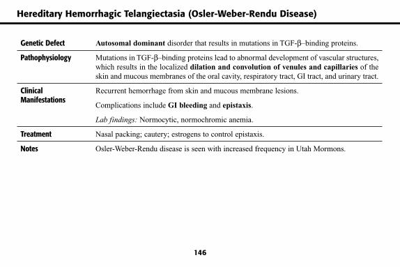
Hereditary Hemorrhagic Telangiectasia (Osler-Weber-Rendu Disease)
146
Genetic Defect Autosomal dominant disorder that results in mutations in TGF-β–binding proteins.
Pathophysiology Mutations in TGF-β–binding proteins lead to abnormal development of vascular structures, which results in the localized dilation and convolution of venules and capillaries of the skin and mucous membranes of the oral cavity, respiratory tract, GI tract, and urinary tract.
Clinical Manifestations
Recurrent hemorrhage from skin and mucous membrane lesions.
Complications include GI bleeding and epistaxis.
Lab findings: Normocytic, normochromic anemia.
Treatment Nasal packing; cautery; estrogens to control epistaxis.
Notes Osler-Weber-Rendu disease is seen with increased frequency in Utah Mormons.

9
147
A 15-year-old girl presents to the emergency room complaining of decreased vision in both eyes.
She also tells you that she has been experiencing worsening headaches over the last 2 months and
that she has been tripping more when she walks. You immediately order a CT scan of her head,
which demonstrates the presence of multiple cystic lesions throughout the cerebellum and brainstem
consistent with hemangioblastomas. You begin to suspect that this patient has a genetic disorder,
which results in the deletion of the VHL gene.

von Hippel-Lindau Disease
147
Genetic Defect Autosomal dominant disorder that results in the deletion of the VHL gene (tumor suppressor gene) on chromosome 3p.
Pathophysiology The VHL gene encodes for a protein that is involved in inhibiting RNA synthesis. With the mutation of this gene, the ability to inhibit RNA synthesis is hindered and vascular malfor-mations and other tumors result.
Clinical Manifestations
Initially presents with headaches, ataxia, or loss of vision. The disease is characterized by hemangioblastomas or cavernous hemangiomas (large vascular spaces filled with blood) of the cerebellum, brainstem, and retina as well as adenomas and cysts of the liver, kidneys, and pancreas.
Complications include increased incidence of renal cell carcinoma, pheochromocytoma, and ocular hemangioblastomas.
Treatment Surgical removal of tumor and radiation therapy.
Notes

9
148
A baby boy is born to a 29-year-old woman. Although there were no complications with the birth, you
become concerned when, 48 hours after birth, the child still has not passed any stool. Radiographic
imaging demonstrates the presence of a meconium ileus. On further questioning, the mother also tells
you that her younger sister suffers from a severe genetic pulmonary disease that also affects her
pancreas. You immediately decide to test the level of chloride ions in this child’s sweat because you
fear that the boy may have a genetic disorder, which involves a mutation on chromosome 7.

Cystic Fibrosis
148
Genetic Defect Autosomal recessive disorder, caused by a mutation (most common is ΔF508, but there are 230+
recognized mutations) on chromosome 7, which results in a defective membrane Cl– channel (CFTR).
Pathophysiology The mutated Cl– channel causes defective chloride and water transport in epithelial cells. This results in the secretion and trapping of thick mucous plugs in the lungs, liver, and pancreas. The mucous plugs obstruct various secretory ducts (bile canaliculi, bronchioles, pancreatic ducts), leading to inflammation and eventual tissue damage. In the lung, the thick mucous in combination with high concentrations of DNA (results from remnants of destroyed neutrophils, which flood the chronically inflamed lung) leads to increased viscosity of sputum.
Clinical Manifestations Chronic lung disease causing productive cough, pulmonary infections, bronchiectasis, cyanosis, and a “barrel-shaped” chest; pancreatic insufficiency causing steatorrhea, malabsorption, and abdominal pain; meconium ileus (small bowel obstruction by mucous plugs in newborn); infertility in males.
Complications include pneumothorax and cor pulmonale.
Lab findings: High Cl– concentrations in sweat test; hypoxia; increased ratio of residual volume to TLC.
Treatment and Prognosis
Antibiotics; bronchodilators; techniques to clear airway secretions; lung transplantation.
Median age of survival is 31 years; death occurs from pulmonary complications.
Notes Cystic fibrosis is the most common fatal inherited disorder in the United States; 1 in 3200 people is affected.

9
149
A 29-year-old man presents to your infertility clinic with his wife. The couple has been trying to
conceive for 3 years with no success. His medical history is significant for recurrent sinusitis and
several bouts of pneumonia that have required hospitalization in the past. He also tells you that his
heart is on the right side. When laboratory tests reveal defective sperm motility, you begin to sus-
pect that this patient might suffer from a rare genetic disorder.

Kartagener Syndrome
149
Genetic Defect A rare autosomal recessive disorder that results in a defect in dynein arms within cilia.
Pathophysiology Dynein arms are responsible for the ciliary movement; therefore, defective dynein arms result in deregulated movements of cilia. In the respiratory tract, impaired ciliary motility results in decreased clearance of bacteria, leading to increased incidence of infection in the lungs and sinuses. In the reproductive system, sperm are unable to travel effectively because of the defective motility of the sperm tail, thereby resulting in infertility. During embryo-genesis, defective ciliary motion can lead to situs inversus.
Clinical Manifestations
Bronchiectasis as manifested with a productive cough, hemoptysis, and cyanosis; sterilityin males; recurrent sinusitis; situs inversus (dextrocardia) is seen in 50% of patients.
Imaging: Findings consistent with bronchiectasis (dilated bronchioles with “signet ring” appearance on CT scan).
Lab findings: Decreased FEV1/FVC ratio.
Treatment Treatment of bronchiectasis with antibiotics, bronchodilators, and surgical resection.
Notes

9
150
A 25-year-old man presents to the emergency room complaining of severe right-sided abdominal
pain. He also reports that his urine has been darker over the last couple of days. His medical history
is significant for emphysema, although he denies any smoking history. Physical examination of the
abdomen reveals scleral icterus and tenderness in the RUQ of his abdomen. Laboratory tests reveal
elevated liver enzymes and hyperbilirubinemia. His viral hepatitis serologies are negative. You
admit him to the hospital for treatment of acute hepatitis and wonder whether an autosomal reces-
sive genetic disorder might account for both his hepatitis and his emphysema.

`1-Antitrypsin Deficiency
150
Genetic Defect An autosomal recessive disorder causes a mutation on chromosome 14 at the α1-antitrypsin gene, leading to low levels of `1-antitrypsin. There are multiple different alleles for the α1-antitrypsin gene. The most common allele combination associated with clinical α1-antitrypsin deficiency is the PiZZ genotype.
Pathophysiology α1-Antitrypsin is a liver-synthesized protease inhibitor, which has the important role of inhibiting neutrophil elastase. At sites of inflammation, neutrophil elastase is released and acts to destroy pathogens as well as the affected tissue. If `1-antitrypsin is deficient, neutrophil elastase is uninhibited and damages delicate tissues, such as the lung(destroys elastin in the alveolar wall), leading to emphysematous changes. α1-Antitrypsin deficiency also results in liver damage. In the liver, the mutated α1-antitrypsin is unable to be secreted by the hepatocyte and instead accumulates, leading to variable hepatic disease.
Clinical Manifestations
Symptoms of emphysema: Dyspnea; cyanosis; “barrel-shaped” chest; use of accessory respiratory muscles. Hepatic disease ranging from intermittent attacks of hepatitis to the development of cirrhosis. Patients also are at an increased risk for hepatocellular carcinoma.
Lab findings: Decreased FEV1/FVC ratio; elevated LFTs.
Treatment Avoid cigarette smoking (worsens emphysema) and alcohol use; bronchodilators as needed; symptomatic treatment of liver disease with liver transplantation if necessary.
Notes The frequency of this disorder is as high as 1:5000 in the US population.

9
151
A 24-year-old woman is referred to your hepatology clinic for recurrent episodes of jaundice and
RUQ abdominal pain. She tells you that the painful episodes usually resolve spontaneously.
According to her medical chart, LFTs during these episodes are consistent with conjugated hyper-
bilirubinemia and elevations in her aminotransferases. After an ultrasound does not elucidate the
cause of her symptoms, you decide to perform a liver biopsy. When the pathologist reports to you
that the gross specimen of liver is darkly pigmented, you begin to suspect that this patient might
suffer from a rare autosomal recessive condition that is caused by defective transport of bilirubin out
of the liver.

Congenital Hyperbilirubinemias
151
Gilbert Syndrome Genetic defect: Autosomal dominant condition resulting in decreased UDPGT activity.
Clinical manifestations: Often asymptomatic; mild jaundice; unconjugated hyperbilirubinemia.
Crigler-NajjarSyndrome Types Iand II
Genetic defect: Autosomal recessive (type I) or autosomal dominant with variable penetrance (type II) conditions resulting in absent UDPGT activity.
Clinical manifestations: Type I: Fatal within 2 years of birth; presents early with jaundice, kernicterus, and unconjugated hyperbilirubinemia. Type II: nonfatal; presents with jaundice and unconjugated hyperbilirubinemia; kernicterus rarely occurs.
Treatment: Plasmapheresis and phototherapy (type I); phenobarbital (type II).
Dubin-JohnsonSyndrome
Genetic defect: Autosomal recessive condition resulting from defective bilirubin transport out of the liver because of a mutated canalicular membrane carrier.
Clinical manifestations: Intermittent jaundice; RUQ and epigastric pain; conjugated hyperbili-rubinemia; mildly elevated LFTs; black liver on gross pathology.
Rotor Syndrome Genetic defect: Autosomal recessive disorder resulting in defective uptake and excretion of bili-rubin by hepatocytes.
Clinical manifestations: Usually asymptomatic; may have bouts of intermittent jaundice; conjugated hyperbilirubinemia.
Notes UDPGT is a protein in the liver responsible for conjugating bilirubin, thus allowing for urinary excretion. If this enzyme is deficient, bilirubin will build up and damage susceptible tissues, such as the brain.

9
152
A 58-year-old immigrant from Ireland presents to your clinic complaining of weakness and gener-
alized abdominal pain over the past couple of months. On further questioning, he tells you that he
has been thirstier lately and is urinating more frequently than usual. He also comments on how tan
he has become, even though he has not been out in the sun that often. On physical examination,
you note mild scleral icterus, hyperpigmentation of the skin on his trunk and extremities, and
hepatomegaly. Laboratory tests reveal a fasting blood glucose level of 200 mg/dL and mildly
elevated LFTs. You wonder whether he may have an autosomal recessive genetic disorder that
would explain his constellation of symptoms, and you decide to order iron studies to test your
hypothesis.

Primary Hemochromatosis
152
Genetic Defect An autosomal recessive condition involving a mutation on chromosome 6 that results in excessive absorption of iron in the intestinal mucosa (exact mechanism is unknown).
Pathophysiology Elevated circulating levels of iron results in iron deposition in tissues, including the liver, pancreas, heart, adrenals, and skin. Iron is toxic to organs, causing DNA damage through an increase in free radical formation, thereby leading to tissue damage and fibrosis. Fibrosis in the pancreas leads to decreased insulin production and results in diabetes. Fibrosis in the liver leads to cirrhosis. Fibrosis in the adrenals leads to an increase in ACTH release from the pituitary through a loss of negative feedback. By increasing ACTH levels, MSH also increases (MSH and ACTH are from the same precursor molecule), resulting in increased melanin deposition in skin.
Clinical Manifestations Tends to present in northern Europeans males after age 50 with the classic triad of cirrhosis,diabetes, and skin hyperpigmentation (“bronze diabetes”). Other symptoms include arthropa-thy, hepatomegaly, and cardiac disease.
Complications include an increased risk of hepatocellular carcinoma.
Lab findings: Mildly elevated LFTs; increased serum iron; decreased total iron-binding capacity; transferrin saturation >80%; serum ferritin 1000 lg/L.
Treatment Weekly phlebotomy and deferoxamine; prevention by neonatal screening for increased transferrin saturation.
Notes There is a strong association of primary hemochromatosis with the HLA-A3 haplotype as a result of linkage disequilibrium.

9
153
A 46-year-old woman presents to your gastroenterology office for her first routine screening
colonoscopy. As you take her history, you notice that she has several melanotic macules on her
hands and lips. Colonoscopy reveals multiple hamartomatous polyps scattered over the entire length
of her colon. You assure the patient that these polyps are benign and that she is not at increased risk
for colon cancer, but you wonder whether she may have a genetic condition that would predispose
her to developing other types of GI and gynecologic cancers.

Multiple Polyposis Syndromes
153
Familial Adenomatous Polyposis
Genetic defect: Autosomal dominant condition caused by mutation of the APC gene on chromosome 5.
Clinical manifestations: There are 500 to 2500 colonic adenomas present at puberty.
Treatment: Prophylactic colectomy (100% will evolve into colon cancer if not resected).
Hereditary NonpolyposisColorectal Cancer
Genetic defect: Autosomal dominant condition caused by defect in DNA mismatch repair genes on chromosome 2, 3, or 7.
Clinical manifestations: Appearance of colonic adenomas in early adulthood; increased risk for colorectal cancer and other cancers (especially endometrial cancer).
Treatment: Surgical resection.
Peutz-Jeghers Syndrome
Genetic defect: Autosomal dominant condition caused by defect in LKB1 (tumor suppressor gene).
Clinical manifestations: Multiple hamartomatous (nonneoplastic) polyps of the colon and small intestine; melanotic macules in the mouth, lips, hands, and genitalia; no increased risk for colon cancer but increased risk of gastric, breast, gynecologic, pancreatic, or lung cancer.
Treatment: Regular screening for gynecologic and GI cancers.
Gardner Syndrome and Turcot Syndrome
Genetic defect: Autosomal dominant conditions associated with defects in APC gene on chromosome 5.
Clinical manifestations: Gardner syndrome: Adenomatous polyps along with osteomas and soft tissue tumors. Turcot syndrome: Adenomatous polyps with CNS tumors. Both conditions are associated with an increased risk for colorectal cancer.
Treatment: Surgical resection.
Notes

9
154
A 12-year-old boy is brought to your pediatrics office complaining of right-sided abdominal pain
over the past 2 days. On further questioning, his mother tells you that he has seemed more emotion-
ally labile than usual and that he has occasionally made statements that are out of context with the
conversation. On physical examination, you note the presence of Kayser-Fleischer rings on both
corneas, tenderness in the RUQ of his abdomen, and a resting tremor of both hands. LFTs reveal
the presence of mild hepatitis. You immediately order laboratory studies to test for serum cerulo-
plasm levels because you suspect that this boy may have a rare disorder that necessitates treatment
with penicillamine.

Wilson Disease
154
Genetic Defect Rare autosomal recessive disorder involving a mutation of chromosome 13 that results in a defective copper-transporting membrane protein in the liver.
Pathophysiology Copper is absorbed in the intestine and transported to the liver, where hepatocytes conju-gate the copper to an α2-globulin to form ceruloplasmin. Ceruloplasmin can then circulate in the plasma and is eventually broken down by lysosomes and secreted into the bile for excretion. In Wilson disease, the copper cannot be transported into the hepatocytes fortransformation into ceruloplasmin. The increased copper then accumulates throughout the body, especially in the parenchymal cells of the liver, kidney, brain (especially the basal ganglia), and cornea.
Clinical Manifestations
Usually presents between the ages of 10 and 30. Symptoms include liver disease (beginning with hepatitis and progressing to cirrhosis), hemolytic anemia, portal hypertension, psychosis, or dementia, Kayser-Fleischer rings (thin brown rings around the corneas on eyeexamination), and choreiform movements (extrapyramidal motor signs similar to those in Parkinson disease). Complications include increased risk of hepatocellular carcinoma.
Lab findings: Decreased serum ceruloplasmin; hypercupriuria (copper in urine).
Treatment Penicillamine (chelates copper for removal from body).
Notes

9
155
A 15-year-old boy is brought to the pediatrician’s office for his annual checkup. He reports feeling
relatively healthy over the last year, although he does note that his vision and hearing seem to have
deteriorated recently. On further probing, you learn that his uncle suffers from a genetic disorder
characterized by deafness and kidney disease. When routine urinalysis of this patient reveals hema-
turia with erythrocyte casts, you begin to worry that this boy may suffer from a rare genetic disorder
characterized by a mutation of type IV collagen.

Alport Syndrome
155
Genetic Defect Genetic disorder with heterogenous inheritance (usually X-linked dominant, although autosomal recessive and dominant variations exist) that results in the mutation of type IV collagen.
Pathophysiology Type IV collagen is an important component of the cochlea, the anterior lens capsule of the eye, and forms the scaffolding of the glomerular basement membrane. Defective type IV collagen leads to irregularities in the glomerular basement membrane and results in malfunctioning of the glomerular filtration barrier, leading to eventual sclerosis.
Clinical Manifestations
Triad of nephritis, sensory deafness, and ocular disorder (cataracts, lens dislocation, corneal dystrophy). Often initially presents with hematuria and erythrocyte casts during adolescence and will usually progress to renal failure by middle age.
Treatment ACE inhibitors; renal transplantation.
Notes
9
156
A 30-year-old woman presents to your clinic for a routine physical. She reports that she has been
in good health except for an occasional headache. Physical examination reveals a blood pressure of
160/96, mild costovertebral angle tenderness with palpation, and a murmur consistent with mitral
valve prolapse. When her urinalysis reveals hematuria, you question your patient further regarding
her family history, and she tells you that her aunt is on dialysis for kidney disease. You decide to
send the patient for renal ultrasounds to confirm your suspicions, and you worry that this patient’s
likely genetic disorder will put her at an increased risk for berry aneurysms.

Autosomal Dominant and Autosomal Recessive Polycystic Kidney Disease
156
Genetic Defect ADPKD: Autosomal dominant disorder caused by a mutation of PKD gene on chromosome 16 that results in a defective protein called polycystin, which is involved in cell-to-cell matrix interactions.
ARPKD: Autosomal recessive disorder caused by a mutation on chromosome 6 that results in a defective protein called polyductin, which is present in the cilia of renal epithelial cells.
Pathophysiology ADPKD: Although the exact mechanism is unknown, it is thought that defective polycystin results in abnormal cell differentiation, which may lead to cyst formation.
ARPKD: The exact mechanism is unknown, but it is thought that polyductin may be involved in cell-to-cell signaling during renal tubular differentiation.
Clinical Manifestations ADPKD: Hypertension, hematuria, and palpable renal masses. CT shows multiple large cysts in both kidneys. Associated with secondary polycythemia, polycystic liver disease, berryaneurysms, and mitral valve prolapse. Patients eventually progress to ESRD.
ARPKD: Hypertension, growth failure, bilateral abdominal masses, and progressive renal failure during childhood. Congenital hepatic fibrosis develops in older children. Imaging shows multiple renal cysts at birth.
Treatment and Prognosis
ADPKD: No therapy can prevent renal failure, although blood pressure control and a low-protein diet may slow progression of ESRD.
ARPKD: Treatment of hypertension; dialysis for ESRD; disease course is variable, but most patients die during childhood.
Notes

9
157
A 25-year-old man presents to your endocrinology office for further evaluation of abnormal labora-
tory testing done by his primary care physician. As you read his medical chart, you find that he
suffers from hypercalcemia in the setting of increased levels of parathyroid hormone, suggesting
primary hyperparathyroidism. You also note that his blood pressure is 180/110, and he mentions
that he often experiences pounding headaches and heart palpitations. You send for a 24-hour urine
collection study, which reveals increased 24-hour urinary VMA, metanephrine, and catecholamine
levels. On the basis of these findings, you begin to think that this patient will likely develop medul-
lary carcinoma of the thyroid and that he should undergo a prophylactic thyroidectomy to avoid
this malignancy.

MEN Syndromes
157
Genetic Defect MEN I: Autosomal dominant disorder caused by a mutation on chromosome 11 in the MEN1 gene.
MEN IIa and IIb: Autosomal dominant disorders caused by differing mutations on chromosome 10 in the RET proto-oncogene.
Pathophysiology MEN I: Although the exact function of the protein coded for by the MEN1 gene is unknown, mutations in the gene have been linked to tumor formation.
MEN IIa and IIb: The RET proto-oncogene is a tyrosine kinase receptor that is involved in cellular growth signaling. The MEN mutations in this protein result in a constitutively active receptor, thereby promoting uncontrollable growth and neoplasia.
Clinical Manifestations
MEN I: Triad of parathyroid hyperplasia/adenoma, pituitary adenoma, and pancreatic islet cell tumors, which often manifests as Zollinger-Ellison syndrome (peptic ulcers secondary to gastrinoma).
MEN IIa: Triad of pheochromocytoma, parathyroid hyperplasia, and medullary carcinoma of the thyroid.
MEN IIb: Triad of pheochromocytoma, medullary carcinoma of the thyroid, and mucocutaneous neuromas of the skin, eyes, and GI tract.
Treatment Treat symptomatically; surgery when appropriate for thyroid carcinoma and pheochromocytoma; genetic screening of family members for preventive thyroidectomy in MEN IIa and IIb.
Notes

9
158
A 5-year-old boy presents to your pediatrics office for a routine visit as a new patient. His mother
tells you that the child is mentally retarded and has recently been complaining of muscle pain in his
arms and legs. While speaking to the mother, you note that the child is short and obese and has very
short metacarpals on his fourth and fifth fingers of both hands. Physical examination is significant
for positive Trousseau and Chvostek signs. Laboratory tests reveal low serum calcium levels in the
setting of elevated parathyroid hormone levels. You begin to suspect that the child suffers from a
rare autosomal recessive disorder, and you prescribe calcium and vitamin D supplements to relieve
the child’s symptoms.

Pseudohypoparathyroidism
158
Genetic Defect Autosomal recessive disorder, caused by a chromosome 20 mutation leading to a faulty PTH receptor.
Pathophysiology PTH binds to PTH receptor, which then interacts with a G protein to activate AC and increase cAMP production in bone, kidney, and intestine. In type 1, there is a deficiency in Gs-α, which leads to decreased coupling of PTH receptor to AC, such that activation of PTH receptor does not activate the target cell. In type 2, the regulation of Gs-α is altered such that the levels of cAMP produced by PTH stimulation are inadequate to activate the target cell. In both forms of thedisease, the target cell is not activated and it is as though PTH is not present.
ClinicalManifestations
Symptoms of hypocalcemia: Tetany or other signs of neuromuscular irritability; prolonged QT interval on ECG; Trousseau sign (carpal spasm 2 min after inflation of blood pressure cuff above systolic blood pressure); Chvostek sign (twitching of the facial muscles on superficial tap-ping of the facial nerve). Patients with type 1 also have Albright hereditary osteodystrophy: short stature, mental retardation, shortened fourth and fifth metacarpal and metatarsal bones, obesity.
Lab findings: Hypocalcemia; increased PTH levels; increased serum phosphate levels.
Treatment Calcium and vitamin D supplements.
Notes PTH acts to increase serum calcium and decrease serum phosphate through effects on the bone (increases osteoclastic activity), the kidney (promote calcium reabsorption and formation of activated vitamin D), and the intestine (increased levels of activated vitamin D promotes increased calcium reabsorption in the gut).

9
159
A 3-year-old boy is brought to your office by his mother, who is concerned that her child’s gait seems
to be deteriorating. The child’s medical history is significant for several sinus infections. The child’s
medical file does not reveal any indication that he had any difficulty walking in the past. However,
as you observe the child walk now, you note that his gait is distinctly ataxic. A full neurologic
examination reveals decreased DTRs in both legs. When close examination of the child’s conjunctiva
reveals multiple telangiectasias, you begin to suspect that this child suffers from a condition that is
ultimately fatal.

Ataxia-Telangiectasia
159
Genetic Defect An autosomal recessive disorder that results in a mutation on chromosome 11 in the ATM gene.
Pathophysiology The gene product of the ATM gene is involved in sensing DNA that has been damaged by radiation and then signaling p53 to delay the cell cycle to allow for DNA repair. If the ATMgene is mutated, p53 is not activated and the cell cycle continues, allowing for replication of damaged DNA. This may lead to abnormal cellular development, especially in the neuro-logic, vascular, and immune systems. The cerebellum is particularly affected (pathologic examination reveals loss of Purkinje and granule cells), thereby accounting for the ataxia.
Clinical Manifestations
Presents in early childhood with neurologic symptoms (cerebellar ataxia with eventual wheelchair confinement; decreased DTRs; dysarthria) and telangiectasias of the face and conjunctiva. Some patients have various immunodeficiencies, leading to recurrent respira-tory infections or various endocrinologic abnormalities.
Complications include a predisposition to developing cancers (breast, lymphoma, leukemia).
Lab findings: Increased α-fetoprotein levels.
Treatment and Prognosis
Vitamin E (antioxidant) and folic acid supplementation; avoid radiation when possible. Most patients do not live past 25; many die much earlier.
Notes

9
160
A 2-year-old girl is brought to your pediatric clinic for evaluation of a delay in walking with repeti-
tive falling. Upon taking a history from the child’s mother, you learn that the patient was slow to walk
initially and now is only able to walk for several feet before falling down. Physical examination
reveals absent deep tendon reflexes in legs, mild scoliosis, and an ataxic gait. When a cardiac workup
reveals signs of hypertrophic cardiomyopathy, you decide to order a study to evaluate for a deficiency
in the protein, frataxin.

Friedreich Ataxia
160
Genetic Defect An autosomal recessive disorder caused by a deficiency in the protein, frataxin.
Pathophysiology Frataxin is coded for by a gene locus on chromosome 9. Frataxin is involved in maintaining normal mitochondrial function as well as iron homeostasis. In Friedreich ataxia, there is a GAA trinucleotide repeat expansion at the gene locus for frataxin, which results in decreased expression of the gene. With decreased levels of frataxin, iron accumulates in the mitochondria and leads to decreased mitochondrial function and eventually cell death. Neuronal tissues and cardiac tissue are particularly sensitive to frataxin deficiencies;hence damages to the nervous system and cardiac system are prominent symptoms of Friedreich ataxia.
Clinical Manifestations
Presents in childhood with an ataxic gait. The patient’s ataxia becomes progressive until the torso and arms are involved and the patient needs a wheelchair. Other symptoms include dysarthria, sensory neuropathy, absent deep tendon reflexes, kyphoscoliosis, and cardiac disease (particularly hypertrophic cardiomyopathy).
Treatment and Prognosis
Supportive treatment for neurologic degeneration; treatment of cardiac disease if present. Most patients become wheelchair bound within 5 years of diagnosis and die prematurely.
Notes

9
161
A 43-year-old woman presents to the emergency room complaining of uncontrollable, jerking move-
ments of both her arms, which has been progressing over the past 3 months. Her husband also tells
you that her memory function has declined and that she seems more irritable than usual. On taking
a family history, you learn that the patient’s father suffered from similar symptoms, which eventually
progressed to dementia. When imaging of the patient’s brain reveals atrophy of the caudate nucleus
with dilation of the lateral and third ventricles, you suspect that this patient has a hereditary neuro-
logic degenerative disorder that will eventually be fatal.

Huntington Disease
161
Genetic Defect Autosomal dominant disorder associated with increased number of CAG repeats in the HD gene on chromosome 4. Exhibits variable penetrance.
Pathophysiology The exact function of the HD gene product is unknown, but researchers theorize that the protein is involved in neuronal apoptosis, which is consistent with the finding that the neu-rologic system is severely affected in this disease. GABAergic striatal neurons of the basal ganglia are damaged, leading to atrophy of the caudate nucleus and putamen. Because the caudate and putamen are part of the extrapyramidal motor system, destruction of these structures gives rise to motor abnormalities, such as those observed in Huntington disease.
Clinical Manifestations
Progressive disorder that initially manifests between the ages of 40 and 50; chorea(involuntary jerky movements); cognitive impairment; mood disturbances. Eventually progresses to severe dementia.
Imaging: MRI demonstrates caudate atrophy and dilation of lateral and third ventricles.
Treatment and Prognosis
Symptomatic treatment for dyskinesia and mood disturbances.
Usually fatal within 15-20 years of diagnosis.
Notes Huntington disease, along with fragile X syndrome and myotonic dystrophy, demonstrates anticipation, a phenomenon in which the number of repeats increases with each generation and results in more severe disease manifestations.

9
162
A 9-year-old boy is taken to your clinic by his mother, who is concerned by the development of
multiple nodules on her child’s skin. The child is adopted so you are unable to ascertain his entire
family history, but adoption records show that his biological father suffered from some sort of dis-
figuring genetic disorder. Examination reveals multiple coffee-colored macules on the boy’s torso
and limbs and distinctive pigmented nodules on his irises. You suspect that his condition is due to
an autosomal dominant genetic disorder caused by a mutation on chromosome 17, and you refer the
patient to a medical genetics clinic and a neurologist.

Neurofibromatosis Type 1
162
Genetic Defect Autosomal dominant disorder that is caused by a mutation in the NF1 gene, which is a tumor suppressor gene located on chromosome 17. The disease can also occur through sporadic mutations of the NF1 gene.
Pathophysiology NF1 encodes for a protein (neurofibromin) that acts as a tumor suppressor gene by decreas-ing the activity of p21 ras oncogene. With mutation of NF1, the p21 ras oncogene is uninhibited and can trigger unhindered cellular growth, which results in the formation of neurofibromas (masses of spindle cells, occurring in the dermis, peripheral nerve, or large nerve trunk). Neurofibromas in the dermis or in the iris can cause hyperpigmentation of the overlying cells, leading to observation of café au lait spots and Lisch nodules, respectively.
Clinical Manifestations
Neurofibromas (may cause neurologic symptoms) and gliomas of the optic nerve; Lisch nodules (pigmented nodules of the iris); café au lait spots (cutaneous pigmented macules); various skeletal abnormalities.
Complications include an increased risk for other tumors (Wilms tumor, meningiomas, pheochromocytomas, chronic myeloid leukemia).
Treatment Surgery to remove neurofibromas if disfiguring or causing neurologic abnormalities.
Notes Neurofibromatosis 2 is an autosomal dominant disorder caused by a mutation in the NF2gene (also a tumor suppressor gene), located on chromosome 22. It is rarer than NF1and presents with bilateral acoustic schwannomas, multiple meningiomas, and otherneoplasms.

9
163
A 6-month-old boy is brought to the emergency room after his parents witnessed him having a
seizure. The child has had multiple seizure episodes since birth. You decide to send the baby for
radiographic imaging of his brain, which reveals multiple cortical tubers through the cerebral
cortex. You worry that this child suffers from an autosomal dominant genetic disorder, and you
order an echocardiogram to check for a cardiac rhabdomyoma and a renal ultrasonogram to check
for renal angiomyolipomas.

Tuberous Sclerosis
163
Genetic Defect Autosomal dominant disorder resulting from a mutation in one of several different genes, including the TSC1 gene on chromosome 9 and the TSC2 gene on chromosome 16.
Pathophysiology The exact mechanism of the TSC genes is unknown, but they are believed to act as tumorsuppressor genes. With the mutation of these genes, multiple different neoplasms result, including brain hamartomas (nodules composed of disorganized neurons in the cerebral cortex; also called cortical tubers), cardiac rhabdomyomas, adenoma sebaceum on the face (lesion consisting of malformed blood vessels), renal angiomyolipomas, and cysts of the bone and lung. The neurologic tumors lead to mental retardation and seizures.
Clinical Manifestations
Seizures and mental retardation in infancy; red nodules on face (adenoma sebaceum), which appear between the ages of 5 and 10; presence of cardiac rhabdomyoma and renal angiomyolipoma.
Treatment Seizure control; regular surveillance for renal angiomyolipomas; genetic counseling.
Notes

9
164
A 15-year-old boy presents to the emergency room, complaining of enlarged cord-like lumps on both
of his legs. He states that these lumps have been there over the last 6 months, but have gotten progres-
sively larger. On review of systems, he also notes a history of frequent tripping and ankle sprains.
Family history is notable for a neurologic disorder in his father, paternal aunt, and two cousins,
although he cannot remember the name of this disorder. Physical examination reveals multiple
palpable cords in the legs, likely consistent with enlarged nerves. You also note that he has evidence
of pes cavus deformity, mild scoliosis, and decreased deep tendon reflexes. You tell the patient that
his symptoms are likely reflective of a relatively common inherited neuropathy and you refer him to
a neurologic clinic for further treatment.

CMT Disease
164
Genetic Defect A collection of diseases resulting from a mutation in one of several different genes involved in nerve myelination and function. There are seven major types of CMT disease, each with multiple subtypes. The two most common forms of CMT are types 1 and 2, both of which are mostly inherited in an autosomal dominant fashion.
Pathophysiology CMT-1: Associated with a collection of mutations involved in formation and stabilization of myelin. Mutation in one of several genes on chromosomes 1, 8, 10, 16, or 17 results in abnormal myelin formation and subsequent myelin breakdown. In response to demyelination, Schwann cells proliferate at an abnormal rate in an attempt to remyelinate the nerve—the repetition of this process can result in a thickened layer of myelin around the nerve, giving rise to an “onion bulb”appearance to the nerve.
CMT-2: Associated with a collection of defects on chromosomes 1, 3, 7, 8, and 12, which result in neuronal cell death and degeneration.
Clinical Manifestations CMT-1: Distal muscle weakness; sensory neuropathy; pes cavus deformity (high arch); decreased deep tendon reflexes; “stork leg” deformity (secondary to calf muscle atrophy); scolio-sis; enlargement of peripheral nerves such that they are palpable (secondary to “onion bulb” thickening of nerve).
CMT-2: Presents with predominantly sensory neuropathy, although distal muscle weakness is also present; tremor
Treatment Supportive treatment of neuropathies; surgical therapy of joint deformities if indicated; genetic counseling.
Notes Charcot-Marie-Tooth disease is the most common inherited neurologic disorder.

9
165
A 24-year-old man presents to the emergency room complaining of acutely decreasing vision in both
eyes. Physical examination reveals decreased vision in the central fields of both eyes. You also
observe a mildly ataxic gait. As you question him further regarding his personal and family history,
he tells you that he has been healthy his whole life but that his older brother and sister as well as his
mother became blind before the age of 35. You begin to suspect that this patient will also become
blind in the near future as a result of a rare genetic disorder.

LHON
165
Genetic Defect A maternally inherited disorder that is caused by a mutation in the mtDNA, encoding for components of the electron transport chain.
Pathophysiology There are many different mutations in the mtDNA that can lead to LHON, and most involve the alteration of the electron transport capacity and ATP production of complexes I and III of the electron transport chain. It is not known how these defects lead to the optic symptoms of LHON; however, some researchers have theorized that the retinal ganglion cells become damaged by the oxidative stress or ischemia that results from defective oxidative phosphorylation.
Clinical Manifestations
Usually presents between the ages of 15 and 35 with central vision loss that is acute in onset and affects both eyes, eventually leading to blindness within 1 year. Occasionally, patients may have other neurologic signs (dystonia, decreased intelligence, ataxia, hearing loss, multiple sclerosis-like symptoms) or cardiac conduction defects.
Treatment Supportive treatment.
Notes

9
166
A 7-year-old girl presents to your pediatric ophthalmology clinic complaining of worsening vision
and pain in her right eye. The patient’s mother tells you that the girl’s father has suffered from
ocular neoplasms in both eyes as well as osteosarcoma. Ophthalmologic examination reveals a
“cat’s eye” pupillary reflex, and recent orbital MRI demonstrated an intraocular mass of the left eye.
You immediately refer this patient to both an oncologist and a medical genetics clinic because you
fear that this patient likely suffers from an autosomal dominant disorder that is associated with a
defective tumor suppressor gene.

Retinoblastoma
166
Genetic Defect An autosomal dominant condition that is caused by homozygous deletion in both alleles of the Rb gene, a tumor suppressor gene located on chromosome 13. The disease is transmitted as an autosomal dominant condition even though homozygosity is necessary for disease, because more than 90% of heterozygous carriers develop the disease. Sporadic cases can also occur.
Pathophysiology When Rb is activated, it serves to halt the cell cycle in the G1 phase by inhibiting E2F transcription factors. When Rb is mutated, the checkpoint at G1 is lost and the cell cycle can continue unhindered into the S (replication) phase. Uninhibited progression to S phase in the face of an Rb gene mutation has been shown to lead to cell apoptosis in most tissues, except for retinoblasts. Instead, neoplastic changes accumulate in the retinoblast and a tumor arises.
Clinical Manifestations
Classically occurs in young children who present with diminished visual acuity, eye pain,strabismus, intraocular mass on fundoscopic examination, and white “cat’s eye” pupillary reflex.
Patients will develop bilateral retinoblastomas with a risk of metastasis to the brain, spinal cord, bone, and lymph nodes. They are also at an increased risk for other cancers (eg, osteosarcoma).
Treatment and Prognosis
Surgery (removal of tumor or eye if necessary) and radiation.
Tumor is fatal once it has spread beyond the eye.
Notes Retinoblastoma is the prototype of Knudson “two-hit” hypothesis: two mutations are required for disease. One deletion is either inherited (familial) or occurs sporadically. The second mutation results from a somatic mutation in both familial and sporadic cases.

9
167
A newborn boy is found on the steps of the police station. He is brought to the hospital for evaluation.
As you examine the child, you notice that he has midface hypoplasia, short limbs, and macrocephaly.
Neurologic examination reveals mild hypotonia of his limbs. You begin to suspect that the child has
an autosomal dominant condition caused by a mutation in the fibroblast growth factor receptor 3 gene.

Achondroplasia
167
Genetic Defect An autosomal dominant condition that is caused by a mutation in the FGFR3 genelocated on chromosome 4.
Pathophysiology FGFR3 is a tyrosine kinase receptor that acts to inhibit chondrocytes at the growth plate of bones, thereby leading to the decreased cartilage proliferation and subsequent decreased growth. In achondroplasia, FGFR3 is always active; therefore, cartilage proliferation at the growth plate is inhibited 100% of the time. This results in short thick bones withnarrow epiphyseal plates, consisting of disorganized chondrocytes.
Clinical Manifestations
Dwarfism (short limbs with normal trunk); macrocephaly with frontal bossing; midface hypoplasia; hypotonia early in life that resolves spontaneously; may have neurologic symp-toms, which result from a small foramen magnum.
Treatment Genetic counseling for parents of affected patient regarding future offspring; symptomatic treatment of neurologic complications.
Notes

9
168
A 5-year-old boy is brought to your clinic complaining of weakness in his legs. Physical examination
is significant for decreased strength in his thighs as well as enlarged calves. While you are speaking
with the boy’s mother, you observe the boy playing with toys on the floor and notice that he uses his
arms to assist himself in rising from a crouching position. When his mother tells you that her brother
died at a young age from a genetic muscle disease, you suspect that this child suffers from an
X-linked recessive muscular disorder and recommend that the patient be seen by a geneticist.

Duchenne Muscular Dystrophy
168
Genetic Defect An X-linked recessive disorder that is caused by a deletion in the DMD gene on the short arm of the X chromosome, which results in an absence of dystrophin synthesis.
Pathophysiology Dystrophin is believed to be involved in maintaining the membrane integrity of the muscle cell. Absence of dystrophin leads to muscle fiber destruction and muscle atrophy.
Clinical Manifestations
Presents in males with onset of disease by age 5. Manifests with weakness in proximal muscles of extremities (usually the pelvis), which progresses superiorly and eventually leads to immobilization, pseudohypertrophy of calves (because of replacement of atrophied muscle with fibrous and fatty tissue), and the presence of Gowers maneuver (useof arms to rise from crouching position).
Lab findings: Increased serum creatine kinase.
Treatment and Prognosis
No treatment.
Death via respiratory failure in adolescence because of involvement of respiratory muscles.
Notes Becker muscular dystrophy is an X-linked recessive disorder that is also characterized by a mutation in the dystrophin gene, which leads to reduced synthesis of dystrophin.It presents similarly to Duchenne muscular dystrophy but is clinically less severe.

9
169
A 2-year-old girl is brought to your pediatrics office by her parents, who are concerned that their
daughter has not started to walk. They also report that when she stands, she tends to fall easily, the
trauma of which leads to large, gaping cuts. When you examine the child, you notice that she has
hypermobile joints and thin, hyperextensible skin. When the girl’s father tells you that he suffers
from a genetic disorder involving faulty collagen synthesis, you begin to suspect that this child’s
delay in walking is the result of an inherited connective tissue disorder, and you refer the family to
a medical genetics clinic.

EDS
169
Genetic Defect A disorder with 10 different variants, all of which are associated with the defective synthesis of collagen. The mode of inheritance is also variable and includes autosomal dominant (types I-IV, VIIA-B, VIII), autosomal recessive (types VI, VIIC, X), and X-linked recessive (types V, IX).
Pathophysiology Collagen is involved in the formation of skin, bone, tendons, vessel walls, ocular structures, and cartilage. Various forms of collagen are mutated in the different variants of EDS, and the muta-tions result in various defects ranging from mutations in structural components of collagen (eg, defective pro α1 chains in collagen type III in EDS IV) to mutations in enzymes involved in posttranslational modification of collagen (defective lysyl hydroxylase in EDS type VI). The abnormal collagen is weak and results in weakness in the structures that it composes, thereby leading to hyperextensible joints through weak tendons, skeletal abnormalities through abnormal bone formation, and susceptibility to various organ injury through fragile skin and vessel walls.
Clinical Manifestations Presentations vary depending on the syndrome. Common symptoms include thin hyperextensible skin, hypermobile joints complicated by dislocation, easy bruising, mitral valve prolapse (I), uterine or intestinal wall rupture (IV), scoliosis (VI), ocular globe fragility with resulting blindness (VI), congenital hip dislocation (VII), periodontal disease (VIII), and the development of premature osteoarthritis (I, VII).
Treatment Vitamin C supplementation (involved in collagen synthesis); symptomatic care for osteoarthritis; routine eye examinations.
Notes

9
170
A 13-year-old girl presents to the emergency room complaining of an acute change in the vision in
her left eye. She denies any trauma to the eye. Ophthalmologic examination reveals displacement of
the lens from the center of the pupil. While you are calling an ophthalmologist to the emergency room,
you note that the girl is extremely tall for her age with long extremities and long fingers on both hands.
You wonder whether this patient may have an autosomal dominant genetic condition that will predis-
pose her to aortic dissection, and you decide to refer her to a medical geneticist for further evaluation.

Marfan Syndrome
170
Genetic Defect An autosomal dominant condition caused by mutation in the fibrillin gene on chromosome 15. Although most mutations are hereditary, 20% are sporadic.
Pathophysiology Fibrillin is a glycoprotein constituent of microfibrils, which are present in great quantities in the extracellular matrix of the aorta, ligaments, perichondrium, and the ocular zonules (attach lens to ciliary body). Mutation of fibrillin leads to a defective extracellular matrix and weakening of the involved structures, leading to hyperextensible joints, cystic medial necrosis of the aorta (results from dilation of aortic valve and weakening of media with increased risk of intimal tear and dissection), mitral valve prolapse (resulting from loss of connective tissue support of valvular leaflet), and a predisposition to lens dislocation or subluxation. These patients also suffer from bony overgrowth and other skeletal abnormalities, but the exact pathogeneses for these changes are unknown.
Clinical Manifestations Tall stature with long extremities; hyperextensible joints; long tapering digits (arachno-dactyly); ectopia lentis (dislocation of lenses); aortic incompetence; dissecting aortic aneurysm;mitral valve prolapse; skeletal abnormalities (kyphosis, scoliosis); spontaneous pneumothorax.
Treatment and Prognosis
Spine brace; endocarditis prophylaxis; aortic valve replacement if necessary; β-blockers.If untreated, death is common between ages 30 and 40 from aortic dissection or CHF secondary to aortic regurgitation.
Notes

9
171
A 19-year-old girl presents to the emergency room after suffering a generalized seizure. She tells
you that she has been recently diagnosed with myoclonic epilepsy. As you continue to assess her,
she tells you that she has been having muscle weakness in all of her extremities and that she often
stumbles while walking. Physical examination reveals decreased muscle strength, an ataxic gait,
and decreased hearing in both ears. When she tells you that all of her siblings and her mother suffer
from some sort of seizure disorder, you immediately begin to suspect that she too may have a rare
genetic disorder that would cause the appearance of ragged-red fibers on muscle biopsy.

Mitochondrial Myopathies (MERRF and MELAS)
171
Genetic Defect MERRF and MELAS: Maternally inherited disorders caused by point mutations in thetRNA gene of mtDNA.
Pathophysiology The mtDNA genes are involved in the production of the cellular apparatus of oxidative phosphory-lation. Defects in any component of this cellular apparatus can lead to defective oxidative phos-phorylation. Defective oxidative phosphorylation will lead to mitochondrial proliferation withconsequent destruction of muscle fibers (“ragged-red fibers”) and can lead to neuronal damage to the spinal cord, cerebellum, and motor cortex. NADH accumulates, leading to inhibition of pyruvate dehydrogenase and thereby resulting in the accumulation of lactate.
Clinical Manifestations
MERRF: Presents during adolescence; ataxia; myoclonic epilepsy with seizures; muscle weakness;hearing loss; progressive mental status decline.
MELAS: Presents in childhood; strokelike episodes with blindness and hemiparesis; vomiting; hearing loss; seizures; lactic acidosis.
Treatment and Prognosis
MERRF: Seizure control; eventually results in encephalopathy and death.
MELAS: Symptomatic treatment; eventually results in dementia and death before age 20.
Notes There are multiple other disorders that are characterized by mutations in mitochondrial DNA, including Kearns-Sayre syndrome (ophthalmoplegia; heart block; retinal degeneration), chronic progressive external ophthalmoplegia (myopathy; ophthalmoplegia), Leigh disease (necrotizing encephalopathy), and Barth syndrome (myoglobinuria; cardiomyopathy).
9
172
A 7-year-old girl is brought to your pediatrics office by her father, who is concerned about the
child’s abnormal hand grip. The father tells you that he suffers from a muscle disease. You notice
that the father is bald, even though he is relatively young. Physical examination of the girl reveals
delayed relaxation of her hand grip. Slit-lamp examination reveals the beginnings of bilateral
cataracts. After prescribing phenytoin for symptomatic relief, you refer the girl and her father to
a geneticist for further evaluation of this disorder.

Myotonic Dystrophy
172
Genetic Defect Autosomal dominant disorder resulting in increased CTG repeats in the myotonin protein kinase gene on chromosome 19.
Pathophysiology The exact function of myotonin protein kinase has not yet been elucidated. Thus, it is still unclear how dysfunction in this enzyme leads to the clinical symptoms of the disease or to the microscopic pathologic findings in muscle (“ring fiber” [cytoplasmic band within the center of the fiber]; fiber splitting and necrosis of intrafusal fibers of muscle spindles).
Clinical Manifestations
Presents between the ages of 20 and 30 but may manifest in childhood. Symptoms include myotonia (inability to relax contracted muscles), often presenting as muscle stiffness as well as weakness and wasting of distal limb and facial muscles.
Also associated with cataracts, testicular atrophy, baldness, cardiac disease, and decreased glucose tolerance.
Treatment Phenytoin to treat myotonia.
Notes Myotonic dystrophy, along with fragile X syndrome and Huntington disease, demonstrates anticipation, a phenomenon in which the number of repeats increases with each generation and results in more severe disease manifestations.

9
173
A 2-month-old baby girl is brought to the emergency department by her parents, who are concerned
that the child seems to cry whenever her right leg is moved. While speaking with the parents, you
notice that the child does not seem to react much to any sudden loud noises of the emergency room.
Physical examination is significant for the presence of a hypoactive and seemingly tender right leg,
likely decreased hearing in both ears, and blue sclera. A radiograph of the baby’s leg reveals a new
fracture of the femur as well as several old healed fractures. You begin to suspect that her condition
is likely related to an inherited deficiency of type I collagen.

Osteogenesis Imperfecta
173
Genetic Defect A genetic disorder caused by mutations in the COLIA1 and COLIA2 genes on chromosomes 17 and 7, respectively, which results in the deficient synthesis of type I collagen. There are four variants of the disorder, each of which is associated with different mutations of type I collagen. Inheritance is variable, although mostly autosomal dominant (types I and IV; some cases of types II and III), with some autosomal recessive inheritance in types II and III.
Pathophysiology Type I collagen comprises the bone, teeth, ears, eyes, and skin. A deficiency of type I collagen causes abnormal bone formation with resulting pathologic changes in the bone (thinning of the trabeculae) that predispose the bone to fracture. Decreased collagen in the eye leads to a transparent sclera, which appears blue caused by the underlying choroids. Patients may have hearing loss as well, which is due to abnormal ear bone formation.
Clinical Manifestations
The four variants of the disorder tend to manifest with skeletal fragility leading to multiple fractures from minimal trauma, blue sclerae (types I-III only), hearing loss, and dental imperfections. Type II is a particularly severe form of the disorder and is fatal within the first week of life.
Treatment Pneumatic bracing; avoidance of trauma.
Notes

9
174
A newborn boy is brought to your pediatrics office for his 2-week checkup. You immediately notice
that the child is extremely pale with hypopigmented skin and hair. An ophthalmologic examination
reveals hypopigmentation of both retinas. You tell the mother that you think her child likely has an
autosomal recessive condition, which results in deficient melanin production, and you advise her to
protect her son from sun exposure while referring the patient to a geneticist. You also advise the
mother that her son’s vision will have to be monitored closely over time.

Albinism
174
Genetic Defect An autosomal recessive condition caused by a mutation on chromosome 11, leading to defective tyrosinase protein synthesis.
Pathophysiology Tyrosinase is an enzyme involved in melanin production. It catalyzes the rate-limiting step converting tyrosine to DOPA. If tyrosinase is defective, melanin cannot be produced, thereby resulting in hypopigmentation of the skin, hair, and retina.
Clinical Manifestations
Hypopigmentation of the skin and hair; ocular abnormalities (hypopigmentation of the retina; strabismus; nystagmus; decreased visual acuity as a result of misrouting of the optic nerves from the retina to the lateral genicular nucleus).
Complications include increased risk for actinic keratoses and skin cancers.
Treatment Protection to sun exposure; regular eye examinations.
Notes Ocular albinism is an X-linked recessive disorder caused by a mutated membranous glycoprotein in the melanocytes of the eye. It presents with only the ocular symptoms of albinism (hypopigmentation of the retina).

9
175
A 2-year-old boy is brought to your office by her mother, who is concerned about what happens
when the child plays outside. The mother tells you that her son seems to sunburn extremely easily,
even with sunblock use. Physical examination of the child is significant for decreased hearing in
both ears as well as heavy freckling of his face, neck, and extensor portions of his arms. You begin
to wonder whether this child has a genetic defect in his cellular DNA repair system, and you advise
the mother to keep her son inside until he is evaluated by a geneticist.

Xeroderma Pigmentosum
175
Genetic Defect An autosomal recessive disorder caused by mutations that result in a defect in the nucleo-tide excision repair pathway of DNA repair.
Pathophysiology Ultraviolet light causes pyrimidine dimers to form in DNA. Pyrimidine dimer formation can lead to chromosomal abnormalities, mutation formation, DNA-strand breakage, and apoptosis. The nucleotide excision repair system acts to eliminate the pyrimidine dimers. When this system is defective, pyrimidine dimers build up, causing multiple DNA abnormalities in sun-exposed tissues (skin) or tissues that rely heavily on the nucleotide excision repair system (neurologic system).
Clinical Manifestations
Onset of disease is usually between the ages of 1 and 2. Symptoms include extreme photosensitivity with severe sunburns, freckling on sun-exposed areas, premature aging of skin, variable occurrence of neurologic degeneration (mental deterioration, hearing loss), and ocular abnormalities.
Complications include a 2000-fold increase in the development of all forms of skin cancer.
Treatment Avoid sunlight; treatment of skin cancers.
Notes

9
176
A 10-month-old boy is brought to the emergency room by his parents, who tell you that the child is
coughing and has a fever. On speaking further with the parents, you discover that this baby has had
two episodes of otitis media and a bout of sinusitis over the last 4 months. Physical examination and
a chest x-ray film reveal the presence of pneumonia. As you prepare to admit the child to the hospital,
the mother mentions that her brother (the patient’s uncle) suffers from an immune deficiency disorder.
You decide to order serum studies to assess levels of B cells, T cells, and immunoglobulin levels.
These tests reveal an absence of serum B cells and low levels of all classes of immunoglobulins,
thereby confirming your suspicion that the child suffers from an inherited immunologic disease.

Bruton Agammaglobulinemia
176
Genetic Defect An X-linked recessive disorder caused by a mutation in Btk gene.
Pathophysiology Btk is involved in the cell-to-cell signaling that leads to the maturation of B-cell precursors. When Btk is mutated, B-cell precursors do not mature into B cells, leading to an absence of B cells in the serum.
Clinical Manifestations
Presents as recurrent pyogenic bacterial infections (otitis media; sinusitis; pneumonia) in boys after 6 months of age (when levels of maternal IgG begin to decline). Cell-mediated immunity function is normal.
Lab findings: Low levels of all classes of immunoglobulins; absence of serum B cells;absent or poorly defined germinal centers in lymph nodes and tonsils on microscopic examination.
Treatment Pooled gamma globulin.
Notes

9
177
A 2-year-old boy is brought to your dermatology clinic for evaluation of his eczema. The child’s father
states that the patient has suffered from eczema essentially his entire life. Past medical history is also
significant for multiple episodes of epistaxis, three bouts of sinusitis, and two ear infections in the
last year alone. Physical examination is notable for extensive eczema. When routine laboratory test-
ing reveals the presence of thrombocytopenia, you begin to wonder if the child may suffer from a
rare X-linked recessive disorder that affects the normal functioning of the cytoskeleton of T cells.

Wiskott-Aldrich Syndrome
177
Genetic Defect An X-linked recessive disorder caused by a mutation in the Wiskott-Aldrich syndrome (WAS) gene.
Pathophysiology The WAS gene codes for a protein (Wiskott-Aldrich syndrome protein [WASp]), which is present in hematopoietic cells. WASp has been shown to be involved in the normal func-tioning of the actin cytoskeleton in T cells and myeloid lineage cells. When WASp is defective, T-cell function is impaired leading to abnormal interaction of the T cell with antigens. With abnormal T-cell function, B-cell homeostasis is altered and, together, this results in immunodeficiency as well as autoimmunity. Thrombocytopenia results from reduced platelet survival secondary to immune mediated mechanism or faulty platelet migration.
Clinical Manifestations
Presents with triad of recurrent pyogenic infections, eczema, and bleeding. Complicationsinclude a predisposition to autoimmune diseases or lymphomas and leukemias.
Lab findings: thrombocytopenia; decreased number and function of T cells.
Treatment Supportive treatment with prophylactic antibiotics and platelet transfusions as needed; IV immune globulin, bone marrow transplant, and splenectomy can be considered for severe cases.
Notes

9
178
A 7-month-old boy is brought to your immunology clinic by his mother. The child’s pediatrician
referred the patient to you because the child has suffered from several infections, including UTIs,
meningitis, and Staphylococcus pneumonia, since birth. You order several laboratory tests on the child.
When the nitroblue tetrazolium dye reduction test comes back negative, you tell the patient’s mother
that her son suffers from an inherited immune deficiency disorder that is associated with leukocyte
dysfunction.

Chronic Granulomatous Disease of Childhood
178
Genetic Defect Caused by inherited X-linked or autosomal recessive defects in genes encoding compo-nents of NADPH oxidase.
Pathophysiology Neutrophils use the myeloperoxidase-halide system to combat bacteria. The myeloperoxidase-halide system requires H2O2 to function. H2O2 is produced by bacterial metabolism and NADPH oxidase. Catalase-positive organisms (eg, Staphylococcus) can destroy the H2O2produced by bacterial metabolism but not the H2O2 produced by NADPH oxidase. Without NADPH oxidase activity, there is no alternative source of H2O2, and the myeloperoxidase-halide system is unable to kill catalase-positive bacteria.
Clinical Manifestations
Presents in childhood with marked susceptibility to opportunistic catalase-positive bacterial infections, including Escherichia coli, S aureus, Serratia, and Aspergillus.
Lab findings: Negative nitroblue tetrazolium dye reduction test because of absence of reactive O2 intermediates.
Treatment Gamma interferon; TMP-SMX prophylaxis.
Notes
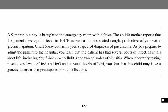
9
179
A 9-month-old boy is brought to the emergency room with a fever. The child’s mother reports that
the patient developed a fever to 101 ° F as well as an associated cough, productive of yellowish-
greenish sputum. Chest X-ray confirms your suspected diagnosis of pneumonia. As you prepare to
admit the patient to the hospital, you learn that the patient has had several bouts of infection in his
short life, including Staphylococcus cellulitis and two episodes of sinusitis. When laboratory testing
reveals low levels of IgA and IgG and elevated levels of IgM, you fear that this child may have a
genetic disorder that predisposes him to infections.

Hyper IgM Syndrome
179
Genetic Defect Primarily caused by inherited X-linked defect in gene encoding for T-cell molecule, CD40 ligand.
Pathophysiology CD40 ligand is a receptor that is located on the activated T cell. When the CD40 ligand binds to the CD40 molecule on the surface of the B cell, several immune processes are stimulated, including immunoglobulin class switching. If CD40 ligand is defective, then immunoglobulin class-switching is severely hindered, hence resulting in elevated levels of IgM and decreased levels of IgG and IgA.
Clinical Manifestations
Presents by age 2 with recurrent upper and lower respiratory infections as well as cellulitis, osteomyelitis, and sepsis. Hepatosplenomegaly and lymphadenopathy are often noted. Complications include increased rates of lymphomas, hepatocellular carcinoma, or neuroendocrine carcinomas.
Lab findings: Elevated levels of IgM; low levels of IgA and IgG; pancytopenia.
Treatment Intravenous immunoglobulin infusion or G-CSF injections to prevent infections; bone marrow transplant for severe forms of the disease.
Notes A rarer form of hyper-IgM syndrome is caused by a defect on chromosome 20 that codes for CD40. This disease is inherited in an autosomal recessive manner and is clinically similar to the X-linked form of hyper-IgM syndrome.

9
180
A 2-month-old girl presents to your urgent care clinic with symptoms of a viral upper respiratory
tract infection. Her mother tells you that this is the third time that her daughter has developed an
illness since birth. On physical examination, you notice that the girl has a small jaw as well as a
positive Chvostek sign (twitching of the facial muscles with superficial tapping of the facial nerve).
You decide to order several serum studies, which reveal hypocalcemia in the presence of decreased
levels of parathyroid hormone as well as an absence of serum T cells. You refer the patient and her
family to a geneticist to evaluate for a chromosomal abnormality and to a cardiologist to assess for
possible congenital cardiac defects, which are often associated with this patient’s condition.

DiGeorge Syndrome
180
Genetic Defect A chromosomal disorder that results in a microdeletion on chromosome 22.
Pathophysiology The chromosomal abnormality results in the failure of development of the third and fourth pharyngeal pouches. Without the fourth pharyngeal pouch, which normally gives rise to the thymus and parathyroids, the thymus and parathyroids do not develop, leading to T-cell deficiency and hypoparathyroidism. Disruption in the third and fourth pharyngeal arches also occurs, which causes abnormal neural crest cell migration and results in cardiac malformations.
Clinical Manifestations
T-cell deficiency manifests as recurrentviral, fungal, and protozoal infections.Hypoparathyroidism manifests with signs of hypocalcemia (tetany). Patients will also often have congenital cardiovascular defects and facial abnormalities (cleft palate; small jaw).
Treatment Fetal thymus transplanted to restore T-cell immunity.
Notes Common variable immunodeficiency is a disorder of variable inheritance that results in the inability for B cells to mature into plasma cells, thereby leading to a deficiency in secreted immunoglobulins. It presents with recurrent pyogenic infections, decreased immuno-globulin levels in the setting of normal serum B-cell levels, and an increased incidence of B-cell neoplasms, gastric cancer, and skin cancers.

9
181
A 3-year-old girl presents to your pediatric office for establishment of care. She and her family have
recently moved to the area from a rural community. The child’s mother reports that the child has
suffered from several bouts of cellulitis as well as sinusitis and pneumonia in the past. As you are
speaking with the child’s mother, you observe that the child has fair skin, extremely light blond hair,
and light-colored eyes. Physical examination is further notable for hepatosplenomegaly and several
ecchymoses. When routine blood work reveals the presence of neutropenia and thrombocytopenia,
you decide to send the patient for genetic testing to see if the patient has a mutation for a gene
associated with lysosomal transport on chromosome 1.

Chediak-Higashi Syndrome
181
Genetic Defect An autosomal recessive disorder that results from a mutation in the CHS1/LYST gene onchromosome 1.
Pathophysiology The CHS/LYST gene codes for a protein, which is responsible for the regulation of lysosomal content transportation to other cells, including neutrophils, neuronal cells, and melanocytes. When the CHS/LYST protein is defective, lysosomes do not function normally in these cells, thereby leading to impaired cytotoxic function of neutrophils (which results in infections) and impaired melanin transport to keratinocytes (which results in albinism).
Clinical Manifestations
Patients present in early childhood with recurrent pyogenic infections (particularly with Staphylococcus and Streptococcus organisms) and oculocutaneous albinism. Bleeding diatheses and hepatosplenomegaly are often noted. If patients survive to early adulthood, they develop debilitating neurologic disease, including neuropathies, ataxia, cognitive decline, and seizures.
Lab findings: Neutropenia; thrombocytopenia; elevated bleeding time; hypergammaglobulinemia.
Treatment Prophylactic antibiotics and G-CSF to ward off infection; steroid courses or bone marrow transplant has been used to treat severe forms of the disease.
Notes

9
182
A 19-year-old woman presents to the emergency room, complaining of abdominal pain, muscle and
joint aches, and fever. She and her family recently moved to the area from Greece and have yet to
establish primary care. The patient tells you that, for the past 5 years, she has had intermittent
episodes with symptoms similar to her current episode. On vital signs, the patient is febrile to
101.4° F. She has significant diffuse tenderness to abdominal palpation, and you notice a slight rub
on cardiac examination. She also has a lacy, reddish rash on her lower extremities. You initiate the
standard workup for abdominal pain and fever and, as you await the results, you wonder whether
the patient’s symptoms are related to a genetic deficiency in pyrin.

Mediterranean Familial Fever
182
Genetic Defect An autosomal recessive disorder that results from mutation in the MEFV gene on chromosome 16.
Pathophysiology The MEFV gene is responsible for producing a protein known as pyrin. Although the exact function of pyrin is unknown, it is thought to be involved in the inhibition of IL-8 and/or chemotactic factor 5a, two inflammatory cytotoxins. In patients with Mediterraneanfamilial fever, pyrin levels are decreased. When an inflammatory response is triggered, the lack of pyrin results in excessive inflammatory activity of cytotoxins, thereby leading to inflammation in the visceral tissues and joints.
Clinical Manifestations
Patients present with 2- to 4-day-long episodes of fever, abdominal pain (from peritoneal inflammation), chest pain (from pleural and pericardial inflammation), muscle and jointpain (from synovial inflammation), and rashes on the lower extremities.
Treatment Colchicine (to treat inflammation); steroids or etanercept for more severe attacks.
Notes As the name suggests, this disorder is more common in patients of Mediterranean descent.

9
183
A 22-year-old man presents to your oncology office for further evaluation of a large mass in his
left femur. The patient had noticed pain in his left thigh over the last several months, and this
symptom prompted his primary care provider to order a CT scan, which subsequently demonstrated
a suspicious mass in the femur, concerning for osteosarcoma. Upon taking a detailed history from
the patient, you learn that his mother is currently undergoing treatment for breast cancer, his older
sister died of leukemia, and that he has two maternal uncles with adrenal carcinomas. Given the
significant family history of malignancy, you decide to send this patient for genetic testing to see
if he has a mutation in a specific tumor-suppressive gene.

Li-Fraumeni Syndrome
183
Genetic Defect An autosomal dominant disorder that results from mutation in the tumor-suppressive gene p53 on chromosome 17.
Pathophysiology The p53 gene is responsible for multiple regulatory processes of the cell cycle and DNA repair. While the mechanisms of p53 are complex, in short, the p53 gene product senses DNA damage and then activates expression of DNA repair proteins as well as proteins that halt the cell cycle at the G1 phase while repairing takes place. If the DNA damage is too extensive, p53 can also trigger cell death. If the p53 gene is mutated, the above stop mecha-nisms are ineffective and cells with damaged DNA continue to proliferate leading to tumor formation.
Clinical Manifestations
Patients with Li-Fraumeni syndrome are at a significantly increased risk of cancers (inparticular sarcomas, osteosarcoma, breast cancer, leukemia, brain tumors, and adrenal carcinoma) at an early age.
Treatment Treatment of cancers with chemotherapy/radiation; genetic testing.
Notes

9
184
A 3-year-old girl presents to your pediatrics office for evaluation of a facial rash. The child’s mother
states that the child developed this rash over the last year and that it has become more prominent over
the last several months. As you review the child’s medical chart, you note that she is well below the
expected growth curve for her age and that she has already suffered from a bout of bronchitis and
gastroenteritis during her short life. On physical examination, you note that the child has a small jaw
and bird-like facies in addition to a butterfly-like rash over the nasal bridge and bilateral cheeks. You
also note scleral telangiectasias as well as other telangiectasias on the child’s limbs. Based on the
constellation of symptoms and physical findings, you worry that the child may suffer from a genetic
disorder that will lead her to have an increased susceptibility to developing malignancy later in life.

Bloom Syndrome
184
Genetic Defect An autosomal recessive disorder that results from mutation in the BLM gene on chromosome 15.
Pathophysiology The BLM gene codes for a protein that is part of the DNA helicase family. DNA helicase isinvolved in unwinding the DNA helix as well as stabilizing the DNA strands during replica-tion. If DNA helicase is faulty, DNA replication is defective and an inordinate number of mutations occur during DNA replication.
Clinical Manifestations
Physical characteristics include short stature, butterfly-shaped rash on cheeks, distinctive facial features (narrow face with bird-like facies; small mandible), and telangiectasias ofthe skin and eyes. Patients also suffer from hypogonadism, immunodeficiency (manifestsas recurrent respiratory and gastrointestinal infections), and increased susceptibility to malignancy.
Lab findings: decreased levels of IgA and IgM.
Treatment Avoid sun exposure; treatment of cancers with chemotherapy/radiation; genetic testing.
Notes

185
REFERENCES
Berg JM, Tymoczko JL, Stryer L. Biochemistry. 7th ed. New York, NY: W. H. Freeman; 2010.
Bhushan V, Pall V, Le T. Blackwell’s Underground Clinical Vignettes. 3rd ed. Malden, MS:
Blackwell Science; 2002.
Champe PC, Harvey RA, Ferrier DR. Lippincott’s Illustrated Reviews: Biochemistry. 5th ed.
Philadelphia, PA: Lippincott Williams & Wilkins; 2010.
Horowitz, M. Basic Concepts in Medical Genetics. 1st ed. New York, NY: McGraw-Hill; 2000.
Kumar V, Abbas AK, Aster J. Robbins Pathologic Basis of Disease, 8th ed. Philadelphia, W. B.
Saunders, 2010.
Le T, Bhushan V, Tolles J. First Aid for the USMLE Step I. New York, NY: McGraw-Hill; 2011.
Longo D, Fauci A, Kasper D, Hauser S. Harrison’s Principles of Internal Medicine. 18th ed.
New York, NY: McGraw-Hill; 2011.

185
McPhee SJ, Lingappa VR, Ganong WF. Pathophysiology of Disease: An Introduction to Clinical
Medicine. 6th ed. New York, NY: McGraw-Hill; 2009.
McPhee SJ, Papadakis MA, Rabow MW. Current Medical Diagnosis & Treatment 2010. 50th ed.
New York, NY: McGraw-Hill; 2011.
Schneider AS, Szanto PA. Board Review Series Pathology. 4th ed. Philadelphia, PA: Lippincott
Williams & Wilkins; 2008.
Schriver CR, Beaudet AL, Sly WS, et al. Metabolic and Molecular Bases of Inherited Disease.
New York, NY: McGraw-Hill; 2001.
Wilcox RB. High-Yield Biochemistry. 3rd ed. Philadelphia, PA: Lippincott Williams & Wilkins; 2009.

186
α1-Antitrypsin deficiency, 150
α-Glycerol phosphate shuttle, 4
α-Thalassemia, 141
β-Thalassemia, 141
11β-Hydroxylase deficiency, 93
17α-Hydroxylase deficiency, 92
21β-Hydroxylase deficiency, 91
3β-Hydroxysteroid dehydrogenase deficiency, 94
Achondroplasia, 167
Acute intermittent porphyria, 83
Adenosine deaminase deficiency, 76
Adrenal leukodystrophy, 54
Alanine shuttle, 58
Albinism, 174
Alcoholism, 114
Aldosterone, 90
Alkaptonuria, 63
Alport syndrome, 155
Amino acid degradation, 56, 60, 61
Amino acid derivatives, 60
Amino acid function, 56
Amino acid metabolism, 55-68
Amorphic mutation, 126
Andersen disease, 16
Androgen insensitivity syndrome, 95
Androgens, 90
Angelman syndrome, 130
Anticipation, 125
Antimorphic mutation, 126
Apolipoprotein function, 31
Ascorbic acid deficiency, 108
Ataxia-telangiectasia, 159
Autosomal dominant inheritance, 120
Autosomal recessive inheritance, 121
Base structure, 70
Becker muscular dystrophy, 168
Bernard-Soulier syndrome, 143
Biotechnology, techniques of, 116
Biotin deficiency, 109
Bloom syndrome, 184
Bruton agammaglobulinemia, 176
Calcium deficiency, 112
Carbohydrate metabolism, 5-25
Index

186
Carbon monoxide poisoning, 82
Carnitine shuttle, 29
Cell cycle, 118
Cellular energy, 1-4
Charcot-Marie-Tooth disease, 164
Chediak-Higashi syndrome, 181
Chemiosmotic hypothesis, 3
Cholesterol synthesis, 32
Chromosomal disorders, 124, 128-134
Chronic granulomatous disease of childhood, 178
Chylomicron, 31
Citrate shuttle, 29
Citric acid cycle, 2
Classic galactosemia, 23
Cobalamin deficiency, 107
Codominance, 125
Common variable immunodeficiency, 180
Congenital erythropoietic porphyria, 85
Congenital hyperbilirubinemia, 151
Cori cycle, 11
Cori disease, 15
Cortisol, 90
Cri-du-chat syndrome, 133
Cystic fibrosis, 148
Cystinosis, 64
Cystinuria, 64
Deamination of amino acids, 57
Deoxyribonucleotide synthesis, 75
DiGeorge syndrome, 180
DNA repair, 117
DNA replication, 117
DNA structure, 116
Down syndrome, 128
Dubin-Johnson syndrome, 151
Duchenne muscular dystrophy, 168
Dysbetalipoproteinemia, 38
Edwards syndrome, 129
Ehlers-Danlos syndrome, 169
Electron transport chain, 3
Essential amino acids, 56
Essential fructosuria, 21
Ethanol intoxication, 114
Ethanol metabolism, 98
Expressivity, 125
Fabry disease, 47
Factor V Leiden, 135
Familial adenomatous polyposis, 153
Familial combined hyperlipidemia, 39
Familial hypercholesterolemia, 35
Familial hyperchylomicronemia, 37
Familial hypertriglyceridemia, 36
187
Familial hypertrophic cardiomyopathy, 144
Fanconi anemia, 136
Farber disease, 50
Fatty acid oxidation, 28
Fatty acid synthesis, 27
Folic acid deficiency, 110
Fragile X syndrome, 134
Frameshift mutation, 126
Friedreich ataxia, 160
Fructose intolerance, 22
Fructose metabolism, 10
Galactokinase deficiency, 24
Galactose metabolism, 10
Gardner syndrome, 153
Gaucher disease, 48
Gel electrophoresis, 116
Genetic code, 116
Genetic disorders
of the cardiovascular system, 144-147
of the cell cycle, 183-184
of the endocrinologic system, 157-158
of the eye, 165-166
of the GI system, 150-154
of the hematologic system, 135-143
of the immune system, 176-182
of the musculoskeletal system, 167-173
of the nervous system, 159-164
of the renal system, 155-156
of the respiratory system, 148-149
of the skin, 174-175
Gilbert syndrome, 151
Glanzmann thrombasthenia, 143
Gluconeogenesis, 11
Glucose-6-phosphate dehydrogenase deficiency, 20, 139
Glutamine shuttle, 58
Glycogen storage diseases, 12-20
Glycogenesis, 6
Glycolysis, 7
GM1 gangliosidosis, 46
Gout, 77
Hardy-Weinberg population genetics, 127
Hartnup disease, 65
Hgb-O2 dissociation curve, 82
HDL, 31
Heme degradation, 81
Heme metabolism, 79-87
Heme synthesis, 80
Hemochromatosis, 152
Hemoglobin, 82
Hemoglobin C disease, 140

Hemoglobin regulation, 82
Hemophilia A and B, 137
Hereditary coproporphyria, 84
Hereditary hemorrhagic telangiectasia, 146
Hereditary nonpolyposis colorectal cancer, 153
Hereditary spherocytosis, 138
Hers disease, 13
Histidinemia, 68
Homocystinuria, 66
Hunter disease, 41
Huntington disease, 161
Hurler disease, 40
Hyper IgM syndrome, 179
Hypercalcemia, 112
Hypermorphic mutation, 126
Hypomorphic mutation, 126
I-cell disease, 51
Imprinting, 125
Incomplete dominance, 125
Inherited hyperlipidemias, 35-39
Iodine deficiency, 113
Iron deficiency, 111
Kartagener syndrome, 149
Klinefelter syndrome, 131
Knudson “two-hit” hypothesis, 166
Krabbe disease, 53
Kwashiorkor, 99
Lactase deficiency, 25
Lactose intolerance, 25
LDL, 31
Lead poisoning, 87
Leber hereditary optic neuropathy, 165
Lesch-Nyhan syndrome, 77
Li-Fraumeni syndrome, 183
Linkage disequilibrium, 125
Lipid metabolism, 26-54
Lipid transport, 30
Lipoprotein function, 31
Liver phosphorylase deficiency, 13
LOD score, 125
Macronutrient deficiency, 99
Macronutrients, 98
Magnesium deficiency, 113
Malate shuttle, 4
Maple syrup urine disease, 67
Marasmus, 99
Marfan syndrome, 170
McArdle disease, 17
187

188
Mediterranean familial fever, 182
Meiotic nondisjunction, 124
MELAS, 171
MEN syndromes, 157
MERRF, 171
Metachromatic leukodystrophy, 54
Methemoglobin, 82
Methionine metabolism, 62
Micronutrients, 97
Mineral deficiency, 111-113
Minerals, 97
Missense mutation, 126
Mitochondrial inheritance, 123
Mitochondrial myopathies, 171
Mixed hypertriglyceridemia, 37
Mosaicism, 125
mRNA processing, 118
Multiple endocrine neoplasia syndromes, 157
Multiple polyposis syndromes, 153
Myotonic dystrophy, 172
Neomorphic mutation, 126
Neurofibromatosis type 1 and 2, 162
Niacin deficiency, 105
Niemann-Pick disease, 49
Nonpolar amino acid, 56
Nonsense mutation, 126
Noonan syndrome, 145
Northern blotting, 116
Nucleotide metabolism, 69-78
Nucleotides, 70
Nutrition, 96-114
Orotic aciduria, 78
Osler-Weber-Rendu Disease, 146
Osteogenesis imperfecta, 173
Osteomalacia, 101
Oxidative deamination, 57
Pantothenic acid deficiency, 104
Patau syndrome, 129
Penetrance, 125
Pentose phosphate pathway, 9
Peutz-Jeghers syndrome, 153
Phenylalanine metabolism, 62
Phenylketonuria, 68
Phospholipid synthesis, 34
Phosphorus deficiency, 113
Pleiotropy, 125
Polar amino acid, 56
Polycystic kidney disease, 156
Polymerase chain reaction, 116
Pompe disease, 14
Porphyria cutaneous tarda, 86
Prader-Willi syndromes, 130
Pseudohypoparathyroidism, 158
Purine nucleotide degradation, 74
Purine ring, 70
Purine salvage pathway, 72
Purine nucleotide synthesis, de novo, 71
Pyridoxine deficiency, 106
Pyrimidine nucleotide degradation, 74
Pyrimidine nucleotide synthesis, 73
Pyrimidine ring, 70
Pyruvate dehydrogenase complex deficiency, 19
Pyruvate metabolism, 8
Restriction fragment length polymorphism (RFLP) analysis, 116
Retinoblastoma, 166
Retinol deficiency, 100
Riboflavin deficiency, 104
Ribosome, 119
Rickets, 101
RNA structure, 116
Rotor syndrome, 151
Sandhoff disease, 45
Sanfilippo disease, 42
Scheie syndrome, 40
SCID, 76
Sialidosis, 52
Sickle cell anemia, 140
Sickle cell trait, 140
Sly syndrome, 43
Southern blotting, 116
Southwestern blotting, 116
Sphingolipid degradation, 33
Sphingolipid storage diseases, 40-54
Sphingolipid synthesis, 32
Steroid hormone synthesis, 88-95
Steroidogenesis, 89
Tarui disease, 18
Tay-Sachs disease, 44
Thalassemia, 141
Thiamine deficiency, 103
Thymidylate synthesis, 75
Transamination, 57
Transcription, 118
Transfer RNA, 119
Translation, 119
Tuberous sclerosis, 163
Turcot syndrome, 153
Turner syndrome, 132
188

189
Urea cycle, 59
Vitamin A deficiency, 100
Vitamin B1 deficiency, 103
Vitamin B12 deficiency, 107
Vitamin B2 deficiency, 104
Vitamin B3 deficiency, 105
Vitamin B5 deficiency, 104
Vitamin B6 deficiency, 106
Vitamin C deficiency, 108
Vitamin D deficiency, 101
Vitamin D toxicity, 101
Vitamin E deficiency, 102
Vitamin K deficiency, 102
Vitamins, 97
VLDL, 31
von Gierke disease, 12
von Hippel-Lindau disease, 147
von Willebrand disease, 142
Western blotting, 116
Wilson disease, 154
Wiskott-Aldrich syndrome, 177
Xeroderma pigmentosum, 175
X-linked recessive inheritance, 122
XYY syndrome, 131
Zinc deficiency, 113