larvicidal, super hydrophobic and antibacterial properties
TRANSCRIPT

RSC Advances
PAPER
Ope
n A
cces
s A
rtic
le. P
ublis
hed
on 2
5 A
ugus
t 201
7. D
ownl
oade
d on
12/
24/2
021
2:07
:25
AM
. T
his
artic
le is
lice
nsed
und
er a
Cre
ativ
e C
omm
ons
Attr
ibut
ion
3.0
Unp
orte
d L
icen
ce.
View Article OnlineView Journal | View Issue
Larvicidal, super
aCentre for Nano Science and Technology,
Tiruchengode-637215, Tamil Nadu, India.
+91-4288-274880; Tel: +91-4288-274741-4bDepartment of Chemistry, National Univ
Singapore
† Electronic supplementary informa10.1039/c7ra05697d
Cite this: RSC Adv., 2017, 7, 41763
Received 20th May 2017Accepted 29th July 2017
DOI: 10.1039/c7ra05697d
rsc.li/rsc-advances
This journal is © The Royal Society of C
hydrophobic and antibacterialproperties of herbal nanoparticles from Acalyphaindica for biomedical applications†
Karthik S., a Suriyaprabha R., a Vinoth M.,a Srither S. R.,a Manivasakan P.,a
Rajendran V. *a and Suresh Valiyaveettil b
The present study is aimed at developing a biocompatible nanomaterial with excellent medicinal properties
using herbs. The herbal nanoparticles were prepared from shade dried leaves of Acalypha indica using the
ball-milling technique. The prepared nanoparticles were characterized using X-ray diffraction, Fourier
transform infrared spectroscopy, ultraviolet-visible spectroscopy, particle size analysis, scanning electron
microscopy, X-ray fluorescence spectroscopy and transmission electron microscopy. The amorphous
herbal AINPs posses an average particle size distribution of 54 � 3 nm and a UV-absorption maximum at
434 nm, and are superhydrophobic (151�) in nature. The prepared herbal AINPs were tested for their
antimicrobial properties against Staphylococcus aureus and Escherichia coli. Mosquito repellent
properties were investigated against three disease vectors, namely, Aedes aegypti, Anopheles stephensi
and Culex quinquefasciatus, and showed significant larvicidal activity due to the existence of
phytochemical compounds in the herbal nanoparticles. The acute toxicity of the herbal nanoparticles
was tested with an in vivo animal model, zebrafish (Danio rerio), to ensure biocompatibility. The
observed results confirmed that herbal AINPs play a dominant role in enhancing the medicinal properties
for different biomedical applications.
Introduction
Everyday human life is affected by skin infections and mosquitorelated diseases such as boils, impetigo, cellulitis, malaria,dengue, lymphatic lariasis and yellow fever.1 Recent develop-ments in nanotechnology demonstrate the practical applicationsof nanoparticles from plant extracts, due to their enhanced anti-bacterial, antifungal, hydrophobic, mosquito repellent and UVprotection properties.2 Many natural products are widely used todevelop newer medicines with potent biological and pharmaco-logical activities. Medicinal plant based drugs have been exten-sively used in skin allogra, cornea allogra, intestinal allogra,cardiac allogra, and in the treatment of various diseases such asmalaria, symptomatic endometriosis, uterine adenomyosis, leio-myoma, etc.3–5 The herbal nanoparticle treated materials areknown in the eld of biomedical applications for their exoticproperties, e.g., non-toxic, mosquito repellent, hydrophobic,ultraviolet protection (UV) and antimicrobial action.6–9
K. S. Rangasamy College of Technology,
E-mail: [email protected]; Fax:
ersity of Singapore, Singapore 117543,
tion (ESI) available. See DOI:
hemistry 2017
Acalypha indica (A. indica) is a common weed that belongs tothe Euphorbiaceae family. It grows in common farmlands,gardens and uncultivated lands. All parts of A. indica (leaves,root, stalk and ower), which include constituents like acaly-phine, triacetoneamine, cyanogenic glucosides, and alkaloids,are highly valuable for medicinal applications, due to their anti-inammatory and antimicrobial properties.10 The earlierstudies on the extracts of Acalypha indica conrm its antimi-crobial properties against pathogenic bacteria such as Escher-ichia coli (E. coli), Salmonella typhi (S. typhi), Pseudomonasaeruginosa (P. aeruginosa), Staphylococcus aureus (S. aureus) andBacillus subtilis (B. subtilis).11,12
As reported by Suresh et al.,13 a GCMS study revealed that theA. indica leaf consists of 2,5-pyrrolidinedione, 1-methyl-3-o-methyl-D-glucose, tetradecanoic acid, 3,7,11,15-tetramethyl-2-hexadecen-1-ol, n-hexadecanoic acid, phytol, 9,12,15-octadeca-trienoic acid, (Z,Z,Z), oleic acid, 1,2-benzenedicarboxylic aciddiisooctyl ester, and squalene. Along with the aforementionedcompounds, Chandra Mohan et al.14 reported that 1H-pyrrole-2,5-dione,1-ethenyl-, 3,8-nonadien-2-one, (E)-, 3,4-didehydro-proline, 4-amino-3-methoxypyrazolo[3,4-d]pyrimidine, pro-panenitrile, and 3-(5-diethylamino-1-methoxy-3-pentynyloxy)-compounds are also present in A. indica leaves.
Mosquito vectors are mainly responsible for endemic andpandemic diseases. The Ae. aegypti vector causes dengue feverin tropical and sub-tropical regions.1 An. strephensi is the
RSC Adv., 2017, 7, 41763–41770 | 41763

Fig. 1 Synthesis protocol for herbal nanoparticles.
RSC Advances Paper
Ope
n A
cces
s A
rtic
le. P
ublis
hed
on 2
5 A
ugus
t 201
7. D
ownl
oade
d on
12/
24/2
021
2:07
:25
AM
. T
his
artic
le is
lice
nsed
und
er a
Cre
ativ
e C
omm
ons
Attr
ibut
ion
3.0
Unp
orte
d L
icen
ce.
View Article Online
primary vector that causes malarial infection.15 Filariasisdiseases are caused by the Cx. quinquefasciatus vector in tropicaland sub-tropical areas.16 Attempts are being made to eradicatemosquito vector borne diseases.17,18 Bio prospecting of thelarvicidal properties is one of the approaches to solving theabove issues. Even though different nanocomposites andchemicals provide excellent medicinal values, the propertiessuch as non-biodegradability, bioaccumulation, bio controlaction, and toxicity keep them from being used for medicalapplications.2,19 Among the different herbal plants, A. indicashows good antibacterial, antimalarial, parasiticide, protisti-cide, plasmodicide, pesticide, antimutagenic, cancer preventiveproperties, and hence nds wide applications as a diuretic,antifungal, purgative, antihelmintic, anti-inammatory and inthe treatment of insect bites.20–22
The medicinal applications of herbal nanoparticles withexotic textural characteristics are validated by coating thesenanoparticles onto cotton fabrics. The different antimicrobial,hydrophobic and wound healing properties are explored innanoparticles coated fabrics.23,24 The recent studies on the self-cleaning, water repellent, high durability, antibacterial and UVprotection properties, etc., of the herbal nanoparticles coatedtextiles strongly suggest their application in medicine.25,26
In the present study, we focus on the development of biomedically important herbal nanoparticles using the greensynthesis method. The herbal nanoparticles are prepared fromA. indica leaves using a ball mill without the addition ofchemicals for aggregation and template shaping. In addition tothe toxicity, antimicrobial and hydrophobic properties, thelarvicidal properties of herbal nanoparticles are exploredagainst three mosquito vectors, Aedes aegypti, Anopheles ste-phensi and Culex quinquefasciatus.
Materials and methodsCollection of samples and preparation of herbal nanoparticles
The middle leaves of A. indica were collected and thoroughlywashed several times using deionized (D.D.) water to removedust on the leaf surface. The leaves were then shade dried for 2weeks. The dried leaves were ground using ball milling for 15 hat 300 rpm. Zirconium balls of 10 mm diameter were used formilling. The milling with a ball ratio of 20 : 1 was carried out for10 g of leaves in a grinding jar with a zirconium protective jacket.Aermilling, the nanoparticles were taken for biological studies,followed by different characterization studies as reported in ourprevious investigations.27–29 The protocol for the preparation ofherbal nanoparticles from A. indica leaves is shown in Fig. 1.
The X-ray diffraction patterns of the prepared A. indicananoparticles (AINPs) were obtained using a powder X-raydiffractometer (XRD; X'Pert PRO, PANalytical, Almelo, theNetherlands) operated with long ne focus of the Cu anode at40 kV and 30 mA in Bragg–Brentano geometry. The XRD patternwas obtained in the 2q range from 10� to 80� in a step-scanmode with a step size of 0.02�. Fourier transform infrared(FTIR) spectra of the nanoparticles were recorded using an FTIRspectrophotometer (Spectrum 100; PerkinElmer, USA) in therange of 400–4000 cm�1 using KBr (90 wt% IR Grade KBr)
41764 | RSC Adv., 2017, 7, 41763–41770
matrix for making transparent disks. The green synthesized leafnanoparticles were monitored periodically using a UV-visible(UV-vis) spectrophotometer (Agilent Cary 8454, Singapore)operated in a wide range from 180–800 nm using a step size of5 A at different time intervals. A particle size analyzer(Nanophox, Sympatec, Germany) was used to determine theparticle size distribution based on the dynamic light scattering(DLS) technique with a sub micrometer at a scattering angle of90�. SEM, coupled with energy-dispersive X-ray (SEM-EDX, JSM6360 JEOL, Japan) analysis, was used to identify themorphology, microstructure, and elemental composition of theprepared nanoparticles, fabrics coated with herbal nano-particles and un-coated fabrics. Grain size and surfacemorphology of AINPs were examined through transmissionelectron microscopy (TEM, CM200; Philips, Eindhoven, TheNetherlands) operated at a potential of 120 kV.
Larvicidal activity/repellent property
Larvae of mosquito vectors, namely, Ae. Aegypti, An. stephensi,and Cx. quinquefasciatus, were collected from Namakkal, TamilNadu, India. Mosquitoes were cultured in the laboratory andwere maintained continuously in laboratory conditions as re-ported by WHO.30 The second and fourth instar larvae wereexposed to the treatment of herbal nanoparticles with differentconcentrations such as 100, 200, 300, 400 and 500 mg L�1. Eachtreatment was carried out in triplicate, each comprising of 25larvae. The hatching of mosquitoes under different doses ofherbal nanoparticle treatments was closely monitored from 24 to48 h, and the LC50 value and mortality percentage were calcu-lated using the relation, (T–C/100� C)� 100, where T is the totalnumber of treated larvae and C the total hatchability of larvae. Astandard larvicidal compound, prallethrin, was used to comparethe larvicidal properties of the prepared herbal nanoparticles.
This journal is © The Royal Society of Chemistry 2017

Paper RSC Advances
Ope
n A
cces
s A
rtic
le. P
ublis
hed
on 2
5 A
ugus
t 201
7. D
ownl
oade
d on
12/
24/2
021
2:07
:25
AM
. T
his
artic
le is
lice
nsed
und
er a
Cre
ativ
e C
omm
ons
Attr
ibut
ion
3.0
Unp
orte
d L
icen
ce.
View Article Online
In vivo toxicity
The invertebrate animal model, with zebrash (Danio rerio)embryos, was used to assess the acute toxicity of the herbalnanoparticles of biomedical importance. Experiments werecarried out with 15 zebrash for each dose (12.5, 25, 50, 75 and100 mg L�1) along with a control as per OECD-203 guidelines.31
All the animal model experiments were approved by theprocedure of the Committee for the Purpose of Control andSupervision of Experiments on Animals (CPCSEA), as instructedby Institutional Animal Ethics Committee (IAEC) guidelines,32
K. S. Rangasamy College of Technology (Reg. no. 1826/PO/EReBi/S/15/CPCSEA), Tiruchengode, Tamil Nadu, India. Thehatching rate, mortality and developmental defects weredetermined for each tested dose in sh embryos, as reported inour earlier studies.33,34
Coating of herbal nanoparticles
Bleached cotton fabric (100%, mass 138.84 g m�2, 116 ends perinch, 84 picks per inch) was used as a substrate for coating theherbal nanoparticles through a padding mangle at a rate of35 rpm for 5 min to collect the uniformly coated fabrics35,36 inorder to explore the hydrophobicity and UV-protection proper-ties. Then, the coated fabrics were studied for their enhancedphysico–mechanical and antimicrobial properties such ascoating thickness, tensile and tear strength, crease recoveryangle, air permeability and bacterial growth reduction; thesedata are given in detail in the ESI le (S1).†
Fig. 2 XRD spectra of AINPs.
Hydrophobicity and UV-protection properties
Hydrophobicity and UV-protection properties of herbal nano-particles were examined for self-cleaning and UV radiationprotection applications, respectively. The hydrophobic natureof the herbal nanoparticle treated and untreated fabrics wasascertained based on the water contact angle (VCA Optima, ACTProduct Inc., Japan). Digital photographic analysis of waterdroplets placed on the fabric surface was conducted beforewashing and aer the 10th wash. Similarly, UV resistant prop-erties of herbal nanoparticles were also studied on the nano-particle coated fabrics using UV transmission spectra (Lambda35; PerkinElmer, USA), in the wavelength range of 280–400 nmas per the ASTM D6603 standard.
Fig. 3 FTIR spectrum of A. indica leaf nanoparticles.
Antimicrobial studies
Two bacterial cultures, namely, Gram-positive S. aureus (ATCC6538P) and Gram-negative E. coli (ATCC 9677), were obtainedfrom the National Collection of Industrial Microorganisms(NCIM) (National Chemical Laboratory, Pune, India). The ob-tained bacterial cultures were further sub-cultured several timesat 37 �C for 24 h. Inoculation of a loopful of test organisms intonutrient broth was carried out to prepare fresh bacterial inoc-ulums, and then incubated at 37 �C for 5–8 h till a fair turbiditywas obtained. Fresh cultures were swabbed on a nutrient agarplate and then herbal particles of different concentrations(25, 50 and 100 mg mL�1) were loaded onto the well puncturedin the nutrient agar plate. Aer 24 h of incubation, zones of
This journal is © The Royal Society of Chemistry 2017
inhibition around the herbal nanoparticles loaded onto theagar well were measured using a millimeter ruler.
Results and discussionCharacterization
Herbal nanoparticles of AINPs were synthesized and compre-hensively characterized for evaluating the inuence of nano-scale particles on the medicinal, antimicrobial and functionalproperties. The XRD pattern of AINPs is shown in Fig. 2, whichconrms the absence of diffraction peaks i.e., amorphousnature, except for the observed broad band at 2q values (20–30�).Generally, the amorphous nature of the particles is non-toxic toliving organisms and hence, amorphous herbal nanoparticlesenhance the biocompatibility for clinical applications.
The FTIR spectrum obtained from AINPs is shown in Fig. 3.The peaks observed between 3200 cm�1 and 3500 cm�1 areassigned to the presence of supercially absorbed water and thestretching mode of the OH/NH group, respectively. The bandsobserved at 2920 cm�1 and 2858 cm�1 correspond to thestretching vibrations of aliphatic and aromatic C–H bonds inthe nanoparticles.37 The peak at 1626 cm�1 is identied as the
RSC Adv., 2017, 7, 41763–41770 | 41765

RSC Advances Paper
Ope
n A
cces
s A
rtic
le. P
ublis
hed
on 2
5 A
ugus
t 201
7. D
ownl
oade
d on
12/
24/2
021
2:07
:25
AM
. T
his
artic
le is
lice
nsed
und
er a
Cre
ativ
e C
omm
ons
Attr
ibut
ion
3.0
Unp
orte
d L
icen
ce.
View Article Online
carbonyl (C]O) stretching vibration in the amide linkage of theplant protein.38 The band at 1384 cm�1 is assigned to theprimary amine (N–H) bending mode, which is the appropriatecharacteristic peak of avanones and terpenoids present in A.indica plant leaves.39 The peaks at 1317 cm�1 and 1243 cm�1
signify the carboxylic acid (C–O) group40 and the obtained peakat 1060 cm�1 discloses the C–N stretching vibrations ofaliphatic amines. The band observed at 660 cm�1 is due to the
Fig. 6 Particle size distribution of A. indica leaf nanoparticles (a) SEM im
Fig. 5 Particle size distribution of A. indica leaf nanoparticles.
Fig. 4 UV-visible spectrum of A. indica leaf nanoparticles in water.
41766 | RSC Adv., 2017, 7, 41763–41770
deformation of a-glucopyranose rings of carbohydrates.40,41 TheFTIR spectrum conrms the presence of major plantcompounds that are responsible for the antimicrobial, larvi-cidal and UV-protection properties.
Fig. 4 shows the UV-vis spectrum of the synthesized herbalAINPs dispersed in water. In the present study, A. indica has anabsorbance in the UV region at around 434 nm, which favours theanti-reective and UV-adsorption properties for textile applica-tions. Similar to silver nanoparticles, the plant based nano-materials also show antimicrobial activity, but with lower toxicity.To determine the stability of the nanoparticles, we obtained theUV-vis spectra of the leaf nanoparticles at different time intervalsfor 8 days. It is interesting to see from Fig. 4 that there is nodifference in absorption spectra, which conrms the higherstability of A. indica nanoparticles. The antireective properties ofthe nanoparticles are favorable for developing UV-resistantbiomaterials in an eco-friendly way. Fig. 5 shows the particlesize distribution curve of the prepared AINPs. The averageparticle size of the prepared nanoparticles is around 54 � 3 nm.
SEM and TEM images of the AINPs are shown respectively inFig. 6 and 7. The topographical characterization of the nano-particles observed in the SEM image shows that the herbal AINPsare uniform in structure. The SEM image shows the discretedistribution of herbal nanoparticles at higher magnication.The elemental composition of AINPs analyzed using EDX showsC and O peaks corresponding to elements such as Na, Mg, Si, Cl,K, and Ca metal ions, which conrm the inorganic compounds.A similar observation was reported in our previous study onobtaining the herbal nanoparticles from Tridax procumbensunder different milling periods.26 The TEM (Fig. 7) image of theprepared AINPs conrms the spherical nature with highdispersion. The average particle size of the herbal AINPs ob-tained from the TEM image is about 50 nm. This is in closeagreement with the measured particle size (54 nm) of the AINPsfrom the particle size distribution measurements.
Larvicidal activity
The larvicidal activities of the ve different concentrations ofAINPs to Aedes aegypti, Anopheles stephensi and Cx. quinque-fasciatus are shown in Table 1. Aer 48 h of exposure, the AINPswere tested for their larvicidal activities according to the
age and (b) EDX analysis.
This journal is © The Royal Society of Chemistry 2017

Table 2 In vivo toxicity of herbal nanoparticles used to treat zebrafish
NanoparticlesConcentrations(mg mL�1)
Mortality (%)
24 h 48 h 72 h
A. indica Control 0 0 025 0 0 3.350 0 3.3 6.675 0 6.6 13.3100 0 6.6 13.3200 0 6.6 6.6
Prallethrin 25 100 0 0
Fig. 7 TEM image of A. indica nanopowders.
Paper RSC Advances
Ope
n A
cces
s A
rtic
le. P
ublis
hed
on 2
5 A
ugus
t 201
7. D
ownl
oade
d on
12/
24/2
021
2:07
:25
AM
. T
his
artic
le is
lice
nsed
und
er a
Cre
ativ
e C
omm
ons
Attr
ibut
ion
3.0
Unp
orte
d L
icen
ce.
View Article Online
treatment concentrations. The larvicidal activities of the herbalnanoparticles were found to be high in terms of mortality rate ata higher concentration of AINPs against Aedes aegypti, Anophelesstephensi and Cx. Quinquefasciatus (LC50 ¼ 500 mg L�1), asshown in Table 1. However, lower mortality was observed whileexploiting both the Ist and IVth instars, due to the low amount ofAINPs against these three mosquito vectors.
Several Euphorbiaceae plant extracts are known to exhibitlarvicidal activity against these three mosquito vectors.42 Previ-ously, extracts of A. indica using methanol, ethyl acetate,benzene and chloroform were studied for their larvicidal andovicidal activities against Anopheles stephensi. In fact, crudenanoparticles of A. indica revealed comparatively higher larvi-cidal action than the extracted compounds of A. indica (by 15percent), against the three mosquito vectors.43 The observedenhanced larvicidal activities are due to the exposure of thehighly reactive surface area of the herbal AINPs. It is inferredfrom the present study that the obtained crude herbal nano-particles with high surface areas act as an effective mosquitorepellent and larvicide. The effectiveness of the prepared herbalA. indica nanoparticles was compared with a commercially usedlarvicide, prallethrin, with the least effective concentrations ofA. indica nanopowder against mosquito larvae (120 mg L�1).Table 1 shows similar larvicidal properties for Prallethrin andprepared herbal nanoparticles. There are reports that show thatPrallethrin is a poisonous material at lower concentration(25 mg mL�1).44 Our study shows that the use of herbal
Table 1 Larvicidal activity of herbal nanoparticles against Ae. Aegypti, An
Mosquito species Instar
A. indica nanoparticles at different
Prallithrin
(120 mg L�1) 100 mg L�1
Ae. aegypti I 100.0 � 0.00 16.07 � 2.71IV 100.0 � 0.00 8.11 � 3.11
An. stephensi I 100.0 � 0.00 19.07 � 3.12IV 100.0 � 0.00 9.32 � 2.05
Cx. Quinquefasciatus I 100.0 � 0.00 19.07 � 3.12IV 100.0 � 0.00 9.32 � 2.05
This journal is © The Royal Society of Chemistry 2017
nanoparticles with good larvicidal action is amenable formosquito control as an alternative larvicide to syntheticchemicals.
In vivo toxicity
The toxicity of AINPs was explored to determine the safe use forhumans and other animals. Most herbs are non-toxic and goodfor health, due to the presence of numerous organiccompounds. In order to explore the toxicity level of the nano-particles, zebrash embryos were treated with the AINPs andtheir developmental stages were monitored for 72 h as shown inTable 2. Among the ve different concentrations of nano-particles, particles of 200 mg L�1 concentration were highlyeffective with no mortality of the zebrash embryos. In addi-tion, a high concentration of nanoparticles does not affect theembryogenesis or hatching rate. Interaction of herbal particlesassociated with the biocompatibility of the plant compoundsdoes not affect the sh developmental stages.
A study investigating several metal oxide nanoparticles fortheir acute toxicity studies revealed signicant toxicity at higherconcentration when the embryos were tested for 72 h.45 Ourobservation reveals the non-toxic nature of the prepared AINPsagainst embryos, due to the samples being devoid of any pro-cessing chemicals like polar and non-polar solvents. Oncomparison of AINPs with other nano metal oxides, the herbalnanoparticles were found to be highly biocompatible withinvertebrate animal model zebrash by means a manifoldreduction in mortality. Unfortunately, exposure concentrationsof herbal nanoparticles are different from other metal oxidenanoparticles,41 since herbal nanoparticles are of biological
. Stephensi and Cx. Quinquefasciatus
concentrations (ppm)
200 mg L�1 300 mg L�1 400 mg L�1 500 mg L�1
33.12 � 1.41 64.43 � 3.17 97.15 � 2.32 100.0 � 0.0025.14 � 2.04 47.03 � 2.10 93.03 � 1.15 100.0 � 0.0035.07 � 2.51 63.14 � 2.71 99.42 � 2.11 100.0 � 0.0027.16 � 1.74 52.11 � 1.76 94.01 � 3.02 100.0 � 0.0038.05 � 2.72 69.32 � 2.43 100.0 � 0.11 100.0 � 0.0026.42 � 1.65 54.27 � 1.54 96.01 � 0.31 100.0 � 0.00
RSC Adv., 2017, 7, 41763–41770 | 41767

Fig. 8 Photographic images of water droplets placed on un-coatedand coated fabrics before and after washing. (a) Un-coated, (b and c)un-coated 5th & 10th wash, (d) C-CF, (e and f) C-CF 5th & 10th wash, (g)AI-C-CF, (h and i) AI-C-CF 5th & 10th wash.
RSC Advances Paper
Ope
n A
cces
s A
rtic
le. P
ublis
hed
on 2
5 A
ugus
t 201
7. D
ownl
oade
d on
12/
24/2
021
2:07
:25
AM
. T
his
artic
le is
lice
nsed
und
er a
Cre
ativ
e C
omm
ons
Attr
ibut
ion
3.0
Unp
orte
d L
icen
ce.
View Article Online
origin and need to be tested at higher concentrations. Even athigher concentrations, the particles do not cause signicanttoxicity, which deserves wider use of the AINPs in the biomed-ical eld for multidisciplinary applications as antimicrobial,mosquito repellent and biocompatible materials. Oncomparing the larvicidal activity of A. indica nanoparticles withthe commercially used larvicide, prallethrin, it was observedthat the synthetic oil prallethrin was highly toxic, since itshowed 100% mortality at 24 h. From the aforementionedstudy, it can be said that A. indica nanoparticles can be used asa natural larvicide in place of prallethrin, due to its lowertoxicity and better larvicidal properties.
The surface characteristics of the AINPs like hydrophobicityand adherence properties were explored for biomedical appli-cations, using cotton fabric. The ultraviolet protection factor(UPF) in the wavelength ranges from 280 to 400 nm was studiedaer coating the herbal particles onto the fabric, and the resultsare shown in Table 3. The percentage of transmittance for UV-Aand UV-B is almost similar to that of the untreated fabrics.46 Asignicant reduction in transmittance was observed for herbalnanoparticle coated fabrics, which is ascribed to the blocking ofUV-B and UV-A radiation due to the coating. In addition, thepercentage blocking of UV-B radiation by the herbal nano-composite is high (57%) as compared to that of chitosan-coating.47 Furthermore, the resistance rate of UV radiation forthe herbal nanocomposite aer the 5th and 10th washes ismoderately reduced (4%) in herbal AINPs treated fabricscompared to that of the untreated cotton fabrics. On the basis ofthe ASTM D6603 standard data, the UPF value for the fabrics ismore than 50%, demonstrating better protection of fabric fromUV rays. The calculated UPF value for the AINPs coated fabricswas observed to be 57.7 � 0.06, i.e., higher than the limitation(i.e., 50) ascribed to the higher resistance to UV irradiation.However, the values for un-coated and chitosan-coated fabricsexhibit lesser UV protection (<50) compared to those of theherbal nanocomposite coated fabrics. Thus, the herbal nano-particles have an increment of nearly 50% in UV protection,compared to the un-coated fabric. This is attributed to the
Table 3 UV protection and water repellent properties of un-coated,chitosan and herbal nanocomposite coated fabric samples
Sample names UPF valueContact angle(�)
Before washUn-coated fabrics 13.9 � 0.63 0Chitosan 42.8 � 0.46 119 � 1Nanocomposite 57.7 � 0.06 151 � 3
Aer 5th washUn-coated fabrics 11.2 � 0.93 0Chitosan 40.1 � 0.77 101 � 3Nanocomposite 55.6 � 0.70 135 � 3
Aer 10th washUn-coated fabrics 10.8 � 0.61 0Chitosan 39.5 � 0.55 93 � 6Nanocomposite 53.1 � 0.47 124 � 3
41768 | RSC Adv., 2017, 7, 41763–41770
ability of herbal nanoparticles to provide protection from UV-rays, which would be an additional advantage for developingradiation protective clothing.48
Hydrophobic activity
The water repellent property of the particles is one of the mostinteresting and desired properties of medical textiles, especiallyin pharmaceuticals and biomedicine.47,48 Researchers have re-ported that the hydrophobic or water repellent property isa function of the textile surface morphology and reducedsurface energy.4,49–51 In our studies, we have investigated thewater repellent efficiency of herbal nanoparticles, in terms ofcontact angle, by coating the herbal AINPs onto cotton fabrics.Generally, the fabrics with angle greater than 90� are consideredto be hydrophobic, while those with angle greater than 150� are
Fig. 9 Antimicrobial activity of A. indica nanoparticles for differentconcentrations: (A) 25 mg mL�1, (B) 50 mg mL�1 and (C) 100 mg mL�1
against E. coli and S. aureus.
This journal is © The Royal Society of Chemistry 2017
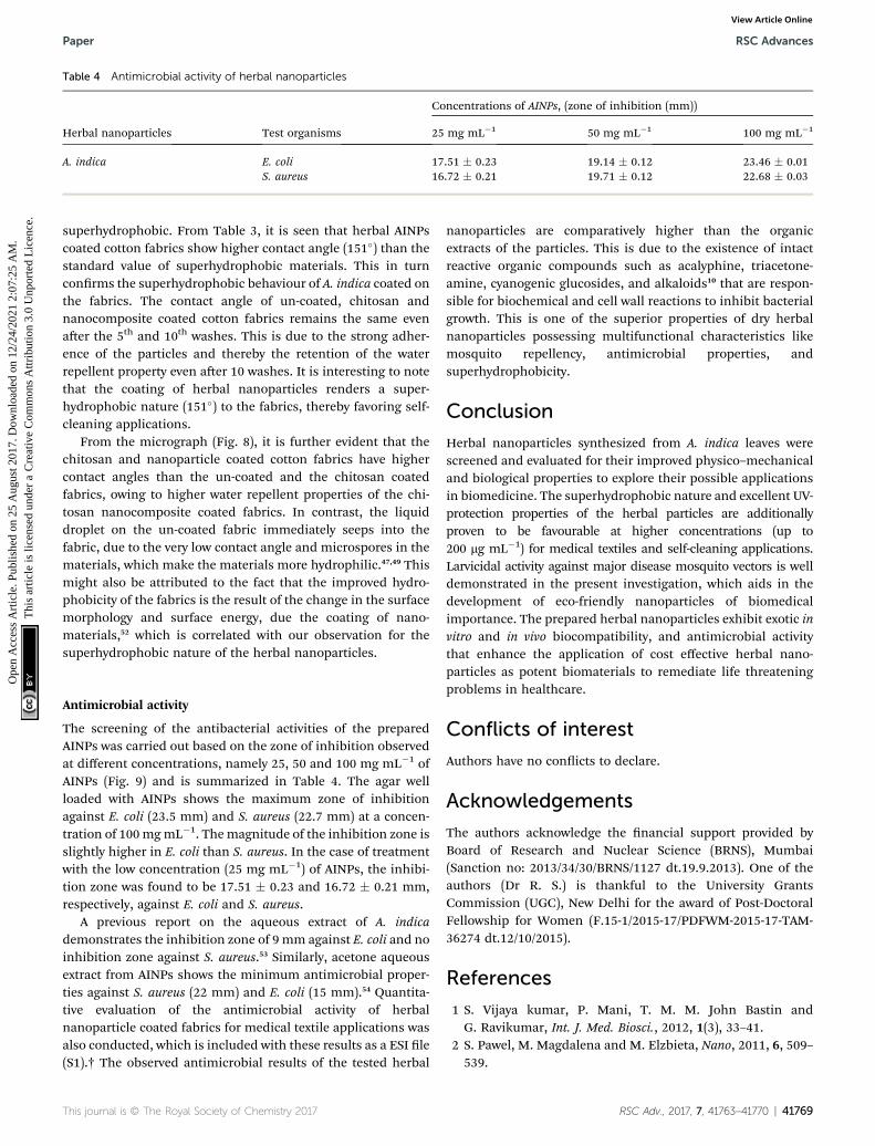
Table 4 Antimicrobial activity of herbal nanoparticles
Herbal nanoparticles Test organisms
Concentrations of AINPs, (zone of inhibition (mm))
25 mg mL�1 50 mg mL�1 100 mg mL�1
A. indica E. coli 17.51 � 0.23 19.14 � 0.12 23.46 � 0.01S. aureus 16.72 � 0.21 19.71 � 0.12 22.68 � 0.03
Paper RSC Advances
Ope
n A
cces
s A
rtic
le. P
ublis
hed
on 2
5 A
ugus
t 201
7. D
ownl
oade
d on
12/
24/2
021
2:07
:25
AM
. T
his
artic
le is
lice
nsed
und
er a
Cre
ativ
e C
omm
ons
Attr
ibut
ion
3.0
Unp
orte
d L
icen
ce.
View Article Online
superhydrophobic. From Table 3, it is seen that herbal AINPscoated cotton fabrics show higher contact angle (151�) than thestandard value of superhydrophobic materials. This in turnconrms the superhydrophobic behaviour of A. indica coated onthe fabrics. The contact angle of un-coated, chitosan andnanocomposite coated cotton fabrics remains the same evenaer the 5th and 10th washes. This is due to the strong adher-ence of the particles and thereby the retention of the waterrepellent property even aer 10 washes. It is interesting to notethat the coating of herbal nanoparticles renders a super-hydrophobic nature (151�) to the fabrics, thereby favoring self-cleaning applications.
From the micrograph (Fig. 8), it is further evident that thechitosan and nanoparticle coated cotton fabrics have highercontact angles than the un-coated and the chitosan coatedfabrics, owing to higher water repellent properties of the chi-tosan nanocomposite coated fabrics. In contrast, the liquiddroplet on the un-coated fabric immediately seeps into thefabric, due to the very low contact angle and microspores in thematerials, which make the materials more hydrophilic.47,49 Thismight also be attributed to the fact that the improved hydro-phobicity of the fabrics is the result of the change in the surfacemorphology and surface energy, due the coating of nano-materials,52 which is correlated with our observation for thesuperhydrophobic nature of the herbal nanoparticles.
Antimicrobial activity
The screening of the antibacterial activities of the preparedAINPs was carried out based on the zone of inhibition observedat different concentrations, namely 25, 50 and 100 mg mL�1 ofAINPs (Fig. 9) and is summarized in Table 4. The agar wellloaded with AINPs shows the maximum zone of inhibitionagainst E. coli (23.5 mm) and S. aureus (22.7 mm) at a concen-tration of 100mgmL�1. Themagnitude of the inhibition zone isslightly higher in E. coli than S. aureus. In the case of treatmentwith the low concentration (25 mg mL�1) of AINPs, the inhibi-tion zone was found to be 17.51 � 0.23 and 16.72 � 0.21 mm,respectively, against E. coli and S. aureus.
A previous report on the aqueous extract of A. indicademonstrates the inhibition zone of 9 mm against E. coli and noinhibition zone against S. aureus.53 Similarly, acetone aqueousextract from AINPs shows the minimum antimicrobial proper-ties against S. aureus (22 mm) and E. coli (15 mm).54 Quantita-tive evaluation of the antimicrobial activity of herbalnanoparticle coated fabrics for medical textile applications wasalso conducted, which is included with these results as a ESI le(S1).† The observed antimicrobial results of the tested herbal
This journal is © The Royal Society of Chemistry 2017
nanoparticles are comparatively higher than the organicextracts of the particles. This is due to the existence of intactreactive organic compounds such as acalyphine, triacetone-amine, cyanogenic glucosides, and alkaloids10 that are respon-sible for biochemical and cell wall reactions to inhibit bacterialgrowth. This is one of the superior properties of dry herbalnanoparticles possessing multifunctional characteristics likemosquito repellency, antimicrobial properties, andsuperhydrophobicity.
Conclusion
Herbal nanoparticles synthesized from A. indica leaves werescreened and evaluated for their improved physico–mechanicaland biological properties to explore their possible applicationsin biomedicine. The superhydrophobic nature and excellent UV-protection properties of the herbal particles are additionallyproven to be favourable at higher concentrations (up to200 mg mL�1) for medical textiles and self-cleaning applications.Larvicidal activity against major disease mosquito vectors is welldemonstrated in the present investigation, which aids in thedevelopment of eco-friendly nanoparticles of biomedicalimportance. The prepared herbal nanoparticles exhibit exotic invitro and in vivo biocompatibility, and antimicrobial activitythat enhance the application of cost effective herbal nano-particles as potent biomaterials to remediate life threateningproblems in healthcare.
Conflicts of interest
Authors have no conicts to declare.
Acknowledgements
The authors acknowledge the nancial support provided byBoard of Research and Nuclear Science (BRNS), Mumbai(Sanction no: 2013/34/30/BRNS/1127 dt.19.9.2013). One of theauthors (Dr R. S.) is thankful to the University GrantsCommission (UGC), New Delhi for the award of Post-DoctoralFellowship for Women (F.15-1/2015-17/PDFWM-2015-17-TAM-36274 dt.12/10/2015).
References
1 S. Vijaya kumar, P. Mani, T. M. M. John Bastin andG. Ravikumar, Int. J. Med. Biosci., 2012, 1(3), 33–41.
2 S. Pawel, M. Magdalena and M. Elzbieta, Nano, 2011, 6, 509–539.
RSC Adv., 2017, 7, 41763–41770 | 41769

RSC Advances Paper
Ope
n A
cces
s A
rtic
le. P
ublis
hed
on 2
5 A
ugus
t 201
7. D
ownl
oade
d on
12/
24/2
021
2:07
:25
AM
. T
his
artic
le is
lice
nsed
und
er a
Cre
ativ
e C
omm
ons
Attr
ibut
ion
3.0
Unp
orte
d L
icen
ce.
View Article Online
3 A. Nasrollahi, K. Pourshamsian and P. Mansourkiaee, Int. J.Nano Dimens., 2011, 1, 233–239.
4 N. R. Dhineshbabu, P. Manivasakan, A. Karthik andV. Rajendran, RSC Adv., 2014, 4, 32161–32173.
5 S. C. Mccombie, Soc. Sci. Med., 1996, 43, 933–945.6 Q. Daoming and N. K. Peter, Drugs R&D, 2003, 4, 1–18.7 N. G. Das, I. Baruah, P. K. Talukdar and S. C. Das, J. VectorBorne Dis., 2003, 40, 49–53.
8 K. Murugan, P. Murugan and A. Noortheen, Bioresour.Technol., 2007, 98, 198–201.
9 B. Gabriela, L. A. Ioana, B. Nicoleta, O. Cristina andM. Aurelia, Ind. Crops Prod., 2015, 67, 18–24.
10 M. A. Rahman, C. B. Sitesh and R. Mohammed, Pak. J.Pharm. Sci., 2010, 23, 256–258.
11 H. Pao-Chuan, M. Jeng-Leun and H. Shu-Hui, FoodMicrobiol., 2001, 18, 35–43.
12 A. Zahir Hussain and S. Kumaresan, Asian J. Plant Sci. Res.,2013, 3, 46–49.
13 M. Suresh, S. A. A. Mohammad, K. R. Pradipta,A. Panneerselvam and N. Thajuddin, Asian Pac. J. Trop.Biomed., 2016, 6(3), 185–191.
14 S. Chandra Mohan, S. Dinakar, T. Anand, R. Elayaraja andB. S. Priya, Int. J. PharmTech Res., 2012, 4(3), 1050–1054.
15 R. Borah, M. C. Kalita, A. Kar and A. K. Talukdar, Afr. J.Biotechnol., 2010, 9, 2527–2530.
16 E. J. Muturi, P. Burgress and R. J. Novak, Am. J. Trop. Med.Hyg., 2008, 78, 536–537.
17 R. Maheswaran, S. Sathish and S. Ignacimuthu, Int. J. Integr.Biol., 2008, 2, 214–217.
18 M. Pavunjarj, K. Baskar, V. Duraipandiyan, N. Abdullah Al-Dhabi, V. Rajendran and G. Benelli, J. Cluster Sci., 2017,1–16.
19 R. Suriyaprabha, K. Gopalu, P. Muthusamy, Y. Rathinam,V. Rajendran and N. Kannan, RSC Adv., 2014, 4, 8461–8465.
20 Y. M. Shivakar and V. L. Kumar, Pharm. Biol., 2003, 41, 263–265.
21 M. Govindarajan, A. Jebanesan, D. Reetha, R. Anisath,T. Pushpanathan and K. Samidurai, Eur. Rev. Med.Pharmacol. Sci., 2008, 12, 299–302.
22 P. Prasad and M. Estari, International InterdisciplinaryResearch Journal, 2014, 4, 175–182.
23 S. Sheila andW. Jakub, Antimicrob. Agents, 2012, 19, 387–406.24 B. Venkatrajah, V. Vanitha Malathy, B. Elayarajah, S. Mohan,
R. Rajendren and R. Rammohan, J. Med. Sci., 2012, 12, 148–160.
25 R. Rajendran, R. Radhai, T. M. Kotresh and C. Emilia,Carbohydr. Polym., 2013, 91, 613–617.
26 N. R. Dhineshbabu, P. Manivasakan, R. Yuvakkumar,P. Prabu and V. Rajendran, J. Nanosci. Nanotechnol., 2013,13, 4017–4024.
27 M. Vinoth, R. SuriyaPrabha, S. Arunmetha, A. Karthik,S. Karthik, P. Paramasivam, P. Prabu, P. Manivasakan,K. Saminathan and V. Rajendran, Synth. React. Inorg., Met.-Org., Nano-Met. Chem., 2015, 46, 1445–1449.
28 S. Karthik, R. Suriyaprabha, K. S. Balu, P. Manivasakan andV. Rajendran, IET Nanobiotechnol., 2016, 11, 12–17.
41770 | RSC Adv., 2017, 7, 41763–41770
29 S. Karthik, M. Vinoth, K. S. Balu, R. Suriyaprabha,P. Manivasakan, V. Rajendran and V. Suresh, J. AlloysCompd., 2017, 723, 698–707.
30 K. Baskar, V. Sudha, G. Nattudurai, S. Ignacimuthu,V. Duraipandiyan, M. Jayakumar, N. Abdullah Al-Dhabiand G. Benelli, Physiol. Mol. Plant Pathol., 2017, 30, 1–5.
31 OECD, Guidelines for the Testing of Chemicals, No. 203: FishAcute toxicity Test, 1992, Adopted 17/07/1992.
32 K. B. Dinesh and C. Desai, Indian J. Pharmacol., 2014, 46(3),257–265.
33 M. Prabhu, R. Suriyaprabha, V. Rajendran, P. Kulandaiveluand S. Valiyaveettil, RSC Adv., 2014, 4, 43630–43640.
34 K. Kavitha, W. Chunyan, D. Navaneethan, V. Rajendran,S. Valiyaveettil and A. Vinoth, RSC Adv., 2014, 4, 43951–43961.
35 B. Gupta, S. Saxena and A. Arora, J. Fibre Text. Res., 2011, 36,272–280.
36 S. S. Ugur, M. Sarıısik and A. H. Aktas, Nanotechnology, 2010,21, 325603–325610.
37 P. Prakash, P. Gnanaprakasam, R. Emmanuel, S. Arokiyarajand M. Saravanan, Colloids Surf., B, 2013, 108, 255–259.
38 K. Rajathi and S. Sridhar, International Journal of GreenChemistry and Bioprocess, 2012, 2, 39–43.
39 B. Priya, S. Mantosh, M. Aniruddha and D. Papita,Bioresources and Bioprocessing, 2014, 1, 1–10.
40 P. Logeswari, S. Silambarasan and J. Abraham, Sci. Iran.,2013, 20, 1049–1054.
41 P. Saravanan, G. Chandramohan, J. Mariajancyrani andP. Shanmugasundaram, Int. J. Environ. Sci., 2013, 2, 1–5.
42 A. A. Rahuman, G. Gopalakrishnan, P. Venkatesan andK. Geetha, Parasitol. Res., 2008, 102, 867–873.
43 M. Govindarajan, A. Jebanesan, T. Pushpanathan andK. Samidurai, Parasitol. Res., 2008, 103, 691–695.
44 M. B. Emmanuel, S. Moorthy, G. Ganeshwala andG. Abraham, J. Med. Toxicol., 2010, 6, 7–30.
45 L. C. Wehmas, C. Anders, J. Chess, A. Punnose, C. B. Pereira,J. A. Greenwood and R. L. Tanguay, Toxicol. Rep., 2015, 2,702–715.
46 S. Agnihotri, S. Mukherji and M. Suparna, RSC Adv., 2014, 4,3974–3983.
47 N. F. Attia, M. Moussa, A. M. F. Sheta, R. Taha and H. Gamal,Prog. Org. Coat., 2017, 106, 41–49.
48 N. F. Attia, M. Moussa, A. M. F. Sheta, R. Taha and H. Gamal,Prog. Org. Coat., 2017, 104, 72–80.
49 K. Autumn, Y. A. Liang, S. T. Hsieh, W. Zesch, W. P. Chan,T. W. Kenny, R. Fearing and R. J. Full, Nature, 2000, 405,681–685.
50 S. Wang, C. Liu, G. Liu, M. Zhang, J. Li and C. Wang, Appl.Surf. Sci., 2011, 258, 806–810.
51 A. Nakajima, K. Hashimoto and T. Watanabe, Monatsh.Chem., 2001, 132, 31–41.
52 B. Liu, L. Wang, Y. Gao, T. Tian, J. Min, J. Yao, Z. Xiang,C. Huang and C. Hu, Text. Res. J., 2015, 85, 795–803.
53 S. S. Ugur, M. Sarıısik and A. H. Aktas, Nanotechnology, 2010,21, 325603–325610.
54 P. Mohanpuria, K. N. Rana and S. K. Yadav, J. Nanopart. Res.,2008, 10, 507–517.
This journal is © The Royal Society of Chemistry 2017