lasik: basic steps for safety and great results
TRANSCRIPT

Handout
LASIK: Basic steps for safety and great results
Senior Instructor: A. John Kanellopoulos Co-instructor: G. Pamel
Synopsis Didactic approach to basic principles in LASIK. Excimer technology will be analysed, as well as microkeratome function and use. Indications, methodology and technique will be approached in a basic didactic function including post-operative care and management of the most common complications. Patient selection and treatment criteria will be discussed as well. This course will focus on the novice clinician, but will reiterate the basic principles for the experienced as well. Objective The participants will share our experience and pearls of indications, patient selection, surgical technique and complication management for safe and effective results.

!"#$%&'()(*&+ LASIK: Basic steps for safety and great results''''"#(*),&+'!'-.!/00/1'21324562/17'8/9'*:%);'7<9=>9?'59>+'"#$%&'()*+',%#!"&%-$%.%/#$*!#()*+',%#!"%$*'0)+#$/)!'.!/'$*#/*!0)$&!1&)!"%$*'0)+#$/)!'.!&,)/*#/0)!1&)!'@.!/00/1'A9/4>3<9>7'6B56'B5C>'A9>4>3>3'*:%);'59>+'
23 45!#$6!758!!93 :45!!;3 <=>7?@!!A3 ?0)#+!0)$&!)B*+#/*%'$!
'D.*:%);'8/9'0?/A25E'5762=056270'513'B?A>9/A25'#C"!<$6%/#*%'$&!DC"@1+-%/#0!*)/E$%F1)!#6#,*#*%'$&!.'+!&,)/%.%/!+).+#/*%G)!)++'+&!/C"!76H1G#$*!()6%/#*%'$&!6C"!,'&*"',)+#*%G)!(#$#-)()$*!)C":'&&%D0)!/'(,0%/#*%'$&!#$6!*E)%+!(#$#-)()$*!!F.*:%);'A562>16'7>G>462/1'#C"!<$6%/#*%'$&!DC"()6%/#0!/'$*+#%$6%/#*%'$&!/C",)+&'$#0%*I!6#$-)+&!6C",+)"',)+#*%G)!)G#01#*%'$!#$6!+).+#/*%G)!)++'+!!H.'*:%);'21769<0>16562/1.0249/I>956/0>7'#C"!D#&%/!,+%$/%,0)&!DC",+)"',)+#*%G)!)G#01#*%'$!,#+#()*)+&!/C"@1+-%/#0!*)/E$%F1)!6C":'&&%D0)!/'(,0%/#*%'$&!#$6!*E)%+!(#$#-)()$*!)C!(#%$*)$#$/)!%&&1)&!'J.'*:%);'21769<0>16562/1.>K420>9'G57>97'#C"!D#&%/!,+%$/%,0)&!DC",+)"',)+#*%G)!)G#01#*%'$!,#+#()*+)&!/C"@1+-%/#0!*)/E$%F1)!6C":'&&%D0)!/'(,0%/#*%'$&!#$6!*E)%+!(#$#-)()$*!)C!(#%$*)$#$/)!%&&1)&!'L.*:%);'21'5462/1'%6>A.M?.76>A'5462/1'/1'7>C>95G'A9/4>3<9>7'/1'65A>E'N<>762/1.517O>9'7>772/1'513'4/C>95=>'/8'M5724'A9/MG>07B//621='with the panelists'

!"#$"!
Through my past 17 years in Ophthalmology, I have been involved in LASIK surgery, and it has been a fascinating journey to experience and learn about the new techniques, technologies, etc. I started working with a femtosecond laser in 2002, in the time of the Intralase FS15, followed by the FS30. We started doing all our LASIK cases on the FS60 in the fall of 2006. We then became the first center in Athens, Greece (and one of the first centers in Europe) to go exclusively to femtosecond LASIK.
Throughout the past five years, we have gained experience with several femtosecond lasers. I feel that there are certain intrinsic surgical pearls that one attains through using a femtosecond laser, that I think would be interesting even for the experienced LASIK surgeon using a microkeratome. So, I will try and summarize these in this short chapter.
In the preoperative evaluation, corneal thickness is of the essence in any LASIK case, and this should be reiterated in a femtosecond LASIK case, so we currently use two modes to evalu-ate corneal thickness (the Pentacam and the Pentacam II). Obviously we study, like most peo-ple, the anterior corneal surface and the posterior elevation, but more importantly I spend most of my time evaluating the normalcy of corneal thickness and looking at the corneal pachymetry map (Figure 11-1), which these tomographies can not give us. If the pachymetry map is round and has a symmetric thickness progression from the center to the periphery, I put more value to that as a diagnostic tool than any irregularity on the anterior curvature that may be the product of dryness or a transparent irregularity of the
cornea. The example presented above is of nor-mal anterior and posterior elevation and good thickness as noted (566 !m). Nevertheless, I con-sider this scan abnormal as the thinnest point is infero-nasal instead of central, and the thickness progression is a distorted elipse.
We additionally use, in cases where the thick-ness is borderline or there is a question about the normalcy of the cornea, a pachymetric map produced by our corneal OCT device. We use the Optovue OCT device for corneal imaging, and the pachymetry map from the Optovue gives us accurate pachymetric measurements of the cornea. The pachymetry distribution of the cor-nea helps make the diagnosis between a normal cornea or a cornea that is more suspicious for ectasia. So, pachymetry is of the essence (Figure 11-2).On clinical evaluation, the difference in preop-erative evaluation of femtosecond LASIK cases is spending careful attention to any superficial corneal scars. It is common that patients who are contact lens wearers—and they are the major-ity of people who decide to undergo LASIK for myopia, astigmatism, and/or hyperopia—may have had a sterile infiltrate or even a bacte-rial keratitis that was not significant enough for them to remember. We know that this may scar Bowman’s membrane (and Bowman’s membrane at that particular point may be absent) so in a regular microkeratome case, this would not be important as the microkeratome blade cuts through the cornea without serious consequenc-
!"#$%&"'%()*+,-./0123'$4'35*1"325&*!2%#*!46"*7"32&*%8*9:;"24"('"
Anastasios John Kanellopoulos, MD
chapter
Buratto L, Slade S, Tavolato M. LASIK: The Evolution of
Refractive Surgery (pp 95-100).© 2012 SLACK Incorporated
11
Copyrighted material. Not for distribution.

!"# $%&'()*#++
es from that corneal scar. In femtosecond LASIK, especially if we choose flaps less than 120 !m in thickness, the significant pressure from the bubble air created within the lamellar cut of the flap may find a Bowman’s membrane scar as a point of least resistance and may “blow” the corneal coat through it and create a vertical gas breakthrough. This is also described as a femto-second buttonhole.
So, great attention to such irregularities is man-dated on slit-lamp biomicroscopy before the femtoLASIK surgery, especially in contact lens users. If this is omitted at the exam, most of these can be evident after the patient interface cone applanates the patient’s cornea (especially in corneas that are overlined dark, iris is brown or dark brown in contrast to light blue or light green). Some actual irregularity of the cornea can be seen at that point and, if the surgeon is quick enough, the thickness of the flap can be changed from, let’s say, 100 !m to 120 !m. This would be a more safe depth to perform a femto flap and avoid a vertical gasp breakthrough. Last, in preoperative evaluation, as we do with most LASIK cases, we access the actual ease with which the globe will be approached by
the femtosecond laser patient interface cone. A lot of Southern Mediterranean men, a com-mon patient that we see in Greece, have very deep-set eyes and very prominent eye brows. The eye brow bone is very prominent, making the distance between the tip of the nose and the actual surface of the cornea quite great and thus, a difficult approach with a femtosecond laser. Of the thousands of patients we have seen over the last 5 years, I had to exclude a few patients from having surgery because of that difficult access (Figure 11-3).
!"#$!%"#$!#$&#'!%%(#%As far as the intraoperative issues, I pay special
attention to explain to the patient that the part of pro-cedure that involves a femtosecond laser is probably the most uncomfortable for the patient. This is because they will feel pressure, especially if it’s a deep-set eye and we have to dilate the eyelids quite significantly with a lid speculum, and there will be a black out time (the time that the suction will be placed and the flap will be created). The “black-out” time has great variability, depending on the femtosecond laser and the diameter of the flap created, as well as the spacing between each individual spots. Our experience with the FS60 was a flap time creation of about 30 seconds, which is quite significant if one considers that a micro-keratome LASIK is under 10 seconds. Our latest fem-
Figure 11-1. This is the most important map, in my opinion, for a LASIK candidate. This pachymetry map on the Pentacam shows inferotemporal skewing of the pachymetry escalation, suggestive of mild ectasia. This map may be irregular in normal anterior and posterior float, suggesting earlier detection. If it is normal and central, it gives me security even in cases with slightly irregural surface.
Figure 11-2. OCT devices can produce similar pachymetry maps of the cornea. This is one from the Optovue (CA) device showing normal cornea pachymetric distribution from center to periphery.
Copyrighted material. Not for distribution.

!"#$%&"'%()*+,-./0*123'$4'35*1"325&*!2%#*!46"*7"32&*%8*9:;"24"('"* <=
tosecond device, the Alcon/WaveLight FS200, takes us about 10 to 12 seconds from docking to flap creation, reducing this black out time significantly and increas-ing patient comfort.
I find that carefully explaining this before the pro-cedure is very helpful, as the patients then tolerate this procedure more comfortably. We also employ a countdown doing the flap creation, which helps limit patient anticipation. This lets patients know when the black-out period will end, which provides reassurance and can prevent the patient from moving
We have found, especially in Southern Mediterranean patients, especially men, that we need to significantly tilt the patient’s head in order to get access to the cornea and avoid the femtosecond device pushing on the nose. This is uncomfortable for the patient and creates a lot of resistance and possible loss of suction during the procedure (Figures 11-4 and 11-5). So, while applanating the eye with the patient interface, I see directly on my screen how well the cornea is being applanated and how broad that area is. During this time, one of my assistants looks directly at any contact between the cone and the nose, and if there is any con-tact that is verbalized to me from the assistant, then we re-dock with the head now tilted a little bit more nasally, in order to avoid such contact. Obviously, after everything has been completed and we are ready to
perform the procedure, the appropriate adjustment on the LASIK flap needs to be done on the laser computer software. I’ve found the need to decenter the flap that I see being planned on the computer, again nasally, maybe some times all the way up to 1 mm. If the head is tilted nasally and I do not perform the decentration during the procedure, my flaps tend to be deviated temporarily, which may be a problem.
So, although the flap may look well-centered on the screen, according to the pupillary image behind the cornea, if the head is significantly tilted to the side then I need to plan my LASIK flap to be exag-gerated nasally (maybe 1 mm). I have found this a helpful pearl in having my flaps more well-centered. Particular care on this point needs to be taken in
Figure 11-3. The patient-interface cone from the FS200 ready to be inserted into it’s lock position.
Figure 11-4. This shows a common issue in deep set eyes. As the patient interface lowers to applanate the cornea, the nose and eyebrow may conflict with its nasal base. This problem is easily bypassed by tilting the head nasally.
Figure 11-5. The FS200 has an automatic feature on it’s head-rest to tilt on either side.
Copyrighted material. Not for distribution.

!"# $%&'()*#++
hyperopic patients, and I will discuss this more exten-sively in Chapter 21.
Last, intraoperatively I find it extremely important to have a very reproducible protocol. One has to con-sider that if we want to be extremely accurate in the flap thickness, we have to take into account the solu-tion or the fluid that is going to be present between the epithelium of the cornea and the patient interface. For instance, if people use anesthetic gel before the procedure, that gel needs to be washed out extremely well. Otherwise, this gel may interfere with the actual thickness we are going to attain. If we recall, the thickness has been determined as the distance between the patient interface and the actual thick-ness planned. If 20 !m of lidocaine gel is between the patient interface and the corneal epithelium, then our flap is going to be cut 20 !m thinner than planned.
So, I always use a drop of alcaine and a drop of ofloxacin right before I start. This has been built into our normogram, as we usually get 5- to 10-!m thin-ner flaps than we actually plan on our femtosecond laser protocol. With thousands of cases now under our belt, both with the FS60 and last year with the FS200 by WaveLight, we feel comfortable that this is a very reproducible technique and does not interfere with the actual flap thickness and measurements afterward.
Also, although LASIK can become a routine, the part where the femtosecond laser generates the lamellar cut is very important, because this is the time where quick reaction is necessary if irregularity, vertical gas breakthrough, a scar, any abnormality, or patient movement can happen. In these cases the surgeon needs to step off the pedal and consider re-docking and completing the procedure or aborting the procedure. With the latest femtosecond lasers, that reaction time has to be immediate. For instance, with the FS200 by WaveLight, the lamellar cut takes about 5 seconds, which is a very short time, and very quick response time needs to be made if any irregu-larity is noticed in the lamellar cut.
We traditionally perform both the femto flaps before we go on to the excimer laser, as we have found it easier than doing the flap first and laser second on one eye and then going back to the other eye to do flap first and excimer laser. In our experi-ence, that has been more uncomfortable to patients, because they invariably find the femto part being the toughest, and we feel that overcoming that part first is more convenient.
Also, the key element here is to explain to the patient the focal point, both with the femto and the excimer laser. Obviously, this is standard with all of our LASIK protocol: recheck the patient’s name, date of birth, and the eye being treated. In a day with mul-tiple patients and a lot of confusion in the office, we have to always anticipate the possibility of having the
wrong patient under the wrong data, a problem that could potentially happen in a busy laser center.
Then the procedure goes on to the laser room with both our Intralase FS60 platform that is coupled with the 400 Hz WaveLight laser, as well as the new refractive suite by WaveLight Alcon, where the fem-tosecond is the FS200 and the excimer laser is the EX500 Concerto by WaveLight. The bed connecting the femto and the laser is the same, so the patient is automatically transferred on to the excimer laser. I wait to lift the flap after the femtosecond laser has finished and the data on the excimer laser are ready to go. I use an aspirating speculum because, right before I lift the flap, I try to irrigate the eye. The pres-sure from the procedure can cause meibomian gland secretions to present in the corneal surface and, if the eyes are not irrigated and that is fluid not evacuated from the surface, it can find its way under the flap or the surface that is going to be treated with the laser. So, an irrigating or aspirating speculum is used to irrigate the surface well and then I lift the flap only after the data on the excimer are ready to go and we have checked that this is the correct patient that is represented on the excimer laser panel.
The online pachymetry present in the EX500 is an excellent tool for double-checking the correct flap thickness and whether there is enough corneal space on the stroma to perform the ablation planned. In our older platform, the FS60 by Intralase and the 400 Hz WaveLight laser, we use a Sonomed pachymetry measurement in order to attain this number. With the EX500, this is done automatically by the laser, opti-cally, and is present on the screen, but right after the flap is lifted. So, then the procedures take place and the flap is repositioned. We use copious irrigation, at least half a bottle of BSS, with a disposable, single-use Hersh Visitec LASIK cannula, to irrigate under the flap and the surface, and again this is where the aspirating speculum is being activated, in order to evacuate all the solution and not have debris from the laser room air or the meibomian secretions re-enter the flap.
The flap is then repositioned. If it is a femto flap, I do not use a wet, Weckcell sponge to “fine tune it” in place, because I have found that LASIK flaps with the femtosecond laser have less slippage on the surface and if I manipulate the flap with a wet spear-sponge I may create striae. So I try to lay the flap down cor-rectly. I use a drop of milky Pred Forte solution, in order to drop it on the surface of the eye, and that drop helps me delineate the flap edges and make sure the flap is in the correct position.
And, finally, I use a Johnston applanator. This is a nice tool to applanate the flap onto the stromal bed and take out microstriae from high myopic cor-rections. The device is available from Rhein, and it
Copyrighted material. Not for distribution.
!"#$%&"'%()*+,-./0*123'$4'35*1"325&*!2%#*!46"*7"32&*%8*9:;"24"('"* <<
is basically a very large, 20-mm lens. It is very flat (about 30 D), and by pushing the lens in the center of the cornea, we make sure that the actual central part of the flap is applanated onto the laser bed that has just been ablated.
Following that, a few drops of ofloxacin are instilled. Then, we always use an Acuvue daily con-tact lens of zero power to lay on the cornea, extra antibiotic drops of ofloxacin and Pred Forte on the contact lens, and then we release the LASIK specu-lum carefully. We leave the contact lens in for approx-
imately 15-18 hours to minimize the friction between the actual eyelids of the patient and the LASIK flap. We have found this measure, performed the past 3 years in our practice, to almost eliminate the chance of flap slippage and flap striae, although it is, I have to admit, sometimes uncomfortable for some patients in the morning as they wake up and have had this lens in for several hours.
So, this is our LASIK protocol for femtosecond laser, and it is illustrated in a video on our website: www.brilliantvision.com/FS200.
Copyrighted material. Not for distribution.

48 CATARACT & REFRACTIVE SURGERY TODAY EUROPE APRIL 2012
COVER STORY
In my 22 years of practicing ophthalmology, I have been involved with LASIK for the past 18. It has been a fascinating journey to learn all the new techniques and technologies and experience performing this
rewarding procedure firsthand. Throughout the time I used a mechanical micro-
kerotome, the idea of the perfect flap matured. It was in 2002, when I first started working with a femtosec-ond laser, that I realized that all the parameters I had fantasized about for an ideal LASIK flap could now be done with the ease of punching numerical buttons on a computer screen. Even so, it is hard to forget the years of calculating corneal diameter, thickness, and curva-ture and choosing keratome rings, microkeratome pass speeds, and blade sharpness, of calibrating blades and timing the suction on the eye—all with a complicated surgeon-specific algorithm to obtain the perfect flap.
After 10 years’ experience creating flaps with the femtosecond laser, and about five femtosecond laser
models later, I believe my preferences and my ability to create the ideal flap have solidified. I use the FS-200 laser (Alcon Laboratories, Inc.), and my ideal flap for a myopic treatment is 8 mm in diameter and 100 µm deep. It has been only with this femtosecond laser that I can consistently create a thin flap without going too thin (under 90 um). Opaque bubble layer (OBL) with femtosecond flaps, however, is an ongoing issue (Figure 1A). Although I cannot claim that I have solved OBL as a problem in general, I seldom see it with the FS-200 (Figure 1B).
The advantage of thin-flap LASIK is a reduction in the biomechanical instability of the cornea induced by the procedure. The preference for a smaller flap diameter is obvious for the same reason, but a smaller flap also reduces the area of the corneal surface that is affected during the procedure. Thus, the risk for dry eye is reduced. With that said, an 8-mm flap is unforgiving, requiring precise centration; significant decentration errors result in postoperative refractive errors.
Over this surgeon’s career, his idea of the perfect LASIK flap matured along
with the technologies available for its creation.
BY A. JOHN KANELLOPOULOS, MD
The LASIK Flap:
Ideal Size and Construction
Figure 1. (A) Opaque bubble layer is visible; (B) no noticeable opaque bubble layer is visible with the FS-200 flap.
A B
Creating a smaller flap reduces biomechanical instability of the cornea as well as the area of the corneal surface that is affected
during the LASIK procedure.

APRIL 2012 CATARACT & REFRACTIVE SURGERY TODAY EUROPE 49
COVER STORY
The LASIK Flap:
Ideal Size and Construction
MYOPIC TREATMENTSA large angle kappa is relatively rare in myopic eyes
in comparison with hyperopic eyes. However, not all myopic eyes have minimal angle kappa. Occasionally a myopic eye will have significant angle kappa, and in these cases the flap must be centered on the visual axis and not the pupillary center. The easiest way to double-check angle kappa in myopic patients is to use Placido–disc-based topography. If the pupil image is decentered from the central Placido reference disc, then significant angle kappa is present. This can be used as a quick reference guide for the busy LASIK surgeon; when the red light goes off on angle kappa, there is the potential for centration issues with a LASIK procedure, even in a myope. Our personal preference for years, which is currently submitted for publication, is to use the topography-guided platform of the WaveLight Allegretto (Alcon Laboratories, Inc.), as it automatically corrects on the visual axis (Figure 2).
There are a few caveats for a thin, small-diameter LASIK flap. Always check for corneal scars, especially in contact lens wearers, as past sterile ulcerations can disrupt Bowman membrane, creating a faint scar that may provoke vertical gas breakthrough during the flap-making process. This can trigger a buttonhole. Therefore, careful slit-lamp evaluation must be performed prior to the procedure. If a corneal scar is identified, a thicker flap should be created to avoid vertical gas break-through.
In addition to selecting the right flap parameters, handling of the flap must also be con-sidered. I prefer to use dispos-able instruments, including a
disposable irrigating cannula (Figure 3) to lift and reposition the flap (no hook or special autoclavable spatula is required). At the end of the pro-cedure, I use a Johnston applanator (Rhein Medical, Inc.; Figure 4) to iron out the potential kinks in the flap over the myopic ablation. Adding this step has greatly improved flap adhesion and patients’ postoperative quality of vision.
I use a drop of steroid suspension at the end of the procedure to delineate the flap gutter and to ensure that the
flap is centered (Figure 5). If the gutter of the flap is thinner on any one side, this indicates that the flap is skewed either to the left or to the right, and if the infe-rior gutter is thicker than the side gutters, this is a sign that fluid is trapped under the flap, usually near the hinge.
A particular advantage of flap creation with the FS-200 is the chimney: This is an initial passage, from the limbus into the lamellar portion of the flap, which allows venting of the intrastromal gas created by the laser (Figure 6). The chimney reduces the occurrence of OBL and avoids the need to create a pocket to vent this gas.
HYPEROPIC TREATMENTSA large angle kappa is almost the rule rather than
the exception in hyperopic eyes. For more than 8 years now, I have performed topography-guided treatment in hyperopic eyes. In hyperopic cases, the flap diameter is 9.5 mm. The larger flap diameter means more cutting time is required; it takes approximately 12 seconds for the FS-200 to create a 9.5-mm flap. Therefore, I increase the flap depth to 130 µm. If there is patient movement, loss of suction, or any other abnormality during the
A B
Figure 2. (A) Using Placido-disc–based topography, the flap should be centered on the visual axis of a myopic eye with significant angle kappa. (B) Postoperative result of the treatment. The arrow points toward the visual axis.
Figure 4. A Johnston applanator is used to iron out the potential kinks in the flap over the myopic ablation.
Figure 3. A disposable irrigating cannula is used to lift and reposition the flap.

50 CATARACT & REFRACTIVE SURGERY TODAY EUROPE APRIL 2012
COVER STORY
longer procedure, the laser activity will be deeper in the cornea, and I will have the room and ability to redo the flap. This would be more difficult if a 100-µm flap was planned.
A deeper flap requires a steeper and wider sidecut (my preference is 70º), posing a barrier to epithelial ingrowth, which is more likely in hyperopic eyes than in myopic eyes. The FS-200 allows the (intentional) decen-tration of the flap toward the visual axis, thus corre-sponding with the decentration of the ablation toward the visual axis, which is usually decentered nasally in hyperopic patients.
OTHER CONSIDERATIONSI evaluate all flaps with the online pachymeter of the
EX-500 excimer laser (Alcon Laboratories, Inc.). Many other modern laser platforms have similar functions. All flaps are created with a femtosecond laser, avoiding the need for complicated calculations and the risk of pro-ducing a flap that is too thick or thin. Our experience
now includes more than 4,000 cases performed with femtosecond laser flap creation, all with pris-tine results.
One of the promises of the femtosecond laser, per-forming ultra-thin flaps, has not come to fruition. Any time I have created a flap under 90 µm, I have seen late haze resembling the haze observed after PRK. Perhaps 90 µm is too close to Bowman membrane, exciting keratocyte activity in
that location and leading to subepithelial haze. Therefore, in my opinion, the gold standard for myopic LASIK is the 100-µm flap, given that the femtosecond laser to be used can reproducibly create flaps with this thickness; alternatively, I would move toward 110- to 120-µm flaps, especially dur-ing the learning curve with a new femtosecond laser. On a sidenote, I have increased the minimum thickness of the residual stromal bed to about 340 µm, and a thinner flap allows me to do that.
It may sound like an extreme prerequisite to have a residual bed thickness of 340 µm for LASIK cases, but practicing in an area where keratoconus is ram-pant, and treating a lot of patients under the age of 30 who may appear topographically normal but may not in fact be normal, a residual stromal bed of 340 µm is safe. If I cannot confirm this thickness, I prefer to implant a phakic IOL such as the AcrySof Cachet (Alcon Laboratories, Inc.) or the Artiflex (Ophtec).
CONCLUSIONIn my practice, LASIK is still the premiere refractive
procedure for the correction of myopia up to 10.00 D and for hyperopia up to 6.00 D. My preferred parame-ters for myopia are an 8-mm flap diameter and 100-µm flap thickness; for hyperopia, it is for a 9.5-mm diam-eter, 130-µm thick flap centered on the patient’s visual axis (usually infranasally).
A. John Kanellopoulos, MD, is the Director of the LaserVision.gr Eye Institute in Athens, Greece, and is a Clinical Professor of Ophthalmology at New York University School of Medicine. Dr. Kanellopoulos is also an Associate Chief Medical Editor of CRST Europe. He states that he has no financial interest in the products or companies mentioned. He may be reached at tel: +30 21 07 47 27 77; e-mail: [email protected].
TAKE-HOME MESSAGE
Figure 6. An initial passage (arrows), from the limbus into the lamellar portion of the flap, allows venting of intrastromal gas.
Figure 5. A drop of steroid suspension is applied at the end of the procedure to delineate the flap gutter (arrows) and to ensure that the flap is centered.
!"#$%"!
Refractive surgery has gained significant popular-ity in the past 2 decades by eliminating the need for spectacles or contact lenses.
I have come to realize that hyperopia is not just the opposite of myopia. Hyperopic patients have several distinct differences:
They are almost always undercorrected. This is due to variable degree of accommodating spasm, resulting in common “regression” fol-lowing LASIK, and second and third treat-ments to be necessary.The have large amounts of angle kappa. As we will expand herein, the visual axis of hyper-opes is usually 0.5 to 1.5 mm off the geometric center of the pupil. This is in stark contrast to myopes and needs special attention during laser correction.In our population of Southern Mediterraneans, PRK appears to be a poor option for hyperopic correction, even of small amounts, due to the increased regression percentages (up to 35%) and the invariable scarring associated with it, despite MMC use. LASIK therefore becomes the only laser correction option in my opinion in our patient population.The mean age of the hyperope corrected in our practice is near or over the presbyopic age mak-ing overcorrection in the non-dominant eye a serious consideration in our practice to be dis-cussed and demonstrated by contact lenses in all hyperopes as will be discussed below.
These are the basic reasons of why I feel hyperopic laser corrections with LASIK deserve a special chapter.
1.
2.
3.
4.
While the lasers and software that drives them become more sophisticated, there are still alignment errors during photoablation that could lead to decen-tration and nonhomogenous ablation patterns.
Decentered ablations can lead to negative visual effects including irregular astigmatism, reduced best-corrected visual acuity, glare, etc.1,2 Thus, proper centration of the ablation zone during refractive pro-cedures is an essential goal. However, a large angle kappa, which is defined as the difference between the primary line of sight and the pupillary axis, may cause alignment errors during photoablation. This issue is more important in hyperopic patients who tend to have larger angle kappas. Because of this, some refrac-tive surgeons prefer to alter the location of the ablation to take into account a large angle kappa. However, there are few data in the literature concerning the nor-mative values of angle kappa in healthy subjects.
!"#$%&'!((!Angle kappa is determined by the intersection
between the line of sight and the pupillary axis. Because the fovea lies slightly temporal to the point at which the pupillary axis intersects with the pos-terior pole of the globe, the normal angle kappa is slightly positive. Evaluation of angle kappa before refractive surgery has gained significant importance because ablation zone centration during refractive surgery is a critical step. Uozato and Guyton asserted that centering on the pupil is the proper method of centration because the photoreceptors are aimed toward the center of a normal pupil; their method has
)*+,-."!/0'&1-2&345*2-567&789&345*2-56:&!;,6<+7,6;+=&
#27:,6:7>&#*72>;&)2-+&?&@*72;&-1&$A5*26*8:*&B6,C&D-5-<275C4.%E69*9&F-22*:,6-8;&1-2&345*2-56:&"!/0'
Anastasios John Kanellopoulos, MD
chapter
Buratto L, Slade S, Tavolato M. LASIK: The Evolution of
Refractive Surgery (pp 139-144).© 2012 SLACK Incorporated
21
Copyrighted material. Not for distribution.

!"#$ %&'()*+$,!
since become the standard practice. However, Pande and Hillman stated that the optimal centration is the corneal intercept of the visual axis because this is the line joining the fovea to the fixation point. They have concluded that coaxially sighted corneal reflex was nearest the corneal intercept of the visual axis. Recently Nepomuceno and colleagues6,7 performed hyperopic LASIK with the ablation centered on the coaxially sighted corneal light reflex. They have con-cluded that the traditional centering method based on the entrance of the pupil could lead to decentra-tion in the presence of a large angle kappa, especially in hyperopic patients. There are various methods that measure the amount of angle kappa. Synoptophore is one of the most widely used methods in the clinical practice. It measures angle kappa depending on the corneal reflection methods. Recently, newer devices such as Orbscan II (Orbtek Inc., Bausch + Lomb, Rochester, NY) have become commercially available and can be used in the evaluation of angle kappa. But, there are no published data evaluating the reli-ability of Orbscan in measuring angle kappa. We have reported the significance and magnitude of angle kappa when studying a normal hyperopic population.8 Other authors have also underlined the importance of angle Kappa in refractive surgery.9-12 We have also previously reported our experience in hyperopic LASIK utilizing a mechanical micro-keratome (Moria M2) and the Wavelight Allegretto excimer laser.13
In a Journal of Refractive Surgery article in 2007 titled “Measurement of angle kappa with synoptophore and Orbscan II in a normal population” by Basmak, Sahin, Yildirim, Papakostas, and Kanellopoulos,8 we found that there is a significant correlation between positive refractive errors and large positive angle kappa val-
ues. Refractive surgeons must take into account angle kappa especially in hyperopic patients in order to avoid complications related to decentration of ablation zone (Figures 21-1 and 21-2).
The LASIK flap may need to be de-centered as well to accommodate the angle kappa. This is challenging for the surgeon (Figures 21-3 and 21-4).
We have presented our experience in compar-ing hyperopic standard to topography-guided LASIK (AAO, 2007) (Figure 21-5).
Topography-guided femto-LASIK and standard LASIK appear to be both safe and effective for hyper-opia. Topography-guided femto-LASIK appears be superior in regard to regression, residual astigmatism, contrast sensitivity, and estimated ablation decentra-tion in regard to the visual axis. Hyperopic LASIK utilizing the topography-guided WaveLight Allegretto excimer laser appears to be safe and effective in the correction of low, moderate, and high hyperopia and/or hyperopic astigmatism. The results appear to be safe and predictable for the low and moderate hypero-pia groups; they appear to carry similar promise in the high hyperopia/high cylinder group. The topogra-phy-guided platform appears to better center the abla-tion to the visual axis vs the pupillary center with high level of reproducibility in all cases, and compared to our previously published results with standard hyper-opic treatments with this laser.
!"!"#$"#"%$"We currently use as our treatment values the dilated
(with one drop of mydriacyl 1%) pupil measurements and not cycloplegic measurements as we feel that they do not represent a physiologic condition. We make
Figure 21-1. This figure depicts the same planned excimer profile for the correction of hyperopic astigmatism, centered on the pupillary center.
Figure 21-2. This figure depicts the same planned excimer profile for the correction of hyperopic astigmatism, adjusted by topogra-phy to take into consideration and adjust for angle kappa.
Copyrighted material. Not for distribution.

!"#$%&'()*+,-%.,/01".%123,345,/01".%126,(7$28#3$27#, 9:9
it an effort to try contact lenses on all hyperopic patients prior to LASIK in order to appreciate the proposed correction better. This is the point that we showcase to them—overcorrecting the non-dominant eye by 0.5 D and how well they can tolerate this. This is well-toler-ated by the majority and offers a great near advantage up to ages of 55!
We use placido disk topography along with the Pentacam, a tool that better illustrates the degree of angle kappa.
As with most hyperopic measurements, the dilated pupil measurement is usually 0.5 to 1 D more hyper-opic. We use an optical zone of 6.5 mm (the treatment zone is 6.5 to 8.5 mm) and use the upper limit of 6 D of spherical
equivalent—meaning up to +6 of hyperopia or up to +6 in spherical equivalent for mixed astigmatim (it is possible to treat a +7 -2.5 D astigmatism as this would be the equiva-lent of +4.5 +2.5 D of astigmatism). We treat with these parameters, but discuss with patients prior to their surgery the fact that they may be myopic 0.5 to 1 D 1 to 3 weeks following the femto-LASIK, until their accom-modative spasm relaxes. We have a 3% re-treatment rate with this protocol, a fact that greatly reinforces this approach. We have to share though the fact the hyperopes become extremely anxious during this 1- to 3-week myopic interval, and they require ample reas-surance and plenty of discussion time.
We feel that correcting for angle kappa gives more accurate results and prohibits the induction of astig-matism, a common occurrence with hyperopic correc-tions due to the “eccentric” placement of the ablation on the pupillary center and not on the visual axis.
We have had the opportunity to enhance our hyper-opic femtoLASIK treatments these last 12 months with the new Alcon/WaveLight refractive suite platform. This platform utilizes the FS200 femtosecond laser with a distinct advantage for hyperopes: a very large applannation area on the cornea allowing for comfort-able “nasal” decentration of the flap in order to accom-modate the angle kappa-corrected ablation center.
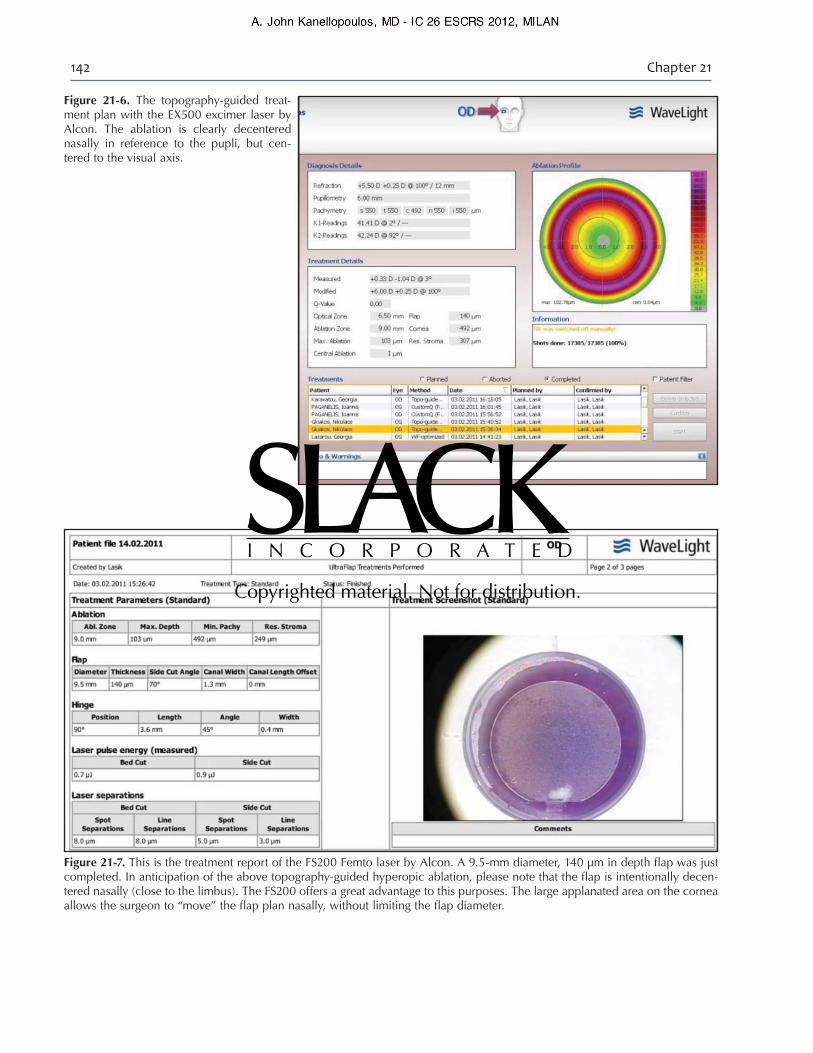
In Figures 21-6 and 21-7, the reader can appreci-ate the excimer treatment plan (the refractive suite utilizes the ultra-fast EX500 excimer that is linked to the Pentacam HD, called the Oculyzer II as a platform device) in reference to the pupil and can clearly understand the necessity of an equally nasally placed ablation. The actual 9.5-mm flap created by the FS200 femtolaser is noted in the second picture. As an additional safety feature, the refractive suite is inter-connected via its network not only between the
Figure 21-3. A hyperopic LASIK flap created with the M2 microkeratome by Moria showing the gross nasal decentration of the flap in order to accommodate the applicable ablation decentration in reference to the pupillary center.
Figure 21-4. A hyperopic LASIK flap created with the M2 microkeratome by Moria showing the gross nasal decentration of the flap in order to accommodate the applicable ablation decentration in reference to the pupillary center.
Figure 21-5. This is the poster presentation from the AAO meeting in 2007 on comparing standard to topography-guided LASIK in hyperopia.
Copyrighted material. Not for distribution.
!"#$ %&'()*+$#!
Figure 21-6. The topography-guided treat-ment plan with the EX500 excimer laser by Alcon. The ablation is clearly decentered nasally in reference to the pupli, but cen-tered to the visual axis.
Figure 21-7. This is the treatment report of the FS200 Femto laser by Alcon. A 9.5-mm diameter, 140 µm in depth flap was just completed. In anticipation of the above topography-guided hyperopic ablation, please note that the flap is intentionally decen-tered nasally (close to the limbus). The FS200 offers a great advantage to this purposes. The large applanated area on the cornea allows the surgeon to “move” the flap plan nasally, without limiting the flap diameter.
Copyrighted material. Not for distribution.

!"#$%&'()*+,-%.,/01".%123,345,/01".%126,(7$28#3$27#, 9:;
two lasers but additionally to the Pentacam device (the Oculyzer) for enhanced safety in data processing. The actual Pentacam scans of each eye of the patient are fed seamlessly to the EX500 and are processed by surgeon and staff. The treatment plan is viewed on the EX500 screen, but also superimposed over the femto flap planned on the relative flap-making process that precedes on the FS200 femtosecond laser. This is an extremely helpful tool for the learning curve of per-forming topography-guided hyperopic LASIK.
The picture below represents the final FS200 report documenting all flap parameters used, as well as the actual flap position at the termination of the side-cut. Please note the nasal (to the left) decentration of the flap in regard to the pupil.
A last consideration for hyperopes, especially the over 40 age group, is the increased incidence of narrow angles. We caution clinicians that hyperopes of over 3 D may have significantly narrow angles, a fact that needs to be evaluated thoroughly; especially when considering that the progressive cataract formation in these patients will further narrow the angle and potentially lead to angle closure attack(s). It is reason-able to consider premature cataract extraction in these patients and/or laser peripheral iridotomy as this approach may eliminate both the hyperopic correction and the phacomorphic danger of glaucoma.
!!"!"!##!$$ 1. Terrell J, Bechara SJ, Nesburn A, et al. The effect of globe
fixation on ablation zone centration in photorefractive kera-tectomy. Am J Ophthalmol. 1995;119:612-619.
2. Pande M, Hillman JS. Optical zone centration in keratorefrac-tive surgery; entrance pupil center, visual axis, coaxially sight-ed corneal reflex, or geometric corneal center? Ophthalmology. 1993;100:1230-1237.
3. von Noorden GK. Binocular vision and ocular motility: theory and management of strabismus. St. Louis, MO: Mosby; 1996:163-205.
4. Uozato H, Guyton DL. Centering corneal surgical procedures. Am J Ophthalmol. 1987;103:264-275; correction p. 852.
5. Pande M, Hillman JS. Optical zone centration in keratore-fractive surgery; entrance pupil centers, visual axis, coaxi-ally sighted corneal reflex, or geometric corneal center? Ophthalmology. 1993;100:1230-1237.
6. Nepomuceno RL, Boxer Wachler BS, Kim JM, Scruggs R, Sato M. Laser in situ keratomileusis for hyperopia with the LADARVision 4000 with centration on the coaxially sighted corneal light reflex. J Cataract Refract Surg. 2004;30:1281-1286.
7. Boxer Wachler BS, Korn TS, Chandra NS, Michel FK. Decentration of the optical zone: centering of the pupil versus the coaxially sighted corneal light reflex in hyperopic LASIK. J Refract Surg. 2003;19:464-465.
8. Basmak H, Sahin A, Yildirim N, Papakostas TD, Kanellopoulos AJ. Measurement of angle kappa with synop-tophore and Orbscan II in a normal population. J Refract Surg. 2007:23(5):456.
9. Looper LP. The relationship between angle lambda and the residual astigmatism of the eyes. Am J Optom. 1959;36:365-377.
10. Effert R, Gruppe S. The amount of angle alpha in a normal population. Acta XVII concilii europaeae strabologicae asso-cieatis, Madrid. 1988:17-21.
11. Scott WE, Mash AJ. Kappa angle measurements of strabismic nad nonstrabismic individuals. Arch Ophthalmol. 1973;89:18-20.
12. Clement RA, Dunne MCM, Barnes DA. A method for ray tracing through schematic eyes with off-axis components. Ophthalmic Physiol Opt. 1987;7:149-152.
13. Kanellopoulos AJ, Conway J, Pe L, Comaish I. LASIK for hyperopia with the WaveLight excimer laser. J Refract Surg. 2006:22(1):43-7.
Copyrighted material. Not for distribution.

REFRACTIVE MINI FOCUS ON SURFACE ABLATION VS LASIKLASIK: POINT
MARCH 2007 I CATARACT & REFRACTIVE SURGERY TODAY EUROPE I 21
LASIK requires that a surgeon use a microkeratometo create a corneal flap. This has been the majordeterrent in comparison to PRK and other surfaceablations. As a cornea surgeon, I am generally
comfortable operating this flap—as far as I keep closetrack of the equipment and parameters involved.
A portion of the flap remains attached to the eye (ie, asa hinge), while the remainder is gently lifted up and backto expose the inner cornea. This means that part of thecorneal nerves are not affected, and the rest are incisedwithin the flap and left to regenerate. LASIK is consideredto be the refractive technique that induces the most dry-ness when compared with PRK. It is, though, unfair tocompare dryness between PRK and LASIK in differentrefractive errors. Most PRKs are indicated for up to -5.00D, whereas the average LASIK correction is for -5.00 D.
FIR ST CA SE S EVER PERFORMEDIn 1994, I evaluated the corneal sensitivity of LASIK ver-
sus PRK for significant and extreme myopic corrections ofup to -15.00 D.1 This included some of the first PRK andLASIK cases ever performed! Treating such high refractiveerrors is not a common practice anymore, especially withPRK, however, the LASIK eyes had better sensitivity versusthe PRK eyes. For lower amounts of myopia, PRK mayhave an advantage over LASIK, because the corneal nerveplexus regenerates and less dryness occurs.
In LASIK, the laser resculpts the corneal stroma, and theflap is returned to its original position. No stitches arerequired, as the flap stays in place by the natural currentof fluid within the cornea. The endothelial cell layer onthe underside of the cornea pumps the fluid. Generally,the cornea has no edema within hours of the procedure,and there is minimal endothelial cell compromise.
ADVANTAGE SEyedrop medications—a significant advantage in
LASIK—are used for up to 1 week, and the patient maytypically resume most normal activities the following dayand full activity within 1 week. Most of the healingprocess takes place within 1 week, except for drynessthat reveres within 1 month to 3 months. There are sev-eral other advantages that make this procedure the mostpopular among refractive surgeons and patients. Below ismy Top 10 list:
1. It can treat wide ranges of refractive error with greataccuracy (ie, myopia from -0.50 D to almost -14.00 D;hyperopia from 0.50 D to 6.00 D; and astigmatism up to6.00 D). This is not quite possible with PRK.
2. In contrast, surface ablations are generally not used inhyperopia, as they are invariably associated with regressionand corneal haze—despite the use of mitomycin C. It isquite important to consider that (1) LASIK holds an advan-tage for all hyperopic corrections in a population that I havecarefully studied over the last few years,2 and (2) LASIK mayoffer a similar rehabilitation in hyperopes as in myopes.
3. The visual rehabilitation is very rapid, and the risk forinfection is less than 1 day. In contrast, surface ablationstake 3 days to 6 days to offer reasonable visual function. Itmay take up to 2 weeks to reach the vision level of post-LASIK day 1. PRK is associated with risk of infection up to3 days to 5 days (ie, until the epithelium completely heals).
4. Regarding the preservation of normal corneal anato-my, LASIK preserves the epithelium and Bowman’s mem-brane. Both are removed during PRK, and therefore it issometimes slow to heal. This results in reduced risk oflight sensitivity with LASIK, while it may occur for 1 weekto 2 weeks after PRK.
5. PRK—and generally all surface ablations—is often
Why is LASIK MyPreferred RefractiveSurgery Procedure?
PRK and other surface ablation techniques should only be used as an alternative for thin and irregular corneas.
BY A. JOHN KANELLOPOULOS, MD
0307CRSTEuro_Kanellopoulos_refmini4.qxd 2/28/07 11:18 AM Page 21

associated with severe pain on postoperative day 1, and itmay be associated with significant discomfort for up to 1week. PRK patients usually take pain relief medication forapproximately 1 day to 2 days postoperatively, becausemost pain fibers in the cornea are located in the surfaceportion. With PRK, these are affected and left “uncovered”during surgery and the healing process. This is a majordeterrent for patients considering the procedure.
6. The ability to establish the final refractive outcome isapproximately 1 month with LASIK, but lies between 3months to 6 months with surface ablations. This makes itvery difficult to retreat or to establish postsurgical results.
7. Inflammation is very rare with LASIK—with the excep-tion of diffuse lamellar keratitis that has now become a rari-ty. Significant inflammation and haze is common in PRK,especially when attempting hyperopes and/or refractiveerrors more than -5.00 D. The cornea keratocytes mayrespond strongly to the intervention. Most surgeons usemitomycin C, and some surgeons anecdotally use frozenbalanced salt solution on a weck cell to reduce the possibili-ty of haze. Although the use of highly diluted mitomycin Chas become generally acceptable internationally, the use offreezing is—in my opinion—a dangerous practice, as freez-ing in vivo may affect not only keratocytes but alsoendothelial cells. Significant post-PRK haze poses a difficultproblem, as it requires large and long treatment with corti-costeroids (ie, significant risks for cataract genesis and/orglaucoma) and/or reoperation (Figure 1).
8. Humans are unique among mammals in thatBowman’s membrane is present in the cornea, and I sharethe theory that it may be needed as an important optical“accessory” in the needs of human visual function.Unfortunately, Bowman’s membrane is inadvertentlyremoved in all surface ablative techniques including PRK.The Bowman-less cornea visual properties have not beenwell understood, and therefore have not been carefully eval-uated. The future will determine whether this theory is ofany importance.
9. In my opinion, flap problems have been the sole disad-vantage of LASIK. With accrued experience, most are pre-ventable and treated with good visual outcome. I have beena strong proponent of thin LASIK flaps (ie, 100 µm to 120microns) and have been very consistent in establishing mygoal in most cases. In the past, I used a highly customizedalgorithm with the Moria M2 microkeratome (Moria,Antony, France) and precalibrated blades. In my practice,beside infection, flap striae are the second-largest concernwith LASIK. Nevertheless, I see this complication a coupletimes a year and treat it with immediate hypotonic waterirrigation and ironing followed by seven interrupted tight 10-0 nylon sutures in a fashion similar to a penetrating kerato-plasty. They are usually removed within the week, and the
visual rehabilitation had been excellent and without prob-lems. I still believe that these eyes are better off with theLASIK procedure versus PRK. I have not seen this problemyet in my IntraLase-LASIK cases (IntraLase Corp., Irvine,California).
10. The popularity of femtosecond lasers, and IntraLaseparticularly, has minimized flap-related problems and fur-ther boosted the use of LASIK versus PRK. It is its ability tocreate a planar flap of a precisely desired thickness that thistechnology can now offer with very high levels of safety.Unfortunately, the costs involved are significant. So, though,was our transition from radial keratotomy to excimer laserrefractive surgery. This means less chance of ectasia, less bio-mechanical change and response from the cornea, andrapid visual recovery.
It is for these reasons that I think that today, any refrac-tive surgeon that has access to an IntraLase will haveLASIK as the preferred procedure and reserve PRK (orany other surface ablation technique) as an alternativefor thin and irregular corneas. !
A. John Kanellopoulos, MD, is a Corneal andRefractive Surgery Specialist. Dr. Kanellopoulos isDirector of Laservision Eye Institute in Athens,Greece, and practices in New York. He is AttendingSurgeon for the Department of Ophthalmology atthe Manhattan Eye, Ear, and Throat Hospital, in New York, andClinical Associate Professor of Ophthalmology at New YorkUniversity Medical School. Dr. Kanellopoulos states that he hasno financial interest in the products or companies mentioned.He is a member of the CRST Europe Editorial Board. He maybe reached at +30 21 07 47 27 77; [email protected].
J Cataract Refract Surg
J Refract Surg
REFRACTIVE MINI FOCUS
22 I CATARACT & REFRACTIVE SURGERY TODAY EUROPE I MARCH 2007
Figure 1. A depiction of significant debilitating post-PRK haze.
0307CRSTEuro_Kanellopoulos_refmini4.qxd 2/28/07 11:18 AM Page 22