late morfofunctional alterations of the sertoli cell caused by
TRANSCRIPT

Brilhante et al. Reproductive Biology and Endocrinology 2012, 10:79http://www.rbej.com/content/10/1/79
RESEARCH Open Access
Late morfofunctional alterations of the Sertoli cellcaused by doxorubicin administered toprepubertal ratsOtávio Brilhante1, Fatima K Okada2, Estela Sasso-Cerri3, Taiza Stumpp2* and Sandra M Miraglia2
Abstract
Background: Doxorubicin is a potent chemotherapeutic drug used against a variety of cancers. It acts throughinteraction with polymerases and topoisomerase II and free radical production. Doxorubicin activity is not specificto cancer cells and can also damage healthy cells, especially those undergoing rapid proliferation, such asspermatogonia. In previous studies our group showed that etoposide, another topoisomarese II poison, causesirreversible damage to Sertoli cells. Thus, the aim of this study was to address the effects of doxorubicin on Sertolicell morphology and function and on the seminiferous epithelium cycle when administered to prepubertal rats.
Methods: Prepubertal rats received the dose of 5 mg/Kg of doxorubicin, which was fractioned in two doses:3 mg/Kg at 15dpp and 2 mg/Kg at 22dpp. The testes were collected at 40, 64 and 127dpp, fixed in Bouin’s liquidand submitted to transferrin immunolabeling for Sertoli cell function analysis. Sertoli cell morphology and thefrequency of the stages of the seminiferous epithelium cycle were analyzed in PAS +H-stained sections.
Results: The rats treated with doxorubicin showed reduction of transferrin labeling in the seminiferous epitheliumat 40 and 64dpp, suggesting that Sertoli cell function is altered in these rats. All doxorubicin-treated rats showedsloughing and morphological alterations of Sertoli cells. The frequency of the stages of the seminiferous epitheliumcycle was also affected in all doxorubicin-treated rats.
Conclusions and discussion: These data show that doxorubicin administration during prepuberty causesfunctional and morphological late damage to Sertoli cells; such damage is secondary to the germ cell primaryinjury and contributed to enhance the spermatogenic harm caused by this drug. However, additional studies arerequired to clarify if there is also a direct effect of doxorubicin on Sertoli cells producing a primary damage onthese cells.
Keywords: Doxorubicin, Sertoli cell, Spermatogenesis, Rat, Transferrin
BackgroundDoxorubicin is an anthracyclic antibiotic with potentchemotherapeutic activity against a variety of cancers.The toxicity of this drug is mediated by its interactionwith topoisomerase II, an enzyme that is abundant incells undergoing rapid and constant proliferation. Doxo-rubicin toxicity can also be mediated by the generationof free radicals [1,2] and lipid peroxidation [3]. Bothmechanisms are not specific to cancer cells and can
* Correspondence: [email protected] of Morphology and Genetics, Developmental BiologyLaboratory, Federal University of São Paulo. Vila Clementino, São Paulo, SP,BrazilFull list of author information is available at the end of the article
© 2012 Brilhante et al.; licensee BioMed CentrCommons Attribution License (http://creativecreproduction in any medium, provided the or
damage healthy cells [4-7]. In the testis, spermatogoniaare the main doxorubicin target, as observed after etopo-side treatment, because of their intense and continuousproliferative activity [8]. The administration of doxorubi-cin to prepubertal rats causes damage to spermatogonia[9] and irreversible damages to adult spermatogenesis[9,10]. Extensive injuries such as decrease of spermato-gonia, degeneration and/or decrease of early spermato-cytes, vacuolated seminiferous epithelium, reduction ofepididymis cauda sperm count and sperm motility,which were caused by a single dose of doxorubicin(10 mg/Kg bw), have been characterized in the rattesticular tissue after the schedule’s termination [11].
al Ltd. This is an Open Access article distributed under the terms of the Creativeommons.org/licenses/by/2.0), which permits unrestricted use, distribution, andiginal work is properly cited.

Brilhante et al. Reproductive Biology and Endocrinology 2012, 10:79 Page 2 of 16http://www.rbej.com/content/10/1/79
Other chemotherapeutic drugs, such as cisplatin [12,13]and etoposide [14-16], another topoisomerase-interactingdrug, also cause damage to germ cells, especially whenadministered during prepubertal and peripubertal phases.In previous studies our group showed that etoposide ad-ministration to prepubertal rats causes irreversible damageto Sertoli cells [17,18], leading to severe spermatogenesisimpairment.Sertoli cells play a key role in spermatogenesis control
and germ cell development [19,20] and are pivotal fortesticular homeostasis; this phenomenon is maintainedthrough the complex interactions with germ cells andwith the other testicular somatic cells, such as Leydigand myoid cells [21]. Sertoli cell alterations can cause se-vere damage to spermatogenesis [22-24]. Some Sertolicell toxicants, such as 2,5-Hexanedione [25] and Mono-(2-ethylhexyl) Phthalate [26], have been shown to causemassive germ cell death. On the other hand, germ celldeath can also lead to functional and morphological altera-tions of Sertoli cells [27]. Because Sertoli cells are consid-ered the most resistant cell in the testis, most of thealterations caused to these cells by the administration of dif-ferent toxicants are considered to be secondary effects ofgerm cell death [28,29]. However, previous studies havesuggested that germ cell death caused by etoposide admin-istration to prepubertal rats is not the unique factor respon-sible for the damages observed in Sertoli cells, since evenafter seminiferous epithelium recovery, Sertoli cells stillshowed functional and morphological alterations [17,18].The transferrin is vital to the regulation of spermatogenesis.It has been demonstrated that transferrin constitutes 5% ofall proteins secreted by Sertoli cell [30]. In addition, it is areliable instrument of investigation of the Sertoli cell func-tion, both in vitro and in vivo [31,32] and it has been con-sidered one of the best markers for this scope [33].Consequently, transferrin has been largely utilized for theevaluation of the Sertoli cell function and its labeling hasbeen successfully included in studies that aimed to investi-gate the possible functional alterations produced as a re-sponse to harmful events [34,35]. Germ cells also expresstransferrin receptors [36]. Therefore, transferrin labelingwas utilized in the current study.The studies performed by our group have suggested
that prepubertal testes are more sensitive to chemother-apeutic drugs than adult testes [9,15,16]. Doxorubicin islargely used in child cancer treatments and has beenshown to be very aggressive to germ cells. However, theeffects of doxorubicin administration on the morphologyand function of Sertoli cells have not been detailedlyaddressed yet, especially during prepuberty. Thus, theaim of the present study was to evaluate the effects ofdoxorubicin on Sertoli cell function and morphologywhen administered to prepubertal rats during key peri-ods of testicular and Sertoli cell development.
MethodsAnimals and drug administrationSixty male Wistar rats (Rattus norvegicus albinus) weremaintained under 12/12 hr light/dark cycles, at 21-23°Croom temperature; standardized lab chow (Nuvilab CR1,NuvitalW, Curitiba, PR, Brazil) and water were providedad libitum. The protocol regarding animal care andtreatment was approved by the Ethical Committee forAnimal Research of the Federal University of São Paulo,Brazil (reference number: 0559/08).The animals were distributed into two major groups:
control (C) and doxorubicin-treated (D). The rats fromgroup D received 5 mg/Kg of doxorubicin (RubidoxW,Bergamo – São Paulo, Brazil) by intraperitoneal route.This dose was fractioned into two doses: one of 3 mg/Kgthat was administered at 15 days postpartum (dpp) andone of 2 mg/Kg that was administered at 22dpp. Therats from the control group received 0.9% saline solutionat the same ages and volume administered to thedoxorubicin-treated group. The rats were maintainedunder standard conditions of luminosity (12 hr light/12 hr dark) and temperature (22-23°C). Food and waterwere allowed ad libitum.The C and D groups were subdivided into three sub-
groups of 10 rats each, according to the ages of euthanasia:40dpp (subgroups C40 and D40); 64dpp (subgroups C64and D64) and 127dpp (subgroups C127 and D127). Theseages were chosen because they represent key time pointsof spermatogenesis development, i.e., at 40dpp the rats areconsidered peripubertal [13], at 64dpp they have alreadyreached puberty [37] but are not sexually mature and at127dpp they are adults [14] and sexually mature [38].
Testis collection and analysisAt the ages previously specified, the rats were weighedand submitted to euthanasia through CO2 inhalationaccording to recommendations of the Ethical Committeeof the Federal University of Sao Paulo (UNIFESP). Thetestes were removed, weighed and had their volume mea-sured according to the Scherle’s method [39]. Subse-quently, the testes were immersion-fixed in Bouin’s liquidfor 48 hr. Each testis was transversally cut and one halfwas embedded in Paraplast PlusW with DMSO (Sigma)and the other half was paraffin-embedded. From the frag-ments embedded in Paraplast PlusW, 3μm cross-sectionswere obtained and submitted to the Periodic Acid-Schiffhistochemical method (PAS) and counterstained withHarris’s Hematoxilin (H). From the paraffin-embeddedfragments, 7μm cross-sections were obtained and submit-ted to the transferrin immunolabeling, as described below.
Transferrin immunolabelingThe sections obtained from the paraffin blocks weredewaxed, washed in running tap water for Bouin’s
Brilhante et al. Reproductive Biology and Endocrinology 2012, 10:79 Page 3 of 16http://www.rbej.com/content/10/1/79
elimination and treated with 3% hydrogen peroxide for15 minutes. The slides were washed in phosphate buffer(PBS, pH 7.2) and incubated with 7% BSA for 15 min-utes. The slides were then incubated with the primaryantibody anti-transferrin (1:1000, ICN Aurora, Ohio,USA) for 1 hr, washed in PBS and incubated with thesecondary antibody (LSABW, Dako, California, USA) for30 minutes. After that, the slides were washed in PBSand incubated with Streptavidin-Peroxidase (LSABW,Dako, California, USA) for 30 minutes. The reaction wasrevealed with DAB (DAKO, California, USA) and
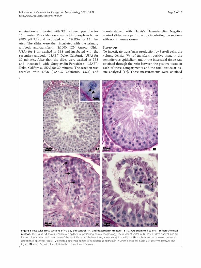
Figure 1 Testicular cross-sections of 40 day-old control (1A) and doxomethod. The Figure 1A shows seminiferous epithelium presenting normallocated close to the basal membrane of the seminiferous epithelium (insetdepletion is observed. Figure 1C depicts a detached portion of seminiferouFigure 1D shows Sertoli cell nuclei into the tubular lumen (arrows).
counterstained with Harris’s Haematoxylin. Negativecontrol slides were performed by incubating the sectionswith non-immune serum.
StereologyTo investigate transferrin production by Sertoli cells, thevolume density (Vv) of transferrin-positive tissue in theseminiferous epithelium and in the interstitial tissue wasobtained through the ratio between the positive tissue ineach of these compartments and the total testicular tis-sue analyzed [17]. These measurements were obtained
rubicin-treated (1B-1D) rats submitted to PAS+H histochemicalmorphology. The nuclei of Sertoli cells show evident nucleoli and are; arrowheads). In the Figure 1B, a tubular section showing germ cells epithelium in which Sertoli cell nuclei are observed (arrows). The

Brilhante et al. Reproductive Biology and Endocrinology 2012, 10:79 Page 4 of 16http://www.rbej.com/content/10/1/79
using a Leica QWin Analysis System (Leica - Cambridge,England) with a x20 objective lens.
Histopathological analysis and frequency of the stages ofthe seminiferous epithelium cycleFor the histopathological analysis, the testicular sectionsfrom the Paraplast blocks were totally analyzed and thealterations, especially the Sertoli cell alterations, weredescribed.The frequencies of the stages of the seminiferous epi-
thelium cycle were obtained from Parablast-embeddedtestes and the analysis was performed according to Hesset al. [40]. Two hundred seminiferous tubule cross-sections were analyzed per rat, which is the minimumnumber of sections that must be analyzed when thenumber of animals per group is 10 (n = 10) [32]. Theidentification of the stages of the seminiferous epithe-lium cycle was based on the classification of Leblondand Clermont [41]. The frequency of the stages, inpercentage, was calculated by the ratio between thenumber of sections in each stage and the total num-ber of analyzed sections multiplying by 100 [42].Because the stages II and III as well as the stages XIIand XIII are very similar between each other, theywere grouped according to previously described[43-45]. This analysis was performed under a lightmicroscope using x50 and x100 objective lenses.
Statistical analysisThe data were submitted to t test. The results were con-sidered significant when p ≤ 0.05.
ResultsTesticular histopathologyThe histopathological analysis of the testes (Figures 1, 2and 3) showed that the control rats presented normalmorphology of seminiferous epithelium and of Sertolicells at 40 (Figure 1A), 64 (Figure 2A) and 127(Figure 3A) days of age. The rats treated with doxorubi-cin showed seminiferous epithelium vacuolization(Figure 3B) and germ cell depletion (Figures 1B, 2B and3B–D). These alterations appeared at all ages, but be-came more intense at 64 days. Sertoli cell also showedmorphological alterations in all doxorubicin-treated rats.Their nuclei showed abnormal morphology (Figures 2Cand 3B–C) and some of them were distant from thebasal membrane (Figures 1C, 2B, 3B) or even in thetubular lumen (Figures 1D and 2B–C). Eventually, Ser-toli cell only tubular sections were observed (Figure 2B).The rats from D64 and D127 subgroups showed intensedisorganization of the seminiferous epithelium and elon-gated spermatid retention at stage IX of the seminiferousepithelium cycle (Figure 2D). The rats from D127 sub-group showed partial recovery of spermatogenesis.
Transferrin labeling and Sertoli cell functionTransferrin labeling observed in the seminiferous epithe-lium (Figures 4, 5 and 6) was dependent on the treat-ment (doxorubicin or saline solution) applied, on theeuthanasia age and on the stage of the seminiferous epi-thelium cycle. The rats from C40 (Figure 4A) and D40(Figures 4B-C) subgroups showed few labeled regions inthe seminiferous epithelium. In the C40 subgroup, fewSertoli cells showed weak labeling in the cytoplasm(Figure 4A); however, cells in the basal region of semin-iferous tubules, probably spermatogonia, showed intenselabeling (Figure 4A). The D40 subgroup showed trans-ferrin labeling in the Sertoli cells, but no labeling in thespermatogonia were observed (Figure 4B). Some hypo-trophic tubular sections from D40 rats did not show anytransferrin labeling (Figure 4C). On the other hand, C64(Figure 5A) and C127 (Figure 6A) control subgroupsshowed strong and abundant labeling in the seminifer-ous epithelium. In these subgroups, Sertoli cells(Figures 5A, 6A) and elongated spermatids (Figure 6A)showed intense labeling. In both C64 and C127 sub-groups, transferrin labeling was more abundant in stagesII-III/XIV (Figure 5A) and VII (Figure 6A), respectively.In the D64 subgroup, some tubular sections in stages II-III and VII showed weak labeling in the Sertoli cell cyto-plasm and no labeling in the nucleus (Figure 5B). In theseminiferous tubules with intense germ cell depletion,no transferrin labeling was observed (Figure 5C). In theD127 subgroup, intense transferrin labeling wasobserved in the Sertoli cell cytoplasm and in some germcells located in the basal region of the seminiferous epi-thelium (Figure 6B), as observed in the C127 subgroup(Figure 6A); however, at the stage VII of seminiferousepithelium, the labeling seemed to be less abundant inthe D127 subgroup than in the C127 subgroup (Figures6A–B). In this subgroup (C127), transferrin labeling wasobserved either in the cytoplasm or nucleus of Sertolicells (Figure 6A), while in D127, these cells showed onlycytoplasm immunolabeling. Moreover, in some elon-gated spermatids, the strong transferrin labeling was alsomore abundant in C127 (Figure 6A) than in D127(Figure 6B). In the D127 subgroup, some tubular sec-tions with intense germ cell depletion showed rare trans-ferrin immunolabeling (Figure 6C–D).The pattern of transferrin labeling (i.e., the stages in
which transferrin labeling was observed and the celltypes that were labeled) in the doxorubicin-treated ratswas similar to that observed in the control rats. How-ever, the volume density of transferrin-positive total tes-ticular tissue reduced in the D40 subgroup (Figure 7)when compared to C40 subgroup, whereas in D64 andD127 this parameter was not altered in comparison tothe corresponding control subgroups C64 and C127. Onthe other hand, in the D40 and D64 subgroups, the

Figure 2 Testicular cross-sections of 64 day-old control (Figure 2A) and doxorubicin-treated (Figures 2B-D) rats submitted to thePAS+H method. The Figure 2A depicts part of a tubular section containing Sertoli cell nuclei with normal morphology. These nuclei are locatedclose to the basal membrane (arrows) of the seminiferous epithelium and show evident nucleolus (inset; arrowhead). In the Figure 2B, a Sertolicell only tubular section is observed. In this tubular section, one of the Sertoli cell nuclei is far from the basal membrane (arrowhead) and anotheris sloughed into the tubular lumen (arrow). The Figure 2C depicts a sloughed portion of seminiferous epithelium (arrowheads) into the tubularlumen in which a Sertoli cell nucleus can be seen (arrow). In the inset, this Sertoli cell nucleus with irregular profile shows abnormal clear areas(arrow). Figure 2D shows a tubular section in stage IX of the seminiferous epithelium cycle with retention of step 19 spermatids (arrow). Note thepresence of intraepithelial spaces and free primary spermatocytes associated.
Brilhante et al. Reproductive Biology and Endocrinology 2012, 10:79 Page 5 of 16http://www.rbej.com/content/10/1/79
volume density of transferrin labeling in the semini-ferous epithelium reduced when compared with the cor-responding control subgroups C40 and C64 (Figure 8).No significant alteration of the volume density of trans-ferrin labeling in the seminiferous epithelium wasobserved in the D127 subgroup in relation to C127 sub-group (Figure 8). The labeling pattern in the interstitialtissue was very similar in the control and doxorubicin-
treated rats. Only the D64 subgroup showed volumedensity reduction of the transferrin-positive interstitialtissue (Figure 9).
Frequency of the stages of the seminiferous epitheliumcycleThe frequency of some stages of the seminiferous epi-thelium cycle was altered in all doxorubicin-treated rats

Figure 3 Testicular cross-sections of 127 day-old control (Figure 3A) and doxorubicin-treated (Figures 3B-3D) rats submitted to thePAS+H method. The Figure 3A depicts seminiferous tubule sections showing normal morphology. The Sertoli cells show normal nuclei (inset;arrowheads). In the Figure 3B, tubular sections with severe germ cell depletion and intraepithelial vacuoles (thin arrows) are seen. A displacedSertoli cell nucleus from the basal membrane is also observed (thick arrow). Some Sertoli cell nuclei show abnormal profile (arrowheads). TheFigure 3C shows Sertoli cell nuclei with abnormal morphology, showing round shape (thick arrows); in one of them the nucleolus is not evident.A binucleated formation of round spermatids can also be noted into the lumen (thin arrow). The Figure 3D depicts a portion of a seminiferoustubule showing a clear area without germ cells (arrows) and many primary spermatocytes in the tubular lumen (asterisk).
Brilhante et al. Reproductive Biology and Endocrinology 2012, 10:79 Page 6 of 16http://www.rbej.com/content/10/1/79
of each age studied (Figures 10, 11 and 12). At 40dpp,an increase of the frequency of stages I, XI and XIV anda reduction of the frequency of stages II-III, IV, V and VIwere observed in the doxorubicin-treated rats (Figure 10)when compared to the corresponding C40 subgroup. Inthe D64 subgroup, an increase of the frequency of stagesII-III, VII and IX and a reduction of the frequency ofstages I and VIII were observed when compared withthe C64 subgroup (Figure 11). The D127 subgroup
showed partial recovery of the synchrony of the semin-iferous epithelium cycle. In this subgroup only the fre-quency of stage VII increased and the frequency ofstages XII-XIII decreased when compared with the C127subgroup (Figure 12).
DiscussionThe deleterious action of doxorubicin on male germ cellshas been described [9]. The citotoxity caused by

Figure 4 Testicular cross-sections of 40 day-old control (Figure 4A) and doxorubicin-treated (Figure 4B-C) rats submitted to transferrinlabeling. The Figure 4A shows weak transferrin labeling in the Sertoli cell cytoplasm (arrows) of the control rat and strongly transferrin-positivegerm cells, probably spermatogonia, which are located in the basis of the seminiferous epithelium (arrowheads). In the Figure 4B (doxorubicin-treated rat), although Sertoli cell cytoplasm is positive (arrows), no labeling is observed in the spermatogonia (arrowheads). The Figure 4C depictsa seminiferous tubule cross-section showing germ cell depletion and no transferrin labeling. Sertoli cell nucleus (arrow). Note the intense labelingin the interstitial tissue (Figures 4A-C; asterisks).
Brilhante et al. Reproductive Biology and Endocrinology 2012, 10:79 Page 7 of 16http://www.rbej.com/content/10/1/79
doxorubicin on the seminiferous epithelium can be relatedto its therapeutic activity; it interferes with molecules asso-ciated to the nuclear DNA and with enzymes (RNA andDNA polimerases, topoisomerases I and II) that are activein the cell division process. Then, doxorubicin forms acomplex with chromatin [46], blocking the G2 phase ofthe cell cycle [47,48] and provoking single and/or doublestrand DNA breaks [49]. Doxorubicin also interferes withmembrane lipids [50-53], provoking alterations in theirchemical structure and impairing their function. The pro-duction of reactive species of oxygen, as a consequence offree radicals caused by doxorubicin, can also affect the cel-lular functions, altering the cellular metabolism in
different aspects [52,54]. In fact, the production of freeradicals is one of the factors that limit the therapy withdoxorubicin. This anticancer agent produces, as previouslymentioned, a significant increase of lipid peroxidation andalterations of antioxidant enzyme activities in differentorgans of rats, including testis, as observed in vitro [55]. Itprovokes significant elevation in the testicular malondial-dehyde concentrations and decreases of glutathione con-tent, glutathione reductase (GR), glutathione-S-transferase(GST), superoxide dismutase (SOD) and catalase activities,thus indicating oxidative stress production in doxorubicin-induced testicular toxicity [11]. Besides, the deleteriouseffect of this anticancer agent on adult rat testis lipids and

Figure 5 Testicular cross-sections of 64 day-old control (Figure 5A) and doxorubicin-treated (Figure 5B-C) rats submitted to transferrinlabeling. In the Figure 5A (control rat), intense transferrin labeling is observed in the Sertoli cell cytoplasm (thin arrows) in both seminiferoustubule sections (stages II-III and XIV). Sertoli cell nuclei (thick arrows) and elongated spermatids (arrowhead) are not labeled. The Figure 5B(64 day-old doxorubicin-treated rat) shows very weak transferrin labeling in the Sertoli cell cytoplasm of tubules at stages II-III and VII (thickarrow); however, no labeling is observed in the Sertoli cell nucleus (thin arrow). The Figure 5C depicts a seminiferous tubule cross-sectionshowing intense germ cell depletion in which no transferrin labeling is observed. Interstitial tissue immunolabeling is also noted (Figures 5A;asterisks).
Brilhante et al. Reproductive Biology and Endocrinology 2012, 10:79 Page 8 of 16http://www.rbej.com/content/10/1/79
fatty acids has been observed after single or multiple doseregimes, resulting in a gradual loss of spermatogenesis andin a decrease in phospholipids, including glycerophospholi-pids and sphingomyelin; in this context, glycerophospholi-pids selectively loose their major polyunsaturated fatty acid(PUFA), while sphingomyelin looses its major very long-chain PUFA (VLCPUFA). By contrast, triglycerides andespecially cholesterol esters (CE) tend to accumulate in thetestes undergoing germ cell death, probably in the surviv-ing Sertoli cells. Their fatty acid patterns suggest that ini-tially these lipids retained part of the PUFA coming from,
or no longer used for the synthesis of germ cell glycero-phospholipids. Determining whether this accumulationresults from a physiologic adaptation to the effects ofdoxorubicin or simply reflects another lipid derangementcaused by the drug remains to be investigated [56].Although some of the doxorubicin mechanisms of ac-
tion on germ cell are known, the effects of this drug onother testicular components are poorly understood.Spermatogonia are the preferential doxorubicin targetdue to the presence of the enzyme topoisomerase II, butprobably primary spermatocytes can also be damaged,

Figure 6 Testicular cross-sections of 127 day-old control (Figure 6A) and doxorubicin-treated (Figure 6B-D) rats submitted totransferrin labeling. The Figures 6A-B show intense transferrin labeling in the seminiferous epithelium (stage VII). However, in the Figure 6A(control group), the labeling is more abundant than in the Figure 6B (doxorubicin-treated group). In the control group (Figure 6A), transferrinlabeling is observed in the Sertoli cell cytoplasm (thick arrow) and nucleus (inset, thin arrow) as well as in the elongated spermatids (arrowhead).In the doxorubicin-treated group (Figure 6B) transferrin labeling is observed in the Sertoli cell cytoplasm (thick arrow) and in the elongatedspermatids (arrowhead), but not in the Sertoli cell nucleus (inset, thin arrow). The Figure 6C shows two seminiferous tubule cross-sectionscontaining large portions of sloughed seminiferous epithelium (stars). In one of them, transferrin positive Sertoli cell cytoplasm is observed(arrow). In the Figure 6D, a tubular section with intense germ cell depletion shows weak Sertoli cell cytoplasm immunolabeling (thin arrows) andno labeling in the Sertoli cell nucleus (arrowheads). Intraepithelial vacuole (thick arrow). Note the intense labeling in the interstitial tissue(Figure 6A-D; asterisks).
Brilhante et al. Reproductive Biology and Endocrinology 2012, 10:79 Page 9 of 16http://www.rbej.com/content/10/1/79
although the role of topoisomerase II in pre-mitoticDNA synthesis, at determined stages, is more accentu-ated than in pre-meiotic synthesis as observed afteretoposide-treatment [8]. Thus, DNA synthesis in pre-meiotic spermatocytes is not so vulnerable to the doxo-rubicin action as pre-mitotic DNA synthesis. Moreover,
it is possible that other topoisomerases can be involvedin the process of pre-meiotic synthesis.Thus, considering the different mechanisms of action
of doxorubicin previously mentioned, it is also possiblethat additionally to spermatogonia, other cells, includingSertoli cells, are also targeted by doxorubicin.

Figure 7 Volume density of transferrin-positive total testicular tissue (VvT) in the control and doxorubicin-treated rats. The VvT showedsignificant reduction in the 40dpp doxorubicin-treated rats (*) when compared with the corresponding control subgroup. No alteration of thisparameter was observed in the 64 and 127dpp doxorubicin-treated rats.
Brilhante et al. Reproductive Biology and Endocrinology 2012, 10:79 Page 10 of 16http://www.rbej.com/content/10/1/79
In the present study, it was observed that doxorubicinadministration to rats at early prepubertal phase alterstransferrin production by Sertoli cells at specific phasesof testicular development, indicating a functional alter-ation of these cells. On the other hand, transferrinsynthesis by Sertoli cells is dependent on the presence ofgerm cells [28,30,57]. Thus, it is possible that the dimin-ution of transferrin production by Sertoli cells is a con-sequence of germ cell depletion caused by doxorubicin.The significant recovery of transferrin synthesis bySertoli cells and of the seminiferous epithelium observedin D127 subgroups support this hypothesis.It is important to highlight that doxorubicin reduces the
synthesis of the transferrin receptor and leads to atypicalchanges in intracellular iron distribution and trafficking[58]. In the testes, transferrin receptors are present in thebasal region of the Sertoli cell membrane and are essentialto promote iron transportation from blood to the germ
Figure 8 Volume density of transferrin-positive seminiferous epitheliureduction of this parameter was observed in the doxorubicin-treated rats asubgroups. The 127dpp doxorubicin-treated rats showed an important reco
cells that are localized in the adluminal compartment ofthe seminiferous epithelium. Iron is crucial for germ cellproliferation and differentiation and Sertoli cells are theonly way by which this ion can reach the germ cells at theadluminal compartment. To deliver iron ions to thesecells, diferric plasma transferrin is endocytosed throughthe receptor at the basal region of the Sertoli cell mem-brane. In the cytoplasm of the Sertoli cells, iron isdetached from plasma transferrin, captured by Sertoli celltransferrin and delivered to germ cells [59]. Thus, if trans-ferrin receptors are damaged by doxorubicin, the iron traf-fic through the seminiferous epithelium could have beenaffected. In addition, the germ cells localized in the basalcompartment of the seminiferous epithelium get irondirectly from plasma transferrin [60]. Thus, damages totransferrin receptors could also cause reduction of ironcapture by these cells. Indeed, in the D40 subgroup,spermatogonia were negative for transferrin, what may
m (VvES) in the control and doxorubicin-treated rats. A significantt 40 and 64dpp (*) when compared with the corresponding controlvery of the VvES.

Figure 9 Volume density of transferrin-positive interstitial tissue (VvTi) in the control and doxorubicin-treated rats. Only the 64dppdoxorubicin-treated rats showed an increase of the VvTi (*) when compared with the control subgroup. Although the 40dpp doxorubicin-treated ratshave shown a smaller mean value of this parameter and the 127dpp rats have shown a higher mean value of VvTi, these data were not significant.
Brilhante et al. Reproductive Biology and Endocrinology 2012, 10:79 Page 11 of 16http://www.rbej.com/content/10/1/79
have contributed to the reduction of the volume density oftransferrin-positive seminiferous epithelium.In addition to transferrin receptor alterations, doxorubi-
cin also increases the plasmatic levels of transferrin [61].Since most of the transferrin present in the interstitial tis-sue comes from the blood stream, it could be possible thatthe increase in the volume density of transferrin-labeledinterstitial tissue observed in the D64 subgroup was aresult of an increase of plasmatic transferrin levels. How-ever, it does not seem to be the case, since the totalvolume of the interstitial tissue also increased. Moreover,the volume density of transferrin-positive interstitial tissueof D40 and D127 subgroups was normal, indicating thattransferrin of this testicular compartment was not
Figure 10 Frequency of the stages of the seminiferous epithelium cycand XIV and a decrease of the frequency of stages II to VI were observed aasterisk (*).
affected. This suggests that, in the testis, doxorubicinacted specifically on Sertoli cell transferrin.Although the damages to Sertoli cell function are gen-
erally reversible, the morphological alterations observedafter doxorubicin treatment suggest that these cells mayhave been directly injured in addition to their secondarydamage occurred due to the germ cell primary harm.The presence of Sertoli cell nuclei in the tubular lumen,for example, indicates that the structural integrity ofthese cells was affected by the treatment with doxorubi-cin. Dislocation of Sertoli cell nuclei from basal toadluminal or luminal compartments has also beendemonstrated in adult rats treated with cimetidine[62,63]. In these studies, this alteration was associated to
le in the 40-day-old rats. An increase in the frequency of stages I, XIt this age. Statistically significant alterations are indicated by the

Figure 11 Frequency of the stages of the seminiferous epithelium cycle in the 64-day-old rats. At this age, the frequency of the stages Iand VIII decreased whereas stages II-III, VII and IX showed an increase of their frequencies. Statistically significant alterations are indicated by theasterisk (*).
Brilhante et al. Reproductive Biology and Endocrinology 2012, 10:79 Page 12 of 16http://www.rbej.com/content/10/1/79
Sertoli cell death. Morevover, in another research, reduc-tion of Sertoli cell number followed by a decrease insperm production, normal morphology and motility wasobserved in doxorubicin-treated adult mice, 6 weeksafter the end of the treatment [64]. In the present study,although we did not score the Sertoli cell number, theSertoli cell nuclei detached into the lumen indicate thatthese cells may have been drastically injured. Thus, be-sides the secondary Sertoli cell damage, due to the pri-marily occurred germ cell death, it is also important toconsider that when doxorubicin was administered,Sertoli cells were passing through critical phases of theirdevelopment, what makes a primary damage more likelyto occur. Around 15dpp, when the rats received the firstdose of doxorubicin, Sertoli cells stop to proliferate [65]and the blood-testis barrier start to be formed [66,67].At 22dpp, when the second dose was administered, Ser-toli cells were still undergoing maturation [65,68].Therefore, we could also consider the higher susceptibil-ity of Sertoli to doxorubicin at these phases than adult
Figure 12 Frequency of the stages of the seminiferous epithelium cycincrease of its frequency and the stages XII-XII showed a decrease of its freasterisk (*).
Sertoli cells. Because Sertoli cells are crucial for sperm-atogenesis, damages in these cells at early pubertal phasecould also lead to germ cell death in other later periodsof sexual maturation as peripuberty (40 days) and afterthe completion of puberty (64 days). Hence, consideringthe Sertoli cell morphological alterations observed inthis study, it is possible that the seminiferous epitheliumalterations observed in the doxorubicin-treated ratscould also be consequence of direct Sertoli cell damage.Indeed, some alterations such as intraepithelial vacuoli-zation, spermatid retention and high frequency of Sertolicell nuclei in which the nucleolus was not evident sug-gest that Sertoli cells were damaged independently ofgerm cell death. Moreover, despite the possibility of thedecrease of Sertoli cell transferrin labeling a conse-quence of germ cell depletion, it is important to considerthat doxorubicin can increase the production of freeradicals as previously observed [52,54]. In addition, pri-mary immature Sertoli cell obtained from 18-day-old rattestes and cultured with the anticancer agents cis-
le in the 127-day-old rats. At this age, the stage VII showed anquency. Statistically significant alterations are indicated by the

Brilhante et al. Reproductive Biology and Endocrinology 2012, 10:79 Page 13 of 16http://www.rbej.com/content/10/1/79
diamminedichloroplatinum (CDDP), adriamycin andvinblastin revealed that these agents have direct dam-aging effects on rat Sertoli cell, decreasing the level oftransferrin. In this research, the concentration of trans-ferrin in the culture medium was measured and used asan indicative of Sertoli cell function [69]. The role ofSertoli cell in postchemotherapy azoospermia has alsobeen noticed in a 31-year-old patient who underwentcancer cytotoxic chemotherapy for non-Hodgkin'slymphoma at 13 years of age . In this patient, a fractionof Sertoli cells (13%) in the atrophic tubules re-expressed the intermediate CK-18 filament protein,which is normally absent after puberty, but not theD2-40 antigen, a membrane-linked glycoprotein whichloss of expression at puberty marks an irreversible stepin Sertoli cell maturation. The reversion to a dedifferen-tiated state, marked by the reexpression of CK-18 as aconsequence of chemotherapy, besides the partial inacti-vation of Sertoli cells following the chemotherapeuticdrug cytotoxicity may contribute to the spermatogenicimpairment, then resulting in infertility [70]. Althoughtesticular germ cell products can regulate Sertoli cellfunction [71,72] and alter the production of transferrin,for example, it is also possible that a harmful effect ofdoxorubicin on Sertoli cell might have occurred in thepresent study. Disruption of Sertoli cell structure andshedding of immature germ cells have been observed indoxorubicin-treated adult mice [64]. However, otherexperiments using labeling of house-keeping proteinssuch as actin and/or markers of Sertoli cell differenti-ation as cytokeratin-18 must be conducted to betterclarify this subject. Another relevant hypothesis is thatthe blood-testis barrier injury, caused by doxorubicintoxicity, was mediated by the generation of free radicals[1,2] and lipid peroxidation [3]. In fact, studies in thetestis and other organs have illustrated the role of envir-onmental toxicant-induced oxidative stress in mediatingthe disruption of cell junctions, which is regulated bythe activation of phosphatidylinositol 3-kinase (PI3K)/c-Src/focal adhesion kinase (FAK) and mitogen-activatedprotein kinase (MAPK), signaling pathways involving po-larity proteins and leading to reproductive dysfunction,such as reduced sperm count and semen quality in men[73]. However, the impact of doxorubicin toxicity on in-tegrity and damage of the blood-testis barrier duringprepuberty are still to be established.Important alterations were observed in the frequency
of the seminiferous epithelium cycle after doxorubicintreatment, especially at 64dpp. The seminiferous epithe-lium cycle is a strictly controlled process that is charac-terized by specific germ cell associations, defined asstages of the seminiferous epithelium cycle. During thiscycle, Sertoli cells change their morphology and func-tion, according to the requirements of the spermatogenic
process. Because Sertoli cells are responsible for thesynchronization of the seminiferous epithelium cycle,alterations of these cells can cause problems to the pro-gression of the stages during the cycle. It is also import-ant to consider that the massive loss of germ cellsdisturbs the typical cell association of each stage, leadingto alterations of the frequency of the stages of the semin-iferous epithelium cycle. Another important factor thatshould be considered is that postpubertal and adultdoxorubicin-treated rats showed retention of step 19spermatids. At this step, these cells are released into thetubular lumen through a process called spermiation,which occurs at stage VIII of the seminiferous epitheliumcycle. This process is controlled by Sertoli cells [74] andinjuries to these cells can alter spermiation and causespermatid retention [75].Alterations of the seminiferous epithelium cycle has
been described after administration of chemicals such as1, 3 dinitrobenzene [75] and 2, 5 hexanedione [76],which are referred as Sertoli cell toxicants [77,78]. Ingeneral, chemotherapeutic drugs are not considered Ser-toli cell toxicants. However, previous studies by ourgroup have suggested that etoposide, another che-motherapeutic drug, in addition to causing damage tothe germ cells, may also provoke direct damages to Ser-toli cells [17,18]. The present study also points to a pos-sible effect of doxorubicin on Sertoli cells. Indeed, someSertoli cell alterations suggest that these cell damagesare more severe than those considered be exclusivelysecondary effects resulted from germ cell death.Another important aspect is that the stage-specific gene
expression is a fundamental characteristic of rat spermato-genesis and Sertoli cells [79]. In fact, in adult doxorubicin-treated mice, a quantitative RT-PCR analysis showed adysregulation in the expression of some genes such as Cskand Axl, which are important to the remodeling of semin-iferous tubule during spermatogenesis and to the germcell differentiation respectively [64]. These remarks couldsupport some of our observations, concerning the con-spicuous alterations in the frequency of some seminiferousepithelium stages in doxorubicin-treated rats, in all agesinvestigated (40, 64 and 127 days). In addition, the inacti-vation and delay of the Sertoli cell maturation due to cyto-toxicity of the chemotherapeutic drugs may contribute tothe spermatogenic impairment [70] and could be relatedto the functional alterations of the Sertoli cell, and prob-ably to the changes in seminiferous epithelium cycle aswell. In our report, the delay or interruption of the Sertolicell differentiation could justify, at least in part, the signifi-cant increase of the frequency of stage I and the reductionof subsequent stages such as II -III, IV, V, VI at 40 days, incomparison to the control rats. In addition, it has beenshown that the aforementioned stages II and III are infre-quently pinpointed as being especially vulnerable to agents

Brilhante et al. Reproductive Biology and Endocrinology 2012, 10:79 Page 14 of 16http://www.rbej.com/content/10/1/79
that act on spermatogenesis [80]; therefore, it is possiblethat their frequencies have been altered due to doxorubi-cin direct action on Sertoli cells. Moreover, as previouslymentioned, it is important to remember that doxorubicinwas administered during prepubertal phase, when theSertoli cells were still undergoing maturation.Summarizing, doxorubicin is a very potent drug that
acts through different mechanisms of action. Withoutdoubt, a secondary damage of Sertoli cell occurred due tothe injury caused to the germ cells. On the other hand, thealterations observed in the present study, along with thefact that Sertoli cells were not completely mature whendoxorubicin was administered, suggest that the directdamage to the Sertoli cell observed is likely to be alsoresponsible, at least in part, for some of the testicularalterations noticed. The iron atypical chelator action ofdoxorubicin, which provokes the decrease of transferrinreceptor synthesis, leading to atypical changes in intracel-lular iron distribution and trafficking [58], can also alterthe synthesis of trasferrin by the Sertoli cell, aphenomenon that should also be considered.Measurements of transferrin contents in rat testes can
indicate damage to Sertoli cell function. High doses ofcisplatin (8 mg/kg), for example, affect testicular trans-ferrin concentration, but lower doses (4 mg/kg and2 mg/kg) have no significant effect on Sertoli cell func-tion. Thus, an anti-cancer agent primarily may affect theDNA synthesizing activity of spermatogonia and sper-matocytes, but high doses of these agents have deleteri-ous effects on Sertoli cells [81]. The age of treatmentchosen can also be a determining factor in the type oftesticular damage observed. However, detailed studieswill be necessary to verify the direct damage of Sertolicell by the anticancer agent doxorubicin when adminis-tered in early prepubertal rats.
ConclusionsThe evaluation of harmful action of chemotherapeuticdrugs on Sertoli cell during prepuberty is advisable since,at this phase, Sertoli cells play a pivotal role on spermato-genesis, supplying the factors required for germ cell gener-ation and helping to synchronize the development ofgerm cells at different stages of seminiferous epithelium.These studies can also contribute to a better understand-ing of the side effects of chemotherapeutic drugs upon theprepubertal testis and upon the male fertility, contributingto the development of chemotherapy protocols intendinggerm cell protection. Besides, the relationship between thedelay in Sertoli cell differentiation or its dedifferentiationwith anticancer agents must be scrutinized.
Competing interestsThe authors declare that they have no competing interests.
Authors' contributionsOB performed all the experimental work. SMM and TS intellectuallycontributed to the experimental design, results analysis, writing and revisionof the manuscript. FKO made the statistical analysis of data. ESC contributedto the final revision of this manuscript. All authors read and approved thefinal manuscript.
AcknowledgmentsThis project was financially supported by CAPES.
Author details1Centre for Health and Rural Technology, Academic Unit of VeterinaryMedicine, Federal University of Campina Grande, Patos, Paraíba, Brazil.2Department of Morphology and Genetics, Developmental BiologyLaboratory, Federal University of São Paulo. Vila Clementino, São Paulo, SP,Brazil. 3Department of Morphology, Laboratory of Histology and Embryology,Dental School of São Paulo State University (UNESP), Araraquara, SP, Brazil.
Received: 29 March 2012 Accepted: 27 August 2012Published: 11 September 2012
References1. Hida H, Coudray C, Calop J, Favier A: Effect of antioxidants on adriamycin-
induced microsomal lipid peroxidation. Biol Trace Elem Res 1995, 47:111–116.2. Jahnukainen K, Hou M, Parvinen M, Eksborg S, Söder O: Stage-specific
inhibition of deoxyribonucleic acid synthesis and induction of apoptosisby anthracyclines in cultured rat spermatogenic cells. Biol Reprod 2000,63:482–487.
3. Suominen JS, Linderborg J, Nikula H, Hakovirta H, Parvinen M, Toppari J:The effects of mono-2-ethylhexyl phthalate, adriamycin and N-ethyl-Nitrosourea on stage-specific apoptosis and DNA synthesis in the mousespermatogenesis. Toxicol Lett 2003, 143:163–173.
4. Calabresi P, Parks RE Jr: Quimioterapia das doenças neoplásicas. In Asbases farmacológicas da terapêutica. Edited by Gilman AG, Goodman LS, RallTW, Murad F. Guanabara-Koogan: Rio de Janeiro; 1985:813–856.
5. Gewirtz DA: A critical evaluation of the mechanisms of action proposedfor the antitumor effects of the anthracycline antibiotics adriamycin anddaunorubicin. Biochem Pharmacol 1999, 57:727–741.
6. Kiyomiya K, Matsuo S, Kurebe M: Differences in intracellular sites of actionof adriamycin in neoplastic and normal differentiated cells. CancerChemother Pharmacol 2001, 47:51–57.
7. Kang JK, Lee YJ, No KO, Jung EY, Sung JH, Kim YB, Nam SY: Ginsengintestinal metabolite-I (GIM-I) reduces doxorubicin toxicity in the mousetestis. Reprod Toxicol 2002, 16:291–298.
8. Hakovirta H, Parvinen M, Lähdetie J: Effects of etoposide on stage-specificDNA synthesis during rat spermatogenesis. Mutat Res 1993, 301(3):189–193.
9. Vendramini V, Sasso-Cerri E, Miraglia SM: Amifostine reduces theseminiferous epithelium damage in doxorubicin-treated prepubertal ratswithout improving the fertility status. Reprod Biol Endocrinol 2010, 8:3.doi:10.1186/1477-7827-8-3.
10. Brilhante O, Stumpp T, Miraglia SM: Long-term testicular toxicity causedby doxorubicin treatment during pre-pubertal phase. Int J Med Sci 2011,3(2):52–60.
11. Lee KM, Lee IC, Kim SH, Moon C, Park SH, Shin DH, Kim SH, Park SC, Kim HC,Kim JC: Melatonin attenuates doxorubicin-induced testicular toxicity inrats. Andrologia 2012, 44:796–803.
12. Lirdi LC, Stumpp T, Sasso-Cerri E, Miraglia S: Amifostine protective effecton cisplatin-treated rat testis. Anat Rec 2008, 291(7):797–808.
13. Favareto APA, Fernandez CB, Fossato da Silva DA, Anselmo-Franci JA,Kempinas WG: Persistent impairment of testicular histology and spermmotility in adult rats treated with cisplatin at peri-puberty. Basic ClinPharmacol Toxicol 2011, 109(2):85–96.
14. Freitas FEL, Cordeiro-Mori F, Sasso-Cerri E, Lucas SRR, Miraglia SM:Alterations of spermatogenesis in etoposide-treated rats: a stereologicalstudy. Interciencia 2002, 27:227–235.
15. Stumpp T, Sasso-Cerri E, Freymüller E, Miraglia SM: Apoptosis and testicularalterations in albino rats treated with etoposide during the prepubertalphase. Anat Rec A 2004, 279(1):611–622.
16. Okada FK, Stumpp T, Miraglia SM: Carnitine reduces testicular damage inrats treated with etoposide in the prepubertal phase. Cell Tissue Res 2009,337:269–280.

Brilhante et al. Reproductive Biology and Endocrinology 2012, 10:79 Page 15 of 16http://www.rbej.com/content/10/1/79
17. Stumpp T, Freymüller E, Miraglia SM: Sertoli cell function in albino ratstreated with etoposide during prepubertal phase. Histochem Cell Biol2006, 126(3):353–361.
18. Stumpp T, Freymüller E, Miraglia SM: Sertoli cell morphological alterationsin albino rats treated with etoposide during prepubertal phase. MicroscMicroanal 2008, 14:225–235.
19. Fritz IB: Selected topics on the biochemistry of spermatogenesis. Curr TopCell Regul 1973, 7:125–174.
20. Carreau S, Foucault P, Drosdowsky MA: Sertoli cells: Funcional aspectscompared in rats, pigs and man. Ann Endocrinol (Paris) 1994, 55(6):203–220.
21. DeKretser MK, Loveland KL, Meinhardt A, Simorangkir D, Wreford N:Spermatogenesis. Hum Reprod 1998, 13(Suppl 1):1–8.
22. Hess RA, Moore BJ, Forrer J, Linder RE, Abuel-Atta AA: The fungicidebenomyl (methyl 1-(butylcarbamoyl)-2-benzimidazolecarbamate)causes testicular dysfunction by inducing the sloughing of germcells and occlusion of efferent ductules. Fundam Appl Toxicol 1991,17:733–745.
23. Richburg JH, Boekelheide K: Mono-(2-ethylhexyl) phthalate rapidly altersboth Sertoli cell vimentin filaments and germ cell apoptosis in young rattestes. Toxicol Appl Pharmacol 1996, 137:42–50.
24. Boekelheide K: Damage to fertility by cancer and its treatments. J NatlCancer Inst Monogr 2005, 34:6–8.
25. Rosiepen G, Chapin RE, Weinbauer GF: The duration of the cycle of theseminiferous epithelium is altered by administration of 2,5-Hexanedionein the adult Sprague–Dawley rat. J Androl 1995, 18:127–135.
26. Boekelheide K, Fleming SL, Johnson KJ, Patel SR, Schoenfeld HA: Role ofSertoli cells in injury-associated testicular germ cell apoptosis. Proc SocExp Biol Med 2000, 225:105–115.
27. Griswold MD: Interactions between germ cells and Sertoli cells in thetestis. Biol Reprod 1995, 52(2):211–216.
28. Djakiew D, Dym M: Pachytene spermatocyte proteins influence Sertolicell function. Biol Reprod 1988, 39:1193–1205.
29. Stallard BJ, Griswold MD: Germ cell regulation of Sertoli cell transferrinmRNA levels. Mol Endocrinol 1990, 4(3):393–401.
30. Skinner MK, Griswold MD: Sertoli cell synthesize and secrete transferrin-like protein. J Biol Chem 1980, 255(20):9523–9525.
31. Holmes SD, Bucci LR, Lipshultz LI, Smith RG: Transferrin binds specificallyto pachytene spermatocytes. Endocrinology 1983, 113(5):1916–1918.
32. Holmes SD, Lipshultz LI, Smith RG: Regulation of transferrin secretion byhuman Sertoli cells cultured in the presence or absence of humanperitubular cells. J Clin Endocrinol Metab 1984, 59(6):1058–1062.
33. Macguire SM, Millar MR, Sharpe RM, Gaughan J, Saunders PT:Investigation of the potential role of the germ cell complement incontrol of the expression of transferrin mrna in the prepubertal andadult rat testis. J Mol Endocrinol 1997, 19:67–77.
34. Guitton N, Brouazin-Jousseame V, Dupaix A, Jegou B, Chenal C: Radiationeffect on rat Sertoli cell function in vitro and in vivo. Int Radiat Biol 1999,75(3):327–333.
35. Zhang H, Loughlin KR: The effect of cocaine and its metabolites onSertoli cell function. J Urol 1996, 155(1):163–166.
36. Sylvester SR, Griswold MD: Localization of transferrin and transferrinreceptors. Biol Reprod 1984, 31(1):195–203.
37. Clegg EJ: Studies on artificial cryptorchidism: degenerative and regenerativechanges in the germinal epithelium of the rat testis. J Endocrinol 1963,27:241–251.
38. Robb GW, Amann RP, Killian GJ: Daily sperm production and epididymalsperm reserves of pubertal and adult rats. J Reprod Fertil 1978, 54:103–107.
39. Scherle W: A simple method for volumetry of organs in quantitativestereology. Mikroskopie 1970, 26:57–63.
40. Hess RA: Quantitative and qualitative characteristics of the stages andtransitions in the cycle of the rat seminiferous epithelium: Lightmicroscopic observations of perfusion-fixed and plastic-embeddedtestes. Biol Reprod 1990, 43:525–542.
41. Leblond CP, Clermont Y: Definition of the stages of the cycle of theseminiferous epithelium in the rat. Ann N Y Acad Sci 1952, 55:548–573.
42. Nakai M, VanCleeff JK, Bahr JM: Stages and duration of spermatogenesisin the domestic ferret (Mustela putorius furo). Tissue Cell 2004, 36:439–446.
43. Wing TY, Christensen AK: Morphometric studies on rat seminiferoustubules. Am J Anat 1982, 165:13–25.
44. Russell LD, Ettlin RA, Sinha Hikim AP, Clegg ED: The classification andtiming of spermatogenesis. In Histological and histopathological evaluation
of the testis. Edited by Russell LD, Ettlin RA, Sinha Hikim AP, Clegg ED.Florida: Cache River Press; 1990:41–58.
45. Hoyt JA, Fisher LF, Swisher DK: Short-term male reproductive toxicitystudy with sulfasalazine in the rat. Reprod Toxicol 1995, 9:315–326.
46. Rabbani A, Finn RM, Ausio J: The anthracycline antibiotics: antitumordrugs that alter chromatin structure. Bioessays 2004, 27:50–56.
47. Konopa J: G2 block induced by DNA crosslinking agents and its possibleconsequences. Biochem Pharmacol 1988, 37:2303–2309.
48. VanRosmalen A, Cullinane C, Cutts SM, Phillips DR: Stability ofadriamycin-induced DNA adducts and interstand crosslinks. NucleicAcids Res 1995, 23(1):42–50.
49. Ross WE, Bradley MO: DNA double-stranded breaks in mammaliancells after exposure to intercalating agents. Biochim Biophys Acta1981, 654(1):129–134.
50. Tritton TR, Yee G: The anticancer agent adriamycin can be activelycytotoxic without entering cells. Science 1982, 217:248–260.
51. Tewey KM, Rowe TC, Yang L, Halligan BD, Liu LF: Adriamycin induced DNAdamage mediated by mammalian DNA topoisomerase II. Science 1984,226:466–468.
52. Speth PA, VanHoesel QG, Haanen C: Clinical pharmacokinetics ofdoxorubicin. Clin Pharmacokinet 1988, 15(1):15–31.
53. Stewart CF, Ratain MJ: Topoisomerase II interact agents. In Cancer:principles and practice of oncology. Edited by DeVita V Jr, Hellman S,Rosenberg SA. Philadelphia: Lippincott Williams & Wilkins Publishers;2001:415–428.
54. Cummings J, Anderson L, Willomott N, Smyth JF: The molecularpharmacology of doxorubicin in vivo. Eur J Cancer 1991, 27:532–535.
55. Srdjenovic B, Milic-Torres V, Grujic N, Stankov K, Djordjevic A, Vasovic V:Antioxidant properties of fullerenol C60(OH)24 in rat kidneys, testes, andlungs treated with doxorubicin. Toxicology Mechanisms and Methods 2010,20(6):298–305.
56. Zanetti SR, Maldonado EN, Aveldaño MI: Doxorubicin Affects TesticularLipids with Long-Chain (C18-C22) and VeryLong-Chain (C24-C32)Polyunsaturated Fatty Acids. Cancer Res 2007, 67(14):6973–6980.
57. Pineau C, Syed V, Bardin CW, Jégou B, Cheng CY: Germ cell-conditionedmedium contains multiple factors that modulate the secretionof testins, clusterin, and transferrin by Sertoli cells. J Androl 1993,14(2):87–98.
58. Xu X, Sutak R, Richardson DR: Iron chelation by clinically relevantanthracyclines: alteraction in expression of iron-regulated genes andatypical changes in intracellular iron distribution and trafficking. MolPharmacol 2008, 73:833–844.
59. Sylvester SR, Griswold MD: The testicular iron shuttle: a "nurse" functionof the Sertoli cells. J Androl 1994, 15(5):381–385.
60. Morales C, Clermont Y: Receptor-mediated endocytosis of transferrin bySertoli cells of the rat. Biol Reprod 1986, 35:393–405.
61. Othman AI, El-Missiry MA, Amer MA, Arafa M: Melatonin controls oxidativestress and modulates iron, ferritin, and transferrin levels in adriamycintreated rats. Life Sci 2008, 83:563–568.
62. Sasso-Cerri E, Cerri PS: Morphological evidences indicate that theinterference of cimetidine on the peritubular components is responsiblefor detachment and apoptosis of Sertoli cells. Reprod Biol Endocrinol 2008,6:18. doi:10.1186/1477-7827-6-18.
63. Beltrame FL, Caneguim BH, Miraglia SM, Cerri PS, Sasso-Cerri E: Vitamin B12supplement exerts a beneficial effect on the seminiferous epithelium ofcimetidine-treated rats. Cells Tissues Organs 2011, 193:184–194.
64. Takahashi H, Tainaka H, Masakazu U, Takeda K, Tanaka H, Nishimune H,Oshio S: Evaluation of testicular toxicology os doxorubicin based onmicroarray analysis of testicular specific gene expression. J Toxicol Sci2011, 36(5):559–567.
65. Clermont Y, Perey B: Quantitative study of the cell population of theseminiferous tubules in immature rats. Am J Anat 1957, 100:241–267.
66. Vitale R, Fawcett DW, Dym M: The normal development of the blood-testis barrier and the effects of clomiphene and estrogen treatment.Anat Rec 1973, 176:333–344.
67. Weber MA, Groos S, Aumüller G, Konrad L: Post-natal development of therat testis: steroid hormone receptor distribution and extracellular matrixdeposition. Andrologia 2002, 34:41–54.
68. Sharpe RM, McKinnell C, Kivlin C, Fisher JS: Proliferation and functionalmaturation of Sertoli cells, and their relevance to disorders of testisfunction in adulthood. Reproduction 2003, 125(6):769–784.

Brilhante et al. Reproductive Biology and Endocrinology 2012, 10:79 Page 16 of 16http://www.rbej.com/content/10/1/79
69. Nambu A, Kumamoto Y, Mikuma N: Effects of anti-cancer agents oncultured rat Sertoli cells. Jap J Urol 1995, 86(6):1132–1136.
70. Bar-Shira Maymon B, Yogev L, Marks A, Hauser R, Botchan A, Yavetz H:Sertoli cell inactivation by cytotoxic damage to the human testis aftercancer chemotherapy. Fertil Steril 2004, 81(5):1391–1394.
71. Foucault P, Drosdowsky MA, Carreau S: Germ cell and Sertoli cellinteractions in human testis: evidence for stimulatory and inhibitoryeffects. Hum Reprod 1994, 9(11):2062–2068.
72. Han IS, Sylvester SR, Kim KH, Schelling ME, Venkateswaran S, Blanckaert VD,McGuinness MP, Griswold MD: Basic fibroblast growth factor is a testiculargerm cell product which may regulate Sertoli cell function. MolEndocrinol 1993, 7(7):889–897.
73. Wong EWP, Cheng CY: Impacts of environmental toxicants on malereproductive dysfunction. Trends Pharmacol Sci 2011, 32(5):290–299.
74. Yan HH, Mruk DD, Lee WM, Cheng CY: Blood-testis barrier dynamics areregulated by testosterone and cytokines via their differential effects onthe kinetics of protein endocytosis and recycling in Sertoli cells. FASEB J2008, 22(6):1945–1959.
75. Hess RA, Linder RE, Strader LF, Perreault SD: Acute effects and long-termsequelae of 1,3-dinitrobenzene on male reproduction in the rat II. Quantitativeand qualitative histopathology of the testis. J Androl 1988, 9(5):327–342.
76. Chapin RE, Morgan KT, Bus JS: The morphogenesis of testiculardegeneration induced in rats by orally administered 2,5-hexanedione.Exp Mol Pathol 1983, 38:149–169.
77. Blackburn DM, Gray AJ, Lloyd SC, Sheard CM, Foster PM: A comparison ofthe effects of the three isomers of dinitrobenzene on the testis in therat. Toxicol Appl Pharmacol 1988, 92(1):54–64.
78. Boekelheide K, Fleming SL, Allio T, Embree-Ku ME, Hall SJ, Johnson KJ,Kwon EJ, Patel SR, Rasoulpour RJ, Schoenfeld HA, Thompson S:2,5-Hexanedione-induced testicular injury. Ann Rev Pharmacol Toxicol2002, 43:125–147.
79. Johnston DS, Wright WW, DiCandeloro P, Wilson E, Kopf GS, JelinskySA: Stage-specific gene expression is a fundamental characteristic ofrat spermatogenic cells and Sertoli cells. Proc Natl Acad Sci USA2008, 105(24):8315–8320.
80. Russell LD, Ettlin RA, Sinha Hikim AP, Clegg ED: Staging for laboratoryspecies. In Histological and histopathological evaluation of the testis. Editedby Russell LD, Ettlin RA, Sinha Hikim AP, Clegg ED. Florida: Cache RiverPress; 1990:1–40.
81. Nambu A, Kumamoto Y: Studies of spermatogenic damages induced byanti-cancer agent and anti-androgenic agents in rat testes. NihonHinyokika Gakkai Zasshi 1995, 86(7):1221–1230.
doi:10.1186/1477-7827-10-79Cite this article as: Brilhante et al.: Late morfofunctional alterations ofthe Sertoli cell caused by doxorubicin administered to prepubertal rats.Reproductive Biology and Endocrinology 2012 10:79.
Submit your next manuscript to BioMed Centraland take full advantage of:
• Convenient online submission
• Thorough peer review
• No space constraints or color figure charges
• Immediate publication on acceptance
• Inclusion in PubMed, CAS, Scopus and Google Scholar
• Research which is freely available for redistribution
Submit your manuscript at www.biomedcentral.com/submit