lateral jaw and throat musculature of the cottonmouth ...public.wsu.edu/~kkardong/web of...
TRANSCRIPT

Gegenbaurs morpho Jahrb., Leipzig 119 (1973) 3, S. 316-335
Lateral Jaw and Throat Musculature of the Cottonmouth Snake Agkistrodon piscivorus1, 2
By
Kenneth V. Kardong
With 14 Figures
(Received 26th June 1972)
Introduction
This paper presents a detailed description of the head muscles of Agkistrodon piscivorus. Like all viperids, this species possesses a jaw mechanism, intricate in design, that is adapted for injection of venom and subsequent swallowing of the prey. The descriptions in this paper serve as a basis for future comparisons to other viperids and for an undErstanding of the function and evolution of viperid jaw mechanisms.
Many early treatments of snake jaw musculature are still significant contributions; they are also historically important as the major reference sources for subsequent workers. Among these early treatments are DUGES (1827), D'ALTON (1834), OWEN (1866), HOFFMAN (1890), GOPPERT (1899), HAGER (1906). More recently, treatments of non-venomous snake jaw musculature have appeared that are in part or entirely descriptive (ANTlIONY and SERRA 1950, COWAN and HICK 1951, ALBRIGHT and NELSON 1959, FRAZZETTA 1959, 1966, GIBSON 1966). One of the earliest works on venomous
snakes is by KATHARINER (1900) who described the lateral jaw musculature of several viperids. PHISALIX (1922) includes some anatomical descriptions in her book on poisonous animals and venoms.
In a series of papers, RADOVANOIC (1928, 1935, 1937) describes osteological, myological, and some blood and nervous system aspects of the snake head in the major families of poisonous and nonpoisonous snakes. BOGART (1943) discusses fang modifications in elapids and suggests taxonomic implications. Several papers by DULLEMEIJER (1956, 1958) contain anatomical description and comparisons as well as a discussion of functional and evolutionary aspects of viperid jaws. The paper by BOLTT and EWER (1964) includes anatomical information, but is mainly important for its discussion and clarification of viperid jaw function. Though KOCHVA'S two papers (1958, 1962) discuss function they are primarily signifioant for their comparative anatomical contributions. Work by LUBOSCH 1933) stands even today as an important contribution to the subject of throat muscles in snakes.
1 Part of a dissertation sumbitted in partial fulfillment of the requirements for the degree 0
Doctor of Philosophy, Department of Zoology, University of Illinois, Urbana, Illinois (USA). ~ This study was carried out in the laboratory of T. H. FRAZZETTA.

K. V. Kardong: Lateral Jaw and Throat Musculature of the Cottorunouth Snake. . . 317
LANGEBARTEL'S 1968 monograph contains descriptions of throat muscles in a variety of venomous
and non-venomous snakes.
As well as contributing information on head morphology, several papers have also been instru
mental in establishing and promulgating anatomical nomenclature; LAKJER (1926) is propably most
important among these. He compared the lateral head musculature between reptilian groups and
tlstablished a system of naming muscles based principally upon subdivisions of the trigeminal and
facial nerves. HAAS used this system in his work (1930, 1931, 1938, 1952, 1968) and amended it
to emphasize the supposed homologies between the adductor mandibulae externis muscles. KOCHVA
(1962) change:l and simplified some of the terminology and, in my judgment, made the muscle
nomenclature more manageable, at le::tst for descriptive purposes.
Terminology
The primary purpose of this paper is anatomical description of and not evolutionary derivation of jaw muscles. Thus, I use KOCHVA'S (J 962) muscle nomenclature because it avoids the use of terms such as "la", "Jb", and the like with their implied and probably incorrect (KOCHVA 1963)
homologies. KOCHVA'S terminology is used throughout this paper for the lateral jaw musculat,ure with only these differences - the term M. cervico-mandicularis is useel in place of the term M. retractor quadrat,i for t,he muscle that runs obliquely downward from the cervical region to the retroarticular process of the mandible and to the distal, lateral knob of the quadrate_ The term Jl. retractor quadrati i" instead used here for the muscle that originates in the skin, passes forward beneath M. cervico-mandibularis, and inserts by a slender tendon. to the quadrate. This aligns the muscle nomonclat.ure more nearly with LUBOSCH'S earlier (1933) terminology.
LANGEBARTEL J'ecently (1968) studied the throat muscles of many snakes. I use much of his
nomenclature for the throat musculature, but with some necessary changes. The muscle he terms transversus branchialis (tb) is actually the M. transversus hyoideus and is called such in this paper
(HOFFMAN 1890). The terml\1. transversus branchialis is used here in the sense of ALBRIGHT and
NELSON (1959, pp. 212-213).
For muscle synomony, KOCHVA (1962) and LANGEBARTEL (1968) should be consulted.
Materials and Methods
The muscle descriptions are based upon formalin preserved and frozen specimens. The formalin preserved heads of seven adult cottonmouths (Agkistrodon piscivorus piscivoru8) were dissected completely. Recently sacrificed and frozen heads of two adult A. p. leuco8torna were examined for details of the lateral jaws and throat musculatu,.e. All specimens examined in this study WNe obtained from a reptile supply dealer (Snake-A-Torium, Florida) and ranged in length from 45 cm to 90 cm. Dissections were done under a stereomicroscope at 7 X magnification.
Venom gland (Figs. 1, 2, 3)
The venom gland is situated on the side of the skull immediately behind the orbit. It is wrapped in a capsule of fibrous connective tissue and is fastened securely by short connective tissue ties, sheets of fascia and especially by three strong ligaments each inserting on one of the three corners of the triangular shaped gland.
The ligamentum quadrato-glandulare holds the top corner of the gland. Connective tissue fibers along the surface of the venom gland capsule converge at the dorsal apex of the gland to form this
shiny, chord-like ligament. It passes upward toward.the articulation between the quadrate and supratemporal bones and attaches at three sites: to the profundus near its origin along the lateral edge of the expanded end of the quadrate, to the quadrate, and to the joint capsule of the supt·atemporalquadrate articul~tion.

318 K. V. Kardong: Lateral Jaw and Throat Musculature of the Cottonmouth Snake ...
The ligamentum quadrato-maxillare arises from a small forwardly directed prominance on the
lateral surface of the retroarticular process of the mandible. It runs forward as a thin ribbon oJ
fibers passing over the profundus muscle and attaches to the posterior corner of the gland by liga
mentous fibers that invade the gland's connective tissue capsule. Just behind the large venom
gland, the ligamentum quadrato-maxillare is joined by a short but strong ligamentous tie emerging
from the dermis at the corner of the mouth (Fig. 10). In some species of snakes, an anterior con
tinuation of the ligamentum quadrato-maxillare is present reaching from the venom gland to the
maxilla (KOCHVA 1962). In Agkistrodon, however, such a continuation is absent.
The ligamentum transverso-glandulare runs from the lateral side of the pterygoid bone to the
anterior basal corner of the venom gland. Attachement to the pterygoid begins at the pterygoid
ectopterygoid articulation and extends forward a short distance along the pterygoid (Fig. 12). The
ligament is thin and flattened, and its fibers slant outward as it runs to the base of the venom gland
attaching near the gland's anterior corner. Other attachments hold the gland and its duct in position. At the lower posterior angle of the
gland the cOIUlective tissue capsule gathers into a short tie that fastens this corner of the triangular
shaped gland to the middle of the profWldus muscle. A whitish sheet of loose cOIUlective tissue wraps
the lower anterior angle of the venom gland to the pterygoideus muscle. The base of the venom gland,
duct, and accessory gland are bundled. in a coat of soft fascia that also attaches to the nearby pte~y
goideus muscle and dermis. The duct of the venom gland is slightly coiled. Jt is carried forward in loose connective tissue
from the gland to the maxilla where it dips downward to empty into the base of the fang. A swelling
is present in the duct immediately behind the maxilla. This is the accessory venom gland (KOCHVA
1958, p. 26). The accessory venom gland is also dressed in a tough cOImective tissue capsule and
secured by fibrous ties to the surrounding tissues.
Lateral jaw musculature
Partly upon the basis of nervous innervation (LAKJER 1926) and upon embryological development (KOCHVA 1963), most of the lateral jaw musculature can be divided into two major groups: the adductor mandibulae and constrictor I dorsalis complexes. The adductor mandibulae muscle complex is composed of an anterior
group of muscles including Mm. superficialis, levator anguli oris, pseudotemporalis,
and pterygoideus and a posterior group that includes Mm. medialis, compressor glandulae, profundus, and posterior.
The constrictor I dorsalis musole complex includes Mm. levator pterygoidei, re
tractor pterygoidei, retractor vomeris, protractor pterygoidei, and protractor quadrati.
M. adductor mandibulae externus superficialis (as) (Figs. 3,1,4,5,9,10)
This wide, strap-like muscle originates from the lateral projection and body of the parietal bone. The lateral projection of the parietal is a shelf-like extension that forms the posterior roof over the eye and is tipped by the small postorbital bone. SpEcifically, the superficialis takes origin from a low crest that first courses medially along this parietal shelf, then curves abruptly backward to run lengthwise along the parietal, and finally, near the posterior end of the bone, bends obliquely inward nearly to the dorsal midline of the parietal. This last, short, oblique part of the crest serves
as one of the sites of origin for the medialis muscle. However, the rest of the parietal crest is the. surface from which the superficialis muscle takes origin.
The superficialis, coverod by the venom gland along most of its length, courses downward from the crest of the parietal bone. Soon after emerging from beneath the venom gland, its parallel myofibers cease and the muscle itself continues only as a thin membrane-like aponeurosis that sweeps
K. V. Kardong: Lateral Jaw and Throat Musculature of the Cottonmouth Snake... 319
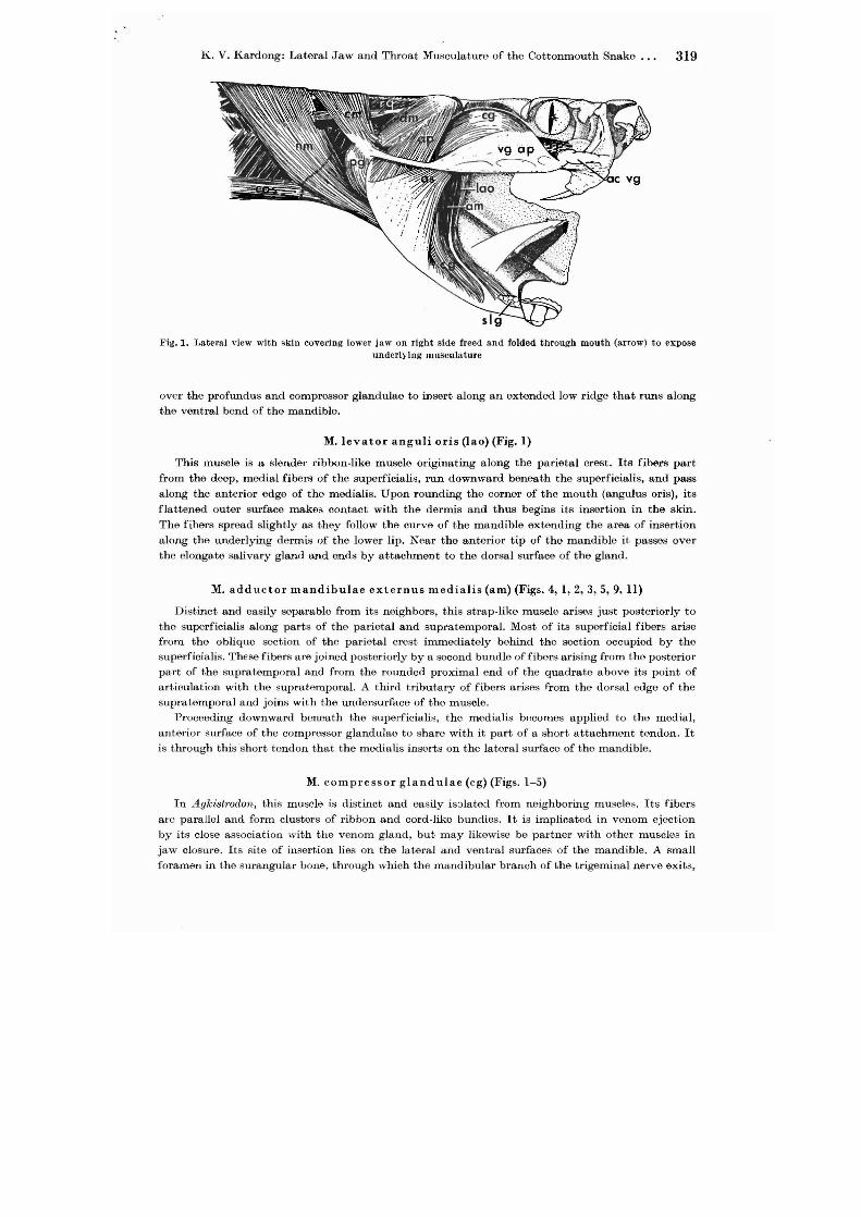
Fig. 1. Lateral view with skin covering lower jaw on right side freed .,nd folded through mouth (arrow) to expose underlying museul. ture
over the profundus and compressor glandulae to insert along an extended low ridge that runs along the ventral bend of the mandible.
M. levator anguli oris (lao) (Fig. 1)
This muscle is a slender ribbon-like muscle originating along the parietal crest. Its fibers part
from the deep, medial fibers of the superficialis, run downward beneath the superficialis, and pass
along the anterior edge of the medialis. Upon rounding the corner of the mouth (angulus oris), its flattened outer surface makes contact with the dermis and thus begins its insertion in the skin.
The fibers spread slightly as they follow the curve of the mandible extending the area of insertion
along the underlying dermis of the lower lip. Near the anterior tip of the mandible it passes over the elongate salivary gland and ends by attachment to the dorsal surface of the gland.
M. adductor mandibulae extern us medialis (am) (Figs. 4, 1,2,3,5,9, ll)
Distinct and easily separable from its neighbors, this strap-like muscle arises just posteriorly to the superficialis along parts of the parietal and supratemporal. Most of its superficial fibers arise from the oblique section of the parietal crest immediately behind the section occupied by the superficialis. These fibers are joined posteriorly by a second bundle of fibers arising from the posterior part of the supratemporal and from the rounded proximal end of the quadrat~ above its point of articulation with the supratemporal. A third tributary of fibers arises from the dorsal edge of the supratemporal and joins with the undersurface of the muscle.
Proceeding downward beneath the superficialis, the medialis becomes applied to the medial, anterior surface of the compressor glandulae to share with it part of a short attachment tendon. It is through this'short tendon that the medialis inserts on the lateral surface of the mandible.
M. compressor glandulae (cg) (Figs. 1-5)
In Agfc,',strodon, this muscle is distinct and easily isolated from neighboring muscles. Its fibers are parallel and form clusters of ribbon and cord-like bundles. It is implicated in venom ejection by its close association with the venom gland, but lllay likewise be partner with other muscles in
jaw closure. Its site of insertion lies on the lateral and ventral surfaces of the mandible. A small foramen in the surangular bone, through which the mandibular branch of the trigeminal nerve exits,

320 K. V. Kardong: Lateral Jaw and Throat Musculature of the Cottonmouth Snake
Fig. 2. Venom gland and neighboring muscles. The M. compressor glandulae (eg) attaehes in Pa.ft to the venom gland by a broad aponeurosis (yg ap) that is iifted from the sllrf'1Ce of the gland by foreeps to show points of attaehment
more clearly
Fig. 3. J~atero-,\]]terio-dorsal view of head. The venom gland's duct has been cut and the gland swung backward and pinned into the position shown so that the underlying muscles can be seen
opens in about the middle of the area of insertion. Fibers of compressor glandulae attach directly to the periosteum, except at the posterior edge of the muscle which here forms a short tendon that disappears into the anterior tip of the mandibular fossa. The posterior edge of the muscle fits into
an olongate groove formed medially by the posterior and laterally by the profundus muscles. The
mandibular branch of the trigeminal nerve can be scen to pass across the posterior and slip between it and the profundus at their line of contact.
Followed dorsally from its insertion, the compressor glandulae bows around the corner of t.he mouth, past the posterior angle of the triangular shaped venom gland, across its top apex, and ends

K. V. Kardong: Lateral Jaw and Throat Musculature of the Cottonmouth Snake. . . 321
Fig. -L Lateral Yiew of head IHu~cles. Venom gland and attachments ha.ve been cOJnp)etely removed except for two short pieces at ligamcnts, one still attached .to the retrOHrticular process at the mandible ann a secoud to the quadratesupratemporal articulation. The M. adductor mandibnlae externus superfieialis (as) and M. compressor glandulae
(cg) h"ve been cut and deflccted to expose underlying muscles
Fig. 5..Latera' view of head mnscles. Quadrate-supmtemporal articulation has been severed thus allowing outward rotation of the quadrate (top arrow) and lower jaw (lower arrow). The M. adductor mandibulne internus pterygoideus
(pg) has also heen cut
abruptly on the anterior side of the gland in a thick tendinous blanket of connective tissue. This covering wraps over the front side of the gland to fuse with the gland's own connective tissue capsule.
M. adductor mandibu1ae extern us profundus (ap) (Figs. 1-7,9,12)
This muscle's size and affiliation with companion muscles give it a special importance. It arises from the dur;;al, lateral, and ventral surfaces of the quadrate (Fig. 9). Though most of its fibers
are long and parallel, a partial pennate construction is suggested by the presence of a superficial
tendon with emanating oblique fibers. Passing downwards, the muscle increases in bulk as more

322 K. V. Kardong: Latera] Jaw and Throat Musculature of the Cottonmouth Snake
A
B
A
I pst
Fig. 6. Lower jaw showing areas of muscle attachment.
A. Medial view of left mandible..13. Lateral view of right mandible
dm
apo
B
dm () c ~r9
pq Iqm
Fig. 7. Posterior region of mandible showing areas of att,whment.
A. Dorsal view. B. Ventn'l view. C. Posterior view of the retroarticular process

K. V. Kardong: Lateral Jaw and Throat Musculature of the Cottonmouth Snake. . . 323
Fig. 8. Latero-posterio-dorsal view of skull showing musclc attachments
A B
Fig. 9. Quadrate bone from dorsal (A) and ventral (B) views showing areas of muscle attachment
fibers, arising from the quadrate, join the main muscle mass. It is thickest mid-way along its length at the angle of the jaw. Beyond this point the muscle flattens, spreads across the outer surface of
the mandible, and inserts along an extensive area of the bone. Some of the muscle's inner fibers spill into the anterior part of the mandibular fossa inserting directly along the interior side and rim of this fossa. However, most fibers continue past this first point of attachment and insert directly to the floor of an elongate depression that lies below the outer rim of the mandibular fossa.
The muscle participates in several unique affiliations with companion muscles and tissues. Its upper half is four sided: its medial side rests on the deep postel'ior muscle; its antel'ior side, together with the posterior muscl~, forms the elongate groove occupied by the posterior edge of the compressor glandulae; its postel'ior and lateral sides are covered by a shiny, fibrous tendon from which some of its own muscle fibers arise. This tendinous coat of the profundus also serves as a surface of origin for fibers that slant backward to contribute to the posterior depressor mandibulae. The middle of the profundus is fastened to the posterior angle of the venom gland by a short cord formed
from strands of connective tissue.

324 K. V. Kardong: Lateral Jaw and Throat Musculature of the Cottonmouth Snake
M. adductor mandibulae posterior (apo) (Figs. 4, 5, 9)
The mutual exchange of fibers occasionally occurs between the posterior and profundLls so the
two muscles are not neatly separable. The mandibular branch of the trigeminal nerve passes across
the outer face of the posterior neal' its anterior base of insertion slipping between it and the more
superficial profundus. Although both heads of the posterior, lateral and medial, are joined at their
origin, they can be peeled apart and their respective originJl estimated. Both arise from the ventral
surface of the quadrate, but the lateral head arises along th~ entire length whereas the medial head
arises only along the distal half of the quadrate. The posterior, composed of parallel fibers, drops downward from its origin as a flat, broad, and
fairly thick curtain toward the mandible. The mandible's coronoid process cleaves the muscle into
two inJlertion heads that spread over its outer and inner surface. The lateral head attaches to the lateral face of the coronoid process and to the sides, floor, and posterior outer rim of the mandibular fossa. The medial head inserts on the rim of the coronoid process itself and across its inner
sLlrface.
M. pseudo temporal is (pst) (Figs. 4,5,6)
The prootic bone bears a roughened depression that is raised along its anterior rim into a crescent shaped ridge. The rounded, proximal end of the supratemporal bone fits into the concave face of this ridge. It is from the middle and lower convex surface of this ridge that the pseudotemporaJis
originates. Its fibers are parallel and form a slender ribbon-like muscle that travels downward in front of the cranial foramen for the maxillary branch of the trigeminal nerve, across the lateral
surfaces of the Mm. levator pterygoidei and pterygoideus, and beneath levator anguli oris and me
dialis. The muscle widens just before inserting directly to a short section of a ridge that itself lies in front of and at the base of the coronoid process_
M. adductor mandibulae internus pterygoideus (pg) and M. adductor mandibulae internus pterygoideus accessorius(pga)
(Figs. 10, 1, 4, 5, 10, 12, 13)
The pterygoideus is an elaborate muscle. Some of its fibers bunch together into distinctive tracts and some form complox, interconnections with each other. Also, it is with the pt,erygoideus that the accessorius is closely associated. Description can be difficult and is often confusing to follow. Thus, a brief introduction is given to help clarify the general arrangement and attachments of these muscles before cOnJlidering their specific structures.
The pterygoideus originates on the ventral surface of the retroarticular process. From its origin, it bends inward beneath the mandible and proceeds directly forward to insert by two heads - one to the ectopterygoid and the other to fascia coating the maxilla. The accessorius is applied to the inside curvature of the pte"ygoideus as a muscular lining and accompanies the pterygoideus as both muscles bend inward b'meath the mandible. The accessorius originates from fibers of the pterygoideus and from the medial side of the mandible just below its point of articulation with the quadrate. The accessorius inJlerts along the ventral, posterior surface of the pterygoid bone.
It is concealed by other muscles and connective tissue. To be seen distinctly, the surrounding pterygoideus must be separated from its insertion and peeled back to thus reveal the accessorius.
The pterygoideus is a curious muscle. Anteriorly, it divides into two heads that insert separately on two diffeI'Ont bones. The dorsal head inserts on the ectopterygoid. This inJlertion site is along
the posterior border of a laterally projecting prominence, the articular knob (Fig. II B), that serves as a major point of articulation with the base of the maxillary bone. Insertion 011 the ectopterygoid is by both a tendon and by direct muscular attachment. The tendon begins forming about halfway along the length of the pterygoideus on its lateral surface. The shiny fibers of tho tendon pass forward and upward to insert on the articular knob of the ectopterygoid. Muscular fibers of the pterygoideus attach to this tendon and also inJlert next to it (medially) directly on the ectopterygoid (Fig. II B, C).

Fig. 10. I,ateml view of head with skin loosened aud pulled forward to expose underlying musclcs. Attachment of ligamentum quadrato-maxillare (Iqm) to the skin is 111so shown. The neuro-mandibularis(um) has been cut and deflected
A
\ \ \, \,,,,,,,,
B
I I I
~ ~
c
Fig. 11. Pterygoidells muscle. Portions of the lateral musculature a.re cut and removed to expose the M. addllcto mandibulae internus pterygoideus (pg) in figure A. E'igures J3 and C show the muscle with the lower jaw rotated outward (arrow) to beller reveal attachments. Three cross-sectional views of the Dluscle are shown in J3. Iu C. the two small solid triangles indicate pmminences on the retroartlcular process and below the articular notch (an) that
serve l1S points of origin for the ptcrygoideus muscle; t. tendon; g. glandulae

326 K. V. Kardong: Lateral Jaw and Throat Musculature of the Cottonmouth Snake ...
The ventral head of the pterygoideus muscle inserts on the maxilla via a soft, strong connective
tissue fascia that covers the maxilla and fang (Fig. lIB). A shiny tendon develops ventrally on
the pterygoideus and moves laterally as it passes forward in the company of fibers that form the
ventral head (Fig. 11 B, cross-section). Most of the ventral head's fibers attach to this tendon,
but some continue anteriorly and insert with the tendon on the connective tissue fascia surrounding
the base of the maxilla. Thus the force of muscular contraction is passed to and distributed upon
the maxilla and fang by the coat of strong fascia. The pterygoideus originates from the posterior end of the mandible. The mandible receives the
quadrate in a C-shaped "articular notch". The origin of the pterygoideus begins beneath this notch
and extends backward along the entire ventral surface of the retroarticular process (Fig. 7B). On
the medial side of the mandible, below the articular notch and continuing on to the retroarticular
process, is a crescent-shaped ridge. The two ends of this ridge form prominences from which strong
tendons arise and spread a short distance over the surface of the pterygoideus (Fig. 11 C).
The pterygoideus has another unusual feature - the pars glandulae. This is a small wedge.shaped
gathering of fibers located midway along the ventro-lateral surface of the M. pterygoideus. Fibers
of the pars glandulae converge into a narrow tendon that passes along the side of the pterygoideus
and inserts on the anterior base of the venom gland (Fig. 11, 12).
The accessorius takes origin from the crescent-shaped ridge carried on the internal, posterior part
of the mandible and from the M. pterygoideus. As described above, the ends of this ridge are raised
into prominences - an anterior prominence and a posterior prominence at the tip of the retroarti
cular process. The accessorius begins its origin immediately behind the anterior prominence and
continues backwards along the ridge. About halfway along this crescent, the site of origin turns
into the M. pterygoideus (Fig. 11 C). The fibers of the accessorius arise from this muscle and share
with it a short tendon arising from the surface of the retroarticular process.' The muscle passes
forward and spreads across the posterior undersurface of the pterygoid bone. A central ridge runs
along the pterygoid bone and is bordered on either side by two fossae - one lateral and one medial
(Fig. 12). Some of the muscle fibers attach to the top of this central ridge, but most find a point
of insertion along the sides or floors of the fossae. The area of insertion in the medial fossa is smaller
than the area of attachment in the lateral fossa. The area of insertion in the lateral fossa extends
forward to overlap the posterior insertion of M. levator pterygoideus. Consequently, the advancing
fibers of accessorius weave through the hindmost fibers of levator pterygoideus to find a site of
attachment along the pterygoid bone (Fig. 12).
M. leva tor pterygoidei (lp) (Figs. 5, 3, 4)
It is concealed by several of the muscles in the adductor mandibulae series, but itself is the
most superficial member of a group of internal muscles that act primarily upon the pterygoid bone.
It originates immediately beneath the thin site of origin of the superficialis in a concave attachment
fossa on the caudal surface of the postorbital process of the parietal bone. The area of origin includes
this attachment fossa, base of the postorbital process, and a small area of the parietal forming the
side of the braincase. It is a stocky, column-like muscle composed of parallel fibers that run obliquely
backward and downward to insert on the middle, upper and lower surfaces of the pterygoid bone
and on the lateral base of the ectopterygoid at its point of articulation with the pterygoid. On the
underside of the pterygoid, its lateral muscular fibers fill a lateral fossa that is, in part, shared
with the anterior fibers of the accessorius muscle, whereas its medial fibers insert on the pterygoid's middle, upper surface.
M. retractor pterygoidei (rp) (Figs. 5, 1, 3, 4)
This is a short but fairly sizeable muscle composed of parallel fibers. It originates in a fossa pressed into the lower side of the braincase just in front of the prootic bone and below the origins of levator pterygoidei and pseudotemporalis. This fossa is hollowed out of the parietal near its suture
with the basisphenoid. The retractor pterygoidei passes obliquely forward and downward to insert
K. V. Kardong: Lateral Jaw and Throat Musculature of the Cottonmouth Snake. . . 327
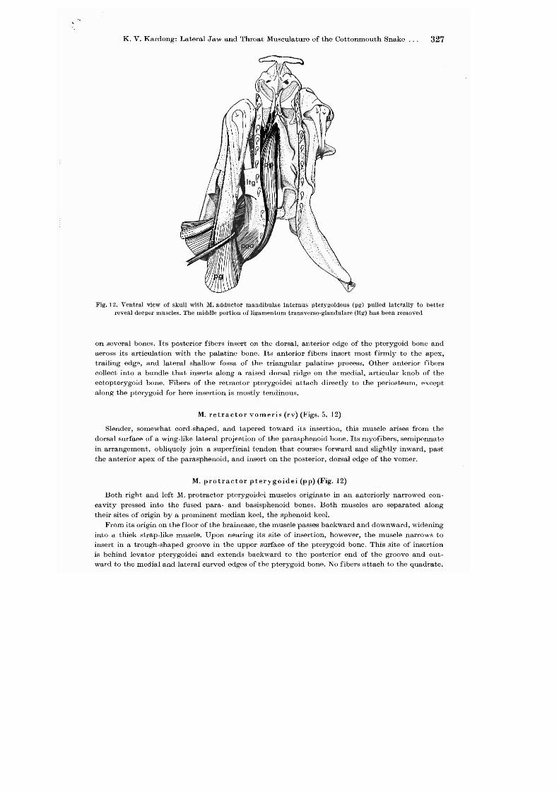
Fig. 12. Ventral view of skull with M. adductor mandibulae intcrnus pterygoideus (pg) pulled laterally to better reveal deeper lUuscles. The middle portion of ligamentum tra.nsverso-glandulare (Jtg) has been removed
on several bones. Its posterior fibers insert on the dorsal, anterior edge of the pterygoid bone and across its articulation with the palatine bone. Its anterior fibers insert most firmly to the apex, trailing edg9, and lateral shallow fossa of the triangular palatine process. Other anterior fibers collect into a bundle that inserts along a raised dorsal ridge on the medial, articular knob of the eetopterygoid bone. Fibers of the retractor pterygoidei attach directly to the periosteum, except along the pterygoid for here insertion is mostly tendinous.
M. retractor vomeris (rv) (Figs. 5, 12)
Slender, somewhat cord-shaped, and tapered toward its insertion, this muscle arises from the
dorsal surface of a wing-like lateral projection of the parasphenoid bone. Its myofibers, semipennate
in arrangement, obliquely join a superficial tendon that courses forward and slightly inward, past
the anterior apex of the parasphenoid, and insert on the posterior, dorsal edge of the vomer.
M. protractor pterygoidei (pp) (Fig. 12)
Both right and left M. protractor pterygoidei muscles originate in an anteriorly narrowed concavity pressed into the fused para- and basisphenoid bones. Both muscles are separated along their sites of origin by a prominent median keel, the sphenoid keel.
From its origin on the floor of the braincase, the muscle passes backward and downward, widening into a thick strap-like muscle. Upon nearing its site of insertion, however, the muscle narrows to insert in a trough-shaped groove in the upper surface of the pterygoid bone. This site of insertion is behind levator pterygoidei and extends backward to the posterior end of the groove and outward to the medial and lateral curved edges of the pterygoid bone. No fibers attach to the quadrate.

328 K. V. Kardong: Lateral Jaw and Throat Musculature of the Cottonmouth Snake
M. protractor quadrati (pq)
A ventrally directed central process of the basioccipital serves as a supportive post for attachment
of much of the neck musculature. The post's anterior, lateral side carries a low, crescent-shaped
"idge from which the protractor quadrati arises by a fan-shaped tendo'n. The tendon is strong at
its origin on the ventral tip of the post, but thins into a weak membranous sheet as it spreads along the low ridge on the side of the post. The muscle is ribbon-like and slips caudally between the
protractor pterygoidei and the deeper neck musculature to insert directly on the posterior half of
the quadrate along a narrow strip on the bone's medial edge.
M. retractor quadrati (rq) (Figs. 1, 3,4,5,9,10)
This small, inconspicuous muscle is concealed beneath the M. cervico-mandibularis. It is half
muscle fiber and half tendon. It originates in the dermis, behind the angle of the jaw, through several strings of muscle fibers. The muscle, formed by the confluence of these fibers, runs forward and slips under cervico-mandibularis. It soon narrows into a long tendon that passes to the medial
cadge of the quadrate, and finds attachment just below this bone's articulation with the squamosal.
M. depressor mandibulae (dm) (Figs. 1, 3, 4, 5)
This muscle is predominantly pennate and separable into two distinct heads at its origin. The
medial head is slender and takes origin in the connective tissue fascia covering the quadrato-supratemporal joint and by a tendon from the crest of the parietal. However, its large, lateral head arises
in a cap of tendinous connecitve tissue that covers the proximal end of the quadrate. Passing down
ward, the depressor mandibulae increases in size as it is joined by muscle fibers that originate in the shiny, tendinous coat over the posterior surface of the profundus.
All fibers of the depressor mandibulae share a common site of insertion. The muscle, passing beneath cervico-mandibularis, narrows slightly and inserts on the dorsal, curved outer surface of the retroarticular process of the mandible. The superficial fibers insert by a short tendon, but the
deeper fibers join directly to the periosteum.
M. eervieo-mandibularis (em) (Figs. 10,1,3,4,5)
This muscle is eas]y distinguished from neighboring muscles. It arises as a sheet of parallel fibers on the dorsal mid-line in the cervical region. Its superficial fibers originate along the dorsal mid-line in a tough, thick connective tissue fascia that occupies a furrow between both sides of the epaxial musculature and that fastens to the tips of the vertebral spines. Its deeper fibers ori
ginate in a short, but wide continuous aponeurosis that in turn arises on the neural spines of vertebrae six through nine.
The muscle narrows as it approaches the angle of the jaw. It passes over the distal end of the
quadrate where a few fibers of depressor mandibulae attach to its inside surface. It inserts by It
short tendon to the lateral knob on the distal end of the quadrate, to the retroarticular process of the mandible, and to the dermis above the ligamentum quadrato-maxillare.
M. constrictor colli (ee) (Fig. 13)
The constrictor colli is present. It originates in the del'mis on the side of the snake and slants downward in an anteroventral course. It inserts on the hyoid and On the posterior tendinous inscription beneath the angle of the jaw.
Throat musculature
The throat musculature is quite variable, and perhaps as a consequence a variety of terms are applied to the same muscles. Also in the literature the anatomical descriptions often treat the muscles differently. For instance, the first three muscles

K. V. Kardong: Lateral Jaw and Throat Musculature of the Cottonmouth Snake. . . 329
l<'ig. 13. Ventral view of throat musculature. The middle of M. constrictor colli (cc) has been cut but its attachment on the skin is shown
here described - M. costo-mandibularis, neuro-mandibularis, and branchiomandibu. laris - are sometimes treated as one composite muscle with three parts (e. g. neurocosto·mandibularis of ALBRIGHT and NELSON 1959) and at other times treated as separate muscles with their own integrity (e. g. LANGEBARTEL 1968). 'Whatever the cause, the result has been the build up of an extensive synonomy around each muscle that can be found reviewed by LANGEBARTEL (1968).
M. costo-mandibularis (cos) (Figs. 13, 1,4,5,10)
The ends of the ribs carry paddle-shaped cartilage discs. This muscle's lateral fibers take origin
from the first through fourth and its medial fibers from the fifth through eight rib tips and associated
discs. Near the first rib, the ribbon-like slips of its lateral fibers are crossed by one of two tendinous inscriptions. This posterior inscription marks a point of union between the muscle's lateral fibers and ]\.1. neuro-mandibularis. A few medial fibers of costo-mandibularis insert on the hyoid, but most
continue farther forward. Upon nearing the angle of the jaw, the parallel flow of both lateral and medial fibers is interrupted by another tendinous inscription. This anterior inscription, like the posterior, meanders into the M. neuro-mandibularis above, but unlike the posterior marks the major terminal line of insertion for the costo-mandibularis and neuro·mandibularis. This anterior inscription
22 Morph. Jb. 119/3

330 K. V. Kal'dong: Lateral Jaw and Throat Musculature of the Cottunmouth Snake
Fig. 14. Ventral view of deep throat muscles. The left !II. branehiomandibularis Is pinned to the side; portions from the !If. intermandibnlaris posterior (imp) and !If. transversus branchialis (tb) are removed to reveal deeper muscles.
On the right side, most of the !II. intermandiblliaris anterior (illla) is removed
also serves as the site of attachment for the branchiomandibularis that continues forward to thl' mandible.
M. neuro-mandibularis (urn) (Figs. 10, 1,3,4,5,13, 14)
This muscle takes origin along the middorsal cervical region arising behind ~L cervico-mandibularis directly from the tips of the neural spines and the supraspinous ligament. This broad, sheetlike muscle courses downward and forward narrowing slightly behind the angle of the jaw. Near the first rib, it is crossed by a posterior tendinous inscription part of which serves as a junction of its ventral fibers with the lateral fibers of M. costo-mandibularis. The neuro·mandibularis continues downward into the throat region to terminate on an anterior tendinous inscription along which it and the costo-mandibularis jointly insert. The branchiomandibularis arises on this same
inscription and continues forward, unaccompanied to the mandible.
M. branchiomandibularis (bm) (Figs. 13, 14)
The line of union between M. costo-mandibularis and M. neuro-mandibularis is marked by an anterior tendinous inscription that also serves as the line of insertion for M. branchiomandibularis.
Additionally, this muscle's medial fibers insert along the hyoid and central raphe. The posterior, lateral comer of the muscle, covering the pterygoideus, thins into a membrane· like aponeurosis. Except for this corner, most of the branchiomandibularis is a broad muscular sheet of parallel fibers
that can be traced forward across the floor of the buccal cavity to its origin along the ventral surface of the mandible by a strong aponeurosis permeated with many tendinous strands.
M. intermandibularis anterior (ima) (Figs. 13, 14)
This short muscle is composed of three parts. Pars anterior arises from the ventral, medial surface of the anterior tip of the dentary just below the site of origin of genioglossus. Its fibers run inward and insert on a connective tissue sheath that coats the medial surface of this muscle and through which it attaches on the ventral midline.
Pars posterior takes origin on the ventral surface of the dentary immediately behind the origin of pars anterior. Its fibers pass to the ventral midline and here insert securely in the dermis between
the pair of epidermal chin scales.

K. V. Kardong: Lateral Jaw and Throat Musculature of the Cottonmouth Snake. . . 331
Judging by its position, the pars glandulo-mandibularis ("constrictor" of LANGEBARTEL 1968,
p. 82) is an apparent derivative of the intermandibularis. It arises on the anterior tip of the dentary
and proceeds backward over the lateral side of the sUblingual gland to insert on the posterior, dorsal
surface of this gland very near the insertion of protractor laryngeus.
M. transversus branchialis (tb) (Figs. 13, 14)
This muscle has two parts, pars mucosalis and pars glandularis ("dilator" of LANGEBARTEL 1968,
p. 82). The pars glandularis is a long, narrow slip of muscle. From its insertion on the posterior
corner of the sublingual gland, it can be traced backward over the M. genio-glossus and M. genio
trachealis then around, inward, and benoath these muscles to attach below them on the median
raphe. It shares this attachment with the anterior fibers of pars mucosalis.
Pars mucosalis arises from the buccal membrane of the floor of the mouth as a delicate, thin
muscular sheet. It passes inward, narrowing slightly and attaches to the membrane-like median
raphe along the vontral midline. This attachment is dorsal to and continues behind M. intermandi
bularis posterior.
M. intermandibularis posterior (imp) (Figs. 13, 14)
This muscle takes origin along a low ridge on the surangular bone just above the posterior tip
of the three-sided angular bone. It passes rostrad and inward inserting first on the ventral midline,
then on the posterior border of the intermandibularis anterior (pars posterior). In some species,
this muscle has two distinct parts - pars anterior and pars posterior (COWAN and HICK 1951). How
ever, in Agkistrodon only the pars anterior is present.
M. transversus h yoideus (th) (Fig. 13)
It arises along the inner edge of the cornua and slants inward where, with its fellow from the
opposite side, it inserts in the modian raphe midway between the cornua of the hyoid. The trans
versus hyoideus occupies only the anterior third of the hyoid and is replaced in the middlo and
po~terior regions of the hyoid by a connective tissue membrane.
M. hyoglossus (hg) (Fig. 14)
This muscle retracts the tongue. It is paired, extends along the tongue, and contributes to the
bulk of the organ. It originates on the cornua of the hyoid and inserts in the tissue of the tongue.
It is sheathed in a coat of thick connective tissue.
M. genioglossus (gg) (Fig. 14)
Just above the origin of the intermandibularis anterior (pars anterior), along the inside bend
of the anterior tip of the dentary, this musclo takes origin. At first cord-like in appearance, it runs
caudally and inward, flattening upon contact with the tongue; now ribbon-like, it becomes embedded
in the connective tissue sheating of the hyoglossus muscle, passes along the sides of the tongue
capsule, and finds firm attachment on the posterior part of the tongue which is protracted by this
muscle. M. hyo-trachealis (ht) (Figs. 13,14)
This thin, flat muscle takes origin on the dorsal surface of the composite neuro-costo-mandibularis,
very near the posterior tendinous inscription below the angle of the jaw. Extending forward and
inward toward the trachea, it becomes applied to the floor of the buccal cavity. It passes over
genio-trachealis and inserts in front of it on the intrinsic laryngeal musculature.
M. genio-trachealis (gt) (Figs. 14, 13)
This slender strap of parallel muscle fibers takes origin on the inside bend of the dentary imme.
diat€ly dorsal to the origin of genio-glossus. Passing backward and inward toward the trachea, it
22*

332 K. V. Kardong: Lateral Jaw and Throat Musculature of the Cottonmouth Snake ...
widens slightly, crosses beneath hyo-trachealis, and inserts immediately behind this muscle on the
side of the trachea. The line of insertion begins on the sixth cartilaginous ring and slants downward
to the ventro-Iateral surface of the thirteenth ring.
M. protractor laryngeus (pI) (Fig. 14)
This muscle.is easily overlooked and was first described by KARLSTROM in 1952 who found it in
colubrids (Natrix taxispilota, Thamnophis ordinoides) and a viperid (Agkistrodon piscivorus) but not
in the boid Charina bottae. I have located it in the viperid Crotalus viridis and colubrid Elaphe
obsoleta, and confirm its absence in CharincJ., bottae. In Agkistrodon, it is very weakly developed and embedded in connective tissue.. This delicate
muscle arises from the anterior end of the larynx and runs forward to attach along the posterior
corner of the sublingual gland.
Summary
The lateral jaw and throat, musculature of the head of the cottonmouth snake (Agkistrodon
piscivorus) are described in detail. Each muscle is treated individually and its position, attachments, and association with neighboring structures described and illustrated. Such a morphological study of head musculature is intended to serve as a basis for future comparisons to other viperids and
for understanding the displacement patterns of the "movable" bony elements in the cottonmouth skull.
Acknowledgements
I am indebted to various persons and institutions for assistance during the course of this study. In particular, I am most grateful to the Department of Zoology, University of Illinois for its provision of facilities and financial support. I have benefited from the conscientious advice of Profs.
EDWIN M. BANKS, ALBERT C. BIANCHINI, JAMES B. KITZMILLER, and DAVID L. STOCl:M. The
understanding and assistance of Ms. WILLEMINA KARDONG is greatly appreciated. My special thanks
go to my graduate sponsor Prof. THOMAS H. FRAZZETTA whose friendship, thoughtful criticism,
and resilient patience throughout this study have been invaluable.
Key to Abbreviations
ac vg accessory venom gland
am M. adductor mandibulae externus medialis
apo M. adductor mandibulae posterior
ap M. adductor mandibulae externus profundus
as M. adductor mandibulae externus superficialis
bm M. branchiomandibularis
cc M. constrictor colli
cg M. compressor glandulae
ch cornu of the hyoid
em M. cervico-mandibularis
cos M. costo-mandibularis
dm M. depressor mandibulae
ec ectopterygoid bone
f frontal bone
g pars glandulae of peterygoideus (pg)
gg M. genioglossus

,
gt
hg
ht
imaa
imap
imag
imp
kb
lao
lp
lqg
lqm
ltg
m
nm
pg
pga
pI
pp
pq
pst
q
rp
rq
rv
sgl
slg
tb
tb g
tbm
th
tr
Vm
vg
vgap
K. V. Kardong: Lateral Jaw and Throat Musculature of the Cottonmouth Snake 333
M. genio-trachealis
M. hyoglossus
M. hyo-trachealis
M. intermandibularis anterior (pars anterior)
M. intermandibularis anterior (pars posterior)
M. intermandibularis anterior (pars glandularis)
M. intermandibular posterior
articular knob
levator anguli oris
M. levator pterygoidei
ligamentum quadrato-glandulare
ligamentum quadrato-maxillare
ligamentum transverso-glandulare
maxilla
M. neuro-mandibularis
M. adductor mandibulae internus pterygoideus
M. adductor mandibulae internus pterygoideus accessorius
M. protractor laryngeus
M. protractor pterygoidei
M. protractor quadrati
M. adductor mandibulae internus pseudotemporalis
quadrate
M. retractor pterygoidei
M. retractor quadrati
M. retractor vomeris
salivary gland
sublingual gland
M. transversus branchialis
M. transversus branchialis (pars glandularis)
M. transversus branchialis (pars mucosalis)
M. transversis hyoideus
trachea
mandibular branch of trigeminus nerve (V)
venom gland
venom gland aponeurosis
References
ALBRIGHT, R. G., and E. M. NELSON: Cranial kinetics of the generalized colubrid snake Elaphe obsoleta quadrivittata. 1. Descriptive morphology. J. Morpho!. 105 (1959) 193-239.
- - Cranial kinetics of the generalized colubrid snake Elaphe obsoleta quadrivittata. II. Functional morphology. J. Morphol. 105 (1959) 241-29L
ANTHONY, J., and R. G. SERRA: Anatomie de l'appareil de la morsure chez "Eunectus murinus" (Boidae). Osteologie, myologie, vaisseux et nerfs. Rev. Brasil. BioI. Rio de J. 10 (1950) 23-44.

334 K. V. Kardong: Lateral Jaw and Throat Musculature of the Cottonmouth Snake ...
BOGERT. C. M.: Dentitional phenomena in Cobras and other Elapids with nGtes on adaptive modi.
fications of fangs. Bull. Amer. Mus. Nat. Rist. 81, Art III (1943) 285-360.
BOLTT, R.' E., and R. F. EWER: The functional anatomy of the head of the puff adder, Bitis arietans
(Men'.). J. Morpho!. 114 (1964) 83-106. COWAK, 1. McT" and W. B. M. RICK: A comparative study of the bead region in three species of
Thamnophis (Reptilia, Ophidia). Trans. Roy. Soc. Canada 45 (1961) 19-60.
D'AI.TOK, E.: Beschreibung des Muskelsystems eines Python bivattatus. Arch. Anat., Physiol., wiss.
Med. (Johannes Muller) 7 (1834) 346-364; 17432-450; 12 628-643.
DuoBs, A.: Recherches anatomique et physiologiques sur Ie deglutition dans les reptiles. Ann. sci.
Nat. 12 (1827) 337-396.
DULI.Ei\tEIJER, P.: 'rhe functional morphology of the head of the common viper, Viper berus (L.).
Arch. Neerlandaises Zool. 2 (1966) 387-497.
- A comparative functional-anatomical study of the heads of some Viperidae. Gegenbaurs morpho
J ahrb. 99 (1958) 881-985.
FRAZZE'l'TA, T. H.: Studies on the morphology and function of the skull in the Boidae (Serpentes).
Part I. Cranial differences between Python sebae and Epicrates cench>-is. Bull. Mus. Camp. Zool.,
Harvard 119 (1959) 453-472. Studies on the morphology and function of the skull in the Boidae (Serpentes). Part II. Morpho
logy and function of the jaw apparatus in Python sebae and Python moluru8. J. Morpho!.1l8 (1966) 217-296.
GIBSON, F. W.: Head muscles of Boa con8trictor. Zoologica 51 (1966) 29-48.
GOl>PERT, E.: Der Kehlkopf der Amphibien und Reptilien. Gegenbaur's Morphol. Jahrb. 28 (II);
Theil, Reptilien (1899) 1-27.
HAAS, G.: Uber die Kaumuskulatur und die Schadelmechanik einiger \Viihlschlangen. Zool. Jahrb.
Anat. 52 (1930a) 95-218.
- Uber die Schiidelmechanik und die Kiefermusklllatur einiger Proteroglypha. Zoo I. Jahrb. Anat. 52
(1930b) 347-404.
- Die Kiefermuskulatur und die Schadelmechanik der Schlangen in vergleichender Darstellllng. ZooI. Jahrb. Anat. 53 (1931 a) 127-198.
- Uber die Morphologie del' Kiefermllskulatllr und die Schadelmechanik einiger Schlangen. Zoo I. Jahrb. Anat. 510 (1931 b) 334-416.
- A note on the origin solenoglyph snak0s. Copeia (1938) 2, 73-78.
- The head muscl0s of the genus Callsus (Ophidia, Solenoglypha) and some remarks on the origin
of the Solenoglypha. 1'l'Oc. Zool. Soc. London 122 (1962) 573-592.
- Anatomical observations on the head of Anomalepis aspinosus (Typlopidae, Ophidia). Acta Zool.
109 (1968) 1-2, 63-139.
HAGER, K. P.: Die Kiefermuskeln der Schlangen und ihre Beziehungen zu den Speicheldriisen. Zool.
Jahrb. Ana-t 22 (1906) 173-224.
HOFFMA..""-, C. K.: Reptilien. In: Bronn's Klassen und Ordnungen des Tier-Reichs. 6. sect. 3, plates. Leipzig 1890.
KATHARINER, L.: Die Mechanik d~s Bissos der solonoglyphen Giftschlangen. BioI. Ctrb!. 20 (1900) 46-53.
KOCHVA, E. T.: The head muscles of Vipe1'a palnestinac and their relat.ion to the venom gl and J. Morpho!. 102 (1958) 23-54.
- On the lateral jaw musculature of the Solenoglypha with remarks on some other snakes. J. Morpho!.
110 (1962) 227-284.
- Development of the venom gland and the trigeminal muscles in Vipem palae.stinae. Acta Anato·
mica 52 (1963) 1-2,49-89.
LAKJER, T.: Studien uber die Trigeminus.versorgte Kaumuskulatur des Sauropsiden. Kopenhagen:
C. A. Reitzel 1926.

" K. V. Kardong: Lateral Jaw and Throat Musculature of the Cottonmouth Snake. . . 335
LANGEBARTEL, D. A.: The hyoid and its assocIated muscles in snakes. Illinois Biological Monographs, K1'. 38. Univ. Illinois Press, Urbana (1968).
LUBoscH, lL: Untersuchungen libel' die Visceralmuskulatur del' Sauropsiden. Gegenbaurs morpho
J ahrb. 72 (1933) 584-666.
OWEN, R.: On the anatomy of vertebrates. Vol. 1, Fishes and Reptiles. London: Longmans, Green
and Co. 1866. PHISALIX, .1\'1.: Animaux venimeux et venins. Vol. 2, Paris: Masson ed Cie. 1922.
RADOVANOVIC, M.: Del' Giftapp~rat del' Schlangen mit besonderer Berlicksichtigung del' Naja tripu
dians. Jena. Z. Naturwiss. 63 (1928) 559-616. Anatonlische Studien am Schlangenkopf. Jena. Z. Naturwiss. 69 (1935) 321-422.
Osteologie des Schlangenkopfes. Jena. Z. Naturwiss. 71 (1937) 179-312.
Dr. KENNETH V. KARDOXG
Department Zoology
Washington State University Pullman, vVash. 99163 (USA)