lcca, an archaeal laccase secreted as a highly-stable glycoprotein into the extracellular medium
TRANSCRIPT

APPLIED AND ENVIRONMENTAL MICROBIOLOGY, Feb. 2010, p. 733–743 Vol. 76, No. 30099-2240/10/$12.00 doi:10.1128/AEM.01757-09Copyright © 2010, American Society for Microbiology. All Rights Reserved.
LccA, an Archaeal Laccase Secreted as a Highly Stable Glycoproteininto the Extracellular Medium by Haloferax volcanii�†
Sivakumar Uthandi,‡ Boutaiba Saad,§ Matthew A. Humbard, and Julie A. Maupin-Furlow*Department of Microbiology and Cell Science, University of Florida, Gainesville, Florida 32611-0700
Received 24 July 2009/Accepted 25 November 2009
Laccases couple the oxidation of phenolic compounds to the reduction of molecular oxygen and thus spana wide variety of applications. While laccases of eukaryotes and bacteria are well characterized, these enzymeshave not been described in archaea. Here, we report the purification and characterization of a laccase (LccA)from the halophilic archaeon Haloferax volcanii. LccA was secreted at high levels into the culture supernatantof a recombinant H. volcanii strain, with peak activity (170 � 10 mU � ml�1) at stationary phase (72 to 80 h).LccA was purified 13-fold to an overall yield of 72% and a specific activity of 29.4 U � mg�1 with an absorbancespectrum typical of blue multicopper oxidases. The mature LccA was processed to expose an N-terminal Alaafter the removal of 31 amino acid residues and was glycosylated to 6.9% carbohydrate content. Purified LccAoxidized a variety of organic substrates, including bilirubin, syringaldazine (SGZ), 2,2,-azino-bis-(3-ethylben-zothiazoline-6-sulfonic acid) (ABTS), and dimethoxyphenol (DMP), with DMP oxidation requiring the addi-tion of CuSO4. Optimal oxidation of ABTS and SGZ was at 45°C and pH 6 and pH 8.4, respectively. Theapparent Km values for SGZ, bilirubin, and ABTS were 35, 236, and 670 �M, with corresponding kcat valuesof 22, 29, and 10 s�1, respectively. The purified LccA was tolerant of high salt, mixed organosolvents, and hightemperatures, with a half-life of inactivation at 50°C of 31.5 h.
Multicopper oxidases (MCOs) are a family of enzymes thatinclude laccases (p-diphenol: dioxygen oxidoreductases; EC1.10.3.2), ascorbate oxidases (EC 1.10.3.3), ferroxidases (EC1.16.3.1), bilirubin oxidases (EC 1.3.3.5), and other enzyme sub-families (27, 65). MCOs couple the oxidation of organic and/orinorganic substrates to the four-electron reduction of molecularoxygen to water. These enzymes often have four Cu atoms clas-sified into type 1 (T1), type 2 (T2), and type 3 (T3) centers, inwhich a mononuclear T1 center on the surface of the enzymeprovides long-range intramolecular one-electron transfer fromelectron-donating substrates to an internal trinuclear T2-T3 cen-ter formed by a T2 Cu coordinated with a T3 Cu pair. The T2-T3cluster subsequently reduces dioxygen to water.
Enzymes of the laccase subfamily oxidize a broad range ofcompounds, including phenols, polyphenols, aromatic amines,and nonphenolic substrates, by one-electron transfer to molec-ular oxygen and thus have a wide variety of applications frombiofuels to human health. The best-known application is theuse of a laccase from the lacquer tree Rhus vernicifera in paintand adhesives for more than 6,000 years in East Asia (29).Laccases have also been used in the delignification of pulp,bleaching of textiles and carcinogenic dyes, detoxification ofwater and soils, removal of phenolics from wines, improving
adhesive properties of lignocellulosic products, determinationof bilirubin levels in serum, and transformation of antibioticsand steroids (60). In addition, laccases have demonstrated poten-tial for use in biosensors, bioreactors, and biofuel cells (61).
Laccases, once thought to be restricted to eukaryotes (fungi,plants, and insects), appear to be widespread in bacteria (10).Laccase-like MCOs are now known to have numerous biolog-ical roles in bacteria, including sporulation, electron transport,pigmentation, metal (copper, iron, and manganese) homeosta-sis, oxidation of phenolate-siderophores, phenoxazinone syn-thesis, cell division, and morphogenesis (9). In contrast to thewidespread occurrence of laccases in bacteria and eukaryotes,only a few MCOs have been identified in archaea, and this isbased only on genome sequences (e.g., the hyperthermophiliccrenarchaeote Pyrobaculum aerophilum and the halophilic eur-yarchaeotes Haloferax volcanii and Halorubrum lacusprofundi).Most archaea with sequenced genomes, however, are anaerobes.Since MCOs reduce molecular oxygen to water, this likely ac-counts for the limited number of MCOs among archaea.
Many archaea thrive under harsh environmental conditions,including high temperature, extreme pH, and/or low wateractivity. Thus, they have many biochemical and physiologicalproperties that are ideal for industrial applications. Here, wereport the identification of a highly thermostable and salt/solvent-tolerant laccase (LccA) from the halophilic archaeonH. volcanii that catalyzed the oxidation of a wide variety ofphenolic compounds. LccA was readily secreted and purifiedfrom the culture broth as a blue multicopper oxidase that wasglycosylated and processed by the removal of 31 amino acidresidues from its N terminus.
MATERIALS AND METHODS
Materials. Biochemicals were from Sigma-Aldrich (St. Louis, MO). Otherorganic and inorganic analytical-grade chemicals were from Fisher Scientific
* Corresponding author. Mailing address: P.O. Box 110700, Bldg.981 Museum Rd., Department of Microbiology and Cell Science, Uni-versity of Florida, Gainesville, FL 32611-0700. Phone: (352) 392-4095.Fax: (352) 392-5922. E-mail: [email protected].
‡ Permanent address: Department of Agricultural Microbiology,Tamil Nadu Agricultural University, Coimbatore, India.
§ Present address: Laboratory of Research on the Arid Area, FSB-Biologic Science Faculty, University of the Sciences and TechnologyH. Boumediene, Algiers, BP no. 32, El-Alia, Algiers, Algeria.
† Supplemental material for this article may be found at http://aem.asm.org/.
� Published ahead of print on 4 December 2009.
733
Dow
nloa
ded
from
http
s://j
ourn
als.
asm
.org
/jour
nal/a
em o
n 01
Jan
uary
202
2 by
27.
2.88
.70.

(Atlanta, GA). Restriction endonucleases, T4 DNA ligase, and Vent DNA poly-merase were from New England Biolabs (Ipswich, MA). AccuPrime GC-richDNA polymerase was from Invitrogen (Carlsbad, CA). Desalted oligonucleo-tides were from Integrated DNA Technologies (Coralville, IA). Agarose forroutine analysis of DNA was from Bio-Rad Laboratories (Hercules, CA).SeaKem GTG agarose, used for the separation and isolation of DNA fragmentsprior to ligation, was from FMC Bioproducts (Rockland, ME).
Strains, media, and plasmids. The strains, primers, and plasmids used aresummarized in Table 1. Escherichia coli DH5� was used for routine recombinantDNA experiments, and E. coli GM2163 was used to generate plasmid DNA fortransformation of H. volcanii (16). E. coli strains were grown at 37°C in Luria-Bertani medium. H. volcanii strains were grown at 42°C in complex medium(ATCC 974 and yeast-peptone-Casamino Acids) and lactate-minimal medium(LMM), as previously described (1, 16, 34). Ampicillin (0.1 mg � ml�1), novo-biocin (0.1 �g � ml�1), and CuSO4 (50 to 500 �M) were included as needed.Cultures were grown in liquid (150 to 200 rpm) and solid (1.5% [wt/vol] agarplates) media.
Genome analysis. NCBI Local BlastP with BioEdit sequence editor softwarev7.0.4.1 (25) was used to compare the theoretical H. volcanii DS2 proteome(http://archaea.ucsc.edu/, April 2007 version) to fungal and bacterial proteinswith known laccase activity. The protein sequences included phenol oxidase A ofthe lignocellulolytic ascomycete Stachybotrys chartarum (42), spore coat proteinA (CotA) from Bacillus subtilis (43), and polyphenoloxidase (PPO) from Strep-tomyces lavendulae (66). Phylogenetic and molecular evolutionary analyses of theprimary protein sequences were conducted using MEGA v3.1 (39). Pairwise andmultiple-sequence alignments were performed using Clustal W (67). Noncon-served regions in the N and C termini were excluded from the protein sequencealignment. Evolutionary distances were estimated from the protein sequencesusing the proportion (p-) distance substitution model. Consensus tree inferencewas by neighbor joining with a bootstrap phylogeny test (1,000 replicates; seednumber for bootstrap, 64,238) and pairwise gap deletion. TatP v1.0 (4), TAT-
FIND v1.4 (57), and EnsembleGly (8) servers were used for prediction of twinarginine translocation (TAT) and glycosylation motifs in the LccA protein.
DNA isolation, analysis, and strain construction. Genomic DNA was pre-pared from H. volcanii DS70 (a strain of DS2 cured of plasmid pHV2) (68) forPCR by transfer of isolated colonies to 30 �l deionized H2O using toothpicks.The cells were boiled (5 min), and the resulting lysate was chilled on ice (10 min)and centrifuged (14,000 � g; 10 min at 4°C). The supernatant (5 to 10 �l) wasused as a template for PCR with primer pairs specific for the H. volcanii laccasegene (lccA; HvoB0205), as listed in Table 1. A mixture of AccuPrime GC-richand Vent DNA polymerase at a 9:1 ratio was used for PCR, with buffer andnucleotide concentrations according to the recommendations of Invitrogen. PCRwas performed with an iCycler (Bio-Rad Laboratories), and the sizes of theproducts were analyzed by electrophoresis using 0.8% (wt/vol) agarose gels inTAE buffer (40 mM Tris acetate, 2 mM EDTA, pH 8.5) with Hi-Lo DNAmolecular weight markers as standards (Minnesota Molecular, Minneapolis,MN). The gels were photographed after being stained with ethidium bromide at0.5 �g � ml�1 with a Mini visionary imaging system (Fotodyne, Hartland, WI).PCR-generated DNA fragments of appropriate size for the lccA gene (1.8 kb)were isolated from 0.8% (wt/vol) SeaKem GTG agarose (FMC Bioproducts,Rockland, ME) gels in TAE buffer using the QIAquick gel extraction kit (Qia-gen) and ligated into the NdeI-to-HindIII sites of pET24b to generate plasmidspJAM821 and pJAM823 for expression of lccA in recombinant E. coli Rosetta(DE3). In addition, the lccA gene was excised from these plasmids and ligatedinto the NdeI-to-BlpI sites of the H. volcanii shuttle vector (pJAM202) to posi-tion the lccA gene downstream of a constitutive rRNA promoter, P2. Theseexpression plasmids (pJAM822 and pJAM824) were transformed into H. volcaniiH26 to generate strains SB01 and US02 for high-level synthesis of LccA with andwithout a C-terminal StrepII tag (-WSHPQFEK), respectively. Plasmid DNAwas isolated using the QIAprep Spin Miniprep Kit (Qiagen, Valencia, CA). Thefidelity of cloned DNA sequences and the 100% identity of lccA genes from DS2
TABLE 1. Strains, plasmids, and oligonucleotide primers used in this study
Strain, plasmid, or primer Description Source or reference
StrainsE. coli
DH5� �� �80dlacZ�M15 �(lacZYA-argF)U169 recA1 endA1 hsdR17(rK� mK
�) supE44 thi-1 gyrArelA1
Life Technologies
GM2163 F� ara-14 leuB6 fhuA31 lacY1 tsx78 glnV44 galK2 galT22 mcrA dcm-6 hisG4 rfbD1 rpsL 136dam13::Tn9 xylA5 mtl-1 thi-1 mcrB1 hsdR2
New EnglandBiolabs
Rosetta (DE3) F� ompT hsdSB(rB� mB
�) gal dcm (DE3) pRARE (Camr) Novagen
H. volcaniiDS70 Wild-type isolate DS2 cured of plasmid pHV2 68H26 DS70 pyrE 1SB01 H26-pJAM822 This studyUS02 H26-pJAM824 This study
PlasmidspET24b Kmr; E. coli expression vector NovagenpBAP5010 Apr; Nvr; 11-kb H. volcanii-E. coli shuttle vector derived from pBAP5009; includes
Halobacterium cutirubrum rRNA P2 promoter (P2rrn); T7 transcription terminator andpHV2 ori for expression of cloned genes in H. volcanii
33
pJAM202 Apr Nvr; pBAP5010 containing P2rrn-psmB-H6 34pJAM202c Apr Nvr; pJAM202 with P2rrn-psmB-H6 deleted 70pJAM821 Kmr; pET24b with lccA-strepII for expression in E. coli Rosetta (DE3) This studypJAM823 Kmr; pET24b with lccA for expression in E. coli Rosetta (DE3) This studypJAM822 Apr Nvr; pJAM202 with P2rrn-lccA-strepII for expression in H. volcanii This studypJAM824 Apr Nvr; pJAM202 with P2rrn-lccA for expression in H. volcanii This study
PrimersHvoB0205 (lccA) up 5�-tgggcgtCATATGacagactggtctaggcggcgg-3�a This studyHvoB0205 (lccA-strepII)
down5�-aaAAGCTTcacttctcgaactgcgggtgcgaccaAGTACTggcgacttcgtcgccgcttc-3�b This study
HvoB0205 (lccA) down 5�-aaAAGCTTtcaggccacttcgtcgccgcttc-3�c This study
a The NdeI site is in italics.b The HindIII and ScaI sites are in italics.c The HindIII site is in italics.
734 UTHANDI ET AL. APPL. ENVIRON. MICROBIOL.
Dow
nloa
ded
from
http
s://j
ourn
als.
asm
.org
/jour
nal/a
em o
n 01
Jan
uary
202
2 by
27.
2.88
.70.

and DS70 strains were confirmed by Sanger DNA Sequencing (ICBR GenomicsFacility, University of Florida).
Enzyme purification. H. volcanii SB01 and US02 cells were grown in mediasupplemented with 100 �M CuSO4 (LMM or YPC; 25 ml per 125-ml Erlenmeyerflask) to stationary phase. Cells were harvested by centrifugation (15,000 � g; 20min at 4°C), and the culture broth was filtered with Whatman 3MM chromatog-raphy paper. The protein was concentrated by gradual addition of 1 volume ofcold absolute ethanol, incubation on ice (1 h), and centrifugation (7,000 � g at4°C for 10 min). The resulting precipitate was suspended in 25 mM Tris-HClbuffer at pH 8.4 (T buffer) to 1/5 the original sample volume and recentrifugedto eliminate insoluble material. US02 precipitate (from an original culture of 360ml) was concentrated to 3 ml using a Centricon PL-30 centrifugal filter accordingto the instructions of the supplier (Millipore, Billerica, MA) and separated intotwo 1.5-ml fractions (where F1 and F2 represent the more viscous lower andupper fractions, respectively) and diluted 6.7-fold with T buffer. For both SB01and US02 samples, proteins were filtered (0.20 �m) immediately prior to appli-cation to an anion-exchange MonoQ HR 5/5 or 10/10 column (Pharmacia)equilibrated with T buffer, as indicated in Table 1. Elution was carried out witha linear NaCl gradient (0 to 1 M in 1 ml T buffer) with fractions containing peakactivity at 625 to 675 mM NaCl. Additional steps were included for purificationof LccA from SB01, including application of active fractions (0.25 ml per run) toa Superdex 200 HR 10/30 column (Pharmacia) equilibrated in T buffer with 150mM NaCl, followed by a MonoQ HR 10/10 column equilibrated in T buffer anddeveloped with a linear NaCl gradient (600 to 800 mM in 1 ml T buffer). Thefractions were monitored for purity by assay with 4-hydroxy-3,5-dimethoxyben-zaldehyde azine (syringaldazine [SGZ]) under standard assay conditions (seebelow for details) and staining with Coomassie blue R-250 after separation byreducing 12% SDS-PAGE according to the method of Laemmli. PrestainedSDS-PAGE standards and Kaleidoscope Standards (Bio-Rad) were used asprotein standards for PAGE. The native molecular mass was determined byapplying samples to a calibrated Superose 200 HR 10/30 column as recom-mended by the supplier (Pharmacia). The molecular mass standards for calibra-tion included cytochrome c (12.4 kDa), carbonic anhydrase (29 kDa), bovineserum albumin (66 kDa), alcohol dehydrogenase (150 kDa), -amylase (200kDa), and apoferritin (443 kDa) (Sigma). Samples were stored at 4°C.
Enzyme activity and protein estimation. Enzyme activity was determined usingthe following substrates: 2,2�-azino-bis-3-ethylbenzthiazoline-6-sulfonic acid(ABTS) (Sigma; A1888), 2,6-dimethoxyphenol (DMP) (Sigma; D135550), 2-me-thoxyphenol (guaiacol; Sigma; G5502), SGZ (Sigma S7896), bilirubin (Sigma;B4126), and ferrous ammonium sulfate. The enzymes were assayed by change inabsorbance using a BioTek Synergy HT multidetection microplate reader with a96-well plate and the following extinction coefficients: ε420 36,000 (ABTS),ε468 49,000 (DMP), ε436 26,600 (guaiacol), ε526 65,000 (SGZ), ε440 56,300 (bilirubin), and ε315 2,200 (ferrous ammonium sulfate) in units of M�1
cm�1. The standard assay conditions were 185 mM NaCl at 45°C with thefollowing buffers and substrate concentrations: 100 mM sodium acetate at pH 6.0for ABTS (5 mM), DMP (2 mM), and guaiacol (5 mM); 25 mM Tris buffer at pH8.4 for SGZ (1 mM); and 200 mM Tris buffer at pH 8.4 for bilirubin (450 �M).Copper sulfate (1 mM) was included for the DMP oxidation assay. Oxidation offerrous ammonium sulfate (1 mM) was monitored with and without salt (185 mMNaCl) in 100 mM MES (morpholineethanesulfonic acid) buffer at pH 5, as wellas 25 mM Tris-HCl buffer at pH 7.0 and 8.4. One unit of enzyme activity isdefined as the amount of enzyme that oxidizes 1 �mol of substrate per min understandard conditions. Kinetic studies were conducted using purified LccA at afinal concentration of 0.61 �g � ml�1 with 5 to 160 �M SGZ, 50 to 1,250 mMABTS, and 90 to 540 �M bilirubin. Kinetic parameters (Km, Vmax, and kcat) weredetermined using the Michaelis-Menten equation. The protein concentrationwas determined based on the method of Bradford using a Bio-Rad protein assaywith bovine serum albumin as the standard (Bio-Rad Laboratories). All assaysand protein purifications were performed at least in triplicate.
For detection of laccase activity in gels, nonboiled samples were separated on7.5% PAGE gels devoid of SDS and -mercaptoethanol. The gels were im-mersed in T buffer containing 1 mM SGZ, and activity was rapidly visualized asa pink color. For detection of total protein, the gels were counterstained withCoomassie blue R-250 stain and destained according to the instructions of thesupplier (Bio-Rad). Images of activity and protein staining were acquired usinga VersaDoc 1000 imaging system (Bio-Rad).
N-terminal sequencing. Purified LccA was separated by reducing 12% SDS-PAGE and transferred by electroblotting to Immobilon-P polyvinylidene diflu-oride (PVDF) membranes (Amersham Biosciences) at 100 V for 100 min at 4°C.Proteins were stained in the membrane with 0.2% (wt/vol) Coomassie blue R-250in 40% acetic acid for 30 s and rinsed with deionized water. The LccA protein
band was excised and subjected to automated Edman degradation for N-terminalsequencing (18).
Glycoprotein analysis. LccA was separated by reducing SDS-PAGE andstained in gel for glycosylation using Pro-Q Emerald 300 (Invitrogen P21857)and total protein was separated using Sypro Ruby (Bio-Rad; 1703126) with aVersaDoc 4000 imaging system according to the instructions of the manufacturer(Bio-Rad Laboratories). The carbohydrate content of purified LccA was esti-mated by the colorimetric method of Dubois et al. (15) using glucose as astandard. Pure LccA was deglycosylated by the trifluoromethanesulfonic acid(TFMS) method (17) for 0 to 10 h on ice and analyzed by reducing SDS-PAGE.Trametes versicolor laccase (Sigma Fluka; 53739) and bovine carbonic anhydrase(Sigma-Aldrich; C7025) were included for glycoprotein analysis as positive andnegative controls, respectively.
Mass spectrometry analysis. Purified LccA was provided for mass spectrom-etry (MS) analysis as an in-solution sample and as protein bands separated byreducing 7.5% SDS-PAGE. Gels were stained with Bio-Safe Coomassie (Bio-Rad), and LccA-protein bands were excised from the gel and destained with 100mM NH4HCO3 (pH 7.5) in 50% (vol/vol) acetonitrile (4°C overnight). Proteinsamples were reduced, alkylated in gel or in solution, digested with trypsin(Promega), and desalted. Peptides were further separated by capillary reverse-phase high-performance liquid chromatography (HPLC) using a PepMap C18
column (15 cm by 75-�m inside diameter [i.d.]) and an Ultimate Capillary HPLCSystem (LC Packings, San Francisco, CA) with a linear gradient of 5 to 40%(vol/vol) acetonitrile for 25 min at 200 nl/min. Tandem MS (MS-MS) analysis wasperformed online using a hybrid quadrupole time-of-flight instrument (QSTARXL hybrid LC/MS-MS) equipped with a nanoelectrospray source (Applied Bio-systems, Foster City, CA) and operated with the Analyst QS v1.1 data acquisitionsoftware. Spectra from all experiments were converted to DTA files and mergedto facilitate database searching using the Mascot search algorithm v2.1 (MatrixScience, Boston, MA) against the theoretical H. volcanii proteome (http://archaea.ucsc.edu/, April 2007 version). The search parameters included trypsinas the cleavage enzyme and carbamidomethylation as a fixed modification. Vari-able modifications included deamidation of asparagine and glutamine; oxidationof methionine; methyl-esterification of aspartate and glutamate; and N-acetyla-tion and phosphorylation of serine, threonine, and tyrosine. The mass tolerancefor all QSTAR analyses was 0.3 Da, and peptides with assigned probability-basedMascot ion scores of less than 30 were excluded.
RESULTS AND DISCUSSION
LccA is related to laccases of the multicopper oxidase fam-ily. Basic local alignment search tool (BLAST) analysis of theH. volcanii theoretical proteome revealed a 63,397-Dapolypeptide of pI 4.34 (HVO_B0205, termed LccA) related tolaccases of the MCO family (Fig. 1). The gene locus encodingLccA resided on pHV3 (440 kb), one of three large repliconswith cdc6- and orc1-associated replication origins separatefrom the 2.848-Mb chromosome (48). Archaeal proteins thatcluster with the MCO family are rare, and none have beencharacterized to date. LccA included the conserved cysteineand histidine residues required for coordination of the type 1,2, and 3 copper centers and activities of blue copper laccases(Fig. 1). In addition to the conserved catalytic residues, the Nterminus of LccA included a twin arginine motif (RRRFL)preceding a hydrophobic patch, suggesting it may be translo-cated in a folded state through the twin arginine translocation(Tat) system. LccA was also predicted to be cleaved betweenAla31 and Ala32 to generate a mature protein of 60.1 kDa andto be glycosylated based on the presence of O- and N-linkedglycosylation motifs, Thr-X5-Pro-X-Pro and Asn-X-(Thr/Ser)(where X represents any amino acid) (Fig. 1).
Consensus tree inference revealed that LccA was closelyrelated to laccases of the Gram-positive bacteria, including 38to 40% identity with and 52 to 53% similarity to the thermo-stable CotA proteins of B. subtilis and Bacillus licheniformis(38, 43) (see Fig. S1 in the supplemental material). Among the
VOL. 76, 2010 ARCHAEAL MULTICOPPER LACCASE 735
Dow
nloa
ded
from
http
s://j
ourn
als.
asm
.org
/jour
nal/a
em o
n 01
Jan
uary
202
2 by
27.
2.88
.70.

FIG. 1. Sequence alignment of H. volcanii LccA (Hvo_B0205) to multicopper oxidases. The vertical lines represent cleavage sites withexperimental evidence based on the N-terminal sequence of the mature LccA (HVO_B0205) and Myrothecium verrucaria bilirubrin oxidase(Mver_Q12737) (37). TAT motifs, indicated by a double underline, are based on in silico analysis using the TatP 1.0 server. Note that TatP alsopredicts both cleavage sites described above. Residues that bind the four copper atoms to form the type 1, 2, and 3 copper sites of knownmulticopper oxidases are indicated by dots above the amino acid residues. Potential O- and N-linked glycosylation sites with EnsembleGly scoresabove 0.8 are indicated by O* and N*, respectively, above the residues, with the Thr-X5-Pro-X-Pro and Asn-X-Thr motifs (where X represents
736 UTHANDI ET AL. APPL. ENVIRON. MICROBIOL.
Dow
nloa
ded
from
http
s://j
ourn
als.
asm
.org
/jour
nal/a
em o
n 01
Jan
uary
202
2 by
27.
2.88
.70.

archaea, LccA was closely related (45% identity) only toHlac_1049, encoded on the main chromosome of Halorubrumlacusprofundi. Other archaeal genomes (completed as of No-vember 2009) did not encode LccA homologs, including thehaloarchaea Halobacterium sp. NRC-1 (Halobacterium salina-rum R1), Haloquadratum walsbyi DSM 16790, Haloarculamarismortui ATCC 43049, and Natronomonas pharaonis DSM2160. Furthermore, LccA had less than 30% identity toPAE1888, an uncharacterized protein with enhanced tran-script levels under denitrifying conditions in P. aerophilum(12). In contrast to LccA, PAE1888 clustered with bacterialMCOs involved in oxidative stress and/or metal homeostasis,such as McoA of Aquifex aeolicus, CueO of E. coli, and Mco ofStaphylococcus aureus (21, 62, 63). Consistent with this, themethionine-rich helix proposed to be involved in metal seques-tration that lies over the type 1 sites of the metal-oxidizingMCOs, such as E. coli CueO (53), is not conserved in LccA.This sparse distribution of LccA homologs among the archaeaand the close relationship of LccA to laccases of the firmicutes(e.g., B. subtilis CotA) suggested the haloarchaeal gene wasacquired by horizontal gene transfer from bacteria and that itencodes a laccase that oxidizes phenolic compounds.
Laccase activity secreted by recombinant H. volcanii. Tocharacterize LccA, our initial efforts focused on modifying theH. volcanii lccA gene for laccase production in recombinant E.coli using the pET expression system with Rosetta (DE3) andplasmids pJAM821 and pJAM823 (Table 1). Although numer-ous H. volcanii proteins have been successfully produced usingthis type of bacterial system (34, 52), LccA protein and laccaseactivity were not detected by SDS-PAGE and SGZ oxidationassay of culture broth and cell lysates of these recombinant E.coli strains. “Salt-loving” haloarchaeal proteins, enzymes withelaborate metal clusters, glycoproteins, and proteins secretedby the TAT system are all notoriously difficult to produce inconventional expression systems, such as E. coli and Saccha-romyces cerevisiae (6, 11, 41). Thus, a number of factors may belimiting the production of LccA in E. coli.
To overcome these limitations, lccA was engineered forhigh-level expression in the native organism H. volcanii, knownfor its halophilic properties and robust TAT system (57). ThelccA gene was positioned downstream of a strong rRNA P2promoter on a multicopy plasmid (pHV2 based) with andwithout coding sequence for a 1-Da C-terminal StrepII tag(-WSHPQFEK), and the resulting plasmids were transformedinto H. volcanii H26 to generate strains SB01 and US02, re-spectively (Table 1). These engineered strains were grown invarious media supplemented with CuSO4 (50 to 500 �M) andcompared to their parent (H26) for the production of laccaseactivity by monitoring the oxidation of SGZ. Using this ap-proach, laccase activity associated with H. volcanii cells wasnegligible for all three strains examined, reaching at most0.69 � 0.12 mU � ml�1 after 80 to 120 h of growth (Fig. 2). In
contrast to these cellular fractions, significant levels of laccaseactivity were detected in the culture broth of H. volcanii SB01and US02, with the highest level of activity detected for strainUS02 in YPC medium with 100 �M CuSO4 (Fig. 2). Underthese conditions, the laccase activity of US02 reached a max-imum of 170 � 10 mU � ml�1 at stationary phase (72 to 80 h),whereas the level of laccase activity secreted by SB01 was40-fold lower, reaching levels of only 4.3 � 0.23 mU � ml�1 at54 h. Laccase activity was not detected in the culture broth ofUS02 and SB01 until stationary phase (Fig. 2) and was notdetected at significant levels throughout all phases of growth
any amino acid) boxed. Amino acid sequences in boldface and underlined represent peptides detected by MS-MS analysis of the laccase purifiedfrom H. volcanii US02 (details of MS-MS analysis are shown in the supplemental material). Black and grey shadings represent identical andfunctionally conserved amino acid residues, respectively. HVO_B0205, H. volcanii laccase LccA (this study); Hlac_ZP-02016827, Halorubrumlacusprofundi putative multicopper oxidase (GenBank accession no. 153896174); Bsub_BAA22774, Bacillus subtilis spore coat protein A (CotA)with laccase activity (28, 43); Scha_AAY23005, Stachybotrys chartarum phenol oxidase A (42); Mver_Q12737, M. verrucaria bilirubrin oxidase (37).
FIG. 2. Laccase activity of H. volcanii SB01 and US02 strains atvarious stages of growth. H. volcanii SB01 (A), US02 (B), and theparent, H26 (data not shown), were inoculated to a final optical densityat 600 nm (OD600) of 0.01 in medium supplemented with novobiocinand 100 �M copper sulfate. LMM and YPC were optimal for produc-tion of laccase activity for SB01 and US02, respectively. The cells weregrown for up to 120 h at 42°C in an orbital shaker (150 to 200 rpm).Samples were withdrawn at intervals and used for estimation of growth(1 OD600 unit � 1 � 109 CFU per ml) and assay of laccase activity. Foranalysis of cell lysates, the cells were harvested by centrifugation (10min at 10,000 � g and 4°C), washed twice in T buffer supplementedwith 2 M NaCl, resuspended in T buffer, and lysed by sonication (15 son, 45 s off at 20% amplitude for 3 min). The culture broth (filtered at0.4 �m) and cell lysate were assayed for laccase (SGZ-oxidizing) ac-tivity under standard conditions. Data are presented as means � stan-dard deviations.
VOL. 76, 2010 ARCHAEAL MULTICOPPER LACCASE 737
Dow
nloa
ded
from
http
s://j
ourn
als.
asm
.org
/jour
nal/a
em o
n 01
Jan
uary
202
2 by
27.
2.88
.70.

for the parent strain H26 (data not shown). The reason for thelag in laccase productivity for US02 and SB01 remains to bedetermined; however, a similar time course has been observedfor the secretion of the Natrialba magadii subtilisin-like Nepprotease from recombinant H. volcanii (14). While the addi-tion of a strong, relatively constitutive promoter upstream oflccA enhanced laccase productivity by over 170-fold, additionof a C-terminal StrepII coding sequence significantly de-creased LccA productivity. Thus, enhanced expression of lccAcoding sequence alone was optimum for secretion of laccaseactivity by H. volcanii.
Although the genetic systems of archaea are not as advancedas those of bacteria or yeast, the productivity of the engineeredH. volcanii strain US02 was surprisingly high, with 170mU � ml�1 laccase activity produced in the medium in lessthan 3 days. This level of productivity is comparable to that ofyeast systems, such as Yarrowia lipolytica and Pichia methan-olica, developed for secretion of the white-rot fungal laccasefrom T. versicolor. Typically, these yeast systems require 5 to 6days of cultivation for production of 230 to 1,260 mU � ml�1 oflaccase activity (24, 32). It should also be noted that the secre-tion of LccA from H. volcanii contrasts with the vast majorityof systems developed for production of bacterial MCOs. Inthese systems, the bacterial MCO is not secreted and is insteadpurified from the soluble and/or insoluble fractions of recom-binant E. coli cells (e.g., production of the Thermus thermophi-lus Tth laccase [45], B. subtilis and B. licheniformis CotA [38,43], Bacillus halodurans Lbh1 [58], S. aureus Mco [63], A.aeolicus McoA [21], and Bacteroides RL5 from the rumenmetagenome [3]).
LccA purified from the culture broth of H. volcanii. LccAwas purified 13-fold from the culture broth of H. volcanii US02to a specific activity of 29.4 U � mg�1 by ethanol precipitation,concentrated using a Centricon PL-30 centrifugal filter, andfractionated by MonoQ chromatography as outlined in Table2. LccA-StrepII was similarly purified from SB01 with theaddition of gel filtration (see Fig. S2 in the supplemental ma-terial) and MonoQ chromatography steps at the final stages ofpurification (Table 2). In contrast to US02-purified LccA, thespecific activity of LccA-StrepII was over 20-fold lower at only 1.3U � mg�1. Thus, addition of a C-terminal StrepII tag not only
reduced the productivity of the strain, but also reduced LccAenzyme activity. Incorporation of StrepTactin chromatographydid not improve the overall activity or yield of LccA-StrepII fromSB01 and thus was not further pursued (data not shown).
The LccA protein isolated from US02 (as described above)was analyzed for purity by reducing SDS-PAGE and MS. Pu-rified LccA migrated as a single protein band of 75 to 80 kDa
FIG. 3. LccA purified to electrophoretic homogeneity from the cul-ture broth of H. volcanii US02. Shown is reducing 10% SDS-PAGE ofLccA at various stages of purification, including ethanol precipitate(EP) (lane 1) and MonoQ 10/10, pH 8.4 (MQ) (lane 2), as indicated.Molecular mass standards are on the left. Proteins were stained withCoomassie brilliant blue R-250.
TABLE 2. Purification of LccA from the culture broth of H. volcanii cellsa
Fraction Total activity (U) Protein (mg) Yield (%) Sp act (U � mg�1) Purification (foldenrichment)
H. volcanii SB0160-h culture broth (130 ml) 740 6.7a 100 0.11 160-h culture broth (filtered) 770 6.4 100 0.12 1.1Ethanol precipitate 1,010 6.3 140 0.16 1.5MonoQ 5/5, pH 8.4 470 0.92 63 0.51 4.6Superdex 200 HR 10/30 170 0.23 23 0.76 6.9MonoQ 10/10, pH 8.4 95 0.07 13 1.3 12
H. volcanii US0272-h culture broth (360 ml) 57 25 100 1.4 1Ethanol precipitate 55 13 94 4.3 2Centricon PL-30 and
MonoQ 10/10, pH 8.4F1, 41.5; F2, 14.2 F1, 1.4; F2, 2.1 F1, 72; F2, 24 F1, 29.4; F2, 6.7 F1, 13; F2, 2.9
a H. volcanii SB01 and US02 were grown for 60 h in HvLMM and 72 h in YPC (42°C; 200 rpm), respectively. LccA protein was purified from the culture broth asindicated. Activity was monitored by oxidation of SGZ under standard assay conditions (see Materials and Methods). F1 and F2 represent the lower (viscous) and upperfractions generated after concentration by centrifugal filtration using a Centricon PL-30, respectively (see Materials and Methods for details).
738 UTHANDI ET AL. APPL. ENVIRON. MICROBIOL.
Dow
nloa
ded
from
http
s://j
ourn
als.
asm
.org
/jour
nal/a
em o
n 01
Jan
uary
202
2 by
27.
2.88
.70.

(Fig. 3) that was specific for LccA based on MS analysis with 38to 68% coverage of the LccA polypeptide sequence (Fig. 1; seeFig. S3 and Table S1 in the supplemental material). The 10- to20-kDa difference in migration of LccA by SDS-PAGE com-pared to that calculated from the polypeptide sequence wasconsistent with its low pI and/or posttranslational modificationby glycosylation.
Multiple isoforms of LccA. LccA (purified as describedabove) was separated by native PAGE and stained for SGZ-oxidizing activity and total protein. In contrast to SB01-puri-fied LccA-StrepII, for which only a single protein band wasdetected, LccA purified from US02 migrated as at least fourseparate isoforms that were all active in SGZ oxidation (Fig.4A). These results suggested that LccA was modified post-translationally and that addition of a C-terminal StrepII tagabolished this modification. It should be noted that mobility innative gels depends on not only the hydrodynamic size, but alsothe charge of the protein. The LccA polypeptide sequence ispredicted to be highly acidic, and this is likely to account forthe rapid migration of all of the LccA isoforms compared toprotein standards of similar molecular mass.
N-terminal cleavage and glycosylation of LccA. Many fungallaccases are modified posttranslationally, including removal ofN-terminal residues, as well as the addition of high-mannose-type glycans to Asn residues (N glycosylation) (5, 22, 37, 41, 51,69). Based on bioinformatics, LccA is predicted to be cleavedbetween Ala31 and Ala32 and to be N and/or O glycosylated(Fig. 1). To investigate these possibilities, the US02-purifiedLccA was N-terminally sequenced by Edman degradation andanalyzed for glycosylation. Purified LccA was cleaved afterAla31 (Fig. 1) and glycosylated based on a carbohydrate con-tent of 6.9% � 0.06%, as determined by a phenol-sulfuric acidmethod (15), and staining using Pro-Q Emerald 300 (26) (Fig.4B). The carbohydrate content of LccA was similar to the 6.9%previously reported for other fungal laccases (51). To furtheranalyze and confirm LccA glycosylation, the purified proteinwas treated with TFMS, a strong acid known to remove glycansfrom polypeptide chains (17). A molecular mass shift of 10 to20 kDa was observed by SDS-PAGE for the TFMS-treatedLccA compared to the untreated control and resulted in itsmigration at 60 kDa, consistent with the molecular masscalculated from the deduced amino acid sequence (Fig. 4C).TFMS treatment did not shift the migration of nonglycosylatedcontrol proteins (e.g., carbonic anhydrase), confirming thatpolypeptide chains remained intact under these conditions(data not shown). Together, these results provide evidence thatLccA is secreted, cleaved, and glycosylated; however, the orderof these events during its maturation remains to be deter-mined.
Interestingly, the LccA-StrepII variant purified from SB01was not associated with carbohydrate and did not stain withPro-Q Emerald (data not shown). Furthermore, the N termi-nus of LccA-StrepII was not amendable to sequencing by Ed-
FIG. 4. LccA purified from US02 is a glycoprotein. (A) Native gelsof LccA at its final stages of purification from US02 (Superdex 200 HR10/30, lanes 1 and 2) and SB01 (MonoQ 10/10, pH 8.4, lanes 3 and 4).The gels were stained for total protein with Coomassie brilliant blueR-250 (CB) and for laccase activity by in-gel oxidation of SGZ (SGZ),as indicated. Prestained kaleidoscope and low-range protein standards(Bio-Rad) were included for US02 and SB01, respectively, as indicatedat the left of each gel. (B) Pro-Q Emerald glycoprotein analysis ofUS02-purified LccA. Proteins were separated by reducing 10% SDS-PAGE. The gels were stained for glycosylation with Pro-Q Emerald(left) and for total protein with Sypro-Ruby (right), as indicated. Theproteins included carbonic anhydrase (CA) (lanes 1); T. versicolorlaccase (TvLc) (lanes 2); LccA MonoQ 10/10, pH 8.4, fractions(HvLcMQ) (lanes 3); LccA ethanol precipitate (HvLcEP) (lanes 4); andCandyCane molecular mass standards (Molecular Probes), including amixture of glyosylated (G) and nonglycosylated proteins, as indicated
(lanes 5). (C) Deglycosylation of LccA. LccA purified from US02 wastreated with TFMS on ice for 0 h (lanes 1), 10 h (lanes 2), and 3 h (lane3) and separated by reducing 6 and 10% SDS-PAGE, as indicated (seeMaterials and Methods for details).
VOL. 76, 2010 ARCHAEAL MULTICOPPER LACCASE 739
Dow
nloa
ded
from
http
s://j
ourn
als.
asm
.org
/jour
nal/a
em o
n 01
Jan
uary
202
2 by
27.
2.88
.70.

man degradation, suggesting it may be blocked by N-terminal� amine group (N�) acetylation. These results are consistentwith the native PAGE analysis and suggest that addition of aStrepII tag to the C terminus of LccA not only reduces itsactivity, but also inhibits its N-terminal cleavage and glycosy-lation.
Spectroscopic and catalytic properties of LccA. LccA (puri-fied from US02) was blue and exhibited an absorbance spec-trum typical of blue multicopper oxidases, including a peak at605 nm and a shoulder at 330 nm (Fig. 5). Many laccasesisolated from fungi and bacteria are blue multicopper oxidasesthat coordinate four copper atoms into three types of copper-binding sites (27). All of these “blue” enzymes produce spectrawith a maximum at 605 nm, corresponding to the T1, or blue,copper atom. The T2 copper site exhibits weakly visible absor-bance, and the T3 copper site has two copper centers and isresponsible for a shoulder at around 330 nm. The spectrum ofLccA is consistent with its clustering with blue laccases, includ-ing the closely related and structurally characterized B. subtilisCotA (20). Thus, the conserved cysteine and 10 histidine res-
idues of LccA are likely to coordinate these three types ofcopper sites.
LccA oxidized a variety of compounds. The four substratesmost commonly used for determination of laccase activity wereevaluated: ABTS, DMP, guaiacol, and SGZ. The oxidation offerrous (Fe2�) ammonium sulfate and bilirubin was also inves-tigated. Although LccA did not oxidize Fe2� or guaiacol in thepresence or absence of copper supplementation, it did oxidizethe remaining substrates, i.e., ABTS at 6.1 � 0.39 U � mg�1,DMP at 12.6 � 1.1 U � mg�1, SGZ at 29.4 � 1.4 U � mg�1, andbilirubin at 25.5 � 1 U � mg�1 (the values reported are forUS02-purified LccA under standard conditions [see Materialsand Methods for details]). Note that the oxidation of DMP washighly stable for several hours but, unlike ABTS, SGZ, andbilirubin, required the addition of CuSO4. LccA presented ahigher specificity for SGZ than for ABTS, with kcat/Km valuesof 0.62 and 0.015 s�1 �M�1, respectively (Table 3). While thespecificity of LccA for SGZ was significantly lower than that ofthe DUF152 RL5 laccases, it is comparable to if not higherthan that of B. subtilis CotA (Table 3). LccA specificity forbilirubin oxidation was also high, with a kcat/Km value of 0.12s�1 �M�1 (kcat, 29 s�1; Km, 236 �M).
Recently, laccases have been grouped based on their DMPoxidase activities (64). The first group oxidizes DMP withoutcopper supplementation (e.g., the laccase of Pyricularia oryzaeand PpoA of Marinomonas mediterranea), the second groupreadily oxidizes DMP after the addition of copper but is alsorapidly inactivated (e.g., CueO of E. coli), and the third typeshows a very low rate of DMP oxidation that lasts for hours,but only in the presence of added copper (e.g., CotA of B.subtilis). LccA oxidized DMP only in the presence of addedCuSO4; hence, it appears to be most similar to the last groupof laccases.
To further evaluate the catalytic properties of LccA, thetemperature, pH, and salt optima of the purified enzyme were
FIG. 5. UV-visible absorbance spectrum of LccA. The absorbanceof LccA (1.5 mg � ml�1) purified from H. volcanii US02 in T buffercontaining 185 mM NaCl was analyzed from 300 to 700 nm.
TABLE 3. Kinetic parameters of H. volcanii LccA compared to those of selected laccase-like enzymes
Enzyme, origin ofencoding gene Source of purified enzyme
ABTS oxidation SGZ oxidation
ReferenceKm (�M) kcat (s�1) kcat/Km
(s�1 �M�1) Km (�M) kcat (s�1) kcat/Km(s�1 �M�1)
LccA, H. volcanii Culture broth of H. volcaniiSB01
700 10.0 0.014 67 19.4 0.30 This study
Culture broth of H. volcaniiUS02
671 9.9 0.015 35 21.7 0.62 This study
CotA, Bacillus subtilis Cell lysate of recombinantE. coli
106 16.8 0.16 26 3.7 0.14 43
CotA, Bacillus licheniformis Cell lysate of recombinantE. coli
6.5 83 13 4.3 100 23 38
McoA, Aquifex aeolicus Refolded from cell lysate ofrecombinant E. coli
128 2.1 0.016 38 0.48 0.013 21
LAC2, white-rotbasidiomycete C30
Culture supernatant of C30 536 683 1.27 6.8 1,093 160 36
LAC1, white-rotbasidiomycete C30
Culture supernatant of C30 56 10.7 5.22 1.8 30 17 36
RL5, metagenome library ofbovine rumen (tentativeBacteroides origin)
Cell lysate of recombinantE. coli
26 18 0.69 0.43 660 1,534 3
BT4348, Bacteroidesthetaiotaomicron
Cell lysate of recombinantE. coli
10 217 21.7 0.83 555 669 3
YifH, E. coli Cell lysate of recombinantE. coli
23 24 1.05 1.1 362 329 3
740 UTHANDI ET AL. APPL. ENVIRON. MICROBIOL.
Dow
nloa
ded
from
http
s://j
ourn
als.
asm
.org
/jour
nal/a
em o
n 01
Jan
uary
202
2 by
27.
2.88
.70.

determined using SGZ and/or ABTS as a substrate. LccAactivity was optimum at 45 to 50°C (see Fig. S4 in the supple-mental material), consistent with the optimal growth temper-ature of the organism (42 to 45°C) (54). Relative to this opti-mum, LccA was 75% active at room temperature (21°C) and40 to 50% active at 60°C. This temperature optimum of LccAis similar to a number of bacterial MCO optima, including 40to 45°C for laccases of Streptomyces sp. (19, 47), 52°C forBacteroides thetaiotaomicron BTH4389, and 44°C for E. coliYifH (3). It is also closely related to the temperature optimumfor simultaneous saccharification and fermentation (SSF) pro-cesses, which include fungal cellulases and second-generationmicrobial biocatalysts (49). However, it is significantly lowerthan the optima of 75°C for B. subtilis CotA (43) and 70°C forA. aeolicus McoA (21).
Most extracellular and intracellular enzymes of haloarchaea,including H. volcanii, require salt for activity and stability.Consistent with this, the activity of LccA was optimal at 200mM salt, with 1.5-fold greater activity in KCl than in NaCl, andthe enzyme displayed reduced activity after the removal of saltby dialysis (see Fig. S4 in the supplemental material). LccA wasalso active at relatively high concentrations of salt, with 65%activity at 1 M NaCl. The ability of LccA to rapidly oxidizephenolic substrates at molar concentrations of salt is not sur-prising, as the enzyme is secreted by a microbe isolated fromthe Dead Sea. Similar to LccA, the activity of Lbh1 from thehalotolerant B. halodurans is stimulated by addition of NaCl(58).
While the oxidation of ABTS by LccA was optimal at low pH(pH 6.0), its optimum for SGZ oxidization was more alkaline(pH 8.4) (see Fig. S4 in the supplemental material). Mostmicrobial laccases display optimal activity at low pH. Thosewith alkaline or near-neutral pH optima are not as common,e.g., Acremonium murorum (23), B. halodurans (58), Coprinuscinereus (69), Streptomyces ipomoea (46), and Melanocarpusalbomyces (35). Unlike LccA, the few microbial laccases thatare active at alkaline pH do not exhibit significant activitybelow pH 6.0. These unique catalytic properties of LccA un-derline the potential of Archaea as a source of high-valuelaccases.
LccA is stable at high temperature and high salt and withsolvent. To assess the thermostability of LccA, the purifiedprotein was preincubated at various temperatures from 37 to70°C for up to 2.5 days. LccA was fully active after 1 h at 55°Cand 5 h at 50°C, with a half-life of inactivation at 50°C ofgreater than 1 day (31.5 h) (see Fig. S5 in the supplementalmaterial). The enzyme retained nearly all of its original activityat 37°C and 35 to 60% of its activity at 45 to 50°C afterincubation for 2.5 days. These results revealed that LccA ishighly thermostable, retaining most of its activity after pro-longed incubation at moderately high temperatures. The mul-ticopper oxidase of A. aeolicus, McoA, has remarkably stableactivity, with the ability to oxidize metals after incubation for5 h at 90°C (21). Likewise, the recently studied recombinantRL 5 laccase from the rumen metagenome has maximal activ-ity at 60°C and is fully stable at this temperature for 3 to 4 h (3).CotA of B. subtilis is also thermostable, with a half-life ofnearly 2 h at 80°C (43).
The purified LccA was also stable at a wide range of saltconcentrations from 100 mM to at least 1.4 M NaCl, retaining
nearly all of its original activity, if not higher activity (see Fig.S5 in the supplemental material). It is noteworthy that pro-longed incubation of LccA in salt (for 24 h) did not alter thisactivity profile (data not shown). Recent analysis of Streptomy-ces psammoticus, a bacterium isolated from a mangroveswamp, revealed that it produces a laccase that is also salttolerant, with full activity after incubation for 24 h in 0.8 MNaCl (47).
Commonly used water-miscible organosolvents (i.e., metha-nol, ethanol, dimethyl sulfoxide [DMSO], and dimethylform-amide [DMF]) were examined for their compatibility withLccA stability and activity. Purified LccA was incubated in Tbuffer with 25% (vol/vol) solvent in the presence of salt (185mM NaCl) for 1.5 to 24 h. The enzyme and solvent mixtureswere diluted 4-fold and assayed for SGZ-oxidizing activity un-der standard conditions (see Fig. S5 in the supplemental ma-terial). LccA was relatively stable in all solvents examined,retaining nearly 75% of its activity after 24 h in methanol orethanol and over 50% of its activity after incubation in DMSOor DMF. Prior studies (7, 40, 44, 55, 56) coupled with ourcurrent findings reveal that a wide variety of laccases, includingLccA, are stable and/or can support catalysis and conversion ofphenolic substrates in water-organic mixed solvents. Sincemost of the substrates and mediators of laccases are insolubleor poorly soluble in water and the degree of polymerization ofphenoxyl radicals is higher in aqueous media (44), solventstability is an important feature to consider in optimizing thisgroup of enzymes. Many industrial applications would alsobenefit from the addition of water-miscible organic cosolventsto laccase-mediated reactions, particularly the conversion ofinsoluble substrates, such as lignin and its derivatives, to usefulproducts (2, 13, 50).
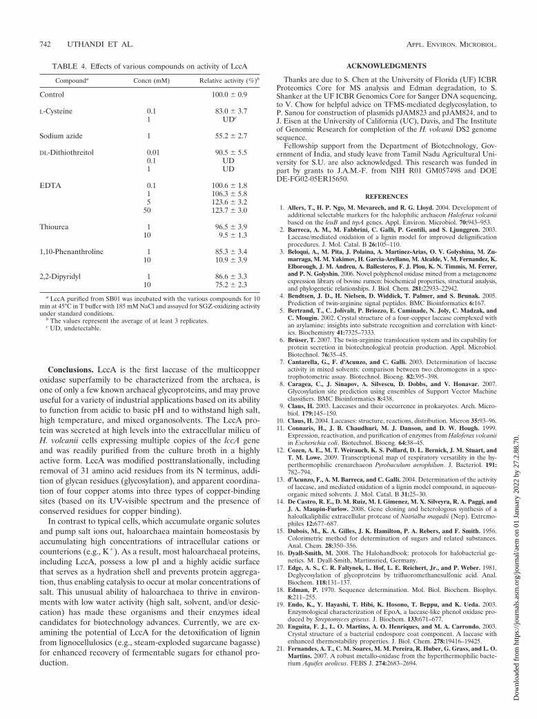
Effects of inhibitors. A number of compounds were exam-ined for their influences on LccA activity, including small ions(sodium azide or NaN3), sulfhydryl group-containing redoxreagents (L-cysteine and dithiothreitol [DTT]), denaturants(thiourea), and chelators (EDTA, 1,10-phenanthroline, and2,2-dipyridyl) (Table 4). The most effective laccase inhibitorsto date are small anions, especially N3
�, which often bind tothe trinuclear copper center and interfere with electron flowand substrate oxidation (31). Consistent with this, LccA wasinhibited nearly 50% by the addition of 1 mM NaN3. In con-trast, some fungal laccases, such as that reported by Saito et al.(59), are relatively unaltered by NaN3 (at 10 mM). Of theredox reagents examined, both L-cysteine and DTT werestrong inhibitors of LccA activity and were effective at rela-tively low concentrations (i.e., 0.1 to 1 mM). As has beenobserved for other laccases, this inhibition is likely caused byreduction of the oxidized substrate by the sulfhydryl groups ofthe redox reagents and not by inhibition of the enzyme (30).The LccA protein was also susceptible to denaturation by thio-urea, with 90% inhibition at 10 mM. Although the generalchelator EDTA did not inhibit LccA activity at 50 mM, thetransition metal chelators 2,2-dipyridyl and 1,10-phenanthro-line were inhibitory at 10 mM, with 25% and 90% reductionsin LccA activity, respectively. These results are consistent withthe recalcitrant nature of the copper-containing laccases to-ward chelation by EDTA (59) yet reveal a likely role of metalsin LccA activity, based on inhibition by phenanthroline.
VOL. 76, 2010 ARCHAEAL MULTICOPPER LACCASE 741
Dow
nloa
ded
from
http
s://j
ourn
als.
asm
.org
/jour
nal/a
em o
n 01
Jan
uary
202
2 by
27.
2.88
.70.
Conclusions. LccA is the first laccase of the multicopperoxidase superfamily to be characterized from the archaea, isone of only a few known archaeal glycoproteins, and may proveuseful for a variety of industrial applications based on its abilityto function from acidic to basic pH and to withstand high salt,high temperature, and mixed organosolvents. The LccA pro-tein was secreted at high levels into the extracellular milieu ofH. volcanii cells expressing multiple copies of the lccA geneand was readily purified from the culture broth in a highlyactive form. LccA was modified posttranslationally, includingremoval of 31 amino acid residues from its N terminus, addi-tion of glycan residues (glycosylation), and apparent coordina-tion of four copper atoms into three types of copper-bindingsites (based on its UV-visible spectrum and the presence ofconserved residues for copper binding).
In contrast to typical cells, which accumulate organic solutesand pump salt ions out, haloarchaea maintain homeostasis byaccumulating high concentrations of intracellular cations orcounterions (e.g., K�). As a result, most haloarchaeal proteins,including LccA, possess a low pI and a highly acidic surfacethat serves as a hydration shell and prevents protein aggrega-tion, thus enabling catalysis to occur at molar concentrations ofsalt. This unusual ability of haloarchaea to thrive in environ-ments with low water activity (high salt, solvent, and/or desic-cation) has made these organisms and their enzymes idealcandidates for biotechnology advances. Currently, we are ex-amining the potential of LccA for the detoxification of ligninfrom lignocellulosics (e.g., steam-exploded sugarcane bagasse)for enhanced recovery of fermentable sugars for ethanol pro-duction.
ACKNOWLEDGMENTS
Thanks are due to S. Chen at the University of Florida (UF) ICBRProteomics Core for MS analysis and Edman degradation, to S.Shanker at the UF ICBR Genomics Core for Sanger DNA sequencing,to V. Chow for helpful advice on TFMS-mediated deglycosylation, toP. Sanou for construction of plasmids pJAM823 and pJAM824, and toJ. Eisen at the University of California (UC), Davis, and The Instituteof Genomic Research for completion of the H. volcanii DS2 genomesequence.
Fellowship support from the Department of Biotechnology, Gov-ernment of India, and study leave from Tamil Nadu Agricultural Uni-versity for S.U. are also acknowledged. This research was funded inpart by grants to J.A.M.-F. from NIH R01 GM057498 and DOEDE-FG02-05ER15650.
REFERENCES
1. Allers, T., H. P. Ngo, M. Mevarech, and R. G. Lloyd. 2004. Development ofadditional selectable markers for the halophilic archaeon Haloferax volcaniibased on the leuB and trpA genes. Appl. Environ. Microbiol. 70:943–953.
2. Barreca, A. M., M. Fabbrini, C. Galli, P. Gentili, and S. Ljunggren. 2003.Laccase/mediated oxidation of a lignin model for improved delignificationprocedures. J. Mol. Catal. B 26:105–110.
3. Beloqui, A., M. Pita, J. Polaina, A. Martinez-Arias, O. V. Golyshina, M. Zu-marraga, M. M. Yakimov, H. Garcia-Arellano, M. Alcalde, V. M. Fernandez, K.Elborough, J. M. Andreu, A. Ballesteros, F. J. Plou, K. N. Timmis, M. Ferrer,and P. N. Golyshin. 2006. Novel polyphenol oxidase mined from a metagenomeexpression library of bovine rumen: biochemical properties, structural analysis,and phylogenetic relationships. J. Biol. Chem. 281:22933–22942.
4. Bendtsen, J. D., H. Nielsen, D. Widdick, T. Palmer, and S. Brunak. 2005.Prediction of twin-arginine signal peptides. BMC Bioinformatics 6:167.
5. Bertrand, T., C. Jolivalt, P. Briozzo, E. Caminade, N. Joly, C. Madzak, andC. Mougin. 2002. Crystal structure of a four-copper laccase complexed withan arylamine: insights into substrate recognition and correlation with kinet-ics. Biochemistry 41:7325–7333.
6. Bruser, T. 2007. The twin-arginine translocation system and its capability forprotein secretion in biotechnological protein production. Appl. Microbiol.Biotechnol. 76:35–45.
7. Cantarella, G., F. d’Acunzo, and C. Galli. 2003. Determination of laccaseactivity in mixed solvents: comparison between two chromogens in a spec-trophotometric assay. Biotechnol. Bioeng. 82:395–398.
8. Caragea, C., J. Sinapov, A. Silvescu, D. Dobbs, and V. Honavar. 2007.Glycosylation site prediction using ensembles of Support Vector Machineclassifiers. BMC Bioinformatics 8:438.
9. Claus, H. 2003. Laccases and their occurrence in prokaryotes. Arch. Micro-biol. 179:145–150.
10. Claus, H. 2004. Laccases: structure, reactions, distribution. Micron 35:93–96.11. Connaris, H., J. B. Chaudhuri, M. J. Danson, and D. W. Hough. 1999.
Expression, reactivation, and purification of enzymes from Haloferax volcaniiin Escherichia coli. Biotechnol. Bioeng. 64:38–45.
12. Cozen, A. E., M. T. Weirauch, K. S. Pollard, D. L. Bernick, J. M. Stuart, andT. M. Lowe. 2009. Transcriptional map of respiratory versatility in the hy-perthermophilic crenarchaeon Pyrobaculum aerophilum. J. Bacteriol. 191:782–794.
13. d’Acunzo, F., A. M. Barreca, and C. Galli. 2004. Determination of the activityof laccase, and mediated oxidation of a lignin model compound, in aqueous-organic mixed solvents. J. Mol. Catal. B 31:25–30.
14. De Castro, R. E., D. M. Ruiz, M. I. Gimenez, M. X. Silveyra, R. A. Paggi, andJ. A. Maupin-Furlow. 2008. Gene cloning and heterologous synthesis of ahaloalkaliphilic extracellular protease of Natrialba magadii (Nep). Extremo-philes 12:677–687.
15. Dubois, M., K. A. Gilles, J. K. Hamilton, P. A. Rebers, and F. Smith. 1956.Colorimetric method for determination of sugars and related substances.Anal. Chem. 28:350–356.
16. Dyall-Smith, M. 2008. The Halohandbook: protocols for halobacterial ge-netics. M. Dyall-Smith, Martinsried, Germany.
17. Edge, A. S., C. R. Faltynek, L. Hof, L. E. Reichert, Jr., and P. Weber. 1981.Deglycosylation of glycoproteins by trifluoromethanesulfonic acid. Anal.Biochem. 118:131–137.
18. Edman, P. 1970. Sequence determination. Mol. Biol. Biochem. Biophys.8:211–255.
19. Endo, K., Y. Hayashi, T. Hibi, K. Hosono, T. Beppu, and K. Ueda. 2003.Enzymological characterization of EpoA, a laccase-like phenol oxidase pro-duced by Streptomyces griseus. J. Biochem. 133:671–677.
20. Enguita, F. J., L. O. Martins, A. O. Henriques, and M. A. Carrondo. 2003.Crystal structure of a bacterial endospore coat component. A laccase withenhanced thermostability properties. J. Biol. Chem. 278:19416–19425.
21. Fernandes, A. T., C. M. Soares, M. M. Pereira, R. Huber, G. Grass, and L. O.Martins. 2007. A robust metallo-oxidase from the hyperthermophilic bacte-rium Aquifex aeolicus. FEBS J. 274:2683–2694.
TABLE 4. Effects of various compounds on activity of LccA
Compounda Concn (mM) Relative activity (%)b
Control 100.0 � 0.9
L-Cysteine 0.1 83.0 � 3.71 UDc
Sodium azide 1 55.2 � 2.7
DL-Dithiothreitol 0.01 90.5 � 5.50.1 UD1 UD
EDTA 0.1 100.6 � 1.81 106.3 � 5.85 123.6 � 3.2
50 123.7 � 3.0
Thiourea 1 96.5 � 3.910 9.5 � 1.3
1,10-Phenanthroline 1 85.3 � 3.410 10.9 � 3.9
2,2-Dipyridyl 1 86.6 � 3.310 75.2 � 2.3
a LccA purified from SB01 was incubated with the various compounds for 10min at 45°C in T buffer with 185 mM NaCl and assayed for SGZ-oxidizing activityunder standard conditions.
b The values represent the average of at least 3 replicates.c UD, undetectable.
742 UTHANDI ET AL. APPL. ENVIRON. MICROBIOL.
Dow
nloa
ded
from
http
s://j
ourn
als.
asm
.org
/jour
nal/a
em o
n 01
Jan
uary
202
2 by
27.
2.88
.70.

22. Giardina, P., F. Autore, V. Faraco, G. Festa, G. Palmieri, A. Piscitelli, and G.Sannia. 2007. Structural characterization of heterodimeric laccases fromPleurotus ostreatus. Appl. Microbiol. Biotechnol. 75:1293–1300.
23. Gouka, R. J., M. van der Heiden, T. Swarthoff, and C. T. Verrips. 2001.Cloning of a phenol oxidase gene from Acremonium murorum and its ex-pression in Aspergillus awamori. Appl. Environ. Microbiol. 67:2610–2616.
24. Guo, M., F. Lu, L. Du, J. Pu, and D. Bai. 2006. Optimization of the expres-sion of a laccase gene from Trametes versicolor in Pichia methanolica. Appl.Microbiol. Biotechnol. 71:848–852.
25. Hall, T. A. 1999. BioEdit: a user-friendly biological sequence alignmenteditor and analysis program for Windows 95/98/NT. Nucleic Acids Symp.Ser. 41:95–98.
26. Hart, C., B. Schulenberg, T. H. Steinberg, W. Y. Leung, and W. F. Patton.2003. Detection of glycoproteins in polyacrylamide gels and on electroblotsusing Pro-Q Emerald 488 dye, a fluorescent periodate Schiff-base stain.Electrophoresis 24:588–598.
27. Hoegger, P. J., S. Kilaru, T. Y. James, J. R. Thacker, and U. Kues. 2006.Phylogenetic comparison and classification of laccase and related multicop-per oxidase protein sequences. FEBS J. 273:2308–2326.
28. Hullo, M. F., I. Moszer, A. Danchin, and I. Martin-Verstraete. 2001. CotA ofBacillus subtilis is a copper-dependent laccase. J. Bacteriol. 183:5426–5430.
29. Huttermann, A., C. Mai, and A. Kharazipour. 2001. Modification of ligninfor the production of new compounded materials. Appl. Microbiol. Biotech-nol. 55:387–394.
30. Johannes, C., and A. Majcherczyk. 2000. Laccase activity tests and laccaseinhibitors. J. Biotechnol. 78:193–199.
31. Johnson, D. L., J. L. Thompson, S. M. Brinkmann, K. A. Schuller, and L. L.Martin. 2003. Electrochemical characterization of purified Rhus verniciferalaccase: voltammetric evidence for a sequential four-electron transfer. Bio-chemistry 42:10229–10237.
32. Jolivalt, C., C. Madzak, A. Brault, E. Caminade, C. Malosse, and C. Mougin.2005. Expression of laccase IIIb from the white-rot fungus Trametes versi-color in the yeast Yarrowia lipolytica for environmental applications. Appl.Microbiol. Biotechnol. 66:450–456.
33. Jolley, K. A., R. J. Russell, D. W. Hough, and M. J. Danson. 1997. Site-directedmutagenesis and halophilicity of dihydrolipoamide dehydrogenase from thehalophilic archaeon, Haloferax volcanii. Eur. J. Biochem. 248:362–368.
34. Kaczowka, S. J., and J. A. Maupin-Furlow. 2003. Subunit topology of two20S proteasomes from Haloferax volcanii. J. Bacteriol. 185:165–174.
35. Kiiskinen, L. L., L. Viikari, and K. Kruus. 2002. Purification and charac-terisation of a novel laccase from the ascomycete Melanocarpus albomyces.Appl. Microbiol. Biotechnol. 59:198–204.
36. Klonowska, A., C. Gaudin, A. Fournel, M. Asso, J. Le Petit, M. Giorgi, andT. Tron. 2002. Characterization of a low redox potential laccase from thebasidiomycete C30. Eur. J. Biochem. 269:6119–6125.
37. Koikeda, S., K. Ando, H. Kaji, T. Inoue, S. Murao, K. Takeuchi, and T. Same-jima. 1993. Molecular cloning of the gene for bilirubin oxidase from Myroth-ecium verrucaria and its expression in yeast. J. Biol. Chem. 268:18801–18809.
38. Koschorreck, K., S. M. Richter, A. B. Ene, E. Roduner, R. D. Schmid, andV. B. Urlacher. 2008. Cloning and characterization of a new laccase fromBacillus licheniformis catalyzing dimerization of phenolic acids. Appl. Micro-biol. Biotechnol. 79:217–224.
39. Kumar, S., K. Tamura, and M. Nei. 2004. MEGA3: integrated software formolecular evolutionary genetics analysis and sequence alignment. Brief.Bioinform. 5:150–163.
40. Luterek, J., L. Gianfreda, M. Wotjas-Wasilewska, N. S. Cho, J. Rogalski, M.Jaszek, E. Malarczyke, M. Staxzczak, M. Fink-Boots, and A. Leonowicz.1998. Activity of free and immobilized extracellular Cerrena unicolor laccasein water miscible organic solvents. Holzforschung 52:589–595.
41. Madzak, C., L. Otterbein, M. Chamkha, S. Moukha, M. Asther, C. Gaillar-din, and J. M. Beckerich. 2005. Heterologous production of a laccase fromthe basidiomycete Pycnoporus cinnabarinus in the dimorphic yeast Yarrowialipolytica. FEMS Yeast Res. 5:635–646.
42. Mander, G. J., H. Wang, E. Bodie, J. Wagner, K. Vienken, C. Vinuesa, C.Foster, A. C. Leeder, G. Allen, V. Hamill, G. G. Janssen, N. Dunn-Coleman,M. Karos, H. G. Lemaire, T. Subkowski, C. Bollschweiler, G. Turner, B.Nusslein, and R. Fischer. 2006. Use of laccase as a novel, versatile reportersystem in filamentous fungi. Appl. Environ. Microbiol. 72:5020–5026.
43. Martins, L. O., C. M. Soares, M. M. Pereira, M. Teixeira, T. Costa, G. H.Jones, and A. O. Henriques. 2002. Molecular and biochemical characteriza-tion of a highly stable bacterial laccase that occurs as a structural componentof the Bacillus subtilis endospore coat. J. Biol. Chem. 277:18849–18859.
44. Milstein, O., A. Huttermann, A. Majcherczyk, K. Schulze, R. Frund, andH. D. Ludemann. 1993. Transformation of lignin-related compounds withlaccase in organic solvents. J. Biotechnol. 30:37–47.
45. Miyazaki, K. 2005. A hyperthermophilic laccase from Thermus thermophilusHB27. Extremophiles 9:415–425.
46. Molina-Guijarro, J. M., J. Perez, J. Munoz-Dorado, F. Guillen, R. Moya, M.Hernandez, and M. E. Arias. 2009. Detoxification of azo dyes by a novelpH-versatile, salt-resistant laccase from Streptomyces ipomoea. Int. Micro-biol. 12:13–21.
47. Niladevi, K. N., and P. Prema. 2008. Effect of inducers and process param-eters on laccase production by Streptomyces psammoticus and its applicationin dye decolourization. Bioresour. Technol. 99:4583–4589.
48. Norais, C., M. Hawkins, A. L. Hartman, J. A. Eisen, H. Myllykallio, and T.Allers. 2007. Genetic and physical mapping of DNA replication origins inHaloferax volcanii. PLoS Genet. 3:e77.
49. Patel, M. A., M. S. Ou, R. Harbrucker, H. C. Aldrich, M. L. Buszko, L. O.Ingram, and K. T. Shanmugam. 2006. Isolation and characterization ofacid-tolerant, thermophilic bacteria for effective fermentation of biomass-derived sugars to lactic acid. Appl. Environ. Microbiol. 72:3228–3235.
50. Potthast, A., T. Rosenau, C. L. Chen, and J. S. Gratzl. 1995. Selectiveenzymic oxidation of aromatic methyl groups to aldehydes. J. Org. Chem.60:4320–4321.
51. Quaratino, D., F. Federici, M. Petruccioli, M. Fenice, and A. D’Annibale.2007. Production, purification and partial characterisation of a novel laccasefrom the white-rot fungus Panus tigrinus CBS 577.79. Antonie Van Leeu-wenhoek 91:57–69.
52. Reuter, C. J., S. J. Kaczowka, and J. A. Maupin-Furlow. 2004. Differentialregulation of the PanA and PanB proteasome-activating nucleotidase and20S proteasomal proteins of the haloarchaeon Haloferax volcanii. J. Bacte-riol. 186:7763–7772.
53. Roberts, S. A., A. Weichsel, G. Grass, K. Thakali, J. T. Hazzard, G. Tollin,C. Rensing, and W. R. Montfort. 2002. Crystal structure and electron trans-fer kinetics of CueO, a multicopper oxidase required for copper homeostasisin Escherichia coli. Proc. Natl. Acad. Sci. U. S. A. 99:2766–2771.
54. Robinson, J. L., B. Pyzyna, R. G. Atrasz, C. A. Henderson, K. L. Morrill,A. M. Burd, E. Desoucy, R. E. Fogleman III, J. B. Naylor, S. M. Steele, D. R.Elliott, K. J. Leyva, and R. F. Shand. 2005. Growth kinetics of extremelyhalophilic Archaea (family Halobacteriaceae) as revealed by Arrhenius plots.J. Bacteriol. 187:923–929.
55. Robles, A., R. Lucas, M. Martinez-Caoamero, N. Ben Omar, R. Perez, andA. Galvez. 2002. Characterisation of laccase activity produced by the hypho-mycete Chalara (syn. Thielaviopsis) paradoxa CH32. Enzyme Microb. Tech-nol. 31:516–522.
56. Rodakiewicz-Nowak, J., J. Haber, N. Pozdnyakova, A. Leontievsky, and L. A.Golovleva. 1999. Effect of ethanol on enzymatic activity of fungal laccases.Biosci. Rep. 19:589–600.
57. Rose, R. W., T. Bruser, J. C. Kissinger, and M. Pohlschroder. 2002. Adap-tation of protein secretion to extremely high-salt conditions by extensive useof the twin-arginine translocation pathway. Mol. Microbiol. 45:943–950.
58. Ruijssenaars, H. J., and S. Hartmans. 2004. A cloned Bacillus haloduransmulticopper oxidase exhibiting alkaline laccase activity. Appl. Microbiol.Biotechnol. 65:177–182.
59. Saito, T., P. Hong, K. Kato, M. Okazaki, H. Inagaki, S. Maeda, and Y.Yokogawa. 2003. Purification and characterization of an extracellular laccaseof a fungus (family Chaetomiaceae) isolated from soil. Enzyme Microb.Technol. 33:520–526.
60. Sakurai, T., and K. Kataoka. 2007. Basic and applied features of multicop-per oxidases, CueO, bilirubin oxidase, and laccase. Chem. Rec. 7:220–229.
61. Shleev, S., J. Tkac, A. Christenson, T. Ruzgas, A. I. Yaropolov, J. W. Whit-taker, and L. Gorton. 2005. Direct electron transfer between copper-con-taining proteins and electrodes. Biosens. Bioelectron. 20:2517–2554.
62. Singh, S. K., G. Grass, C. Rensing, and W. R. Montfort. 2004. Cuprousoxidase activity of CueO from Escherichia coli. J. Bacteriol. 186:7815–7817.
63. Sitthisak, S., K. Howieson, C. Amezola, and R. K. Jayaswal. 2005. Charac-terization of a multicopper oxidase gene from Staphylococcus aureus. Appl.Environ. Microbiol. 71:5650–5653.
64. Solano, F., P. Lucas-Elio, D. Lopez-Serrano, E. Fernandez, and A. Sanchez-Amat. 2001. Dimethoxyphenol oxidase activity of different microbial bluemulticopper proteins. FEMS Microbiol. Lett. 204:175–181.
65. Solomon, E. I., U. M. Sundaram, and T. E. Machonkin. 1996. Multicopperoxidases and oxygenases. Chem. Rev. 96:2563–2606.
66. Suzuki, T., K. Endo, M. Ito, H. Tsujibo, K. Miyamoto, and Y. Inamori. 2003.A thermostable laccase from Streptomyces lavendulae REN-7: purification,characterization, nucleotide sequence, and expression. Biosci. Biotechnol.Biochem. 67:2167–2175.
67. Thompson, J. D., D. G. Higgins, and T. J. Gibson. 1994. CLUSTAL W:improving the sensitivity of progressive multiple sequence alignment throughsequence weighting, position-specific gap penalties and weight matrix choice.Nucleic Acids Res. 22:4673–4680.
68. Wendoloski, D., C. Ferrer, and M. L. Dyall-Smith. 2001. A new simvastatin(mevinolin)-resistance marker from Haloarcula hispanica and a newHaloferax volcanii strain cured of plasmid pHV2. Microbiology 147:959–964.
69. Yaver, D. S., M. D. Overjero, F. Xu, B. A. Nelson, K. M. Brown, T. Halkier,S. Bernauer, S. H. Brown, and S. Kauppinen. 1999. Molecular characteriza-tion of laccase genes from the basidiomycete Coprinus cinereus and heterol-ogous expression of the laccase lcc1. Appl. Environ. Microbiol. 65:4943–4948.
70. Zhou, G. Y., D. Kowalczyk, M. A. Humbard, S. Rohatgi, and J. A. Maupin-Furlow. 2008. Proteasomal components required for cell growth and stressresponses in the haloarchaeon Haloferax volcanii. J. Bacteriol. 190:8096–8105.
VOL. 76, 2010 ARCHAEAL MULTICOPPER LACCASE 743
Dow
nloa
ded
from
http
s://j
ourn
als.
asm
.org
/jour
nal/a
em o
n 01
Jan
uary
202
2 by
27.
2.88
.70.