le polmoniti nell’anziano - grg-bs.it · in-hospital mortality depends not only on the severity...
TRANSCRIPT

LE POLMONITI
NELL’ANZIANO
11 maggio 2018
Andrea Crucitti
Gruppo di Ricerca Geriatrica, Brescia
Journal Club

POLMONITEReazione infiammatoria del parenchima polmonare ad agenti infettivi

DIAGNOSI
Comparsa o peggioramento di un infiltrato polmonare ad esame radiologico
+
Evidenza della sua origine infettiva:- Febbre di nuova insorgenza
- Espettorato purulento- Leucocitosi
- Desaturazione

RX TORACE
- Opacità/addensamenti localizzati o multipli o consolidamento lobare (forme batteriche tipiche)
- Infiltrati interstizio-alveolari, anchebilaterali (forme batteriche atipiche e virali)
ATTENZIONE!Comparsa di reperti più tardivi in pazienti anergici,
neutropenici, etilisti, IRC, diabetici (ripetere RX a 24-48 ore)

QUALI ACCERTAMENTI?
SATURIMETRIADesaturazione periferica
EMOGASANALISI ARTERIOSA
PARAMETRI VITALIFC, FR, PA, ECG, TC
ESAMI EMATOCHIMICIEmocromo con formula, PCR, funzionalità renale ed epatica,
elettroliti

CLASSIFICAZIONE
Polmonite acquisita in comunità (CAP)Esordio in un paziente non ospedalizzato o entro 48 h dal
ricovero
Polmonite nosocomiale (HAP)Esordio o peggioramento di un infiltrato polmonare dopo
almeno 48 h dal ricovero(Se esordio dopo 48 h dall’inizio della ventilazione meccanica – VAP)
HCAP – NHAP …

LE POLMONITI
NELL’ANZIANO
Come "invecchia"
l’apparato respiratorio

Janssens JP, Krause KH. Lancet Infect Dis. 2004; 4: 112-24

CON L’INVECCHIAMENTO CAMBIA LA MECCANICA RESPIRATORIA:A) Ridotta elasticità del polmone (alterazioni nel parenchima polmonare:
aumento del diametro alveolare, "enfisema senile", diminuzione deldiametro delle piccole vie aeree)
B) Ridotta compliance della parete toracica (irrigidimento della parete,alterazioni strutturali della gabbia toracica e cambiamenti nella forma deltorace con cifosi dorsale e aumento del diametro antero-posteriore –conseguenze di osteoporosi e fratture vertebrali)
C) Ridotta forza dei muscoli respiratori (stato nutrizionale, sarcopenia)
CONSEGUENZE:A) ↑ Capacità Funzionale Residua, ↓ Volumi Espiratori Forzati e Picco di
Flusso EspiratorioB) Aumento del lavoro di respirazioneC) Disfunzione dei muscoli respiratori che, in corso di polmonite, può portare
a ipoventilazione e insufficienza respiratoria ipercapnica
Janssens JP, Krause KH. Lancet Infect Dis. 2004; 4: 112-24

INOLTRE, NELL’ANZIANO SI OSSERVA:- Tosse meno efficace- Inefficace clearance delle secrezioni delle vie aeree- Minore sensibilità dei centri respiratori all'ipossia o
all'ipercapnia→ diminuita e ritardata risposta ventilatoria in caso di
malattia acuta
↓ RISERVA FUNZIONALEl'apparato respiratorio rimane in grado di mantenere un
adeguato scambio di gas durante l'intera vita, a meno
di patologie acute che lo colpiscono…
INSUFFICIENZA RESPIRATORIA ACUTA
Janssens JP, Krause KH. Lancet Infect Dis. 2004; 4: 112-24

Meyer KC. Semin Respir Crit Care Med 2010;31:561–574

LE POLMONITI
NELL’ANZIANO
Epidemiologia

INCIDENZACAP. Dopo i 65 anni, 25–44 casi per 1000 persone l’anno (maggiore
di 4 volte che nei pazienti < 65 anni).
RSA. 33-114 casi per 1000 persone l’anno, ed aumenta con l’età.
MORTALITÀ:Tassi di mortalità tra gli anziani ricoverati per CAP > 30%.Per polmoniti acquisite in RSA, la mortalità può raggiungere il 57%.
Janssens JP, Krause KH. Lancet Infect Dis. 2004; 4: 112-24


UO MEDICINAPeriodo 2015-2016
Nr totale pazienti: 1549
Polmonite No polmonite
Nr pazienti 353 (22.8 %) 1196 (77.2 %)
Età media, aa 81.98 80.1
Degenza media, gg 11.7 9.7
Sesso, F (%) 54.1 % 59 %
Mortalità, in Osp. 19 % 8.8 %
Dimesso a casa 54.7 % 67.6 %
Riab. 9.1 % 6.2 %
RSA 7.9 % 9.4 %
0.025
0.000
0.000

Clinical Infectious Diseases 2017;64(11):1486–93

Clinical Infectious Diseases 2017;64(11):1486–93
CVEs complicate a substantialproportion of hospitalized CAPcases and that their occurrencesignificantly increases the mortalityassociated with this infection.
This suggests that CAP should beregarded not only as a diseaselimited to the lung but also as asystemic illness that commonlyand negatively affects thecardiovascular system.

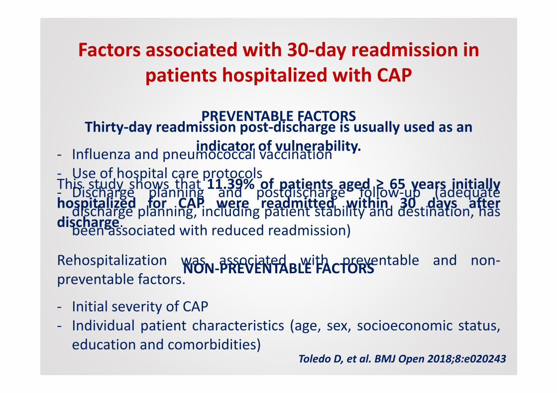
Factors associated with 30-day readmission in patients hospitalized with CAP
PREVENTABLE FACTORS
- Influenza and pneumococcal vaccination- Use of hospital care protocols- Discharge planning and postdischarge follow-up (adequate
discharge planning, including patient stability and destination, hasbeen associated with reduced readmission)
NON-PREVENTABLE FACTORS
- Initial severity of CAP- Individual patient characteristics (age, sex, socioeconomic status,
education and comorbidities)Toledo D, et al. BMJ Open 2018;8:e020243
Thirty-day readmission post-discharge is usually used as an indicator of vulnerability.
This study shows that 11.39% of patients aged ≥ 65 years initiallyhospitalized for CAP were readmitted within 30 days afterdischarge.
Rehospitalization was associated with preventable and non-preventable factors.

Toledo D, et al. BMJ Open 2018;8:e020243
CAP-unrelated causes - 49.5% of readmissionsComorbidities in 91% of readmitted patients
The reason for readmission is generally destabilization of comorbidities.
Pneumonia often occurs in patients with underlying comorbidities and often results in a worsening of such underlying conditions.

In-hospital mortality depends not only on the severity of theacute illness and age, but also on preexisting conditions, suchas loss of functional independence, severe and moderatecognitive impairment and low body mass index.Therefore, we suggest screening all elderly patients for the presenceof delirium and acute mobility impairment both on admission andsystematically during the hospitalization.
Gait speed and walking abilities may be used as clinical indicators offrailty in older subjects. Functional status can predict clinical outcomes(e.g. clinical recovery, re-hospitalization and mortality), and is considered aclinical outcome itself; its deterioration can be triggered by an acuteillness, such as pneumonia.

Davydow S, et al. Am J Med. 2013 ; 126: 615–624.e5

LE POLMONITI
NELL’ANZIANO
Caratteristiche cliniche

SEGNI E SINTOMI DELLA POLMONITE
Eur J Intern Med. 2014; 25: 312-319
• DOLORE TORACICO (pleuritico)
• TOSSE• DISPNEA• FEBBRE• LEUCOCITOSI

SEGNI E SINTOMI DELLA POLMONITENELL’ANZIANO
Eur J Intern Med. 2014; 25: 312-319
• DOLORE TORACICO (pleuritico)
• TOSSE• DISPNEA• FEBBRE• LEUCOCITOSI
• CADUTE• CAMBIAMENTO ACUTO
DELLO STATO FUNZIONALE• INAPPETENZA• INCONTINENZA URINARIA• CONFUSIONE/DELIRIUM

7.803 patients with CAP
Comorbidity present in only half of the younger
patients(46.6% versus 88.2%)
CAP COULD BE A DIFFERENT ENTITY IN THE ELDERLY BECAUSE OF AN ATYPICAL CLINICAL PRESENTATION, MORE SEVERE SYMPTOMS AND HIGHER LONG-TERM
MORTALITY IN COMPARISON TO YOUNGER PATIENTS
Eur Respir J. 2012; 39: 1156-1161

POLMONITE AB-INGESTISLa polmonite ab-ingestis (aspiration
pneumonia) è definita come una polmonite inun individuo con fattori di rischio peraspirazione (alimentare o di secrezioniorofaringee) nelle vie aeree inferiori, conepisodio di aspirazione dimostrato osospettato.
Maggiore severità clinica e tasso di mortalità.
MAGGIORE PROBABILITÀ SE: riscontro radiografico di addensamenti polmonari
in segmenti declivi dove per anatomia e gravità è più facile il passaggio disecrezioni o residui alimentari:- nel caso di pazienti allettati, i segmenti posteriori dei lobi superiori e i
segmenti superiori dei lobi inferiori;- in generale i lobi inferiori, soprattutto a destra.

FATTORI PROTETTIVI- bassa carica batterica delle
secrezioni;- valido riflesso della tosse;- adeguata funzione muco-
ciliare;- normali meccanismi
immunitari
Janssens JP, Krause KH. Lancet Infect Dis. 2004; 4: 112-24
POLMONITE AB-INGESTIS
FATTORI PREDISPONENTI- colonizzazione oro-faringea
- ridotta clearance muco-ciliare;- alterata funzione orale e
neurologica;- alterata meccanica
respiratoria;- comorbilità
Circa 50% di tutti gli adulti sani aspirano piccole quantità di secrezioni
orofaringee durante il sonno.
Alta incidenza di aspirazione silente negli anziani con polmonite: 71% dei
pazienti anziani con CAP.

Journal of Critical Care. 2015; 30 : 40-48
Distinguishing anaerobic pleuropneumonia due to aspiration fromclassic CAP may be very difficult.
Absence of shaking chills and the development of lung abscess arefeatures more common in aspiration pneumonia. Aspirationpneumonia has a more indolent course (slightly longer time beforepresentation to the hospital, 4.5 vs 2.6 days).

Una maggiore frequenza di aspirazione si riscontra nei pazienti con demenza e con patologia cerebrovascolare
L’ultilizzo di presidi per nutrizione enterale non protegge dalla broncoaspirazione (SNG, PEG, PEJ)
Janssens JP, Krause KH. Lancet Infect Dis. 2004; 4: 112-24
POLMONITE AB-INGESTIS
La flora batterica orale negli anziani più fragili può diventare più virulenta
Potenziali patogeni respiratori che colonizzano il cavo orale sonoStaphylococcus aureus (24.5%), Klebsiella pneumoniae (18.1%),Pseudomonas aeruginosa (18.1%) e Enterobacter cloacae (11.6%)
Sumi Y, et al - Arch Gerontol Geriatr 2007; 44: 119–124

FATTORI FAVORENTI COLONIZZAZIONE VIE RESPIRATORIE• Terapie antibiotiche
• Intubazione endotracheale• Fumo
• Malnutrizione• Interventi chirurgici
• Ridotta salivazione (da farmaci, ad es. antidepressivi, antiparkinsoniani,
diuretici, antipertensivi e antistaminici)• Malattie/Igiene cavo orale
La colonizzazione batterica del tratto respiratorio superiore più che all’età si
correla alla gravità delle condizioni cliniche.Colonizzazione da parte di batteri Gram-negativi può riguardare il 60-73% deipazienti anziani in un reparto di Medicina e il 22-37% di quelli istituzionalizzati.
Janssens JP, Krause KH. Lancet Infect Dis. 2004; 4: 112-24
Nei pazienti istituzionalizzati il rischio di polmonite è ridotto da un'adeguata
igiene orale (e negli individui edentuli).

Eur J Intern Med. 2014; 25: 312-319
POLMONITE AB-INGESTIS

La diagnosi di polmonitenosocomiale (Hospital-acquiredpneumonia – HAP) in età moltoavanzata è spesso difficile a causadi presentazioni cliniche atipiche epaucisintomatiche (delirium,
mancata risposta febbrile, tosse
assente, esame obiettivo
scarsamente suggestivo).
POLMONITE NOSOCOMIALE
Talvolta la diagnosi viene formulata in seguito ad approfondimentirichiesti in presenza di cambiamenti non altrimenti giustificati nelle
prestazioni cognitive, peggioramento di patologie croniche, nuova
comparsa di dispnea, tachipnea, tachicardia, ridotta saturazione di
ossigeno arteriosa.

EPIDEMIOLOGIA HAP -VAP

EZIOLOGIA E FATTORI DI RISCHIO


Respiratory Care. 2014; 59: 1078-1085

Many studies have attributed these findings to a nosocomial etiology. Further investigation on microbial composition and MDR infections
risk factors of HCAP are neededSemin Respir Crit Care Med 2009; 30: 239–248

LE POLMONITI
NELL’ANZIANO
In Pronto Soccorso…

PNEUMONIA SEVERITY INDEX FOR COMMUNITY-ACQUIRED PNEUMONIA


Clinical Infectious Diseases 2007; 44: S27–72
Three possible scenarios must be taken into account when evaluating theseverity of a patient with pneumonia:1) onset of severe sepsis;
2) onset of acute respiratory failure;
3) presence of decompensated comorbidities.
Scores and models can help the identification of the most adequate setting of care for patients with CAP,
BUTobjective criteria should always be supplemented with physician evaluation ofsubjective factors when deciding the setting of care, including:- the ability to safely and reliably take oral medication;- availability of outpatient support resources and caregivers in case of
dependent patients;- other medical or psycho-social needs (such as homelessness and poor
functional status);- lack of response to previous adequate empiric antibiotic therapy.

ICU ADMISSION DECISION
• Direct admission to an ICU is required for patients withseptic shock requiring vasopressors or with acuterespiratory failure requiring intubation and mechanicalventilation
• Direct admission to an ICU or high-level monitoring unitis recommended for patients with 3 of the minor criteriafor severe CAP
Clinical Infectious Diseases 2007; 44: S27–72

In some studies, a significant percentage of patients with CAP are
transferred to the ICU in the first 24–48 h after hospitalization.
Mortality and morbidity among these patients appears to be
greater than those among patients admitted directly to the ICU.
Clinical Infectious Diseases 2007; 44: S27–72

Acute respiratory failure can be treated with different levels of intensity of care, including non-invasive ventilation (NIV) and
mechanical ventilation (MV).
However, despite the lack of evidence that these mechanisms of
support are less effective in elderly patients, they are oftenundertreated in clinical practice.
Elderly patients (>75 years) had a similar duration of MV, ICU and hospital stay, and in-hospital mortality (38% vs. 31%), but lower cost of
care when compared to younger patients.
It does not seem appropriate to restrict intensive care and ventilatory support only on the basis of chronologic age.


LE POLMONITI
NELL’ANZIANO
Terapia

DIAGNOSI EZIOLOGICA:(SE CAP SEVERA, RICOVERO IN ICU o COMORBIDITÀ)
- Coltura dell’espettorato/tracheoaspirato/BAL
- Emocolture se febbre
- Antigeni urinari per Pneumococco e Legionella
- Sierologie per Chlamydia spp. e Mycoplasma
pneumoniae (++ se infiltrati interstizio- alveolari)
NB. Avviare antibiotico-terapia dopo esecuzione di emocolture e raccolta di campioni microbiologici

E l’antibiotico?
Polmonite acquisita in comunità (CAP)Esordio in un paziente non ospedalizzato o entro 48 h dal
ricovero
Polmonite nosocomiale (HAP)Esordio o peggioramento di un infiltrato polmonare dopo
almeno 48 h dal ricovero
Polmonite associata alla ventilazione (VAP)Esordio dopo 48 h dall’inizio della ventilazione meccanica

Clinical Infectious Diseases 2007; 44: S27–72
Clin Microbiol Infect 2011; 17(Suppl. 6): E1–E59

Terapia empirica CAP
Clinical Infectious Diseases 2007; 44: S27–72 N Engl J Med 2014;371:1619-28
Clin Microbiol Infect 2011; 17(Suppl. 6): E1–E59
Per es:AMOXICILLINA / CLAVULANATO
+AZITROMICINA
Oppure:LEVOFLOXACINA
Elevata resistenza pneumococchi a CIPROFLOXACINA

Terapia empirica CAP
Clinical Infectious Diseases 2007; 44: S27–72 N Engl J Med 2014;371:1619-28
Clin Microbiol Infect 2011; 17(Suppl. 6): E1–E59

Terapia empirica CAP
Clinical Infectious Diseases 2007; 44: S27–72 N Engl J Med 2014;371:1619-28
Clin Microbiol Infect 2011; 17(Suppl. 6): E1–E59

DE-ESCALATION
QUANDO ISOLAMENTO e ANTIBIOGRAMMA DISPONIBILE:Da terapia empirica ad ampio spettro a terapia mirata
Da terapia di associazione a monoterapia
SE PAZIENTE STABILE, IN MIGLIORAMENTO CLINICO (TC< 37,5 °C, parametri vitali e funzione gastrointestinale normale)
Da terapia endovenosa a terapia per os
DURATA TERAPIA ANTIBIOTICA- Almeno 5 giorni, fino a 10-14 giorni nelle CAP severe
- Paziente sfebbrato da almeno 48 ore- Stabilizzazione clinica
NON INDICATO CONTROLLO RADIOGRAFICO A FINE TERAPIA

E l’antibiotico?
Polmonite acquisita in comunità (CAP)Esordio in un paziente non ospedalizzato o entro 48 h dal
ricovero
Polmonite nosocomiale (HAP)Esordio o peggioramento di un infiltrato polmonare dopo
almeno 48 h dal ricovero
Polmonite associata alla ventilazione (VAP)Esordio dopo 48 h dall’inizio della ventilazione meccanica

To minimize patient harm and exposure to unnecessary antibiotics and reduce
antibiotic resistance, we recommend that the antibiogram data be utilized to
decrease the unnecessary use of dual gram-negative and empiric methicillin-resistant
Staphylococcus aureus (MRSA) antibiotic treatment.
We also recommend short-course antibiotic therapy for most patients with HAP or
VAP independent of microbial etiology, as well as antibiotic de-escalation.

Clin Infect Dis. 2016; 63: 575-82
Terapia empirica HAP

Clin Infect Dis. 2016; 63: 575-82

We recommend that all hospitals regularly generate and disseminate a local antibiogram, ideally one that is
specific to their intensive care population if possible(empiric treatment regimens guided by the local distribution of pathogens associated and their antimicrobial susceptibilities).
For patients with suspected HAP/VAP, we recommend using clinical criteria alone,
rather than using serum PCT or PCR or Pulmonary Infection Score plus clinical criteria,
to decide whether or not to initiate antibiotic therapy
Clin Infect Dis. 2016; 63: 575-82


The rationale for inclusion of the HCAP designation with the HAP/VAP
guidelines in 2005 was that patients with HCAP were thought to be at high
risk for MDR organisms by virtue of their contact with the healthcare
system.
THERE IS INCREASING EVIDENCE FROM A GROWING NUMBER OF STUDIES THAT MANY PATIENTS DEFINED AS HAVING HCAP ARE
NOT AT HIGH RISK FOR MDR PATHOGENS.
Furthermore, although interaction with the healthcare system is potentially a risk for MDR pathogens, underlying patient characteristics are also
important independent determinants of risk for MDR pathogens.
RECOMMENDATIONS REGARDING COVERAGE FOR MDR PATHOGENS AMONG COMMUNITY-DWELLING PATIENTS WHO
DEVELOP PNEUMONIA WOULD LIKELY BE BASED ON VALIDATED RISK FACTORS FOR MDR PATHOGENS
HEALTHCARE-ASSOCIATED PNEUMONIA (HCAP)
Clin Infect Dis. 2016; 63: 575-82


– Cosa prendi per la tosse?
-Generalmente un po’ di freddo.

Grazie