lecture 2 – thoracic walls and breast thoracic wall ... · lecture 2 – thoracic walls and...
TRANSCRIPT
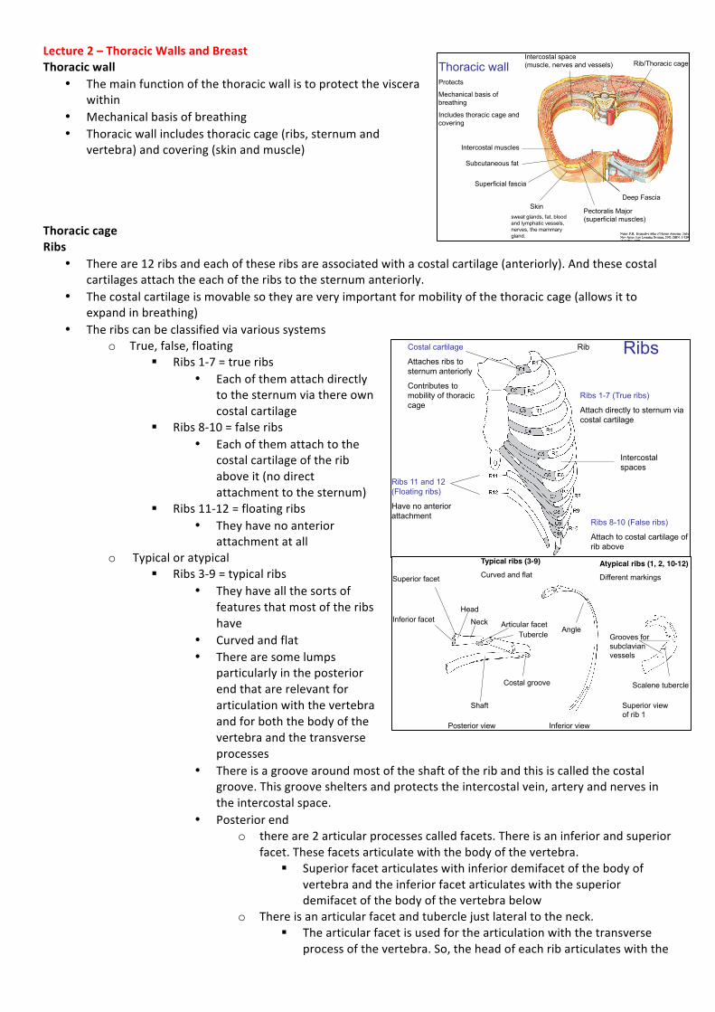
Lecture2–ThoracicWallsandBreastThoracicwall
• Themainfunctionofthethoracicwallistoprotecttheviscerawithin
• Mechanicalbasisofbreathing• Thoracicwallincludesthoraciccage(ribs,sternumand
vertebra)andcovering(skinandmuscle)ThoraciccageRibs
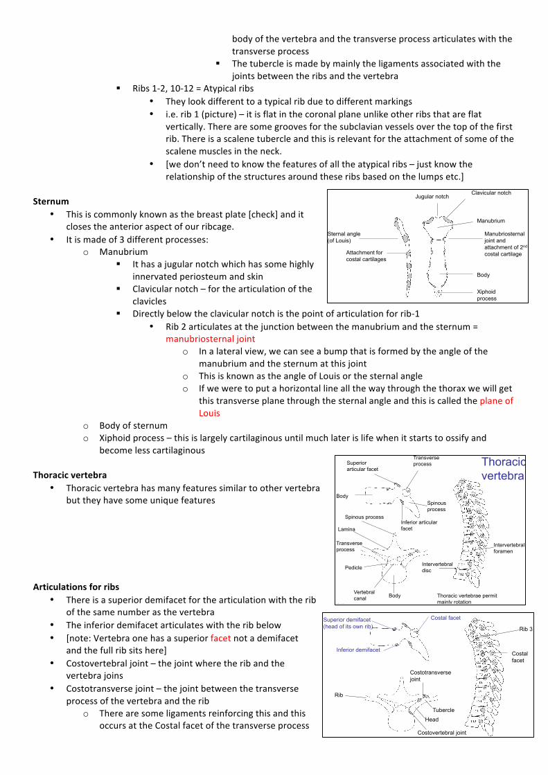
• Thereare12ribsandeachoftheseribsareassociatedwithacostalcartilage(anteriorly).Andthesecostalcartilagesattachtheeachoftheribstothesternumanteriorly.
• Thecostalcartilageismovablesotheyareveryimportantformobilityofthethoraciccage(allowsittoexpandinbreathing)
• Theribscanbeclassifiedviavarioussystemso True,false,floating
§ Ribs1-7=trueribs• Eachofthemattachdirectly
tothesternumviathereowncostalcartilage
§ Ribs8-10=falseribs• Eachofthemattachtothe
costalcartilageoftheribaboveit(nodirectattachmenttothesternum)
§ Ribs11-12=floatingribs• Theyhavenoanterior
attachmentatallo Typicaloratypical
§ Ribs3-9=typicalribs• Theyhaveallthesortsof
featuresthatmostoftheribshave
• Curvedandflat• Therearesomelumps
particularlyintheposteriorendthatarerelevantforarticulationwiththevertebraandforboththebodyofthevertebraandthetransverseprocesses
• Thereisagroovearoundmostoftheshaftoftheribandthisiscalledthecostalgroove.Thisgroovesheltersandprotectstheintercostalvein,arteryandnervesintheintercostalspace.
• Posteriorendo thereare2articularprocessescalledfacets.Thereisaninferiorandsuperior
facet.Thesefacetsarticulatewiththebodyofthevertebra.§ Superiorfacetarticulateswithinferiordemifacetofthebodyof
vertebraandtheinferiorfacetarticulateswiththesuperiordemifacetofthebodyofthevertebrabelow
o Thereisanarticularfacetandtuberclejustlateraltotheneck.§ Thearticularfacetisusedforthearticulationwiththetransverse
processofthevertebra.So,theheadofeachribarticulateswiththe
Netter, F.H. Interactive Atlas of Human Anatomy. 3rd ed. New Jersey, Icon Learning Systems, 2003, ISBN: 1-929007-15-9, Plate #179
Thoracic wall Protects
Mechanical basis of breathing
Includes thoracic cage and covering
Intercostal muscles
Subcutaneous fat
Skin sweat glands, fat, blood and lymphatic vessels, nerves, the mammary gland.
Pectoralis Major (superficial muscles)
Deep Fascia
Superficial fascia
Intercostal space (muscle, nerves and vessels) Rib/Thoracic cage
Ribs 11 and 12 (Floating ribs)
Have no anterior attachment
Costal cartilage
Attaches ribs to sternum anteriorly
Contributes to mobility of thoracic cage
Rib
Ribs 1-7 (True ribs)
Attach directly to sternum via costal cartilage
Ribs 8-10 (False ribs)
Attach to costal cartilage of rib above
Intercostal spaces
Author’s own
Ribs
Posterior view
Superior facet
Inferior facet Head
Neck
Tubercle Articular facet
Typical ribs (3-9)
Curved and flat Atypical ribs (1, 2, 10-12)
Different markings
Shaft
Costal groove
Inferior view
Superior view of rib 1
Angle Grooves for subclavian vessels
Scalene tubercle
Author’s own
bodyofthevertebraandthetransverseprocessarticulateswiththetransverseprocess
§ Thetubercleismadebymainlytheligamentsassociatedwiththejointsbetweentheribsandthevertebra
§ Ribs1-2,10-12=Atypicalribs• Theylookdifferenttoatypicalribduetodifferentmarkings• i.e.rib1(picture)–itisflatinthecoronalplaneunlikeotherribsthatareflat
vertically.Therearesomegroovesforthesubclavianvesselsoverthetopofthefirstrib.Thereisascalenetubercleandthisisrelevantfortheattachmentofsomeofthescalenemusclesintheneck.
• [wedon’tneedtoknowthefeaturesofalltheatypicalribs–justknowtherelationshipofthestructuresaroundtheseribsbasedonthelumpsetc.]
Sternum
• Thisiscommonlyknownasthebreastplate[check]anditclosestheanterioraspectofourribcage.
• Itismadeof3differentprocesses:o Manubrium
§ Ithasajugularnotchwhichhassomehighlyinnervatedperiosteumandskin
§ Clavicularnotch–forthearticulationoftheclavicles
§ Directlybelowtheclavicularnotchisthepointofarticulationforrib-1• Rib2articulatesatthejunctionbetweenthemanubriumandthesternum=
manubriosternaljointo Inalateralview,wecanseeabumpthatisformedbytheangleofthe
manubriumandthesternumatthisjointo ThisisknownastheangleofLouisorthesternalangleo Ifweweretoputahorizontallineallthewaythroughthethoraxwewillget
thistransverseplanethroughthesternalangleandthisiscalledtheplaneofLouis
o Bodyofsternumo Xiphoidprocess–thisislargelycartilaginousuntilmuchlaterislifewhenitstartstoossifyand
becomelesscartilaginousThoracicvertebra
• Thoracicvertebrahasmanyfeaturessimilartoothervertebrabuttheyhavesomeuniquefeatures
Articulationsforribs
• Thereisasuperiordemifacetforthearticulationwiththeribofthesamenumberasthevertebra
• Theinferiordemifacetarticulateswiththeribbelow• [note:Vertebraonehasasuperiorfacetnotademifacet
andthefullribsitshere]• Costovertebraljoint–thejointwheretheribandthe
vertebrajoins• Costotransversejoint–thejointbetweenthetransverse
processofthevertebraandtheribo Therearesomeligamentsreinforcingthisandthis
occursattheCostalfacetofthetransverseprocess
Sternum
Manubrium
Body
Xiphoid process
Clavicular notch Jugular notch
Attachment for costal cartilages
Sternal angle (of Louis)
Manubriosternal joint and attachment of 2nd costal cartilage
Author’s own
Body Vertebral canal
Pedicle
Transverse process
Lamina
Spinous process
Transverse process
Body Spinous process
Superior articular facet
Inferior articular facet
Intervertebral foramen
Intervertebral disc
Author’s own
Thoracic vertebra
Thoracic vertebrae permit mainly rotation
Head
Tubercle
Superior demifacet (head of its own rib)
Inferior demifacet
Costal facet
Rib 3
Costal facet
Costotransverse joint
Costovertebral joint
Rib
Author’s own
Articulations for ribs
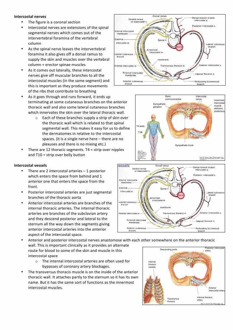
Intercostalspace• Intercostalspace–spacebetweeneachoftheribs• Intercostalmuscles–musclesthatfillupthesespaces.
Intercostalmuscles
• Thereare3layersofintercostalmuscles–theyallruninthesamesortoforientation
• Themostsuperficiallayerofthemusclesiscalledtheexternalintercostalmusclesandtheyrunantero-inferiorly
o Theyprojectallthewaythroughthespaceposteriorlyandanteriorlytheyformamembranesotherearenomusclefibresanteriorly
o Theexternalintercostalmuscleselevatetheribso Theystopsomewhereatthecostochondraljunction
• Thesecondlayerofmusclesiscalledtheinternalintercostalmusclesandtheyrunperpendiculartotheexternalintercostalmuscles–postero-inferiorly
o Posteriorly,thesemusclesbreakdownandformamembranebutanteriorlywecanseeallthemusclesthroughtothesternum
o Thereare2typesofmovementsheredependingonwhereintheintercostalspace§ Ifitislateraltothecostochondraljunction–becausetheyhavetheoppositefibredirection
toexternalintercostalmuscles,theinternalintercostalmuscleswilldepresstheribs(notelevate)
§ Iftheyaremedialtothecostochondraljunction–thentheywillelevatetheribs.Thisisduetotheshapeoftheribs(3Dstructureoftheribs)
• Innermostintercostalmuscles–theytendtobebrokendownandbepatchy
o Posteriorlytheyareverydeficient–thereisnomembraneeither
o Thedirectionofthesemusclefibresarethesameasthoseoftheinternalintercostalmusclessotheybothmusthavethesamefunctions
Intercostalspace
• Intercostalveins,arteriesandnervesareinbetweentheinnermostintercostallayerandtheinternalintercostalmusclelayer.Theyareshelteredhighintheintercostalspaceunderthecostalgroovepftheribabove.
o So,ifwewanttotheaspiratesomefluidinthepleurawewanttomakesurethatwepenetrateabovetheribbelowtoavoidgoingclosetoanddamagingthisneurovascularbundle
Netter, F.H. Interactive Atlas of Human Anatomy. 3rd ed. New Jersey, Icon Learning Systems, 2003, ISBN: 1-929007-15-9, Plate #179
Intercostal muscles
Subcutaneous fat
Skin sweat glands, fat, blood and lymphatic vessels, nerves, the mammary gland.
Pectoralis Major (superficial muscles)
Deep Fascia
Superficial fascia
Intercostal space (muscle, nerves and vessels) Rib/Thoracic cage Intercostal
space
Eizenberg N, Briggs C, Barker P, Grkovic I. An@tomedia: Thorax module. Melbourne An@tomedia Publishing, 2001, ISBN: 0646 407 317
External intercostal muscles
(antero-inferior)
Internal intercostal muscles
(postero-inferior)
External intercostal membrane
Depress
Elevate
Intercostal muscles
Eizenberg N, Briggs C, Barker P, Grkovic I. An@tomedia: Thorax module. Melbourne An@tomedia Publishing, 2001, ISBN: 0646 407 317
Innermost intercostal muscle
(postero-inferior)
Deficient posteriorly
Author’s own
Intercostal vein
Intercostal artery
Intercostal nerve
Innermost intercostal muscle
Internal intercostal muscle
External intercostal muscle
Costal groove of rib
Intercostal space
Intercostalnerves• Thefigureisacoronalsection• Intercostalnervesareextensionsofthespinal
segmentalnerveswhichcomesoutoftheintervertebralforaminaofthevertebralcolumn
• Asthespinalnerveleavestheintervertebralforaminaitalsogivesoffadorsalramustosupplytheskinandmusclesoverthevertebralcolumn=erectorspinaemuscles
• Asitcomesoutlaterally,theseintercostalnervesgiveoffmuscularbranchestoalltheintercostalmuscles(inthesamesegment)andthisisimportantastheyproducemovementsoftheribsthatcontributetobreathing
• Asitgoesthroughandrunsforward,itendsupterminatingatsomecutaneousbranchesontheanteriorthoracicwallandalsosomelateralcutaneousbrancheswhichinnervatestheskinoverthelateralthoracicwall.
o Eachofthesebranchessupplyastripofskinoverthethoracicwallwhichisrelatedtothatspinalsegmentalwall.Thismakesiteasyforustodefinethedermatomesinrelativetotheintercostalspaces.(itisasinglenervehere–therearenoplexusesandthereisnomixingetc.)
• Thereare12thoracicsegments.T4=stripovernipplesandT10=stripoverbellybutton
Intercostalvessels
• Thereare2intercostalarteries–1posteriorwhichentersthespacefrombehindand1anterioronethatentersthespacefromthefront.
• Posteriorintercostalarteriesarejustsegmentalbranchesofthethoracicaorta
• Anteriorintercostalarteriesarebranchesoftheinternalthoracicarteries.Theinternalthoracicarteriesarebranchesofthesubclavianarteryandtheydescendposteriorandlateraltothesternumallthewaydownthesegmentsgivinganteriorintercostalarteriesintotheanterioraspectoftheintercostalspace.
• Anteriorandposteriorintercostalnervesanastomosewitheachothersomewhereontheanteriorthoracicwall.Thisisimportantclinicallyasitprovidesanalternaterouteforbloodtosomeoftheskinandmuscleinthisintercostalspace
o Theinternalintercostalarteriesareoftenusedforbypassesofcoronaryarteryblockages.
• Thetransversusthoracismuscleisontheinsideoftheanteriorthoracicwall.Itattachespartlytothesternumsoithasitsownname.Butithasthesamesortoffunctionsastheinnermostintercostalmuscles.
Moore K L. Clinically Oriented Anatomy. 2nd ed. Williams and Wilkins; 1985. ISBN 0683061321. Ch.1 Page 65
Intercostal nerves
Rami communicantes
Sympathetic trunk
Sympathetic ganglion
Netter, F.H. Interactive Atlas of Human Anatomy. 3rd ed. New Jersey, Icon Learning Systems, 2003, ISBN: 1-929007-15-9, Plate #179
Intercostal nerve Innermost
intercostal muscle deficient posteriorly
Netter, F.H. Interactive Atlas of Human Anatomy. 3rd ed. New Jersey, Icon Learning Systems, 2003, ISBN: 1-929007-15-9, Plate #150
T4 teets
T1O
Moore K L. Clinically Oriented Anatomy. 2nd ed. Williams and Wilkins; 1985. ISBN 0683061321. Ch.1 Page 65
Intercostal vessels
Netter, F.H. Interactive Atlas of Human Anatomy. 3rd ed. New Jersey, Icon Learning Systems, 2003, ISBN: 1-929007-15-9, Plate #179
Descending aorta Posterior intercostal artery
Internal thoracic artery
Netter, F.H. Interactive Atlas of Human Anatomy. 3rd ed. New Jersey, Icon Learning Systems, 2003, ISBN: 1-929007-15-9, Plate #176
Internal thoracic vessels
Transversus thoracis
Anterior intercostal artery