lecture 8 - aau
TRANSCRIPT

Lecture 8
SPR: immobilization strategiesSPR: immobilization strategies

The Aim of the Lecture
Quantitative
In the last lecture:
Quantitative parameters of a binding reaction: concentrationconcentrationof an analyte, reaction rates, affinity etc.
specific for a particular
affinity etc.
specific for a particular sensor type reaction and geometry
In this lecture:• How to make our SPR sensor selective?• How to make our SPR sensor selective?

Surface plasmon sensor• Principle of affinity SP biosensor

Immobilization techniquesDifferent applications might require specific binding techniques, however there are general requirements:
• successful label-free t f ifi
however there are general requirements:
measurement of a specific binding event requires the best possible activity of the ligandpossible activity of the ligand
• conformation and orientation should be close to the in vivoshould be close to the in vivosituation
• non-specific binding should be p gkept low
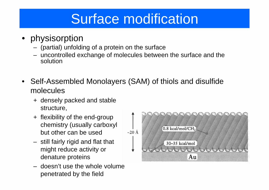
Surface modification• physisorption
– (partial) unfolding of a protein on the surface– uncontrolled exchange of molecules between the surface and the
solution
• Self-Assembled Monolayers (SAM) of thiols and disulfide molecules+ densely packed and stable
structure,+ flexibility of the end group+ flexibility of the end-group
chemistry (usually carboxyl but other can be used
– still fairly rigid and flat that might reduce activity or denature proteinsp
– doesn’t use the whole volume penetrated by the field

Surface modification• Dextran hydrogel:
thin hydrophilic (hydrogel) layer based on highly flexible mainly unbranched dextran molecules
• Advantages:+ highly hydrophilic environment with non-cross-linked structure (keeps
molecules in a solution-like state)+ increased binding capacity+ increased binding capacity+ thickness of the layer can match the penetration depth+ can be tailored using various molecular weight (10k – 1M) and different g g ( )
chemistry• Other chemistries: Polyvinyl alcohol, polyacrylic acid, poly-L-lysine

Immobilisation strategies
covalent capture Hydrophobic (supported bilayer)
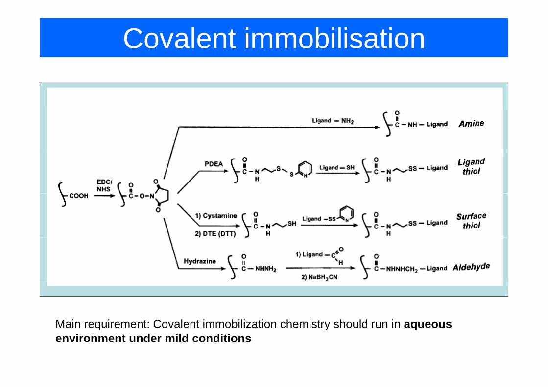
Covalent immobilisation
Main requirement: Covalent immobilization chemistry should run in aqueousMain requirement: Covalent immobilization chemistry should run in aqueous environment under mild conditions

Amine couplingC li b li h id l d• Coupling to carboxylic groups: the most widely used strategy
• Carboxylic groups can be obtained:SAM of mercaptoacid on gold– SAM of mercaptoacid on gold
– carboxilated dextrane (e.g. reaction of with haloacetic acids), normally purchased with given degree of carboxylation
very unstable, hydrolyses quickly
stable for several minutes toquickly minutes to hours

Amine coupling
• Typical protocol:Typical protocol:– injection of EDC mixed with NHS in water, pH=4-6 (natural buffering)– injection of a protein in acetate buffer pH=4-6, low ionic strength.
P iti l h d t i ill b tt t d t th ti l h dPositively charged protein will be attracted to the negatively charged hydrogel (“pre-concentration”, fraction of carboxylic groups should remain unreacted), 1-10min.
Protein concentration up to 50 ng/mm2 (several monolayers) can be• Protein concentration up to 50 ng/mm2 (several monolayers) can be obtained
• Coupling under mild conditions, very few immobilization points, activity is l l d ( I G ll t i f 75% f ti it )largely preserved (e.g. IgG usually retains of approx. 75% of activity)
surface modification withsurface modification with amino groups

Amine coupling
• typical sensogram for amine coupling
Proteins with pI>3.5 can be effectively pre-concentrated at pH~5. However acidic proteins will be repelled from the layer

Amine coupling
• micelle mediated immobilization of negatively charged proteins

Thiol-based cooupling• Coupling to thiol groups
– mainly used in the situations when the ligand lacks amino groups or they are located close to the active centercan be introduced on the sensor surface or on the molecule– can be introduced on the sensor surface or on the molecule
– very specific– disulfide bond can be cleaved by reactive thiols (e.g. mercaptoethanol)
coupling to thiol-modified surface
coupling to pyridyl disulfide surface

Thiol-based cooupling• coupling thiolated binding partner to maleimide-
modified sensor surfacemodified sensor surface

Covalent immobilization• coupling to aldehyde groups
– based on the generation of aldehyde functionality by oxidation of carbohydrate residueses dues
– normally not located near the active site, therefore activity is well preserved– antibodies are particularly well suited for this type of immobilizaation

Capture-based coupling
• can be indispensable when– covalent immobilization might impair activity of the binding
site– particular molecule should be captured from e g cell lysateparticular molecule should be captured from e.g. cell lysate
• analyte can be removed• requirement: affinity should be high• requirement: affinity should be high
• Commonly used pairs:• Commonly used pairs:– avidin-biotin bond: very robust, kd=(10-12-10-15)M, as strong
as covalent bond– His-tagged protein – nitrilotriacetic acid (NTA)+Ni2+. Can
be easily broken by a chelating agent e.g. EDTA– Antibody – antigen– Antibody – antigen

Coupling via Lipid layer• Important for
– targeting membrane protein with drugs (drugtargeting membrane protein with drugs (drug screening)
– targeting lipid membranes (e.g. AMPs)
• bilayer capture:phospholipids modified with a thiol groupn es – phospholipids modified with a thiol group
– histidine modified lipids– oligonucleotide modified lipidscl
uded
ine
vesi
cle
o go uc eo de od ed p ds
hydrophobic groups attached to CM dextran
inc
the
– hydrophobic groups attached to CM dextran– membrane protein attached to CM dextran
uded
in
laye
rin
clu
the
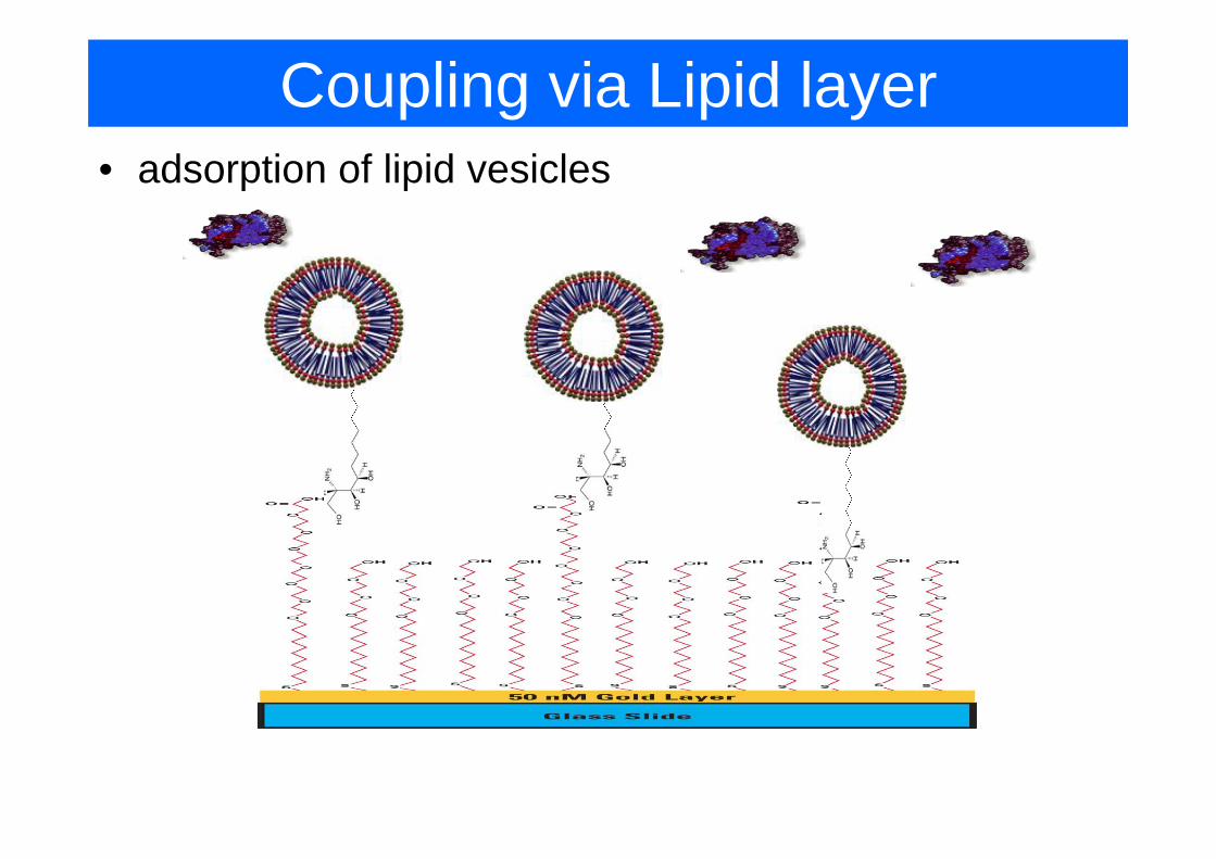
Coupling via Lipid layer• adsorption of lipid vesicles

Coupling via Lipid layer• lipid-detergent method:
1. detergent solubilized membrane proteins are immobilized on the sensor surface with amino couplingsensor surface with amino coupling
2. mixed micelles (with detergent) are injected and captured by proteins3. Detergent is washed away, lipids form membrane-like bilayer
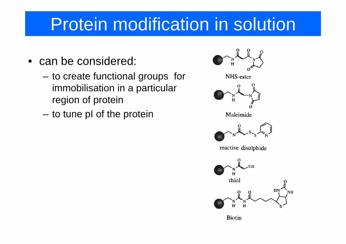
Protein modification in solution
• can be considered:– to create functional groups for
immobilisation in a particular region of protein
– to tune pI of the protein

Protein immobilization• choice of pH might affect a conformation of protein during
binding
• it might be beneficial to protect the active site of the protein by having analyte present during immobilisation
• additional partial EDC/NHS cross-linking might be important to prevent a complex protein from dissociation into monomers

Immobilization of other molecules
• Oligonucleotides: g– binding of thiolated derivatives to gold– use of biotinylated derivatives, binding at neutral pHy , g p– EDC/NHS crosslinking of amino-modified nucleotide in the
presence of CTAB

How to improve signal for small analytes• Sandwich assay
• Competition or inhibition analysis can be used for interaction that are difficult to analyse directly (e.g. due to low molecular mass of analyte).molecular mass of analyte).
Competition Inhibition
peptide 12p1 inhibition of gp120 YU2 binding to MoAb

Molecular imprinting polymers• Molecular imprinting – process of template-induced formation
of specific recognition sites, binding or catalytic. Following l f h l bi di i l f i lremoval of the template binding sites are left in polymer.
Ad t• Advantages• high stability• low cost• unlimited spectrum of
l tanalytes. • easy processing and
integration into sensorintegration into sensor design

Molecular imprinted polymers• Detection of adrenaline using gold NP and MIP
Matsui et al, Anal.Chem 76, 1310 (2004)

Antibodies• Most of immunoassays relay on monoclonal or
polyclonal antibodiesImmunoglobulin fragments
Fab
minimal antigen binding construct
variable
constant

Single domain Antibodies (sdAb)• In addition to standard antibodies, certain animals
such as sharks llamas and camels contain singlesuch as sharks, llamas and camels contain single heavy chain antibodies
single-domain fragment 15kDadiameter 2.4nmlength 4nm
Advantages: g• can inhibit enzymes at active sites due to smaller finger
size• stable against high temperature (up to 900C), chemical
denaturation, can refold after denaturation
SPR Biosensors for Medical diagnostics
• diagnostic methods based on monitoring of d ag os c e ods based o o o g oconcentration of disease biomarkers (e.g. PSA) are gaining popularityPSA) are gaining popularity
• Desirable:– test directly on bodily fluid– sufficient throughputsufficient throughput– continuous monitoring
SPR h t ti l t t th• SPR has a potential to meet those requirements

SPR detection for continous measurements• Molecular detection format: continuous detection with weak affinity antibodies.
• Example: maltose detection using anti-maltose ab.

SPR Biosensors for Medical diagnostics
• cancer biomarker (12 approved by FDA)– PSA– carcinoembrionic antigen (CEA): colorectal& breast cancer
cancer antigen CA15 3 (breast cancer)– cancer antigen CA15-3 (breast cancer)– CA125 (ovarian cancer)– carbohydrate antigen CA19-9 (pancreas, colon, stomach y g (p , ,
cancer)– alpha-fetoprotein AFP (liver&testicular cancer)

SPR Biosensors for Medical diagnostics
• PSA detection: – current clinical assays: ELISA (enzyme-linked
immunosorbent), 0.1ng/mLSPR d i h d 0 15 / L l f– SPR sandwiched assay: 0.15 ng/mL on planar surfaces, 2.4ng/mL on hydrogel
1. mouse monoclonal anti-PSA immobilized2. sample injection3 amplification with polyclonal rabbit anti PSA3. amplification with polyclonal rabbit anti-PSA4. amplification with biotinylated anti-rabbit 5. streptavidin coated latex spheres
oror4. anti-rabbit coated colloidal gold
Why planar surfaces are better for this assay?
Besselink et al, Anal. Biochem 333, p. 165 (2004)
Why planar surfaces are better for this assay?
SPR Biosensors for Medical diagnostics
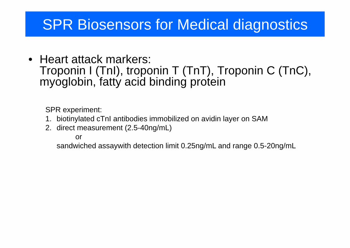
• Heart attack markers: T i I (T I) t i T (T T) T i C (T C)Troponin I (TnI), troponin T (TnT), Troponin C (TnC), myoglobin, fatty acid binding protein
SPR experiment:1. biotinylated cTnI antibodies immobilized on avidin layer on SAM2. direct measurement (2.5-40ng/mL)
or sandwiched assaywith detection limit 0.25ng/mL and range 0.5-20ng/mL

SPR Biosensors for Medical diagnostics• Antibody based assays
– type I diabetes diagnostics using anti-glutamic acid decarboxylase ab (GAD) (GAD immobilized on the surface)( ) ( )
– detection of antibodies against cholera toxin (cholera toxin immobilized on the surface)
– hepatitus C– anti-adenoviral antibodies
• Hormone based assaysy– direct detection of hCG (human chorionic gonadotropin, pregnance
marker) using anti-hCG immobilized with biotinylated oligos. Detection limit below 0.5ng/mLg
– detection of estrone and estradiol using inhibition format
• Drug detection– coumarin (anticoagulant, 7-OHC): competition assay with 7-OHc on the
surface and antibodies injected in a mixture with serumsurface and antibodies injected in a mixture with serum– warfarin (anticoagulant)– morphine and morphine metabolites
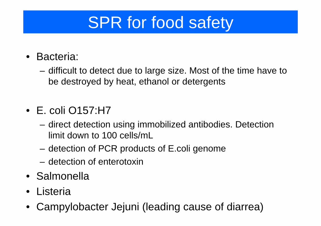
SPR for food safety
• Bacteria:– difficult to detect due to large size. Most of the time have to
be destroyed by heat, ethanol or detergents
• E. coli O157:H7E. coli O157:H7– direct detection using immobilized antibodies. Detection
limit down to 100 cells/mL– detection of PCR products of E.coli genome– detection of enterotoxindetection of enterotoxin
• Salmonella• Listeria• Listeria• Campylobacter Jejuni (leading cause of diarrea)
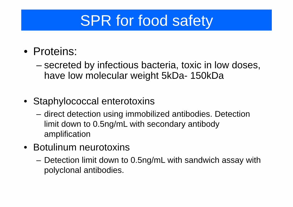
SPR for food safety
• Proteins:– secreted by infectious bacteria, toxic in low doses,
have low molecular weight 5kDa- 150kDag
• Staphylococcal enterotoxinsStaphylococcal enterotoxins– direct detection using immobilized antibodies. Detection
limit down to 0.5ng/mL with secondary antibody g y yamplification
• Botulinum neurotoxinsotu u eu oto s– Detection limit down to 0.5ng/mL with sandwich assay with
polyclonal antibodies.p y

SPR for food safety
• Low molecular weight compoundsg p– large diffusion rate but low molecular weight doesn’t
produce significant change in refractive index
• Domoic acid: neurotoxin originated from algaeDomoic acid: neurotoxin originated from algae– detection using molecularly imprinted polymer. Detection
range 2ng/mL – 3.3 ug/mLg g g– inhibition assay. Detection limit 0.1 ng/mL
• Mycotoxins (produced by Aspergillus Penicillum andMycotoxins (produced by Aspergillus, Penicillum and Fusarium)
Detection limit down to 0 5ng/mL with sandwich assay with– Detection limit down to 0.5ng/mL with sandwich assay with polyclonal antibodies.