lectures 11: trabecular bone transcribed – 3.054 / 3.36 · pdf file− 3d voronoi...
TRANSCRIPT

Figure removed due to co pyright re strictions. See Figure 1: Vajjhala, S., A. M. Kraynik, et al . "A Cellular Solid Model f orModulus Reduction due to Resorption of Trabecular Bone." Journal of Biomechanical Engineering 122, no. 5 (2000): 511–15.
24
Osteoporosis

• As trabeculae thin - buckling easier σ∗ ∝ (ρ/ρs)2
• Once trabeculae begin to resorb, connectivity reduced, strength drops dramatically
• Modeling:
◦ Can’t use unit cell or dimensional analysis (need to model local effects)
◦ Finite element modeling
◦ Initially: − 2D Voronoi honeycombsMatt Silva − 2D representation of vertebral bone
− 3D Voronoi foam — Surekha Vajjbala
Voronoi honeycomb
• Random seed points, draw perpendicular bisectors
• Use a minimum separation distance to get cells of approximately uniform size
• FE analysis — each trabecula a beam element
• First calculated elastic moduli
◦ FEA results close to analytical model for random (isotropic) honeycomb (40 models, all sameρ∗/ρs, about 25x25 cells in each)
◦ Modulus is average of stiffness over entire material
25
}

Silva et al, 1995
26
Source: Silva, M. J., L. J. Gibston, et al. "The Effects of Non-periodic Microstructure on the Elastic Properties of Two-dimensionalCellular Solids." International Journal of Mechanical Sciences 37 (1995): 1161-77. Courtesy of Elsevier. Used with permission.
Modelling: 2D Voronoi

Silva et al, 1995
27
Source: Silva, M. J., L. J. Gibston, et al. "The Effects of Non-periodic Microstructure on the Elastic Properties of Two-dimensionalCellular Solids." International Journal of Mechanical Sciences 37 (1995): 1161-77. Courtesy of Elsevier. Used with permission.
2D Voronoi

• Next, calculated compressive strength of Voroni honeycombs
• Each cell wall 1-3 beam elements
• Model non-linear elasticity and failure behavior
• 15x15 cells in model (random seeds ≈ isotropic)
• Cell wall assumed to be elastic – perfectly plastic σys/Es = 0.01 vs = 0.3
• For this value of σys/Es, transition between elastic buckling and plastic collapse stress at ρ∗/ρs = 0.035in regular hexagonal honeycomb
• Calculated compressive strength of honeycombs with ρ∗/ρs = 0.015, 0.035, 0.05, 0.15
• Generated 5 different Voronoi honeycombs at each ρ∗/ρs
• Compressive σ − E behavior:
ρ∗/ρs ≥ 0.05 -strain softening, permanent deformation on unloading
-plastic hinge formation, cell collapse in narrow localized bands
ρ∗/ρs 0.035 -non-linear elastic deformation - recoverable
Strength: 0.6 to 0.8 of σ∗ periodic
28
<

Relative density = 15% Plastic failure Silva et al, 1997
29
2D Voronoi
Source: Silva, M. J., and L. J. Gibson. "The Effects of Non-periodic Microstructure and Defects on the Compressive Strength of Two-dimensionalCellular Solids." International Journal of Mechanical Sciences 39 (1997b): 549-63. Courtesy of Elsevier. Used with permission.

2D Voronoi
Relative density 1.5%; elastic buckling failure
30
Silva et al, 1997 Source: Silva, M. J., and L. J. Gibson. "The Effects of Non-periodic Microstructure and Defects on the Compressive Strength of Two-dimensionalCellular Solids." International Journal of Mechanical Sciences 39 (1997b): 549-63. Courtesy of Elsevier. Used with permission.

2D Voronoi
31
Silva et al, 1997 Source: Silva, M. J., and L. J. Gibson. "The Effects of Non-periodic Microstructure and Defects on the Compressive Strength of Two-dimensional
Cellular Solids." International Journal of Mechanical Sciences 39 (1997b): 549-63. Courtesy of Elsevier. Used with permission.

• Max. normal strains at nodes in honeycombs (linear elastic).
◦ Voronoi honeycombs — normal distribution
◦ Regular hexagonal honeycombs — dashed lines on plot
◦ Normal strain in vertical cell walls in regular hexagonal honey combs — mean normal strain inVoronoi
◦ Oblique walls — bending — larger strains
◦ Voronoi honeycomb 5% of strain outside of range of strain in regular hexagonal honeycomb
◦ Decrease in strength associated with broader range of strains in Voronoi honeycombs
◦ Minimum strength at ρ∗/ρs = 0.05
— Interaction between elastic buckling and plastic yield
� �2π2EI π2 E π r4 π2 rσcr = = = E
l2 4 l2 π r2 4 l
32

2D Voronoi
Silva et al, 1997
Figure removed due to co pyright r estrictions. See Figure 5; Silva, M. J., and L. J. Gibson. "The Effectsof Non-periodic Microstructure and Defects on the Compressive Strength of Two-dimensional CellularSolids." International Journal Mechanical Sciences 39, no. 5 (1997): 549-63.
33
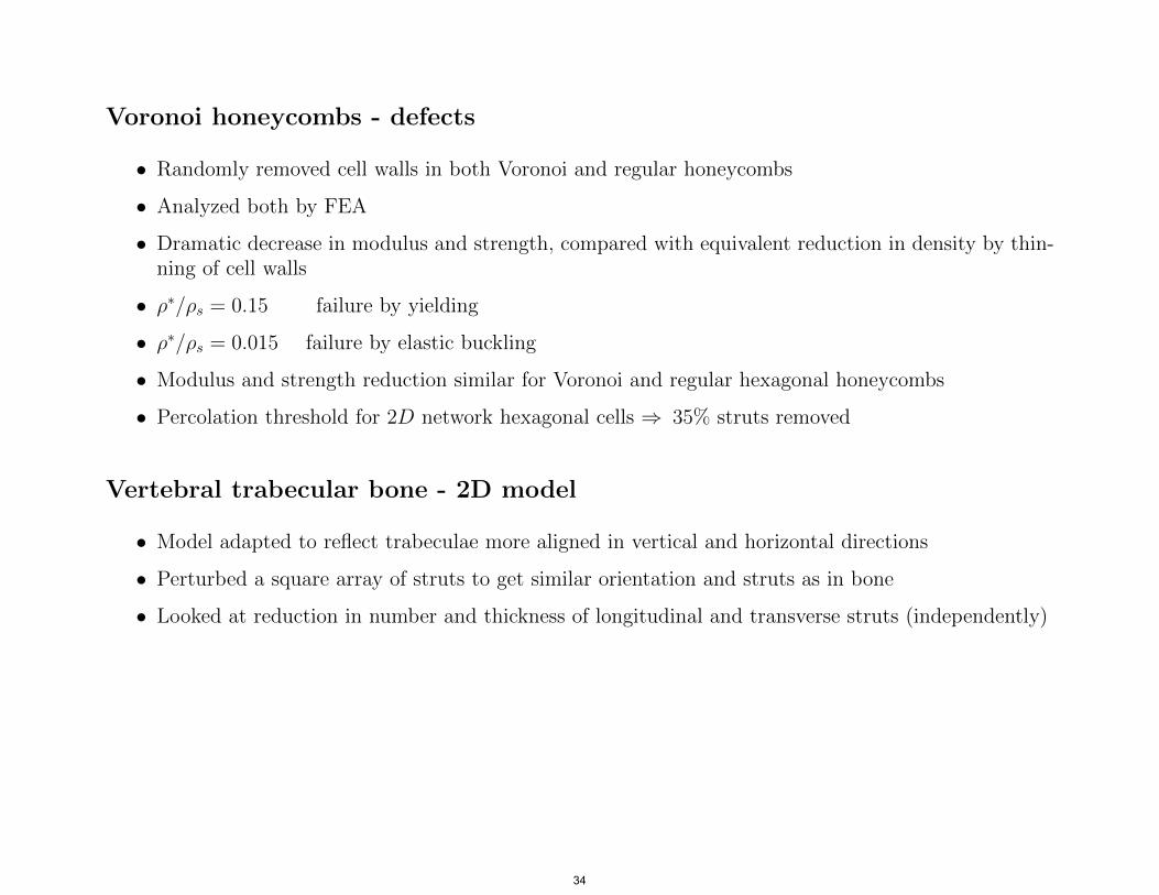
Voronoi honeycombs - defects
• Randomly removed cell walls in both Voronoi and regular honeycombs
• Analyzed both by FEA
• Dramatic decrease in modulus and strength, compared with equivalent reduction in density by thinning of cell walls
• ρ∗/ρs = 0.15 failure by yielding
• ρ∗/ρs = 0.015 failure by elastic buckling
• Modulus and strength reduction similar for Voronoi and regular hexagonal honeycombs
• Percolation threshold for 2D network hexagonal cells ⇒ 35% struts removed
Vertebral trabecular bone - 2D model
• Model adapted to reflect trabeculae more aligned in vertical and horizontal directions
• Perturbed a square array of struts to get similar orientation and struts as in bone
• Looked at reduction in number and thickness of longitudinal and transverse struts (independently)
34

Silva et al, 1997
35
2D Voronoi
Source: Silva, M. J., and L. J. Gibson. "The Effects of Non-periodic Microstructure and Defects on the Compressive Strength of Two-dimensionalCellular Solids." International Journal of Mechanical Sciences 39 (1997b): 549-63. Courtesy of Elsevier. Used with permission.

Vajjhala et al, 2000
Figure removed due to co pyright re strictions. See Figure 2: Vajjhala, S., A. M. Kraynik, et al . "A Cellular Solid Model f orModulus Reduction due to Resorption of Trabecular Bone." Journal of Biomechanical Engineering 122, no. 5 (2000): 511–15.
36
2D Voronoi

Silva et al, 1997
37
Source: Silva, M. J., and L. J. Gibson. "Modelling the Mechancial Behavior ofVertebral Trabecular Bone: Effects of Age-related Changes in Microstructure."Bone 21 (1997a): 191-99. Courtesy of Elsevier. Used with permission.
Vertebral Trabecular Bone

Silva et al, 1997
Figure removed due to co pyright re strictions. See Figure 3: Silva, M. J., and L. J. Gibson. "Modelling the MechancialBehavior of Vertebral Trabecular Bone: Effects of Age-related Changes in Microstructure." Bone 21 (1997): 191-99.
38
Vertebral Trabecular Bone
Silva et al, 1997
Figure removed due to co pyright re strictions. See Figure 4: Silva, M. J., and L. J. Gibson. "Modelling the MechancialBehavior of Vertebral Trabecular Bone: Effects of Age-related Changes in Microstructure." Bone 21 (1997): 191-99.
39
Vertebral Trabecular Bone

Vertebral Trabecular Bone
Silva et al, 1997
Figure removed due to co pyright re strictions. See Figure 5: Silva, M. J., and L. J. Gibson. "Modelling the MechancialBehavior of Vertebral Trabecular Bone: Effects of Age-related Changes in Microstructure." Bone 21 (1997): 191-99.
40

Silva et al, 1997
41
Source: Silva, M. J., and L. J. Gibson. "Modelling the Mechancial Behavior ofVertebral Trabecular Bone: Effects of Age-related Changes in Microstructure."Bone 21 (1997a): 191-99. Courtesy of Elsevier. Used with permission.
Vertebral Trabecular Bone

Silva et al, 1997
Figure removed due to co pyright re strictions. See Figure 7: Silva, M. J., and L. J. Gibson. "Modelling the MechancialBehavior of Vertebral Trabecular Bone: Effects of Age-related Changes in Microstructure." Bone 21 (1997): 191-99.
42
Vertebral Trabecular Bone

Silva et al, 1997
43
Source: Silva, M. J., and L. J. Gibson. "Modelling the Mechancial Behavior ofVertebral Trabecular Bone: Effects of Age-related Changes in Microstructure."Bone 21 (1997a): 191-99. Courtesy of Elsevier. Used with permission.
Vertebral Trabecular Bone
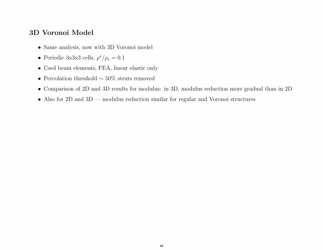
3D Voronoi Model
• Same analysis, now with 3D Voronoi model
• Periodic 3x3x3 cells, ρ∗/ρs = 0.1
• Used beam elements, FEA, linear elastic only
• Percolation threshold ∼ 50% struts removed
• Comparison of 2D and 3D results for modulus: in 3D, modulus reduction more gradual than in 2D
• Also for 2D and 3D — modulus reduction similar for regular and Voronoi structures
44
Vajjhala et al, 2000
Figure removed due to co pyright re strictions. See Figure 7: Vajjhala, S., A. M. Kraynik, et al . "A Cellular Solid Model f orModulus Reduction due to Resorption of Trabecular Bone." Journal of Biomechanical Engineering 122, no. 5 (2000): 511–15.
45
3D Voronoi Model

Vajjhala et al, 2000
Figure removed due to co pyright re strictions. See Figure 7: Vajjhala, S., A. M. Kraynik, et al . "A Cellular Solid Model f orModulus Reduction due to Resorption of Trabecular Bone." Journal of Biomechanical Engineering 122, no. 5 (2000): 511–15.
46
3D Voronoi Model

Metal foams as bone substitute materials
• Metals used in orthopedic implants (e.g. hip, knee)
• Co-Ci, Ti, Ta, stainless steel alloys
• Biocompatible, corrosion resistant
• But moduli of metals are greater than modulus of bone e.g. ETi = 110 GPa, Ecortical = 18 GPa, Etrab.bone = 0.01 − 2 GPa
• Stress shielding can lead to bone resorption
• To improve mechanical interaction between implant and bone:
◦ porous sintered metal beads used to coat implants - promote bone ingrowth
◦ also, wire mesh coatings have been developed, primarily for flat implant surfaces
◦ recently, interest in using metal foams as coatings
◦ longer term, interest in using in replacement vertebral bodies
• Variety of processes for making metal foam implant coatings
47

Ta, replicating PU foam Ti, replication of PU with CVD foam by slurry infiltration and sintering Ti, fugitive phase Ti, foaming agent Ti, expansion of Ar gas Ti, freeze-casting (freeze-drying) Ti, selective laser Ni-Ti, high temperature sintering synthesis (powders mixed, pressed and
Image sources given in ignited by, for example, Cellular Materials in Nature tungsten coil heated by and Medicine electrical current)
48
Images removed due to copyright restrictions. See Figure 8.1:Gibson, L. J., M. Ashby, and B. A. Harley. Cellular Materials inNature and Medicine. Cambridge University Press, 2010.http://books.google.com/books?id=AKxiS4AKpyEC&pg=PA228
Metal Foams: Microstructure

Processing
(a) Replicate open cell polyurethane foam
• Pyrolize PU foam → 2% dense vitreous carbon
• Coat with Ta by CVD ⇒ struts 99% Ta, 1% C
• Cell size 400 − 600 µm; coating thickness 40 − 60 µm, ρ∗/ρs = 0.15 − 0.25
• “Trabecular metal” (Zimmer) trade name
• Ta forms surface oxide Ta2O5 — does not bond to bone
• But, if treat with dilute NaOH, then heat to 300◦ and cool, then submerge in simulated body fluid (ion concentration matches human blood plasma) ⇒ get apatite coating on foam struts, which bonds to bone
(b) Infiltrate slurry of titanium hydride into open cell foam
• Heat-treat to decompose TiH2
• Sinter remaining Ti (also removes initial foam)
(c) Fugitive phase methods
• Mix Ti powder and fugitive phase powder
• Heat to T1 (∼ 200◦ C) to decompose filler, then to T2 (1200◦ C) to sinter Ti powder
49
C
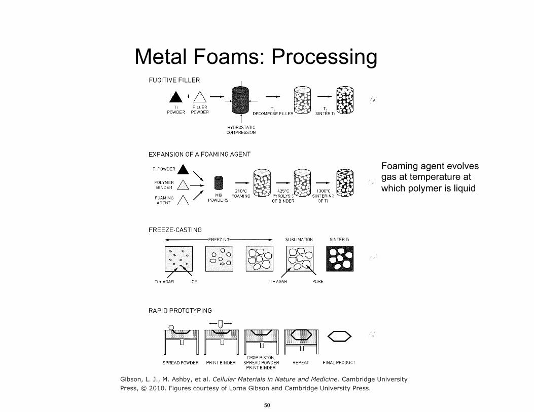
Foaming agent evolves gas at temperature at which polymer is liquid
Gibson, L. J., M. Ashby, et al. Cellular Materials in Nature and Medicine. Cambridge UniversityPress, © 2010. Figures courtesy of Lorna Gibson and Cambridge University Press.
50
Metal Foams: Processing

Processes
(d) Expansion of foaming agent
(e) Freeze casting (freeze dying)
(f) Rapid prototyping (3D Printing, selective laser sintering)
σ − E curves - similar to other foams
Data for E∗ , σ∗ c
51

52
Source: Wen, C. E., M. Mabuchi, et al. "Processing of Biocompatible Porous Ti and Mg."Scripta Materialia 45 (2001): 1147-53. Courtesy of Elsevier. Used with permission.
Ti Foam: Stress-strain

Gibson, L. J., M. Ashby, et al. Cellular Materials in Nature and Medicine. Cambridge UniversityPress, © 2010. Figures courtesy of Lorna Gibson and Cambridge University Press.
53

54
Bone in Evolutionary Studies

Bone structure in evolutionary studies
• Phylogenetic chart — big picture — structural biomaterials (mineralized)
• Sponges — first multi-celled animal
◦ calcarea: CaCO3 spicules (needles)
◦ hexactinellida: SiO2 — “glass sponges”
◦ demospongiae: most sponges — some have SiO2 spicules — spongin (type of collagen)
• Cnidarians - e.g. corals, jellyfish
◦ Corals CaCO3
• Mollusca — bivalves, snails, octopus
◦ if mineralized CaCO3
• Arthropods e.g. hexapoda (insects), arachnide (spiders), crustaceans (shrimp, lobster)
◦ Exoskeleton of insects and spiders: chitin
◦ Crustaceans: chitin may be mineralized with CaCO3
55

Vertebrates
• Cyclostomata
◦ jawless fish — lampreys hagfish
◦ no vertebra — notochord
◦ no bone
• Chondrichthye
◦ sharks, rays, skates
◦ cartilagenous skeleton — some mineralization, but not true bone
• Actinopterygii
◦ ray-finned fish
◦ true bone
◦ 450 million years ago
Bone structure and loading
• Bone grows in response to loading
• Bone structure reflects mechanical loading and function; e.g. quadruped vs. biped
• Evolutionary studies have looked at trabecular bone architecture and density
56

Hedges and Kumar, 2009 From: The Timetree of Life. Hedges, S. B., and S. Kumar (eds.) © 2009 Oxford University Press. All rights reserved. Thiscontent is excluded from our Creative Commons license. For more information, see http://ocw.mit.edu/help/faq-fair-use/.
57

Venus Flower Basket (Euplectella aspergillum) • Hierarchical structure • Remarkably stiff, tough • Joanna Aizenberg (Harvard) • Aizenberg et al (2004) Biological
glass fibers: correlation between optical and structural properties. PNAS
58

Hedges and Kumar, 2009
From: The Timetree of Life. Hedges, S. B., and S. Kumar (eds.) © 2009 Oxford University Press. All rights reserved. Thiscontent is excluded from our Creative Commons license. For more information, see http://ocw.mit.edu/help/faq-fair-use/.
59
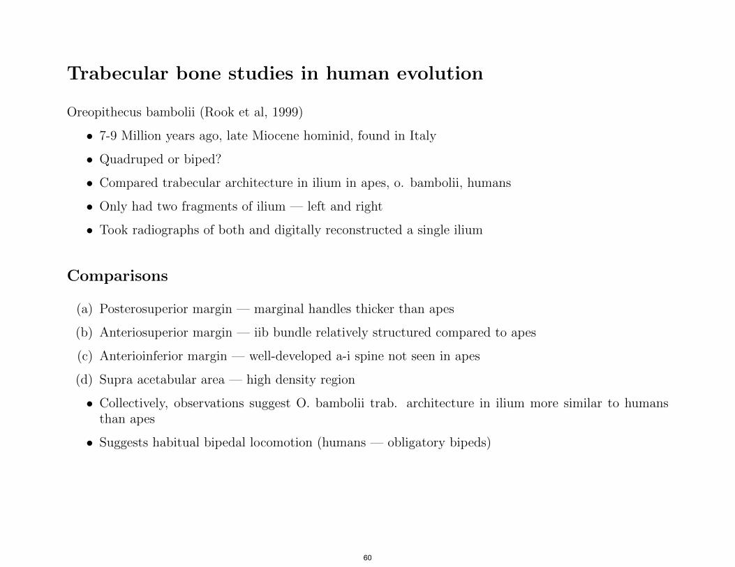
Trabecular bone studies in human evolution
Oreopithecus bambolii (Rook et al, 1999)
• 7-9 Million years ago, late Miocene hominid, found in Italy
• Quadruped or biped?
• Compared trabecular architecture in ilium in apes, o. bambolii, humans
• Only had two fragments of ilium — left and right
• Took radiographs of both and digitally reconstructed a single ilium
Comparisons
(a) Posterosuperior margin — marginal handles thicker than apes
(b) Anteriosuperior margin — iib bundle relatively structured compared to apes
(c) Anterioinferior margin — well-developed a-i spine not seen in apes
(d) Supra acetabular area — high density region
• Collectively, observations suggest O. bambolii trab. architecture in ilium more similar to humans than apes
• Suggests habitual bipedal locomotion (humans — obligatory bipeds)
60

Oreopithecus bambolii: Ilium Rook et al. (1999)
Image is in the public domain. Source: Wikimedia Commons.
hgp://en.wikipedia.org/wiki/Iliac_crest

Trabecular architecture: Ilium
Figure removed due to copyright restrictions. See Figure 1: Rook L., et al. "Oreopithecus was a
Bipedal Ape after All." Proceedings of the Natural Academy of Sciences 96 (1999): 8795-99.
62

Figure removed due to copyright restrictions. See Figure 2: Rook L., et al. "Oreopithecus was a
Bipedal Ape after All." Proceedings of the Natural Academy of Sciences 96 (1999): 8795-99.
63
Digitally reconstructed ilium

Comparison of trabecular architecture
Figure removed due to copyright restrictions. See Figure 3: Rook L., et al. "Oreopithecus was a
Bipedal Ape after All." Proceedings of the Natural Academy of Sciences 96 (1999): 8795-99.
64

MIT OpenCourseWarehttp://ocw.mit.edu
3.054 / 3.36 Cellular Solids: Structure, Properties and ApplicationsSpring 2015
For information about citing these materials or our Terms of Use, visit: http://ocw.mit.edu/terms.